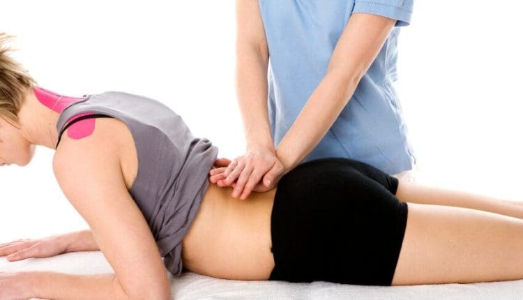
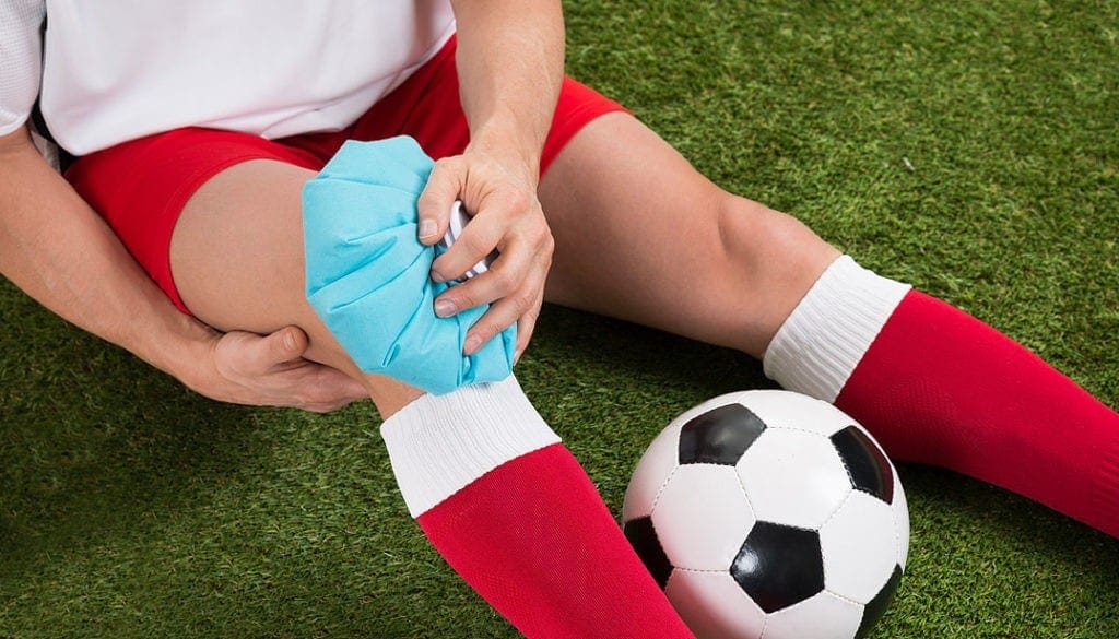
Denise was unfortunately involved in an automobile accident that resulted in lower back pain. When she realized that she could not sit or sleep for lengthy periods of time without having debilitating symptoms, Denise discovered chiropractic care with Dr. Alex Jimenez at El Paso, TX. After she received therapy for her automobile accident injuries, Denise experienced relief from her symptoms and she managed to participate in her regular activities once more. As a result of the education and maintenance Dr. Alex Jimenez supplied, Denise recovered her initial health and overall wellness.
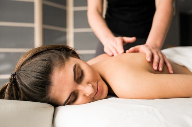
Lower Back Pain Chiropractic Treatment
Back pain is one of the most common health issues today, with approximately nine out of ten adults undergoing it at any time in their lifetime, where about five working adults will develop it annually. Some quote that about 95 percent of Americans will experience back pain at some point in their lifetime. It is undoubtedly the typical cause of chronic pain, since it’s also a substantial contributor of missed work and handicap. In the USA alone, severe cases of lower back pain would be the fifth most frequent reason for doctor visits and triggers 40 percent of missed days off work. What’s more, according to statistics, back pain is the only top cause of disability worldwide.
We are blessed to present to you�El Paso�s Premier Wellness & Injury Care Clinic.
As El Paso�s Chiropractic Rehabilitation Clinic & Integrated Medicine Center,�we passionately are focused treating patients after frustrating injuries and chronic pain syndromes. We focus on improving your ability through flexibility, mobility and agility programs tailored for all age groups and disabilities.
If you have enjoyed this video and/or we have helped you in any way please feel free to subscribe and recommend�us.
Truide Torres, office supervisor, developed facet syndrome from engaging in gymnastics at a young age. As a result of extra strain being put on her backbone, Mrs. Torres has been made to decrease her involvement in physical and exercise actions. To be able to continue being involved in fitness, Truide Torres discovered facet syndrome pain therapy with Dr. Alex Jimenez, D.C. at El Paso, TX. Mrs. Torres was educated and correctly taken care of by Dr. Jimenez because of her facet syndrome and she managed to take part in her physical and exercise actions once more. Truide Torres urges people to consider Dr. Alex Jimenez and his team as the non-surgical selection for facet syndrome pain therapy, describing them as a caring, educated and professional group of caregivers.
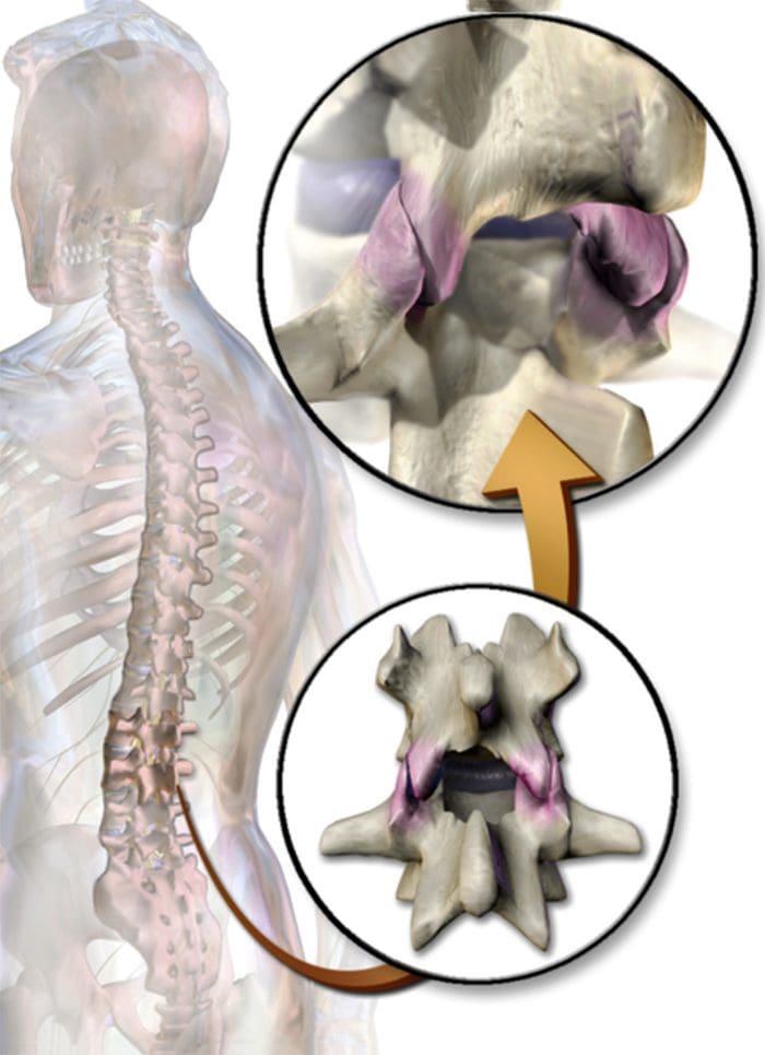
Facet Syndrome Chiropractic Treatment
Facet syndrome (also popularly called facet joint disease, facet osteoarthritis, facet hypertrophy or facet arthritis) is a syndrome in which the side joints (synovial diarthroses, from C2 to S1) degenerate into the role of causing debilitating symptoms. In combination with degenerative disk disorder, a different but associated disorder, facet syndrome is supposed to be among the most common causes of lower back pain. The indications of facet joint syndrome rely almost completely concerning the positioning of the degenerated joint, so the intensity of the injury and the quantity of pressure which has been placed on the surrounding nerve roots. It’s vital to say that the amount of pain a person undergoes doesn’t correlate well with the total amount of degeneration which has happened within the joint. A good deal of women and men experience little if any pain while some, with the particular exact same quantity of harm, experience chronic pain.
We are blessed to present to you�El Paso�s Premier Wellness & Injury Care Clinic.
As El Paso�s Chiropractic Rehabilitation Clinic & Integrated Medicine Center,�we passionately are focused treating patients after frustrating injuries and chronic pain syndromes. We focus on improving your ability through flexibility, mobility and agility programs tailored for all age groups and disabilities.
If you have enjoyed this video and/or we have helped you in any way please feel free to subscribe and recommend�us.
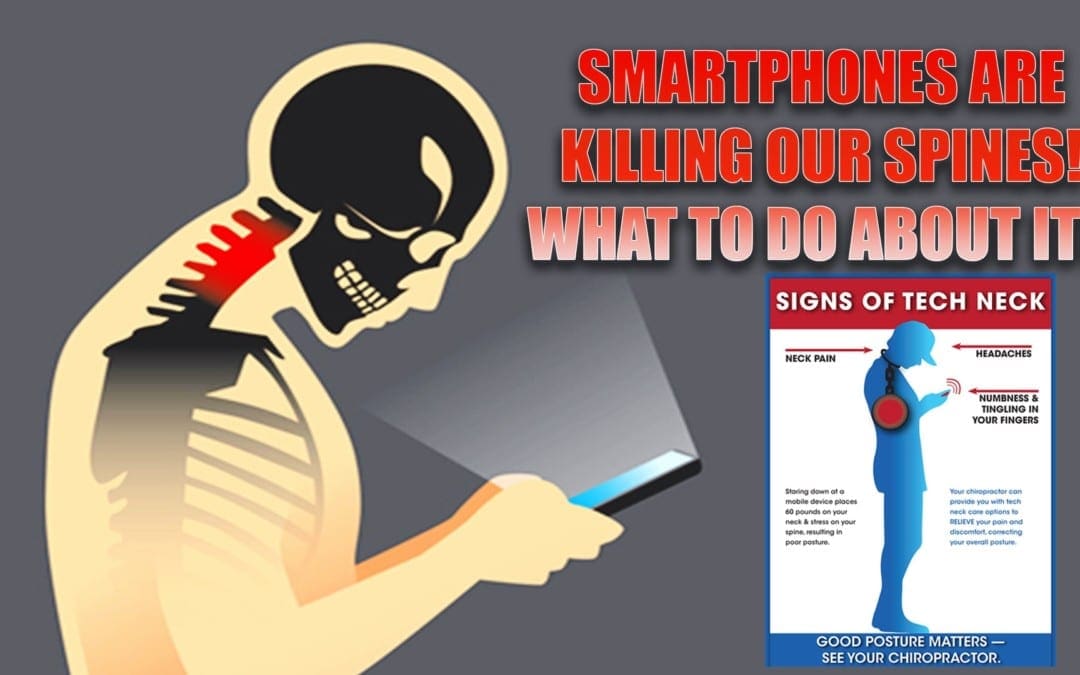
Smartphones! It’s no secret that almost everyone has one. In fact, a series of surveys by The Pew Institute in 2015 showed that 64% of American adults now own a smartphone of some kind. They keep us connected, offer us flexibility, and provide us with access to endless information.
They have their drawbacks. Smartphones also do a number on your spine.
Frequently hunching over your phone, texting, surfing, or reading takes a toll on your spine in numerous ways.
SmartPhones
Pressure
According to spine surgeon Kenneth Hansraj, looking down at your smartphone exerts up to 60 pounds of pressure on your head. This is serious wear and tear, and can set you up to be a prime candidate for degenerative issues.
Pain
The motion of texting or surfing the net on your smartphone can cause tightness in your shoulders, and pain radiating down your arms into your wrists and hands. Too many hours spent on your device can also cause upper and lower back pain.
Muscle Spasms
Over time, your smartphone usage may damage your upper back to the point you begin dealing with muscle spasms, often attributed to “text neck.”
Instead of woefully shutting you beloved smartphone in a drawer, vowing to never use it again, realize there are ways to keep your phone and a healthy spine intact. You just need to take a few simple precautions. Here are four ways to prevent spinal issues from smartphone usage, and handle the ones you may already have.
#1: Be Aware Of Your Posture
A few small changes to the way you use your smartphone can be the difference in ensuring your spine, neck, and back don’t end up suffering. Bring your phone up in front of your face, instead of leaning your head over and bending your neck to see your screen. This will keep the pressure off your neck, and minimize issues that could result.
#2: Take Breaks
Get off that phone, there’s a whole world out there! Avoid the temptation of keeping your nose stuck in your phone for an hour or more. Keep your surfing and texting to a few minutes at a time. Lay down your phone, stand up, or simply look around at your surroundings every few minutes. Frequent breaks give your spine a chance to relax, and relieve the pressure of bending your neck for a long period.
#3: Practice Stretching
Get in the habit of stretching your neck, raising your arms, rolling your shoulders, and twisting your back at the waste. These easy stretches are a quick way to loosen up body parts that can tighten up and cause injury over time.
#4: Visit A Chiropractor
If you begin noticing pain, spasms, or irritation in your neck, shoulders, or back that lingers, make an appointment with a professional chiropractor. Explain when the pain occurred, the severity, and the activity that started it. You may only require a simple adjustment to get re-aligned and gain relief from pain caused by overuse of your smartphone. A good chiropractor can also help decrease the chances of the injury worsening over time.
Not one of us is going to swear off our smartphones because of pain. However, by following these tips, improving our posture, and taking frequent breaks to stretch, we will be able to minimize the impact our phone obsession has on our spine. If you overdo it, make sure you get to an experienced chiropractor for an adjustment, so the issue is kept to a minimum.
I came to him (Dr. Alex Jimenez) and he’s been doing work on me and it’s been like, we’re going on 7 days, and I seem to be improving a lot more with him than what I’ve done with other therapists that I’ve been going to in almost a year. I would recommend him very highly, he’s good at what he does. – Leticia
According to the National Institute of Neurological Diseases and Stroke, or NINDS, lower back pain is one of the most common reasons for premature disability, often amounting to many doctor office visits and missed days at work. Based on these statistics, at least 80 percent of individuals in the world will experience low back pain at some point in their lifetimes, a majority of which could have been prevented.
Most lower back pain results from an injury, such as muscle sprains or strains due to abrupt movements or poor body mechanics while lifting heavy things. Low back pain may also be caused by certain ailments, such as a ruptured or herniated disc, sciatica, arthritis, kidney infections, diseases of the spinal column or cancer of the spinal cord,. Acute back pain can last anywhere from a few days to a few weeks while chronic back pain can last over three weeks to even months.
Low back pain is significantly more likely to happen in people between the ages of 30 and 50. This is partly as a consequence of changes that develop within the whole body with age. As you grow older, the fluid-like substance of the intervertebral discs in the spine reduces. This means that the discs in the spine experience stress more easily. You also lose muscle tone, which makes the spine more vulnerable to harm.
Ask any healthcare professional and you’ll get confirmation that low back pain is the most frequent health issue they are asked to look after. Strengthening your muscles and using good body mechanics are beneficial in preventing lower back pain. Often back pain can decrease on its own, especially through the use of the “RICE” treatment. But whilst rest, ice, compression and elevation can reduce back pain, its important to also seek treatment from a healthcare professional to treat the true underlying cause of your lower back pain.
What are the Symptoms of Low Back Pain?
Low back pain can be different for everyone. It might be sharp or stabbing. It may be dull, achy, or feel as a sort of cramp. The kind of pain you have will be based on the root cause of your low back pain. Many individuals discover that reclining or lying down can enhance their lower back pain, regardless of the underlying reason. Individuals with low back pain might experience a number of these, including:
Back pain that worsens with lifting and bending.
Worsening symptoms when sitting down.
Symptoms that become worse when walking.
Back pain which comes and goes, frequently following an up and down path.
Pain that extends from the back to the buttocks or outer hip, and travels down the leg.
Sciatica, including buttock and leg pain which travels into the foot, along with numbness, weakness or tingling sensations. It’s likely to get sciatica without low back pain.
Because low back pain can develop due to a variety of underlying health issues, symptoms commonly associated with lower back pain may differ from person to person. Irrespective of your age or symptoms, even if your low back pain doesn’t get better over a couple weeks, or is associated with fever, chills, or sudden weight loss, it’s fundamental for you to contact a healthcare professional immediately.
How is Low Back Pain Diagnosed?
Most doctors begin by conducting a physical examination to determine where you’re feeling the pain. A physical exam may also ascertain whether pain is affecting other structures and functions of your body. Your doctor may check your reflexes and your response to certain senses. This determines if your low back pain is affecting your nerves. If you do not have such debilitating symptoms, your doctor will probably monitor your condition before sending you for testing.
Certain symptoms like lack of bowel control, fever, fatigue, and weight loss might demand additional testing. Likewise, if your low back pain persists following home treatment, your doctor may also most likely want to send you for tests. Seek medical attention immediately at the event you observe any of these symptoms in addition to lower back pain.
Imaging evaluations such as X-rays, CT scans, ultrasound, and MRI may be needed in order for your doctor to evaluate bone issues, disk difficulties, or problems with the joints and ligaments in your spine. If your doctor suspects a matter with all the bones in your spine, they could send you to have a bone loss or bone density test. Electromyography, or EMG, as well as nerve conduction tests can help identify a problem with your own nerves.
How Can I Prevent Low Back Pain?
There are plenty of methods to prevent lower back pain. Practicing prevention techniques may also help reduce the seriousness of the symptoms once you have lower back pain. Prevention involves exercising the muscles in your core and back, losing weight if you are overweight or obese, lifting items properly by bending at the knees and lifting with the legs, and maintaining proper posture. Among the most common causes of lower back pain is a misalignment of the spine, or a subluxation, originating from improper posture.
Most office setups don’t provide ergonomic or support positioning desk chairs, while poor work habits prevents us from providing our spines with the much-needed relief we deserve throughout the day. Non-desk jobs also have their own perils. Standing daily, especially when combined with heavy lifting or routine bending, may also cause low back pain. The muscles surrounding the lumbar spine may not acquire the support they need when bending and lifting, resulting in low back pain. In either circumstance, strengthening these back muscles is fundamental to reducing the probability of chronic lower back pain.
Insist on an ergonomic desk chair, or have the opportunity to stand and move around more frequently. If you’re a cashier, invest in shoes with good arch support, which may help keep your entire body aligned. If needed, put on a technical brace to help support heavy lifting. Good habits at home to prevent low back pain can involve sleeping on a firm surface and having seats that are in the proper height. Steer clear from high-heeled shoes. If you smoke, then you may need to quit. Smoking causes the degeneration of spinal discs and reduces blood flow. But when you already have low back pain, a variety of treatment options, such as chiropractic care, can help treat lower back pain.
Dr. Alex Jimenez’s Insight
Many health issues can ultimately affect the spine, causing low back pain. Because of this, an individual’s symptoms are always different, often characterized by the underlying problem affecting them. A chiropractor can diagnose the source of a patient’s symptoms over a series of tests and evaluations, to determine the best treatment approach for the individual’s cause of low back pain. Chiropractic care focuses on naturally correcting any spinal misalignments, or subluxations, to reduce low back pain.
How Can Chiropractic Care Treat Low Back Pain?
Chiropractic care is one of the most well-known treatment options for relieving lower back pain. Medical practitioners normally recommend their patients to consider alternative treatment options before turning to prescription drugs and/or medications or surgery. The reasons are obvious: Many medications and/or drugs can have long-term health consequences. Whatever the advantages of providing temporary pain relief may be, these carry risks of complication throughout the recovery period.
Chiropractic care is a treatment approach which focuses on the diagnosis, treatment and prevention of a variety of injuries and/or conditions associated with the musculoskeletal and nervous system. Through the use of spinal adjustments and manual manipulations, a chiropractor can carefully restore the natural alignment of the spine, reducing stress in the complex structures of the spine and improving function. Chiropractic care may also include other treatment techniques and methods to help reduce low back pain.�Lower back pain may also need the two-pronged way of using both active and passive physical therapeutics, unless the healthcare professional has a reason to recommend one over another.
Passive treatments includes using ice packs and heating pads. The healthcare professional may also use many different forms of pulsing equipment, which triggers nerves and releases pain.
Active treatments comprises the individual to perform stretches and exercises that build the type of flexibility and strength necessary to stop future flare-ups and reduce current pain. Lots of them are done with a chiropracto’s supervision, on specialized equipment, though some might be performed at the patient’s home after they learns the principles.
Chiropractic care can help treat low back pain through the treatment approaches mentioned above. Furthermore, a chiropractor may suggest lifestyle modifications to help promote a faster recovery, including physical activities and nutritional guidelines. It you’re experiencing low back pain, make sure to seek immediate medical attention, in order to receive a proper diagnosis and be able to continue with treatment. Moreover, preventing lower back pain can help avoid future episodes. The scope of our information is limited to chiropractic as well as to spinal injuries and conditions. To discuss the subject matter, please feel free to ask Dr. Jimenez or contact us at�915-850-0900�.
Curated by Dr. Alex Jimenez
Additional Topics: Acute Back Pain
Back pain is one of the most prevalent causes for disability and missed days at work worldwide. As a matter of fact, back pain has been attributed as the second most common reason for doctor office visits, outnumbered only by upper-respiratory infections. Approximately 80 percent of the population will experience some type of back pain at least once throughout their life. The spine is a complex structure made up of bones, joints, ligaments and muscles, among other soft tissues. Because of this, injuries and/or aggravated conditions, such as herniated discs, can eventually lead to symptoms of back pain. Sports injuries or automobile accident injuries are often the most frequent cause of back pain, however, sometimes the simplest of movements can have painful results. Fortunately, alternative treatment options, such as chiropractic care, can help ease back pain through the use of spinal adjustments and manual manipulations, ultimately improving pain relief.
I go back to normal after seeing him (Dr. Alex Jimenez) and I know I can go back to doing whatever I can. But I’m now more careful. I would recommend him. It’s hard to find someone that knows their job and has a love for their job. That’s why I always come back. If I’ve got pain, I’m gonna look for him. – Mike Melgoza
Low back pain is one of the most prevalent reasons why people visit the doctor’s office and miss days from work. Approximately 80 percent of people will experience back pain at some point throughout their lifetime. Low back pain can range from moderate to severe and it can be acute, short-term, or chronic, long-term.�Because back pain can be caused by a variety of factors, the symptoms may also vary from one person to the other.
Most of the time, low back pain is more of an annoyance than anything else.�If an individual’s low back pain becomes intense and persistent, it can be tremendously debilitating and it can ultimately make it a challenge to participate as well as engage in many everyday activities. It’s essential to seek immediate medical attention to receive a proper diagnosis and continue with the best treatment option for each patient’s specific health issue.
Causes of Low Back Pain
Many potential causes can result in low back pain. In our modern world, spinal misalignments, or subluxations, caused by poor posture, have become one of the most common causes for low back pain, probably due to the simple fact that more and more people work in sedentary desk jobs than ever before. Without the appropriate back support from an ergonomic desk chair, low back pain can easily occur as a result of poor posture and due to the limited mobility of the spine throughout the day.
Even people who are up on their feet the vast majority of the day might suffer from low back pain due to a lack of spinal and abdominal support as well as a lack of proper coordination of the back muscles. While lower back pain can’t always be prevented, it’s possible to reduce the risk of suffering from lower back pain by practicing proper posture throughout the day to support the spine.
For individual’s who sit behind a computer screen for extended periods of time, this might mean investing in an ergonomic desk chair. For the more active individuals, it may mean purchasing a good pair of athletic shoes which can provide them with the right level of back and foot support throughout the day. Low back pain can also develop due to a variety of injuries and/or conditions. Fortunately, many treatment options are available to help treat low back pain, including chiropractic care and physical therapeutics.
Dr. Alex Jimenez’s Insight
The spine is made up of small bones, known as vertebrae, intervertebral discs, muscles, ligaments and nerves. With several factors, however, including poor posture, trauma from an injury, or an aggravated condition, the spine can become misaligned, ultimately affecting the complex structures surrounding the spine and resulting in back pain. Low back pain is among the most common types of back pain, particularly due to its increased role in supporting the weight of the human body.
Chiropractic Care for Low Back Pain
Chiropractic care is an alternative treatment option which focuses on the diagnosis, treatment and prevention of a variety of injuries and conditions associated with the musculoskeletal and nervous system, including back pain. Seeking chiropractic care as treatment for lower back pain is always recommended before you opt to start taking any prescription drugs and/or medications.
Pain and anti-inflammatory drugs and/or medications can relieve your low back pain, however, the results are usually temporary and these may also bring about undesirable side effects. Chiropractic care is a non-invasive and drug-free strategy for low back pain relief. It’s recommended to seek alternative treatment options, including chiropractic care and physical therapeutics, before turning to the use of drugs and/or medications as well as surgical interventions.
A doctor of chiropractic, or chiropractor, will commonly use spinal adjustments and manual manipulations to carefully correct any spinal misalignments, or subluxations, which may be causing the patient’s low back pain. Moreover, a chiropractor may utilize�passive and active treatments for low back pain and they often vary considerably in their techniques and methods. Passive and active treatments for low back pain are described as follows:
Passive treatments depend upon techniques and methods to be performed on the person. This may include anything from applying ice or heat packs to the affected area to stimulating the affected region with controlled electricity. Other treatment modalities used here may comprise of ultrasonography, TENS units, and iontophoresis.
Active treatments, on the other hand, describes measures that the patient will take, as instructed by a healthcare professional, to manage their low back pain. Typically, this comes from the type of stretches and exercises that are meant to reduce low back pain and minimize potential flare-ups too. Active treatments could include low-impact aerobic conditioning and back strengthening exercises. These may vary based upon the healthcare professional’s requirements.
Chiropractic care and physical therapeutics might be a wonderful solution for treating nearly any level of low back pain. Through the utilization of spinal adjustments and manual manipulations, as well as a combination of active and passive physical therapeutics, you’re in a position to work towards reducing your stress and increasing your body’s natural capability to prevent future health issues.
Contact a healthcare professional today to find out more about how you can manage your low back pain.The scope of our information is limited to chiropractic as well as to spinal injuries and conditions. To discuss the subject matter, please feel free to ask Dr. Jimenez or contact us at�915-850-0900�.
Curated by Dr. Alex Jimenez
Additional Topics: Acute Back Pain
Back pain is one of the most prevalent causes for disability and missed days at work worldwide. As a matter of fact, back pain has been attributed as the second most common reason for doctor office visits, outnumbered only by upper-respiratory infections. Approximately 80 percent of the population will experience some type of back pain at least once throughout their life. The spine is a complex structure made up of bones, joints, ligaments and muscles, among other soft tissues. Because of this, injuries and/or aggravated conditions, such as herniated discs, can eventually lead to symptoms of back pain. Sports injuries or automobile accident injuries are often the most frequent cause of back pain, however, sometimes the simplest of movements can have painful results. Fortunately, alternative treatment options, such as chiropractic care, can help ease back pain through the use of spinal adjustments and manual manipulations, ultimately improving pain relief.
Truide Torres, office manager, first considered chiropractic care with Dr. Alex Jimenez during her pregnancy as a result of her low back pain. Mrs. Torres experienced aggravating symptoms throughout the different stages of her pregnancy, which led her to seek a natural treatment strategy for her well-being, especially in consideration of her child in the womb. After Truide Torres started chiropractic treatment with Dr. Alex Jimenez, she recovered her overall well-being and managed to return to her very first state of well-being. As a professional manager, Truide Torres additionally receives regular chiropractic care for any lower back pain which may occur as a result of her job. Mrs. Truide expresses how important it is to maintain her spinal maintenance and she urges Dr. Alex Jimenez as the nonsurgical choice for several health difficulties.
Lower Back Pain Pregnancy Chiropractic Treatment
Low�back pain, or LBP, is a normal health issue between the muscles, nerves, and bones of the spine. Pain may be different, often described as a dull persistent pain or some sudden sharp sense. Low back pain could be classified by severity and length, including acute (pain lasting less than 6 months ), sub-chronic (6 to 12 months ), or chronic (over 12 months ). The status could be further categorized together with all the underlying causes as both bodily, non-mechanical, or referred pain. The indications of lower back pain may generally improve in a couple of weeks, but a few cases may require additional treatment. In almost all episodes of lower back pain, a specific underlying cause isn’t identified or properly cared for, and healthcare professionals might feature it to joint or muscle strain.
We are blessed to present to you�El Paso�s Premier Wellness & Injury Care Clinic.
As El Paso�s Chiropractic Rehabilitation Clinic & Integrated Medicine Center,�we passionately are focused treating patients after frustrating injuries and chronic pain syndromes. We focus on improving your ability through flexibility, mobility and agility programs tailored for all age groups and disabilities.
If you have enjoyed this video and/or we have helped you in any way please feel free to subscribe and recommend�us.
It’s been great, my turf toe has been getting a lot better. Actually, I didn’t see a doctor for about 4 months and it just kept getting worse. But when I started seeing Dr. Jimenez, it just, little by little it’s been starting to get better. It feels a lot better when I practice and stuff like that. So, it’s getting better. – Vincent Garcia
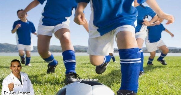
There’s always a particular risk for athletes when it comes to sports-related injuries, or sports injuries, particularly when participating in physical activities. Moreover, contact sports, such as soccer, baseball, football and basketball, tend to have higher injury rates than any other sport.
Twice as many men suffer sports-related injuries in contrast with females as a consequence of the types of sports that they most commonly engage in. Whether you’re a seasoned and experienced athlete or you simply play as a weekend warrior, there’s always a chance of experiencing a sports injury. Below, we will discuss several of the most common types of sports injuries, or sports-related injuries.
Common Sports Injuries
Sprains and strains are the most common sports injuries. Sprains are medically defined as injuries to the ligaments, or the strong bands which connect bones to the joints. Overly stretching these ligaments beyond their natural range can ultimately damage or even tear them.
Strains are medically referred to as injuries to the muscle fibers or tendons, which connect muscles to bones. Strains are known as “pulled muscles” for a reason, overly stretching or overuse of a muscle can cause tears in the muscle fibers or tendons.
�Think of ligaments and muscle-tendon units like springs,� explained Dr. William Roberts, MD, sports medicine physician at the University of Minnesota and spokesman for the American College of Sports Medicine. �The tissue lengthens with stress and returns to its normal length, unless it is pulled too far out of its normal range.� Additionally, sports injuries can result in a variety of other health issues.
Patellofemoral Syndrome
Accidents in sports which can harm an athlete generally are inclined to be knee injuries. Patellofemoral syndrome could be caused by a slide or fall onto the knees. This type of sports injury involves swelling, inflammation and an imbalance of the knee at its groove. Strengthening exercises and stretching can help provide flexibility and mobility to the muscles. Apart from strengthening exercises and stretches, a doctor of chiropractic, or chiropractor, may utilize therapeutic techniques for this specific injury.
Concussion
A blow to the head could lead to a concussion. Concussions are a serious type of sports injury and these should never be disregarded. Symptoms indicating a possible head injury may include nausea, vomiting, confusion, headache, and slurred speech. Any athlete who incurs a concussion must seek immediate medical attention. Chiropractic care can help with several of the symptoms, such as headaches, related to a concussion.
ACL Tear
The anterior cruciate ligament, or the ACL, is a fundamental ligament found in the knee. An ACL tear can be caused due to a sudden change in directions or coming to a sudden stop when playing sports or during exercise and physical activities. There’s typically swelling, inflammation and uncertainty in movement working with an ACL tear. Chiropractic care can assist with the recovery process of an ACL tear, particularly through physical therapeutics and rehabilitation programs.
Hip Flexor Strain
The hip flexor muscles are all located in the upper front area of the thigh. Sprinting, running slopes and sudden movements could lead to a hip flexor strain. There can be pain and discomfort together with swelling and inflammation in the region surrounding the thigh. Stretching and range of motion exercises employing a doctor of chiropractic, or chiropractor, can help aid with recovery. A chiropractor will work closely with a patient to determine the best treatment approach for their sports injuries.
Shin Splints
With shin splints, there’s usually pain and other painful symptoms in the lower leg, particularly along the tibia. Shin splints are the most common type of sports injuries among runners or running athletes. Ice and cold therapy can help reduce swelling and inflammation on the site. Moreover, runners or running athletes can prevent suffering shin splints by purchasing a good pair of shoes with proper arch support. The right equipment can always promote a safe participation in sports and physical activities.
Sciatica
Sciatica is back pain which radiates down the back of the leg and into the foot. This collection of symptoms is often seen in cyclists and athletes who perform a lot of backwards turning and swinging sports like tennis and golf. Sciatica, or sciatic nerve pain, may be caused by a pinched or compressed nerve, frequently due to a bulging or herniated disc. Chiropractic care is a well-known, alternative treatment option which can help alleviate sciatica, or sciatic nerve pain, symptoms.
Shoulder Injury
Shoulder injuries in sports commonly range from dislocations and misalignments to strains and sprains of the shoulder tendons and ligaments. Because the shoulder is frequently referred to as a weak joint, it is often vulnerable to suffering harm from sports injuries during exercise and physical activities, aside from the athlete’s specific sport. Ice and cold therapy as well as chiropractic care and rehabilitation can help ease the symptoms associated with shoulder injuries.
Tennis or Golf Elbow
This issue is known as an overuse sports injury. Repetitive actions inflame the forearm and wrist. Ice and cold therapy as well as rest normally helps with the symptoms, but stretching and strengthening exercises recommended by a chiropractor can also help.
Groin Pull
Additionally known as a groin strain, the groin muscles can get strained with quick side-to-side movements when engaging in exercises and physical activities. Stretching and strengthening exercises can help with the recovery process in this case as well.
Hamstring Strain
The hamstring muscles can be found in the back of the thigh. When athletes fail to stretch or exercise accordingly before engaging in their specific sports, it can cause this muscle to be pulled. If the symptoms of this condition continue over a couple of weeks, a chiropractor, or doctor of chiropractic, can help provide the necessary treatment through the use of other treatment approaches, such as ultrasound, among others, to help encourage the natural healing of the muscle and improve symptoms.
Dr. Alex Jimenez’s Insight
Although many common sports injuries are often beyond our control, athletes can engage in stretches and exercises before participating in their specific physical activities to help prevent a sports injury. Every workout should start with a gentle warm-up to prevent most of these sports injuries. It’s important for athletes to be mindful of the amount of pressure they exert on their bodies in order for them to avoid suffering sports injuries.
If you’ve suffered a sports injury, make sure to seek immediate medical attention from a qualified and experienced healthcare professional. Many skilled sports medicine doctors are dedicated to sports medicine and also focus on providing rehabilitation determined by the performance requirements of athletes. Healthcare professionals will design a treatment plan targeted to your sports injuries.
Get back in the game with the guidance of qualified and experienced healthcare professionals in sports injuries. Contact us and make sure to schedule a consultation.�The scope of our information is limited to chiropractic as well as to spinal injuries and conditions. To discuss the subject matter, please feel free to ask Dr. Jimenez or contact us at�915-850-0900�.
Curated by Dr. Alex Jimenez
Additional Topics: Acute Back Pain
Back pain is one of the most prevalent causes for disability and missed days at work worldwide. As a matter of fact, back pain has been attributed as the second most common reason for doctor office visits, outnumbered only by upper-respiratory infections. Approximately 80 percent of the population will experience some type of back pain at least once throughout their life. The spine is a complex structure made up of bones, joints, ligaments and muscles, among other soft tissues. Because of this, injuries and/or aggravated conditions, such as herniated discs, can eventually lead to symptoms of back pain. Sports injuries or automobile accident injuries are often the most frequent cause of back pain, however, sometimes the simplest of movements can have painful results. Fortunately, alternative treatment options, such as chiropractic care, can help ease back pain through the use of spinal adjustments and manual manipulations, ultimately improving pain relief.
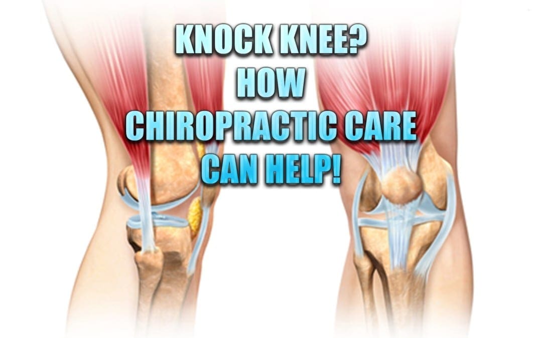
Knock knee is a condition that many children acquire when they are toddlers. Often, within a few years they grow out of it and their legs straighten naturally with no lasting effects.
Occasionally, though, a child�s legs don�t straighten and this is a cause for concern. There are many problems that can stem from knock knees, some of which will follow the child into adulthood and for the rest of his or her life. While there are several recognized treatments for knock knee, including surgery, chiropractic care has an excellent track record in managing and remedying this disorder.
What Is Knock Knee?
Knock knee, or genu valgum, is a condition that causes a person�s knees to bow in toward each other. In other words, when they stand with their knees touching and feet flat, parallel to each other, facing forward, their ankles do not touch. There may be a few inches between them or a foot, depending on its severity.
Most children go through a stage at around 3 or 4 years where they are knock kneed but by around age 8 or 10 they grow out of it and their legs straighten. Many parents become concerned when they first see their child becoming knock kneed. This is why it is vital that they understand a child�s normal growth patterns. It helps them worry less about something completely normal as well as know when to seek help if the condition does not right itself.
Aside from normal physiological child development, the atypical version of knock knees can be caused by several factors including:
Bone deformities
Knee malalignment
Genetics
Infection
Weak knee infrastructure
Injury
Rickets Disease
Scurvy
Blount�s Disease
What Health Problems Can Be Caused By Knock Knee?
Knock knee can cause pain and inflammation in the knees, ankles, and feet, as well as the hip and back. The pain can make mobility difficult. This is exacerbated if the patient is overweight because the added pressure on the joints as they are set at an unnatural position that does not adequately support the body can result in injuries to the bone, ligaments, and tendons.
A difference in leg length, a common issue with knock knee, can also cause the body to become misaligned, leading to back and hip pain. Over the long term and in severe cases, knock knee can lead to arthritis in adults and children.
How Is Knock Knee Treated?
Treatment for knock knee depends on the cause and age of the child. If the child is young and it has been determined that the knock knee is just a normal part of their growth pattern, very little action is taken although some experts advise laying a good foundation for the child by teaching them the importance of a healthy diet, regular exercise, and good posture.
Cases that are caused by some underlying factor, or that extend beyond the age that the child�s legs are expected to straighten, may require bracing. If there is an underlying cause such as infection or injury, that will need to be addressed in order to correct or manage the problem. In severe (and rare) cases, surgery may be necessary.
Is Chiropractic Care An Effective Treatment For Knock Knee?
Chiropractic care is an exceptional treatment for children of all ages who have knock knee. For younger patients who are experiencing it as a normal stage of development, it will help to keep their spine aligned and encourage good, healthy posture. In children who have an underlying cause, it can help to relieve any pain while increasing mobility as well as bringing the spine into proper alignment.
Many of the factors that cause knock knee can be addressed through chiropractic treatment and it has the added benefit of providing a whole-body wellness approach that teaches proper diet, exercise, and lifestyle changes. This noninvasive, gently, natural treatment can give children their best chance at being free from this condition.
Injury Medical Clinic:�Chiropractic Care Knee Injury
I actually feel pretty good right away, just because I know what I need and he knows how to take care of my particular injury. So, I’m usually good to go, next day, after I get re-aligned. Without a shadow of a doubt. I like the way he treats his patients, I like the way he treats me and he gets it done for me. I would recommend Dr. Jimenez to anyone. He is incredible. He is awesome. And I think he can pretty much pinpoint a lot of the issues that you may have. – Carlos Hermosillo
The vast majority of people are capable of dealing with a little bit of pain. Some people may often experience the occasional aggravation from overdoing their workout at the gym or perhaps some people have experienced that sudden twinge out of hitting their shins on the mattress, however, most of the time, these symptoms can be easily dismissed through the use of an over-the-counter anti-inflammatory drug and/or medication and they can go about their everyday life again as usual. But when pain is much more widespread throughout the whole body and it starts to be accompanied with fatigue, changes in mood and sleep patterns, these people may be dealing with a more serious health issue: fibromyalgia.
What is Fibromyalgia?
Fibromyalgia is a medical term utilized to describe a group of symptoms, rather than a single condition. Ordinarily, someone who is experiencing debilitating fatigue, tender areas around their the body, changes in mood or muscular pain, it may be characterized as common symptoms associated with this particular illness. Even though many healthcare professionals are still not sure about what causes fibromyalgia, most believe the pain is due to an overactive sympathetic nervous system, the part of the human body which activates the fight or flight response. A great deal of individuals experience symptoms after a physically or emotionally traumatic event. Other individuals begin having pain after spells of chronic depression or anxiety although others have symptoms that grow gradually over time. Fibromyalgia has been demonstrated to impact more women than men and in the event that they’ve got a relative with the disease, they are more likely to develop it also.
How is Fibromyalgia Treated?
Because fibromyalgia is not considered to be a single illness but rather a collection of symptoms and since it’s cause is unknown, there is currently no known cure for fibromyalgia. Instead, healthcare professionals generally focus on improving as well as managing the symptoms, typically by prescribing pain drugs and/or medications. Frequently, in case the sympathetic nervous system does respond to drugs/medications, the pain will subside. Healthcare professionals usually prescribe a combination of pain drugs and/or medications, antidepressants or anti-seizure medication to achieve this sort of regulation and alleviate symptoms. However, this treatment approach only temporarily relieves the symptoms associated with fibromyalgia instead of treating the health issue at its source. Chiropractic care is a safe and effective, alternative treatment option which focuses on the diagnosis, treatment and prevention of a variety of injuries and conditions associated with the musculoskeletal and nervous system.
Dr. Alex Jimenez’s Insight
Many patients with fibromyalgia seek chiropractic care to help ease neck pain, back pain and leg cramps which are often common symptoms associated with this syndrome. Research studies have also demonstrated that spinal adjustments and manual manipulations can help relieve chronic pain. One research study evaluated the effectiveness of chiropractic care on relieving symptoms related to fibromyalgia. The outcome measures demonstrated a decrease in pain and fatigue as well as an increase in the quality of sleep of the patients. A chiropractor who specializes in patients with fibromyalgia can carefully correct the alignment of the spine, alleviating the chronic pain associated with fibromyalgia.
Chiropractic Care for Fibromyalgia
Although fibromyalgia cannot be cured, one of the most well-known ways of managing and improving your symptoms is with chiropractic care. Chiropractic care can help relieve fibromyalgia pain by�carefully correct any spinal misalignments, or subluxations, through the use of spinal adjustments and manual manipulations. A variety of research studies have also demonstrated that other treatment techniques, such as aerobic exercise together with postural strengthening activities, which not only alleviate widespread pain, but also increase energy levels, improve sleep and elevate mood. Through an appropriate treatment plan, a chiropractor can help balance your own body’s capability to control nerve impulses, thus relieving your fibromyalgia symptoms. Normally, chiropractic care can include the following treatment techniques, besides spinal adjustments and manual manipulations.
Cardiovascular Exercise
One of the best methods to alleviate widespread pain would be by means of low-impact movement. This may include exercises and physical activities, such as walking on a treadmill, water aerobics or other exercises aimed towards improving blood flow, reducing strain and increasing cardiovascular fitness. A doctor of chiropractic, or chiropractic, can prescribe an exercise and physical activities program designed to your abilities, in order to help relieve fibromyalgia pain and symptoms.
Muscle Strengthening and Range of Motion
If you are in pain, the human body’s natural tendency is to protect the area, or limit its motion from fear of causing aggravation. This limited mobility can weaken substantial muscle structures and functions which further limit your range of motion and lift your stress� levels. This vicious cycle can only be improved by working to strengthen weakened muscles and reunite range of motion to the area you have been guarding. But how can you do this if you are in pain? A chiropractor will combine special exercises and physical activities together with other treatment approaches in order to help promote a faster recovery as well as decrease the symptoms of pain. A chiropractor will also closely assess the patient to make sure they are performing the right treatments.
Pain Relieving Modalities
A doctor of chiropractic, or chiropractor, comprises a large number of tools aimed toward relieving pain without the need for drugs and/or medications. These may consist�of ice, heat, trigger point therapy, stretching, massage, electrical stimulation, or other therapeutic tools that can help limit your pain and retrain your body. With concentrated exercise together with a cardiovascular exercise program, it is very likely to retrain your body without triggering widespread pain. Whether you have a disease which cannot be cured, such as fibromyalgia, it is very likely to get relief in the worst indications throughout alternative treatment options. Contact a healthcare professional to schedule your evaluation and find out how we can get you on the road to recovery. The scope of our information is limited to chiropractic as well as to spinal injuries and conditions. To discuss the subject matter, please feel free to ask Dr. Jimenez or contact us at�915-850-0900�.
Curated by Dr. Alex Jimenez
Additional Topics: Acute Back Pain
Back pain is one of the most prevalent causes for disability and missed days at work worldwide. As a matter of fact, back pain has been attributed as the second most common reason for doctor office visits, outnumbered only by upper-respiratory infections. Approximately 80 percent of the population will experience some type of back pain at least once throughout their life. The spine is a complex structure made up of bones, joints, ligaments and muscles, among other soft tissues. Because of this, injuries and/or aggravated conditions, such as herniated discs, can eventually lead to symptoms of back pain. Sports injuries or automobile accident injuries are often the most frequent cause of back pain, however, sometimes the simplest of movements can have painful results. Fortunately, alternative treatment options, such as chiropractic care, can help ease back pain through the use of spinal adjustments and manual manipulations, ultimately improving pain relief.
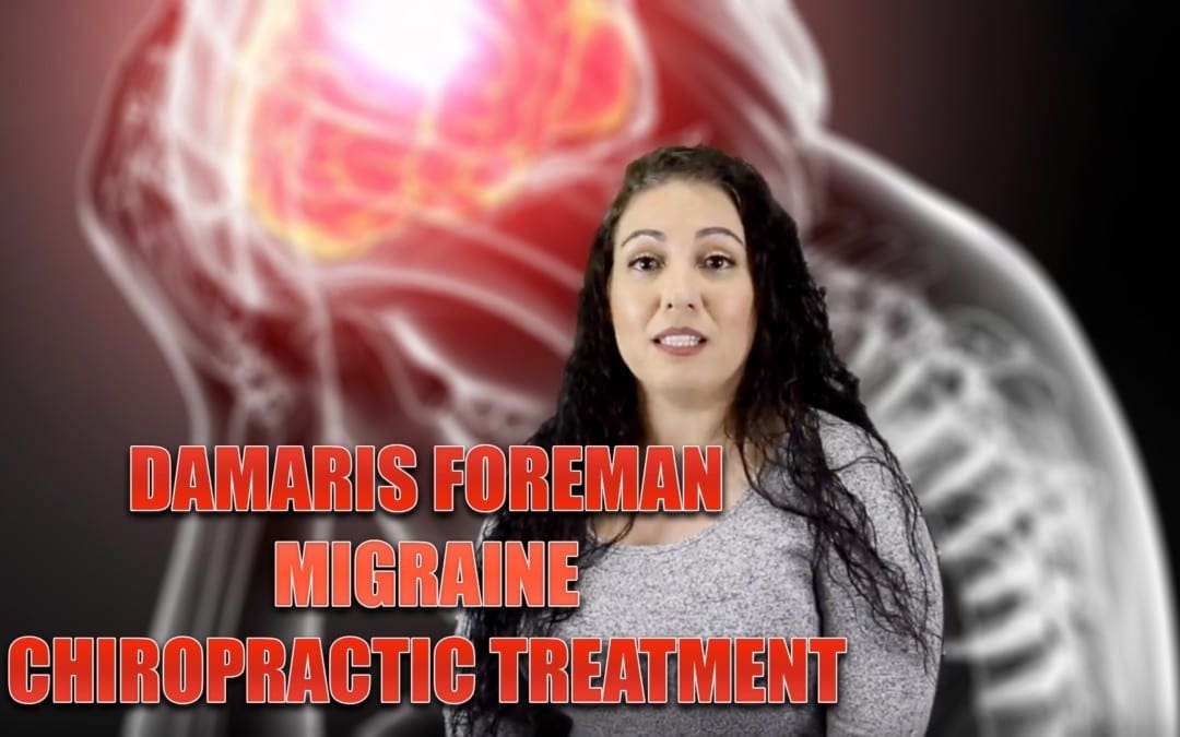
Damaris Foreman experienced migraine headaches for approximately 23 years. After visiting many healthcare professionals due to her migraine pain without seeing a great deal of progress, she was finally advised to find migraine pain treatment with Dr. Alex Jimenez, a chiropractor located in the city of El Paso, TX. Damaris significantly benefitted from chiropractic care and she experienced a massive sense of relief after her first spinal adjustment and manual manipulation. Damaris Foreman was able to confront a great deal of her questions and concerns and she was efficiently taught how to deal with her migraine pain. Damaris clarifies how Dr. Alex Jimenez’s migraine treatment is one of the best treatments she’s received and she highly recommends chiropractic care as the best non-surgical choice for enhancing and healing her migraine headaches.
Chiropractic Migraine Treatment & Relief
A migraine is commonly referred to as a primary headache disorder characterized by recurrent headaches as well as identified by moderate to severe in intensity. Ordinarily, the migraine headaches affect one half of the brain, are pulsating in personality, and might last from two to 72 hours. Associated symptoms may include nausea, vomiting, and sensitivity to light, noise, or odor. The pain could be aggravated by bodily activity. One third of people who suffer with migraines experience migraine with aura: normally a brief number of visual disturbance suggests that the headache will soon happen. It can also occur with minimal if any aggravation pain following it.
We are blessed to present to you�El Paso�s Premier Wellness & Injury Care Clinic.
As El Paso�s Chiropractic Rehabilitation Clinic & Integrated Medicine Center,�we passionately are focused treating patients after frustrating injuries and chronic pain syndromes. We focus on improving your ability through flexibility, mobility and agility programs tailored for all age groups and disabilities.
If you have enjoyed this video and/or we have helped you in any way please feel free to subscribe and recommend�us.
IFM's Find A Practitioner tool is the largest referral network in Functional Medicine, created to help patients locate Functional Medicine practitioners anywhere in the world. IFM Certified Practitioners are listed first in the search results, given their extensive education in Functional Medicine