Gloria experienced considerable foot pain which extended throughout her entire body. Her plantar fasciitis developed due to her everyday physical activities. Gloria describes how her foot pain and plantar fasciitis affected her quality of life. The custom foot orthotics provided to her by Dr. Alex Jimenez, a chiropractor in El Paso, TX, tremendously helped improve her foot pain and plantar fasciitis symptoms. Gloria describes how much comfort the custom foot orthotics provided for her painful symptoms. Gloria highly recommends Dr. Alex Jimenez as the non-surgical choice for foot pain and plantar fasciitis treatment, among other health issues.
Contents
Treat *PLANTAR FASCIITIS & FOOT PAIN* with Orthopedic Foot Orthotics | El Paso, TX (2019)
This condition affects approximately 2 million people in the United States a year.
There are doctors that believe bone spurs are the cause, and surgery is needed. However, bone spurs are not the cause of plantar fasciitis. Surgery will not eliminate the pain but may weaken or even rupture the plantar fascia
Symptoms
Pain on the bottom of the heel
Pain in the arch of the foot
Pain that is usually worse upon arising
Pain that increases over a period of months
Swelling on the bottom of the heel
Causes:
Improper footwear
Strenuous activity
Obesity
Over-pronation
High arches or flat feet
Poor shock absorption shoes
Plantar fasciitis is commonly seen in middle-aged patients.
We also see it often in those who place a great deal of stress on their feet like:
Runners
Athletes�
Soldiers
We are blessed to present to you�El Paso�s Premier Wellness & Injury Care Clinic.
We Are Ready To Help You Get Back To Your Normal Life!
How Chiropractic Helps Plantar Fasciitis
Chiropractic care is a very effective treatment for plantar fasciitis as well as the pain that is caused by the condition. Chiropractic for plantar fasciitis involves a very precise technique that involves adjustments to the feet and ankles as well as spinal alignment. This provides several benefits.
Reduces Stress in the Plantar Fascia�� When a ligament is inflamed or stressed the tissue can develop very small tears that cause the pain of plantar fasciitis. Chiropractic adjustments made to the heel and foot take the pressure off of the plantar fascia, allowing it to relax.
Promotes Healing�� When the stress on the plantar fascia is reduced through these chiropractic adjustments, the foot can begin to heal. The chiropractor may also recommend specific exercises that stretch the ligament and help it heal. They may also advise the patient of lifestyle changes as well as nutritional adjustments that can help with the pain and condition.
Provides Effective Pain Management � Chiropractic is a very effective way to manage pain throughout the body. Spinal adjustments allow better communication between the brain and nerves, allowing the central nervous system to function more effectively. Condition-specific adjustments speak to the root of the problem, not just the symptoms. This means a more effective form of pain management that is longer lasting.
Reduces the Risk of Further Injury�� When a person has a condition like plantar fasciitis, they will often adjust their gait in an effort to avoid the pain. This puts stress on other parts of the body and can lead to back pain, sore joints, strained muscles, and other problems. Chiropractic�s�whole-body approach�helps the person realign their body properly so that they stand and walk properly. This helps them avoid further injury and discomfort.
Foot Levelers Kiosk
The Kiosk helps guide patients in selecting the best custom-made orthotics for their condition and lifestyle. It’s�
Fast:�Push the Start button and the scanner begins.
Easy to use:�User-friendly easy touch screen.
Engaging:�Videos explain the importance of healthy feet and the benefits of custom-made orthotics.
Cloud-based:�Results can be securely accessed from anywhere.
Comprehensive:�Easily retrieve previous scans to compare them to new scans and see the difference.
The Foot Levelers Kiosk saves time so you can spend more time living your life.
What’s Afoot
Plantar fasciitis is caused when the ligament that connects your toes to your heel (the plantar fascia) becomes inflamed, swollen, and weak. This causes the bottom of your foot or heel to hurt when you walk or stand, especially when you first wake or after sitting for a long time. The pain tends to be sharp and stabbing. It is the most common�cause of heel pain and is more prevalent among middle-aged people. However, anyone can get it at any age, especially people who spend a lot of time on their feet.
Feet are important. When you consider what your feet go through, taking 8,000 steps over the course of a day, according to the�Illinois Podiatric Medical Association�(IPMA), it�s easy to see how 75 percent of all Americans will have some type of�foot pain�at some point in their lives.�Plantar fasciitis�is a common and very painful foot condition that can become chronic if not treated. It is also a condition that responds very well to chiropractic care.
Cerebral perfusion pressure, or CPP, is the net pressure gradient which carries oxygen to brain tissue. It is measured by the difference between the mean arterial pressure, or MAP, and the Intracranial Pressure, or ICP,� which is measured in millimeters of mercury (mm Hg). Regulating CPP is fundamental in the treatment of patients with intracranial pathology, including shock, hemodynamic distress, and traumatic brain injury. �
Although the average CPP is generally between 60 and 80 mm Hg, these values may change to the left or to the right depending on individual physiology. MAP and ICP has to be measured together because CPP is a calculated measure. Regulating CPP at hemodynamically unstable conditions with abnormal ICP or in cases of intracranial pathology will reduce the chance of ischemic brain injury. �
CPP = MAP – ICP
Contents
Cerebral Perfusion Pressure Physiology
CPP and ICP
At its own average range of 60 to 80 mm Hg, the CPP is determined by the ICP and the mean arterial pressure. Under regular standards, the ICP is between 5 and 10 mm Hg which has a reduced effect on the CPP than the MAP in clinical circumstances not associated with intracranial pathology. ICP is generally measured through intracranial pressure transduction.
Physiologically, the ICP is a function of intracranial compliance. Intracranial compliance is the relationship between the ICP and the volume of the intracranial cavity including cerebrospinal fluid, or CSF, brain tissue as well as arterial and venous blood volume. Because the skull is a fixed and rigid anatomic space, the ICP can increase if the intracranial volume increases while intracranial compliance decreases. As the ICP increases or intracranial compliance decreases, CPP also decreases. �
Several processes determine that ICP continues to stay within the average range for the longest extended period of time possible, especially throughout periods of affected intracranial volume and compliance. As volume adds to the intracranial space, CSF can shift into the spinal subarachnoid space, causing the ICP to continue significantly unchanged. As volume increases due to a growing space-occupying lesion, brain tissue edema or blood, this process ultimately becomes overwhelming, and ICP begins to increase substantially. �
Cerebral blood flow, or CBF, is also a fundamental factor in ICP homeostasis. Cerebral auto-regulation makes sure that steady blood flow is maintained in the brain over a wide range of physiologic alterations. When blood pressure decreases, auto-regulation causes cerebral vasodilation and an increase in CBF and cerebral blood volume, maintaining ICP and CPP. However, when blood pressure increases, auto-regulation causes cerebral vasoconstriction and a decrease in CBF with a decrease in cerebral blood volume, also regulating ICP and CPP. Too many changes outside of average CBF ranges can cause brain ischemia and injury. �
CPP and MAP
Because ICP in its average ranges is a considerably small number, the CPP generally depends on the mean arterial pressure. MAP is the normal blood pressure during one cardiac cycle which can be measured through invasive hemodynamic monitoring or calculated by the systolic blood pressure, plus two times the diastolic blood pressure, divided by three. The average range of MAP is 70 to 100 mm Hg. �
The average arterial pressure can be affected due to everyday activities, such as rest, stress, and exercise or physical activities. However, if the ICP continues to stay the same, the average arterial pressure can change across its significantly wide range without tremendously decreasing or increasing the CPP. As a matter of fact, CPP and CBF will continue to stay considerably unchanged across a wider range of MAP (50 � 150 mm Hg) than normal due to cerebral auto-regulation and vasoconstriction or vasodilation of cerebral vasculature. �
For patients with hypertension, the auto-regulation setpoint changes, decreasing the average arterial pressure associated with the patient�s normal arterial pressure, which causes vasodilation to increase CBF. Patients with lower than normal average arterial pressure at baseline will have auto-regulatory vasoconstriction as a reaction to an increase in their significant average MAP, to return CBF to baseline. When looking at CBF and CPP in the context of the patient�s average MAP, it is clinically significant based on the regulation of intracranial pathology and hemodynamic derangements. �
Cerebral Perfusion Pressure Complications
Diagnosing and treating cerebral perfusion pressure complications necessitates measuring both the ICP and the MAP. The MAP may be quantified through the utilization of invasive hemodynamic processes, most frequently cannulation of a peripheral artery such as the radial or femoral artery. The MAP may also be measured with a non-invasive blood pressure cuff by applying the formula mentioned above utilizing the systolic and diastolic blood pressures. � Intracranial pressure is generally measured through an intracranial pressure transduction device. The most common and most accurate method or technique is utilizing an intraventricular monitor. The intraventricular dimension of ICP is the normal standard. An intraventricular catheter is inserted into a hole drilled in the skull and into the lateral ventricle to gauge the pressure of the CSF. The benefit of an intraventricular catheter is that CSF could be eliminated, if needed, to decrease ICP. Considerable complications for the ICP include a possibility of bleeding, infection, and difficulty with proper placement. Options include sub-dural and intra-parenchymal monitors. �
The ICP can be measured non-invasively through several methods and techniques, including transcranial Doppler ultrasonography or TCD. TCD utilizes a temporal window to evaluate the speed of blood flow through the middle cerebral artery. Systolic and diastolic average flow velocity is utilized to determine a pulsatility index. The pulsatility index was determined to be closely associated with ICP in several research studies as well as be associated with ICP in other research studies. Therefore, it is not suggested to use TCD as a substitute for direct ICP dimension. Invasive diagnosis and treatment of the MAP through an arterial cannula and the ICP through an intraventricular catheter will give a continuous and accurate calculation of CPP. �
Cerebral Perfusion Pressure Clinical Significance
Two general types of pathologic health issues can ultimately occur where the regulation of the CPP is fundamental, such as intracranial pathology, where ICP regulation is essential and hemodynamic instability/shock where MAP regulation is the most essential. Intracranial pathology involves space-occupying lesions, such as tumors, epidural and subdural hematoma or severe intraparenchymal hemorrhage and cerebral edema as seen after ischemic injury, traumatic brain injury or acute hepatic encephalopathy. In these circumstances, average CPP depends on decreasing the ICP into a normal range as soon as possible while regulating the MAP. When CPP is normal, it’s fundamental to keep in mind that every individual’s brain tissue has a CPP that is “normal” in the context of that individual patient’s physiology, which may be affected by other health issues, such as hypertension or cardiovascular disease. Moving towards a more dynamic direction of the average CPP utilizing the patient’s personal auto-regulatory capacity. These diagnosis and treatment approaches involve more frequent and sophisticated monitoring and might not be readily available for widespread utilization. �
In the instance of considerable traumatic brain injury, significant cerebral edema can decrease intracranial compliance and CSF, developing an increased ICP or intracranial hypertension. Auto-regulatory mechanisms and techniques may or may not function normally and when ICP continues to be elevated, CPP will decrease causing further injury through an ischemic process. In circumstances such as these, together with starting the measures for decreasing the ICP, it is essential to prevent hypotension (MAP – ICP = CPP) and in some instances, allowing hypertension to reasonably occur. �
In circumstances of instability, the ICP is considerably stable as cerebral auto-regulation is undamaged. In the instance of hypotension, the MAP decreases due to blood loss, or hemorrhagic shock, intravascular leak, or distributive shock, and decreased cardiac output, or cardiogenic shock, and the CPP also decreases. It’s the association between MAP and CPP which carries resuscitation guidelines to recommend regulating a MAP greater than or equal to 65 mm Hg. With a normal ICP, this threshold must make sure that a CPP of 55 to 60, the minimum necessary to stop cerebral ischemic injury, is ultimately maintained. As in the circumstance of ICP and cerebral auto-regulation, the goal of MAP is to be within the context of an individual patient’s evaluation hemodynamic function. Patients with untreated hypertension must have increased MAP goals to maintain proper CBF and CPP. �
As previously mentioned in the following article, cerebral perfusion pressure, or CPP, is the net pressure gradient which affects cerebral blood flow to the brain, also known as brain perfusion. According to healthcare professionals, the CPP, or cerebral perfusion pressure, must be constantly regulated within a specific limit because too little pressure or too much pressure could potentially cause a variety of brain health issues. Cerebral perfusion pressure may be associated with a variety of neurological diseases. – Dr. Alex Jimenez D.C., C.C.S.T. Insight
The purpose of the article is to discuss cerebral perfusion pressure and its association with neurodegenerative diseases. Neurological diseases are associated with the brain, the spine, and the nerves. The scope of our information is limited to chiropractic, musculoskeletal and nervous health issues as well as functional medicine articles, topics, and discussions. To further discuss the subject matter above, please feel free to ask Dr. Alex Jimenez or contact us at 915-850-0900 . �
Curated by Dr. Alex Jimenez �
Additional Topic Discussion: Chronic Pain
Sudden pain is a natural response of the nervous system which helps to demonstrate possible injury. By way of instance, pain signals travel from an injured region through the nerves and spinal cord to the brain. Pain is generally less severe as the injury heals, however, chronic pain is different than the average type of pain. With chronic pain, the human body will continue sending pain signals to the brain, regardless if the injury has healed. Chronic pain can last for several weeks to even several years. Chronic pain can tremendously affect a patient’s mobility and it can reduce flexibility, strength, and endurance.
Formulas for Methylation Support
XYMOGEN�s Exclusive Professional Formulas are available through select licensed health care professionals. The internet sale and discounting of XYMOGEN formulas are strictly prohibited.
Proudly,�Dr. Alexander Jimenez makes XYMOGEN formulas available only to patients under our care.
Please call our office in order for us to assign a doctor consultation for immediate access.
If you are a patient of Injury Medical & Chiropractic�Clinic, you may inquire about XYMOGEN by calling 915-850-0900.
�
For your convenience and review of the XYMOGEN products please review the following link.*XYMOGEN-Catalog-Download �
* All of the above XYMOGEN policies remain strictly in force. �
�Achilles injury, which turns out to be a ruptured Achilles tendon.
This is a devasting injury for anyone, especially world-class athletes. Take NBA player Kevin Durant and his injury that could keep him out for some time. To be fair Durant was dealing with a calf injury on the same leg, for some weeks before this injury.
But it was his first game back in action and it led to a crushing injury!
Calf & Achilles Tendon Relationship
The calf and Achilles tendon are so interrelated that when one has an issue so does the other.
The fascia of these muscles segue ways to form the Achilles tendon.
If the calf is tight, then the Achilles is going to be tight.
The relationship between calf injury and an Achilles injury.
Cycle of Injury
Going through any area of the cycles can lead to a devastating injury.
Symptoms
Pain and stiffness around the lower leg, right above the back of the heel
Begins as a mild ache and worsens throughout the day
Jumping,� running, stair climbing, and sprinting can spark intense pain
Tenderness or stiffness especially in the morning, but improves with movement/activity
The key is to intervene to stop the cycle.
A few ways:
Rest
Massage
Physical Therapy
Chiropractic
Custom orthotics
Active Release Therapy (ART)
ART is extremely�effective�for breaking up scar tissue and improving issues with soft tissues. Treatment can do a lot to improve the health of your tendon and the surrounding tissues to avoid further complications. It will also do a lot to help relieve the pain you are experiencing.
Chiropractic Adjustments/Manual Manipulations
Many times, the issues with your Achilles tendon are the result of misalignment in other parts of your body. When your joints are misaligned it tends to put extra stress on your feet and Achilles tendons. To minimize the stress on your joints, your chiropractor will adjust your spine and other joints to ensure proper alignment.
Get Help for Your Achilles Injury & Call Us Today!
If you are experiencing heel pain, please get in touch with our chiropractic team. We can help to alleviate your pain and help you avoid experiencing further problems with your Achilles tendon.
Difference Foot Orthotics Make to *REDUCE FOOT PAIN* & Correct Posture | El Paso, TX (2019)
Custom made foot orthotics can help control foot motion and posture. Healthcare professionals prescribe custom foot orthotics to help patients focus on their foot posture and mobility control. Research studies have ascertained that using custom foot orthotics for posture and mobility control can help fix excessive foot pronation and supination to prevent a variety of foot health problems. The subsequent video describes how custom foot orthotics will help control foot posture and mobility to improve health and wellness.
What’s Afoot
If you have low back pain�or have had it, you are not alone. Experts estimate that around�80% of people�will experience some type of back problem at some point in their lives.�The Global Burden of Disease 2010�lists low back pain as the number one cause of disability worldwide. The good news is the majority of back pain is mechanical in origin or is not organic. This means that infection, cancer, fracture, inflammatory arthritis, and other serious conditions are not the cause. In fact, you may benefit by looking to your feet, knees,�and hips as the culprits.
NCBI Resources:
The large, thick tendon that travels up from the base of the heel and into the calf muscle is the Achilles tendon. It connects the calf muscle and heel. It allows you to walk, run and jump. The Achilles tendon is strong and durable, but it is possible to overwork it and cause enough overuse injury.
The phrase, �You are what you eat� implies that the way we are defines us as the food we all consumed. However, our gut tells us otherwise as the food we eat, may in fact be leading us to obesity. Our gut plays a role in our overall health, when we eat good food our gut is happy and when we eat bad food our gut will tell us by fighting off the bad food. A recent study showed us that the bacteria in our gut produce amyloid and lipopolysaccharides. These two microbiomes seem to show us that together, with proper dieting that these microbiomes can prevent Alzheimer�s Disease.
As the microbiomes and the bacteria that co-exist in our gut, there are the two most predominant groups that have also played a key role in our lifestyle: gram-positive Firmicutes and gram-negative Bacteroidetes- both play a huge role in obesity. Firmicutes are bad bacteria that lead us to obesity. When we eat processed food and sugars, our body starts to crave it more, thus leading us to be overweight.
Junk Food
When we eat junk food, all that sugar and fat are feeding the Firmicutes. Firmicutes thrive on sugar since our bodies need it and it can be both good or bad While Bacteroidetes are the good bacteria that leads us to a healthy gut. Bacteroidetes are in the stomach regions as well as the Firmicutes. These two predominant bacteria groups tell us that the food we eat can actually affect our bodies when we eat bad foods or good foods.
However, Dr. Kristen Senella mentioned that we all have a different balance of Firmicutes and Bacteroidetes since we are all different shapes and sizes. Depending on our health and food lifestyle, we can have either a low Firmicutes and a high Bacteroidetes or a high Firmicutes and a low Bacteroidetes. Plus, having either a high or low count of Firmicutes can lead to weight gain or weight loss; depending on which healthy lifestyle and exercise regime you are following.
Gram-Positive
Gram-positive bacteria will appear blue or violet, while gram-negative bacteria will appear red or pink under the microscope. When studying the gut and the bacteria groups that it is hosting, scientist use mice to study how their guts react to different diets they are put through so that way we, as humans, can take either pills to help our bodies maintain a healthy lifestyle or to read and do our own research. One group is fed in a healthy lifestyle and doesn�t experience diseases or ailments that we face. And the other group is fed with a bad lifestyle where they are prone to many of the diseases and fatigue as their life span is shortened very quickly. In order for us to actually maintain a healthy lifestyle and importantly feel good is to make sure our Firmicutes are not too dangerously low, but we can control it with probiotics.
Probiotics
Probiotics can vary from yogurt, fermented vegetables, kombucha, and miso. But there are certain companies that also reign supreme in the probiotic market. Activia yogurt and Yakult are two of the most well-known companies that use the live microorganisms to help us maintain a healthy lifestyle as well as keeping our gut�s microbiome in check. When we have some sort of probiotic foods in our system, we are preventing certain ailments and diseases going out of control. Like our cholesterol, blood pressure, being lactose intolerant, or recurring abdominal pains.
When we mix probiotics into our food when we are trying to maintain a healthy lifestyle, we can see a vast improvement in how we have more energy, we feel full that we don�t have to overeat or mindless snacking, and overall we feel good in our gut as we go through our daily routine. From 2007 to now, roughly 3.9 million Americans use probiotics to maintain a healthy gut, however, those probiotics are just a fraction of what the six types of foods that can help maintain a healthy gut microbiome to help support a healthy lifestyle.
Healthy Lifestyle
For instance, a good healthy lifestyle is eating your basic food groups; whether it be plant-based or omnivorous, as well as, exercising a couple of times out of the year. A bad healthy lifestyle is eating processed food and not exercising, which leads to obesity and cardiac arrest. Depending on the person and the efforts that they are willing to maintain a healthy lifestyle, they can achieve longevity by taking care of their gut first and foremost.
Family In Kitchen Making Morning Breakfast Together
Protein
Let�s start with protein. Protein can vary with lean meats like chicken and beef or plant-based like beans, legumes or tofu. Any of these types of protein can help our bodies by making us make our muscles grow, but also control the bacteria in our guts. Next up is fats. Fats can vary like good and bad bacteria. There are good fats like fish, nuts, olive oil, and avocado; as well as, bad fats like butter, lard, and fatty foods. Granted that we can overindulge on the trans fats as there are many fast-food chains, but we can moderate ourselves to not eat out at fast food joints all the time.
Yes, they are cheap and easy to access, however now and days, we as humans are now cooking more in our homes and meal prepping our meals to be healthier. Digestible and Non-Digestible Carbohydrates are mostly starch, sugars, and fibers. These two food groups can make our gut feel happy or upset depending on the food we consume. Sugars, starches, and fibers help our bodies by feeling full with the starches, the fibers help our bowel movements in case our gut feels bloated, and the sugars gives us microburst of energy for our fast-paced lives.
Fermented & Polyphenols
The last two food groups are fermented food and Polyphenols. Both of these food groups have amazing properties since we see them everywhere in the food market, hiding in plain sight. Fermented foods like yogurt and kimchi are a few examples of ways of keeping our guts happy and stopping many diseases. Polyphenols are antioxidant foods like dark chocolate, berries, dark greens, and certain fruits. These help our gut curb that sugar hunger and all in all taste really good.
All in all, our gut microbiomes are important to us and our overall health as we all try to maintain and achieve a healthy lifestyle. The phrase �we are what we eat� still implies to all of us, however, it is up to us to actually put in the work and constantly try out different foods to make sure that our gut is still functioning properly. No matter which diet you choose, pick one that will work with your body and your gut since we all are made differently. But our gut should be the first thing that we should listen to.
You have probably heard about the “gray matter” of the brain which is made up of cells known as neurons, however, a lesser-known type of brain cell is ultimately what makes up the “white matter” of the brain.� These are known as glial cells. �
Glial cells, also known as glia or neuroglia, were only considered to simply offer structural support. The term “glia” literally translates to “neural adhesive.” However, relatively recent research studies have demonstrated that they play a variety of roles in the brain and the nerves which run throughout the entire human body. However, there is more left to find out. �
Contents
Types of Glial Cells
Glial cells commonly offer support to the neurons. Without them, several of the most fundamental roles would never be achieved although they may not perform these roles themselves. Glial cells come in numerous forms, each of which performs certain functions to keep the brain functioning properly or not, in case of a neurological disease which affects the glial cells. �
The central nervous system, or CNS, is made up of the brain, the spinal cord, and the nerves. Five types of glial cells include: �
Astrocytes
Oligodendrocytes
Microglia
Ependymal cells
Radial glia
Moreover, there are also glial cells on the peripheral nervous system, or PNS, which is made up of the nerves in the upper and lower extremities, away from the spine. The two types of glial cells found in the peripheral nervous system include: �
Schwann cells
Satellite cells
� �
Astrocytes
The most common type of glial cell in the central nervous system is the astrocyte, also known as astroglia. The “astro” part of the name refers to how they look like stars with projections coming out all over the glial cell. Protoplasmic astrocytes have thick projections with lots of branches. Fibrous astrocytes have long, slender arms. The fibrous ones are found in the white matter while others are found among neurons in the gray matter.� Astrocytes play several major roles, including: �
Developing the blood-brain barrier or BBB. The BBB is similar to a strict security system which only allows substances which are supposed to be in the brain. This filtering system is essential for maintaining brain health.
Regulating the substances around neurons. Neurons communicate utilizing chemical messengers known as neurotransmitters. Once a chemical has transmitted a message to a cell, it essentially stays there cluttering things up until an astrocyte recycles it through a process known as reuptake. The reuptake process is generally the main target of numerous medications, including anti-depressants. Astrocytes also clean up what’s left behind when a neuron dies, as well as excess potassium ions, which are chemicals that play a fundamental role in nerve function.
Regulating blood flow to the brain. For the brain to process information accordingly, it needs a certain amount of blood to flow throughout all of its different regions. An active region receives more blood flow than an inactive one.
Synchronizing the activity of axons. Axons are characterized as long, thread-like elements of the neurons and the nerve cells which ultimately conduct electricity to help transmit messages from one cell to another.
Astrocyte dysfunction has been potentially connected to a wide variety of neurological diseases, including: �
Amyotrophic lateral sclerosis (ALS or Lou Gehrig’s disease)
Huntington’s chorea
Parkinson’s disease
Animal models of astrocyte-related disorders are helping researchers learn more about these neurological diseases. �
Oligodendrocytes
Oligodendrocytes develop from stem cells. The term is made up of Greek words which, all together, mean “cells with several branches.” Their main role is to help information move faster. Oligodendrocytes appear like white spikey balls. Their purpose is to make a protective layer, similar to the plastic insulation on electric wires. This layer is known as the myelin sheath. �
The myelin sheath is not constant. There is a gap between each membrane which is known as the”node of Ranvier,” and it is the node which helps electrical signals move effectively along neural cells. The signal is transmitted from one node to the next, which increases the velocity of the nerve conduction whilst also reducing how much energy it takes to transmit it. �
Messages along myelinated nerves may travel as fast as 200 miles per second. At birth, you only have a few myelinated axons, and the quantity of these keeps growing until you’re about 25 to 30 years old. Myelination is thought to play an important role in intelligence. Oligodendrocytes also supply stability and transmit energy from blood cells into the axons. �
The expression “myelin sheath” may be familiar to you because of its association with multiple sclerosis. In multiple sclerosis, it is believed that the human body’s immune system attacks the myelin sheaths, which leads to the breakdown of these neurons and ultimately causes impaired brain functioning. Spinal cord injuries may also cause damage to these structures. � Other neurological diseases believed to be associated with oligodendrocyte dysfunction include: �
Leukodystrophies
Tumors known as oligodendrogliomas
Schizophrenia
Bipolar disorder
Several research studies suggest that oligodendrocytes may become affected by the neurotransmitter glutamate, which, among other functions, stimulates regions of the brain so that you’re able to focus and learn new information. Nonetheless, in high levels, glutamate can be considered an “excitotoxin,” which means that it may overstimulate cells until they die. �
Microglia
Microglia are tiny glial cells. They act as the brain’s dedicated immune system, which is necessary since the BBB isolates the brain from the rest of the human body. Microglia are attentive to indications of disease and injury. If they find a problem, they are in charge of taking care of it, even if it ultimately means clearing away dead cells or getting rid of a toxin or pathogen. �
If they respond to an injury, microglia cause inflammation as part of the recovery process. In some cases, such as in Alzheimer’s disease, they might become hyper-activated and cause too much inflammation. That is thought to cause amyloid plaques and other health issues connected with the neurological disease, among a variety of other brain health issues. � Along with Alzheimer’s disease, other neurological diseases which may be associated with microglial malfunction include: �
Fibromyalgia
Chronic neuropathic pain
Autism spectrum disorders
Schizophrenia
Microglia have been thought to play many fundamental roles beyond that, including learning-associated plasticity and guiding the development of the brain. The brain produces many connections between neurons which allow them to pass information back and forth. The brain produces a lot more of these than we need, which is not always efficient. �
Microglia detect unnecessary synapses and they clean them out. Microglial research has really taken off in recent decades, leading to an ever-increasing comprehension of their roles in both health and disease in the central nervous system. �
Ependymal Cells
Ependymal cells are primarily known for creating a membrane known as the ependyma, and it can be described as a thin membrane lining the central canal of the spinal cord and the ventricles or passageways of the brain. They also create cerebrospinal fluid. Ependymal cells are extremely small and they lineup closely together to make the membrane. �
Inside the ventricles, are the cilia, which look like small hairs which move back and forth to help circulate the cerebrospinal fluid. Cerebrospinal fluid provides nutrients and removes waste products in the brain. Additionally, it serves as a cushion and shock absorber between the skull and the brain. It’s also essential for homeostasis in the brain, regulating its temperature along with other attributes which keep its potential and functioning. Ependymal cells are also included in the BBB. �
Radial Glia
Radial glia are believed to be a type of stem cell, which means that they create other types of cells. In the developing brain, they’re the”parents” of neurons, astrocytes, and oligodendrocytes. They also supply scaffolding for developing neurons, thanks to long fibers which direct young brain cells into position as the brain forms in a human embryo. Their role as stem cells, especially as founders of neurons, is ultimately what makes them the focus of research studies regarding how to repair brain damage from injury or illness. Later in life, the radial glia perform important roles in neuroplasticity as well. �
Schwann Cells
Schwann cells are known after the physiologist Theodor Schwann, who discovered them. They function a lot like oligodendrocytes in which they supply myelin sheaths for axons, but they develop in the peripheral nervous system, or PNS, rather than in the central nervous system or CNS. However, Schwann cells form spirals directly across the axon. �
Ranvier’s nodes are found between the membranes of oligodendrocytes and these help in neural transmission in precisely the same exact way. Schwann cells can also be part of the PNS’s immune system. They ultimately have the ability to consume the axons of the nerve and give a protected path for a brand new axon to develop when another nerve cell is damaged. Neurological diseases involving abnormal Schwann cells include: �
Guillain-Barre’ syndrome
Charcot-Marie-Tooth disorder
Schwannomatosis
Chronic inflammatory demyelinating polyneuropathy
Leprosy
Several research studies on bronchial Schwann cells for spinal cord injury and other types of peripheral nerve damage have been promising. Schwann cells are implicated in certain types of chronic pain. Their activation following nerve damage may contribute to dysfunction in a type of nerve fiber known as nociceptors, which feel external factors like heat and cold. �
Satellite Cells
Satellite cells get their name due to the way they surround certain neurons, with several satellites forming a sheath around the cellular surface. Researchers have only just started to learn about these cells but they’re believed to be similar to astrocytes. The main role of satellite cells is believed to be the regulation of the surroundings around the nerves. �
The nerves which have satellite cells make up something known as ganglia, which are clusters of nerve cells in the autonomic nervous system and sensory apparatus. The autonomic nervous system regulates internal organs, even while the sensory system is what enables people to see, hear, taste, touch, and smell. Satellite cells provide nourishment to the neuron and absorb heavy metal toxins, such as lead and mercury, to stop them from damaging the nerves and other structures. �
They are also believed to assist transport several neurotransmitters and other substances, including: �
Glutamate
GABA
Norepinephrine
Adenosine triphosphate
Substance P
Capsaicin
Acetylcholine
Much like microglia, satellite cells detect and respond to injury and inflammation. However, their role in repairing cell damage isn’t yet fully well understood. Satellite cells have been connected to chronic pain between peripheral tissue injury, nerve damage, and a systemic heightening of pain, or hyperalgesia, which can ultimately result from chemotherapy. �
Glial cells, also known as glia or neuroglia, are characterized as non-neuronal cells which are ultimately found in the central nervous system, or CNS, and the peripheral nervous system, or PNS. There are various types of glial cells, including astrocytes, oligodendrocytes, microglia, ependymal cells, and radial glia in the CNS and Schwann cells and satellite cells in the PNS. The glial cells play many fundamental roles in the human nervous system. – Dr. Alex Jimenez D.C., C.C.S.T. Insight
The purpose of the article is to discuss the types of glial cells associated with the brain and neurodegenerative diseases. Neurological diseases are associated with the brain, the spine, and the nerves. The scope of our information is limited to chiropractic, musculoskeletal and nervous health issues as well as functional medicine articles, topics, and discussions. To further discuss the subject matter above, please feel free to ask Dr. Alex Jimenez or contact us at 915-850-0900 . �
Curated by Dr. Alex Jimenez �
Additional Topic Discussion: Chronic Pain
Sudden pain is a natural response of the nervous system which helps to demonstrate possible injury. By way of instance, pain signals travel from an injured region through the nerves and spinal cord to the brain. Pain is generally less severe as the injury heals, however, chronic pain is different than the average type of pain. With chronic pain, the human body will continue sending pain signals to the brain, regardless if the injury has healed. Chronic pain can last for several weeks to even several years. Chronic pain can tremendously affect a patient’s mobility and it can reduce flexibility, strength, and endurance.
Formulas for Methylation Support
XYMOGEN�s Exclusive Professional Formulas are available through select licensed health care professionals. The internet sale and discounting of XYMOGEN formulas are strictly prohibited.
Proudly,�Dr. Alexander Jimenez makes XYMOGEN formulas available only to patients under our care.
Please call our office in order for us to assign a doctor consultation for immediate access.
If you are a patient of Injury Medical & Chiropractic�Clinic, you may inquire about XYMOGEN by calling 915-850-0900.
�
� For your convenience and review of the XYMOGEN products please review the following link.*XYMOGEN-Catalog-Download �
* All of the above XYMOGEN policies remain strictly in force. �
Athletes both recreational and fully competitive can be impacted by injuries to the muscles and ligaments around the hip.
These injuries interfere with performance levels and sometimes end participation completely.
�Excessive pronation along with shoes with poor shock absorption has been found to be an underlying cause for various leg/hip injuries.
Custom made Orthotics improve the biomechanics of the feet and reduce the extent of pronation helping to prevent sport-related leg/foot injuries.
Over Foot Pronation
Research has determined that athletes with more foot pronation have a higher probability of sustaining a leg injury, including iliotibial band syndrome that comes from excessive tightness of the hip muscles.
People involved in sports or recreational activities lower their likelihood of developing traumatic and overuse hip injuries through chiropractic treatment and using custom foot orthotics.
The amount of pronation during standing and while running at a standard speed is determined by measuring the angles of the footprints.
Athletes with more pronation have a higher likelihood of an overuse injury.
Standing (static) and running (dynamic) prints show the amount of pronation and is a predictor of developing an overuse injury.
Athletic performance and injury prevention involve regularly checking the alignment of patients� feet in the standing position.
Hip Injuries & The Hamstring
Many hip injuries develop from poor biomechanics and improper movement, especially when running.
Smooth muscle coordination provides balance and support for the pelvis and is needed for optimum sports performance.
This includes:
Hamstring muscles
Hip abductor muscles
Tensor fascia lata or the iliotibial band
When there is an issue with the feet and ankles, abnormal motion like over-rotating the entire leg is the perfect set-up for pulls, sprains, and strains.
50% of standing consists of heel strike and maximum pronation.
The hamstring muscles function to control the knee and ankle when the heel strikes and absorb the impact.
The theory behind orthotic support is that orthotics help the hamstrings control the position of the calcaneus and knee, so there is less stress on the hip and pelvis.
Hip Injuries & Over-Pronation
Orthotics can correct excessive pronation and treatment of hip problems. These are some of the problems/pathologies that can develop.
These conditions develop in athletes who push their body’s to the limit going for optimal performances.
Conclusion
Overpronation and poor shock absorption contribute to leg injuries � from:
Foot
Lower leg
Knee
Thigh
Hip
These conditions can be prevented with custom-made orthotics.
Foot biomechanics evaluation is a must
To avoid hip injuries, athletes need regular evaluations of foot alignment and function
Wear well-designed and solid-constructed shoes
Chiropractors can prevent arch breakdown and foot problems with custom orthotics, and also treat numerous injuries to the lower extremities, especially the hips.
Excessive Foot Pronation can Affect *FOOT POSTURE & MOBILITY* | El Paso, TX (2019)
The following video discusses how excessive foot pronation can ultimately affect foot posture and mobility. Several factors can affect foot posture and mobility, such as excessive foot pronation. Excessive foot pronation is most prevalent among the general population, therefore, it is considered to be one of the most common factors for abnormal foot posture and mobility, which can lead to a variety of health issues like overuse injuries. Excessive foot pronation and even supination can ultimately affect overall health and wellness.
Hip Labrum tears in athletes can occur from a single event or recurring trauma. Running may cause labrum tears due to the labrum being utilized more for weight-bearing and taking excess forces while at the end-range motion of the leg. Sporting activities are probable causes, specifically those that require frequent hip rotation or pivoting to a loaded femur as in ballet or hockey. Constant hip rotation places increased strain on the capsular tissue and harm to the iliofemoral ligament. This subsequently causes hip instability putting increased stress on the labrum and causing a hip labrum tear.
What’s Afoot
Chiropractic�seeks to find the cause of the conditions it is used to treat, including pain, instead of just treating symptoms. Because of this, the chiropractor will work to find the cause of the pain, in this case, overpronation and overpronation, and correct it � or the effects of the condition � in addition to treating the back pain.
Overpronation and oversupination can cause a variety of injuries and conditions that affect not only the feet and ankles, but also the knees,�hips, and back as well. Some of the more common injuries and conditions include:
Flat feet or posterior tibial tendon dysfunction
Ankle Sprains
Achilles tendinitis
Arch pain
Plantar fasciitis
Corns
Shin splints
Heel pain
Tight calves
Calluses
Knee pain
Patellar tendonitis
Tight hip flexors
Back pain
Sciatica
Herniated disks
NCBI Resources
Muscle imbalances in the hip, such as tight hip flexors, can cause low back pain � or at least contribute to it. When the hip flexor muscles are too tight, it causes what is known as an anterior pelvic tilt.�Hip flexors�can become too tight if the person sits for extended periods of time or engages in activities like cycling and jogging. A chiropractor can guide you through exercises that will help release the tight muscles and stop the micro spams that occur as a result.
Neural cell death can occur both during the development and throughout the pathophysiology of the nervous system. Two different types of cell death, known as necrosis and apoptosis, are involved in pathological neuronal loss, however, apoptosis is the process of programmed cell death during development. All types of cells will go through apoptosis. This mechanism controls neuronal growth where an excess of neurons is produced and only those which form connections with the target structures will receive enough survival factors. The remaining neurons will then ultimately go through death and removal. �
Apoptosis continues throughout life and it is the main process involved in the elimination of surplus, unwanted, damaged or aged cells. Dysregulation of apoptosis is demonstrated after damage or injury as well as in neurodegeneration and in tumorigenesis. Treatment approaches which influence the apoptotic pathway offer valuable therapeutic options in a wide variety of pathological states. The purpose of the article is to describe the significance of apoptosis in neurological diseases. �
Contents
What is Apoptosis?
Apoptosis is the well-conserved and highly controlled process of cell death involved in the removal of unnecessary, surplus, aged or damaged cells. Dysregulation of apoptosis can ultimately develop mutated cells which can result in malformations, autoimmune diseases, and even cancer. Abnormal apoptosis can also result in the elimination of healthy cells which can occur in health issues such as infection, hypoxic-ischaemic injury, neurodegenerative or neuromuscular diseases, and AIDS. �
Apoptosis is different from necrotic cell death. In necrosis, cell death is caused by an external factor and involves the early loss of tissue, damage to organs, and the leakage of cytoplasmic contents, leading to the recruitment of phagocytes which can cause an acute inflammatory reaction. In contrast, apoptosis is often considered cell suicide. According to research studies, cells which die due to apoptosis retain membrane and organelle structure and function until late in the process while still developing plasma membrane blebbing, reduced cytoplasmic volume, chromatin condensation, and nuclear fragmentation. �
In the final phases, cell fragments wrapped in plasma membrane pull away as apoptotic bodies which are then phagocytosed by healthy cells. The removal of cell debris also occurs in the absence of an inflammatory response, and this silent, quick, and efficient elimination of apoptotic cells mean that apoptosis can be difficult to find in cells. However, as many as 50 percent of the cells in developing adulthood may go through apoptosis where less than 1 percent of cells are apoptotic at any one time. �
Apoptosis in the Nervous System
Programmed cell death by apoptosis occurs in several developmental processes, such as body sculpting and removal of self-reacting resistant cells as well as sexual organ growth and gamete formation. The general principle of growth in multicellular organisms involves the development of excess numbers of cells, where the excess or unwanted cells are then removed by apoptosis through the development of functional organs. In the developing nervous system, apoptosis has been demonstrated to occur in neural tube formation and continues throughout terminal differentiation of the neural system. �
A growing number of neurotrophic factors, such as nerve growth factor family, including both the neurokines and development factors like insulin-like growth variables (IGF-I and IGF-II), encourage the survival of several types of neurons. Targeted disruption of genes encoding these factors or their receptors demonstrate that neurotrophic factors are significant for the development of specific neuronal populations. Neurotrophic factors function by binding to specific receptors in the cell membrane. Moreover, the effects of NGF offer a good illustration of the subtle command the system permits. �
The nerve growth factor receptor has high and low affinity components. It will function as a survival factor if it binds to the high-affinity trkA receptor but it will also cause apoptosis of retinal neurons or oligodendrocytes once it binds to the low-affinity receptor p75 in the absence of trkA. Nerve growth factor in the extracellular environment is consequently able to control neural development by both boosting the growth of several types of cells as well as the removal of other cells. �
In some cases, however, concentrated genetic disruption of neurotrophic factors or their receptors may leave the central nervous system seemingly unaffected, demonstrating that these variables can ultimately become biased. According to research studies, it has now become evident that the control of neuronal survival does not only depend on the supply of trophic molecules by the targets but also on activity, humoral factors, and trophic support from glia or glial cells. �
Furthermore, neurons don’t simply undergo programmed cell death during differentiation. Apoptosis appears to regulate cell numbers in systems as diverse as the disappearance of the germinal layer during the third trimester of pregnancy, the sexual differentiation of the medial preoptic nucleus where apoptosis is controlled by testosterone, lineages throughout the olfactory epithelium, oligodendrocyte development in the optic nerve, and the development of Schwann cells in the peripheral nervous system. Programmed cell death occurs in a variety of other processes in the developing nervous system. �
Apoptosis in Nervous System Injuries & Diseases
Although apoptosis is a fundamental process involved in the developing nervous system, apoptosis can ultimately be involved in a variety of nervous system injuries and diseases. In most cases, the connection between a specific mutation or trauma as well as the activation of apoptotic cascades remains evasive. An overview of a developing list of neurological diseases in which apoptosis has been implicated as a significant pathological mechanism is provided below. �
Neuronal Injury
Cerebral hypoxic-ischaemic injury is a cause of neurological injury and death. Magnetic resonance spectroscopy studies have demonstrated that transient hypoxia-ischemia contributes to a biphasic disturbance of cerebral energy metabolism. Related to the biphasic energy collapse, two waves of cell death appear to follow hypoxic-ischaemic injury in the developing brain. Immediate neuronal death is most likely due to necrosis resulting from the accumulation of calcium ions. �
Delayed cell death caused by hypoxic-ischemic injury appears to involve further mechanisms with increasing data which demonstrates that in the delayed phase, cell death occurs by apoptosis. The amount of apoptosis is directly associated with the magnitude of ATP depletion during hypoxia-ischemia. Apoptosis can occur in the brains of newborn babies following birth asphyxia and sudden intrauterine death. Apoptosis can also be notable in white matter injury in newborn babies. �
Apoptosis may continue for months after an hypoxic-ischaemic injury due to constant changes in cerebral energy metabolism in infants during the months after birth asphyxia. Following focal neural injury, apoptosis has been discovered in remote regions from the initial damage. After severe spinal cord injury in reptiles, apoptosis of oligodendrocytes occurs in distant degenerating fiber tracts and after forebrain injury in rats, apoptosis was demonstrated in the cerebellum. �
The apoptotic loss of oligodendrocytes could consequently be a potential source of secondary demyelination in paraplegia and in the chronic degeneration related to multiple sclerosis. Further research studies must be performed in order to provide further evidence on the role of apoptosis in this type of injury which begins from the report of which Bcl-2 expression boosts the growth and regeneration of retinal axons. Apoptosis in neuronal injury can be demonstrated in a variety of ways. �
Neural Cancers
A connection between apoptosis and the cell cycle is demonstrated in carcinogenesis where proto-oncogenes, such as c-fos, c-jun, and c-myc, can activate apoptosis and promote cell division while inactivation of the pro-apoptotic p53 tumor suppressor gene is a frequent mark of human neoplasia. By way of instance, in a number of gliomas, the reduction of wild p53 activity was connected to tumor progression, possibly leading to resistance to chemotherapy and radiotherapy. �
Although there have been reports of Bcl-2 overexpression in glioma cell lines, the correlation between the anti-apoptotic effect of this gene and malignancy is not yet clear. However, a homolog of Bcl-2, the brain associated apoptosis gene (BRAG-1), is found predominantly in the brain, and it is upregulated in human gliomas as a rearranged transcript. As demonstrated above, the process of apoptosis can also be significant in the development of neural cancers, according to research studies. �
Infectious Disease
Apoptosis may play a role in HIV encephalopathy. In the brain, the virus reproduces primarily in microglia which it enters through the CD4 receptor. Although the activation of microglia is believed to be the main reason for adrenal loss and demyelination, neurons die by apoptosis in HIV encephalopathies because of HIV mediated alterations in astrocyte function and aberrant stimulation of NMDA receptors or due to nitric oxide from the activation of inducible nitric oxide synthase. �
In subacute sclerosing panencephalitis, widespread apoptotic death was demonstrated to develop in the brain, although no correlation was observed between viral load, lymphocyte infiltration, and the number of apoptotic cells. DNA fragmentation indicative of apoptosis was detected in scrapie-infected sheep and mice brains, suggesting a function associated with cell death in spongiform encephalopathies. Apoptosis may also ultimately be involved in another infectious disease. �
Neurodegeneration
Spinal muscular atrophy is associated with mutations in the survival of motor neuron and neuronal apoptosis inhibitory protein (NAIP) enzymes. NAIP is closely related to the baculovirus inhibitor of apoptosis protein and inhibits apoptosis in many cell types. This implies that mutations in NAIP could deregulate apoptosis in spinal motor nerves, causing their death. Recent studies emphasize the importance of anti-apoptotic genes in cerebral protection which can rescue neurons. �
Apoptosis has also been implicated in retinal dystrophies such as retinitis pigmentosa. In this case, apoptosis results from mutations in the three photoreceptor genes, rhodopsin, peripherin, and the ?-subunit of cyclic guanosine monophosphate di esterase, resulting in photoreceptor degeneration. The absence of c-fos prevents apoptosis in those cells is unknown. Moreover, defined neurotrophins and growth factors injected intraocularly in animal models of retinal degeneration improve photoreceptor survival, suggesting that the apoptotic cascade can be obstructed by supplying exogenous survival signs. �
The mutation underlying Huntington’s disease is an expanded trinucleotide which is fundamental for normal development and can be regarded as a cell survival gene. Transgenic models demonstrated increased apoptosis in the neurons of an embryonic neuroectoderm. During apoptosis, caspase-3 (apopain) is improved by a gain of function associated with the triplet expansion. This is supported by the overexpression of specific trinucleotide repeats in transgenic mice. �
Most cerebellar ataxias are associated with neuronal loss. Ataxia-telangiectasia, caused by mutations in the ATM gene, is considered to have an apoptotic component. ATM shares extensive and significant homology with the DNA dependent protein kinases involved in DNA damage responses at different cell cycle checkpoints and is downregulated in most patients with ataxia-telangiectasia. The simple fact that inappropriate p53 mediated apoptosis is the major cause of death in ataxia-telangiectasia cells suggests that the mutation causes improper triggering of apoptosis by otherwise non-lethal DNA injury. �
From the familial form of amyotrophic lateral sclerosis gain of function, mutations in the gene encoding copper-zinc superoxide dismutase (sod-1) develop a dominant pro-apoptotic sign. Although cell harm by the accumulation of free radicals can trigger apoptosis, these mutants can induce apoptosis both in nerve cells in culture and in transgenic mice. Mental retardation in Down’s syndrome has also been associated with abnormal apoptosis. Although cortical neurons from fetal Down’s syndrome brains are different, they then degenerate and undergo apoptosis, according to research studies. �
Degeneration is blocked by treatment with free radical scavengers, suggesting that a defect in the metabolism of reactive oxygen species is the trigger for apoptosis. In Parkinson’s disease, the death of dopaminergic neurons in the substantia nigra was demonstrated to occur through apoptosis and may be obstructed by delivery of glial-derived neurotrophic factor. Alzheimer’s disease is associated with the progressive accumulation of ?-amyloid protein which is the fundamental component of neural plaques. The ?-amyloid peptide can cause neurons to undergo apoptosis in vitro research studies. �
Inherited Metabolic Disease
Furthermore, few data suggest that the acute encephalopathy associated with maple syrup urine disease is because of the induction of apoptosis by an accumulating metabolite of leucine, ?-keto isocaproic acid. This compound is a potent inducer of apoptosis in central nervous system glial cells and the result is significantly enhanced in the presence of leucine. Phenylalanine and leucine do not induce apoptosis in this system, suggesting that this result is ultimately unique. �
There are two ways in which a cell can die, necrosis and apoptosis. While necrosis occurs due to an external factor which harms the cell, apoptosis follows a controlled, predictable routine. Apoptosis is generally known as programmed cell death. Apoptosis, or programmed cell death, has many fundamental functions in the developing structures of the human body, however, research studies have demonstrated that abnormal apoptosis can be associated with the development of a variety of neurological diseases. – Dr. Alex Jimenez D.C., C.C.S.T. Insight
The purpose of the article above is to discuss the process of apoptosis, or cell death, in neurodegenerative diseases. Neurological diseases are associated with the brain, the spine, and the nerves. The scope of our information is limited to chiropractic, musculoskeletal and nervous health issues as well as functional medicine articles, topics, and discussions. To further discuss the subject matter above, please feel free to ask Dr. Alex Jimenez or contact us at 915-850-0900 . �
Curated by Dr. Alex Jimenez �
Additional Topic Discussion: Chronic Pain
Sudden pain is a natural response of the nervous system which helps to demonstrate possible injury. By way of instance, pain signals travel from an injured region through the nerves and spinal cord to the brain. Pain is generally less severe as the injury heals, however, chronic pain is different than the average type of pain. With chronic pain, the human body will continue sending pain signals to the brain, regardless if the injury has healed. Chronic pain can last for several weeks to even several years. Chronic pain can tremendously affect a patient’s mobility and it can reduce flexibility, strength, and endurance.
Formulas for Methylation Support
XYMOGEN�s Exclusive Professional Formulas are available through select licensed health care professionals. The internet sale and discounting of XYMOGEN formulas are strictly prohibited.
Proudly,�Dr. Alexander Jimenez makes XYMOGEN formulas available only to patients under our care.
Please call our office in order for us to assign a doctor consultation for immediate access.
If you are a patient of Injury Medical & Chiropractic�Clinic, you may inquire about XYMOGEN by calling 915-850-0900.
�
For your convenience and review of the XYMOGEN products please review the following link.*XYMOGEN-Catalog-Download �
* All of the above XYMOGEN policies remain strictly in force. �
Candida is a yeast that grows naturally in the human mouth and the intestines.
Small amounts aid in nutrient digestion and absorption.
However, overgrowth of candida can damage the intestinal lining and release toxic byproducts directly into the circulatory system.
If not addressed Candida overgrowth can turn chronic and lead to:
Fungal skin infections
Chronic fatigue syndrome
Fibromyalgia
Autoimmune disease
Brain fog
Mood swings
Vaginal infections
Seasonal allergies
The most damaging fact is that it can puncture holes through the intestinal lining.
Candida grows roots as it spreads.
The roots can tear through the intestinal wall as they search for sustenance that can result in leaky gut.
Leaky gut releases endotoxins from the intestinal lumen, like lipopolysaccharide (LPS), that can enter into circulation, triggering an innate immune response that often results in low-grade inflammation.
These gut bacteria lyse, release LPS into the intestinal lumen, where no damage can be done to a healthy gut.
But if the intestinal lining is damaged, LPS can enter directly into circulation, and trigger off a low-grade inflammation anywhere in the body.
Prevention
One way to protect the microbiome against Candida and LPS is with a combination of probiotic spores and yeast.
This combination has the power to control:
Pathogenic infections
Repair intestinal damage
Strengthen the immune system for future infections
However, most probiotics don’t effectively survive digestion to colonize the large intestine.
But probiotic spores and yeasts come equipped to survive the harsh gastric passage and�safely enter the intestines.
PROBIOTICS
Probiotic spores are likely the most promising therapy for metabolic endotoxemia, while currently there are no other probiotics or compounds that have demonstrated the same effect.
There are many types of probiotics that offer different types of beneficial bacteria to help for the proper functioning of the body. Here are 7 types.
Lactobacillus Acidophilus
Lactobacillus Reuteri
Lactobacillus Bulgaricus
Streptococcus Thermophilus
Bifidobacterium Bifidum
Saccharomyces Boulardii
Bacillus Subtilis
Candida Control
When probiotics are not enough to contain a chronic�Candida overgrowth it may be helpful to incorporate natural compounds like
Propolis is a resinous material that has built-in antifungal properties that are used by honeybees to protect the inside lining of the hive.
Bee propolis supports the immune system and fights infections without having to call on the immune system.
This spares energy, avoids activation of immune cells/responses and prevents inflammatory reactions that cause irritation and pain discomfort.
Undecylenic acid can also control fungal overgrowths in the gut.
It is a monounsaturated fatty acid with antifungal properties.
It can be used as a topical ointment that is safe for the most sensitive places like:
Skin
Mouth
Vaginal cavity
Undecylenic acid has also been found to be highly effective in treating Tinea pedis, better known as, athletes foot.
A total body system approach to Candida overgrowth is ideal
Address gut health as the source of Candida overgrowth
Restore the intestinal barrier
Utilize natural compounds (undecylenic acid and bee propolis), to balance intestinal cultures of Candida
This combination is a natural and effective way to improve gut barrier function and control harmful gut infections.
6 Day *DETOX DIET* Treatment | El Paso, TX (2019)
Fred Foreman is a basketball coach who depends on his well-being to be able to participate in his everyday tasks and responsibilities. That’s when coach Foreman started the 6 Day Detox Program from Xymogen, what was developed to help renew and enhance the human body’s natural cleansing and detoxification capabilities. Fred Foreman discusses his experience with the 6 Day Detox Program, describing the benefits he experienced as well as the effort he had to make, to support his overall well-being through the detox. Fred Foreman feels a great sense of fulfillment with the 6 Day Detox Program and he encourages other people, who also wish to improve their overall health and wellness, to detox their body. Coach Foreman highly recommends the 6 Day Detox Program as an alternative treatment choice for overall health and wellness.
NCBI Resources:
Probiotics are the good bacteria (or friendly bacteria) that line your gut and help in the absorption of nutrients from the food and thus boost up your immune system. Digestive disorders, candida, frequent attack of cold and flu, autoimmune disease, skin problems, etc. are some side effects we will experience due to lack of enough probiotics. In this world, due to unhealthy agricultural practices (little or no probiotics in food) and the intake of antibiotics for every health problem (kill the existing good bacteria). So, we have to include more probiotic-rich foods in our diet.
The human brain is the human body’s control center. It is a fundamental structure in the nervous system, which also includes the spinal cord and a system of nerves and neurons. The nervous system controls every structure and function in the human body. When the brain is damaged, it can ultimately affect the function of the nervous system, including memory, sensation, and even personality. Brain disorders include any health issues which affect the brain. This includes health issues due to: �
genetics
illness
trauma or injury
Contents
What are the Different Types of Brain Disorders?
There is a wide array of different brain disorders which can vary tremendously in symptoms and grade of severity. Below, we will demonstrate the different types of brain disorders and discuss several of the most common types of brain disorders. �
Brain Injuries
Brain injuries are generally caused by blunt trauma or injury. Trauma or injury can damage brain tissue, neurons, and nerves. This damage affects the brain’s capacity to communicate with the rest of the human body. Several brain injuries include: �
hematomas
blood clots
contusions or bruising of brain tissue
cerebral edema or swelling inside the skull
concussions
strokes
Common symptoms of brain injuries include: �
vomiting
nausea
speech difficulty
bleeding from the ear
numbness
paralysis
memory loss
problems with concentration
Furthermore, other common symptoms you may develop include: �
high blood pressure
low heart rate
pupil dilation
irregular breathing
Depending on the type of brain injury, treatment may include medication, rehabilitation, or brain surgery. Approximately half of the people with acute brain injuries require surgery to remove or repair damaged tissue and to relieve stress. Individuals with mild brain injuries may not require any treatment past medication. Many people with brain injuries may also require: �
physical therapy
speech and language therapy
psychiatry
Brain Tumors
Occasionally, brain tumors can develop and they can become quite dangerous. These are known as primary brain tumors. In other instances, cancer from other regions of the body can spread into the brain. These are known as secondary or metastatic brain tumors. Brain tumors may be categorized as either malignant (cancerous) or benign (noncancerous). Healthcare professionals also categorize brain tumors as grades 1, 2, 3, or 4. Higher numbers indicate more severe cancers. �
The main cause of the majority of brain tumors is largely unknown. They can occur in people of all age. Symptoms of brain cancers generally depend on the size and location of the tumor. The most common symptoms of brain tumors include: �
headaches
seizures
tingling sensations or numbness in the arms or legs
nausea
vomiting
changes in personality
difficulty with movement or balance
changes in hearing, speech, or vision
The type of treatment you’ll receive for the brain tumors depends on a variety of different factors, such as the size of the brain tumor, your age, and your overall health and wellness. The main types of treatment for brain tumors include: �
chemotherapy
radiation therapy
surgery
Neurodegenerative Diseases
Neurodegenerative disorders cause the brain and the nerves to gradually deteriorate as people age. They can affect an individual’s personality and cause confusion. They are also able to destroy the brain’s tissue and nerves. Brain disorders like Alzheimer’s disease may develop over time with age. It can slowly impair memory and thought processes. Other diseases, such as Tay-Sachs disease are genetic and can develop at any age. Common neurodegenerative diseases include: �
Huntington’s disease
ALS (amyotrophic lateral sclerosis), or Lou Gehrig’s disease
Parkinson’s disease
all types of dementia
Several of the most common symptoms of neurodegenerative diseases include: �
Memory loss
forgetfulness
apathy
anxiety
agitation
a loss of inhibition
mood changes
Neurodegenerative diseases can ultimately cause irreversible damage and symptoms generally have a tendency of becoming worse as the disease progresses. New symptoms can also continue to develop over time. Unfortunately, there’s no treatment for neurodegenerative diseases, however, treatment can help improve symptoms. The treatment goal for these health issues is to reduce symptoms and maintain quality of life. Treatment often involves the use of medications to control symptoms. �
Mental Disorders
Mental disorders, or mental illnesses, are a wide variety of health issues which affect behavior patterns. Much like the brain disorders previously mentioned, symptoms can also vary. Several of the most commonly diagnosed mental disorders are: �
depression
anxiety
bipolar disorder
post-traumatic stress disorder (PTSD)
schizophrenia
The symptoms of mental disorders can vary based on the health issue. Different people can experience exactly the same mental disorders differently. Make sure to speak with a healthcare professional if you notice any changes in your behavior, thought patterns, or mood. The two major types of treatments for mental disorders are medication and psychotherapy. Different treatments work better for different health issues. Many individuals find that a combination of both is best. �
If you believe that you may have a mental disorder, it’s important to speak to a healthcare professional for diagnosis in order to determine which treatment program is suitable for you. There are many resources available to treat mental disorders. �
What are the Risk Factors for Brain Disorders?
Brain disorders can affect anyone, however, the risk factors can ultimately vary for different types of brain disorders. Traumatic brain injury is most common in children under 4 years old, young adults between 15 and 25 years old, and adults 65 and older. Brain tumors may affect any individual at any given age. An individual’s risk for developing brain disorders generally depends on the individual’s genetics and their vulnerability to environmental risk factors, such as radiation. �
Older age and family history are the most important risk factors for neurodegenerative diseases. Mental disorders are extremely common. About 1 in 5 American adults have experienced a mental health issue. Your risk may be greater if you: �
have a family history of mental illness
have or have had traumatic or stressful life experiences
have a history of misusing drugs or alcohol
have or have experienced a traumatic brain injury
There are a variety of treatment approaches which can help improve brain disorders. The outlook for people with brain disorders depends on the type and severity of the brain disorder. Several of these health issues can be easily treated with the utilization of medication and other therapy methods and techniques. Other brain disorders, such as neurodegenerative diseases and several types of traumatic brain injuries have no cure, however, treatment approaches can help improve symptoms. – Dr. Alex Jimenez D.C., C.C.S.T. Insight
The purpose of the article above is to discuss the different types of brain disorders, including neurodegenerative diseases. Neurological diseases are associated with the brain, the spine, and the nerves. The scope of our information is limited to chiropractic, musculoskeletal and nervous health issues as well as functional medicine articles, topics, and discussions. To further discuss the subject matter above, please feel free to ask Dr. Alex Jimenez or contact us at 915-850-0900 . �
Curated by Dr. Alex Jimenez �
Additional Topic Discussion: Chronic Pain
Sudden pain is a natural response of the nervous system which helps to demonstrate possible injury. By way of instance, pain signals travel from an injured region through the nerves and spinal cord to the brain. Pain is generally less severe as the injury heals, however, chronic pain is different than the average type of pain. With chronic pain, the human body will continue sending pain signals to the brain, regardless if the injury has healed. Chronic pain can last for several weeks to even several years. Chronic pain can tremendously affect a patient’s mobility and it can reduce flexibility, strength, and endurance.
Formulas for Methylation Support
XYMOGEN�s Exclusive Professional Formulas are available through select licensed health care professionals. The internet sale and discounting of XYMOGEN formulas are strictly prohibited.
Proudly,�Dr. Alexander Jimenez makes XYMOGEN formulas available only to patients under our care.
Please call our office in order for us to assign a doctor consultation for immediate access.
If you are a patient of Injury Medical & Chiropractic�Clinic, you may inquire about XYMOGEN by calling 915-850-0900.
�
For your convenience and review of the XYMOGEN products please review the following link.*XYMOGEN-Catalog-Download �
* All of the above XYMOGEN policies remain strictly in force. �
Subluxation describes what happens when the spinal joints get shifted out of alignment. This can be caused by:
Stress
Trauma
Chemical imbalance
The nervous system, which consists of the (spine/nerves/brain) is the central headquarters of the body. Subluxation removal helps the body function at its optimum level.
Contents
Chiropractic adjustments reduce pressure on the nerves and ease the flow of communication and signals going back and forth.
These adjustments trigger the release of endorphins, which cause instant pain relief.
Our hips are made durable, but with age and normal wear and tear, this cartilage wears down, which can lead to injury.
This also includes the:
Muscles
Tendons
Bones in and around the hip
And can be caused by a number of conditions, including:
Arthritis
Avascular necrosis (osteonecrosis)
Cancers
Bursitis
Hip fractures
Hip labral tear
Muscle/tendon strain
Tendinitis
If adjustments seem to be only temporary, this could be an indicator of problems beyond the spine.
Pain or other symptoms in the body start with the body�s foundation: the feet.
When we walk there are incremental-segmental movements of the spine that are small motions but are very important in proper function.
If there are foot problems:
Spinal subluxations
Pain
Degeneration throughout the body can happen.
Research has found that poor foot mechanics can create severe problems that affect the normal functions of the:
Ankle
Knee
Hip
Back
Symptoms of Vertebral Subluxation
Neck and back pain
Headaches
Dizziness
Balance issues
Spine muscle spasms
Tightness
Weakness
Spinal mobility
Pain
Numbness
Tingling
Joint pain
Tenderness
Stiffness
Treatment
If you are experiencing any of these symptoms, subluxation could be the cause.
Chiropractic treatment and custom orthotics are the best way to correct the subluxation.
Chiropractors are experienced in finding and correcting these problems. And the custom orthotics can help keep you and your spine in perfect alignment.
Get Rid of *LOW BACK PAIN* with Custom Foot Orthotics | El Paso, TX (2019)
Approximately 80 percent of the population will experience some type of back pain sometime throughout their lifetime. Low back pain and sciatica are several of the most common complaints frequently reported in a doctor office setting. But, did you know that low back pain and sciatica can be caused due to foot problems? Custom-made functional foot orthotics can help support and promote the natural alignment of the spine. Poor posture associated with foot problems and other health issues can be corrected through the utilization of custom-made functional foot orthotics. Because every individual has unique foot anatomy, custom-made foot orthotics can be beneficial for a variety of people with foot problems and other health issues. Dr. Alex Jimenez is the non-surgical choice for foot problems.
What’s Afoot
Although many people try to treat their low back pain on their own first, one of the fastest and best ways to treat it is through custom orthotics, which actually optimize the performance of your entire body, not just your feet and lower back. Over-the-counter orthotics do not provide proper support and can even cause more damage to the body. Chronic lower back pain is not a normal thing that has to do with age or lifestyle.
If you are experiencing lower back pain, call a chiropractor and ask about how custom orthotics can help you.
NCBI Resources
Chiropractors use specific methods to return the vertebrae in their proper locations or muster them to allow them to go freely when subluxations happen. These techniques are called spinal manipulations or adjustments. During an adjustment, the vertebra is freed in the misaligned location and returned to the right place in the spinal column. The adjustment permits the entire body to cure and preserve homeostasis once performed.
IFM's Find A Practitioner tool is the largest referral network in Functional Medicine, created to help patients locate Functional Medicine practitioners anywhere in the world. IFM Certified Practitioners are listed first in the search results, given their extensive education in Functional Medicine
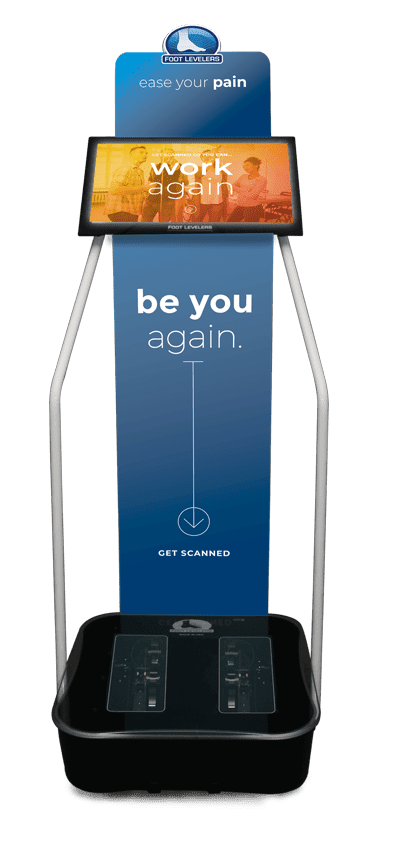
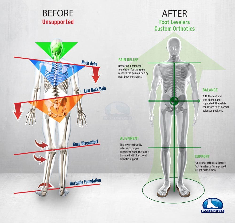


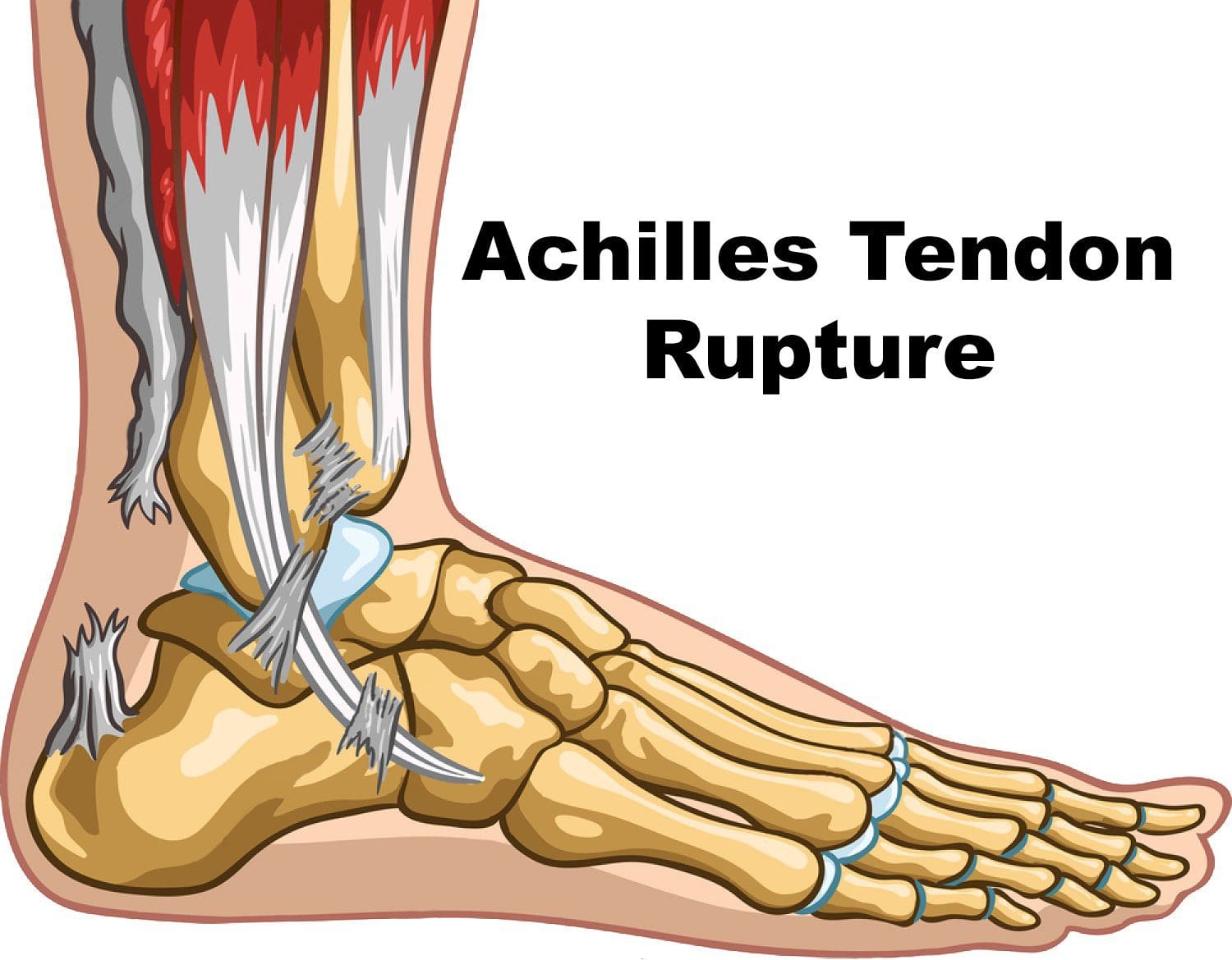 This is a devasting injury for anyone, especially world-class
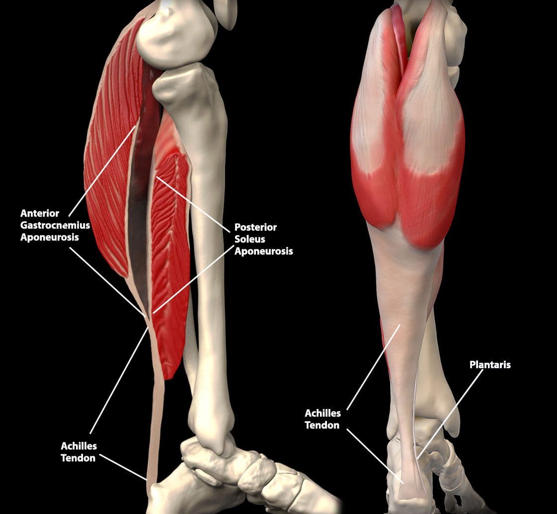
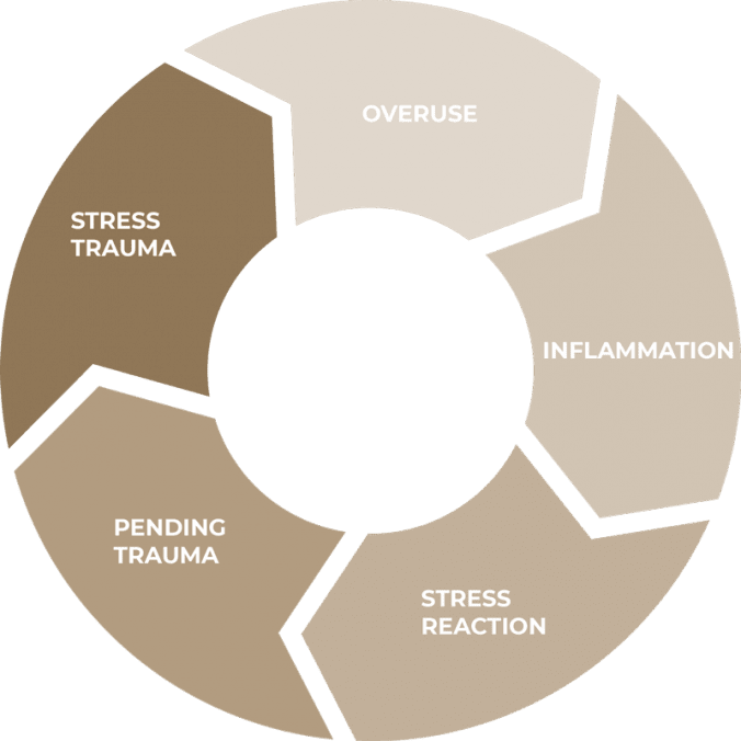
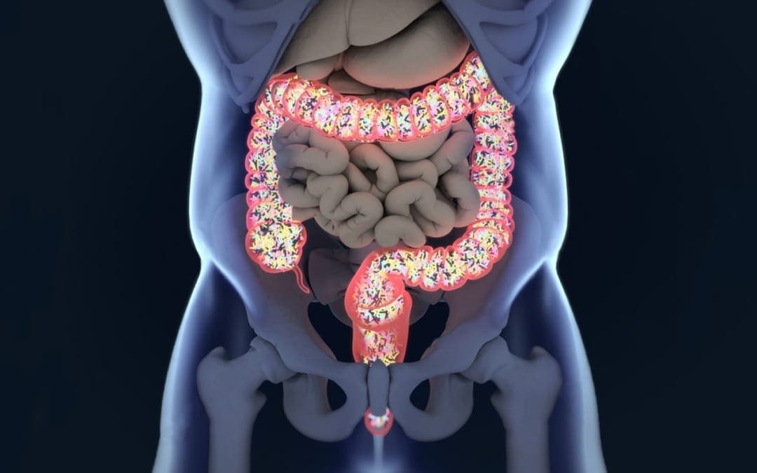
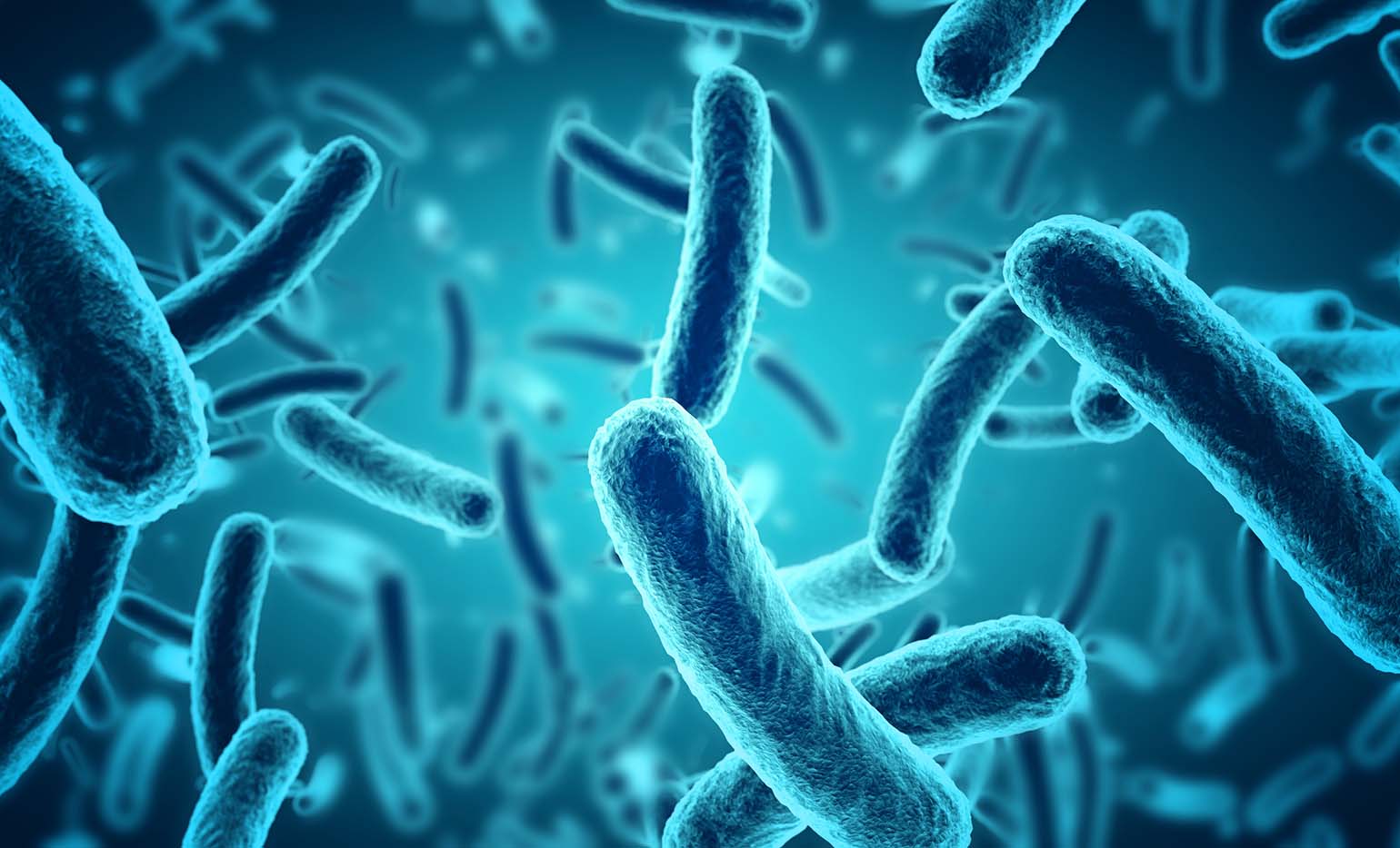

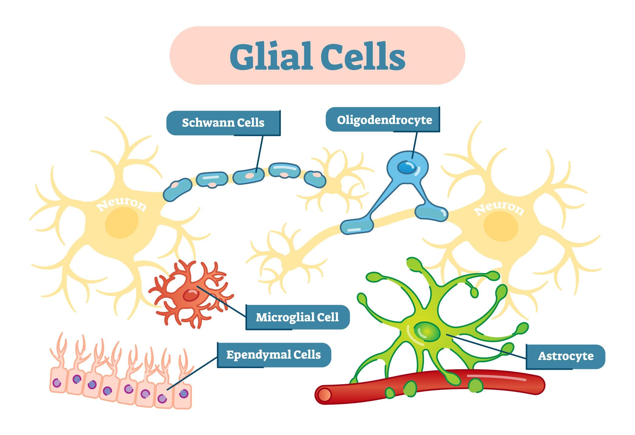 �
�

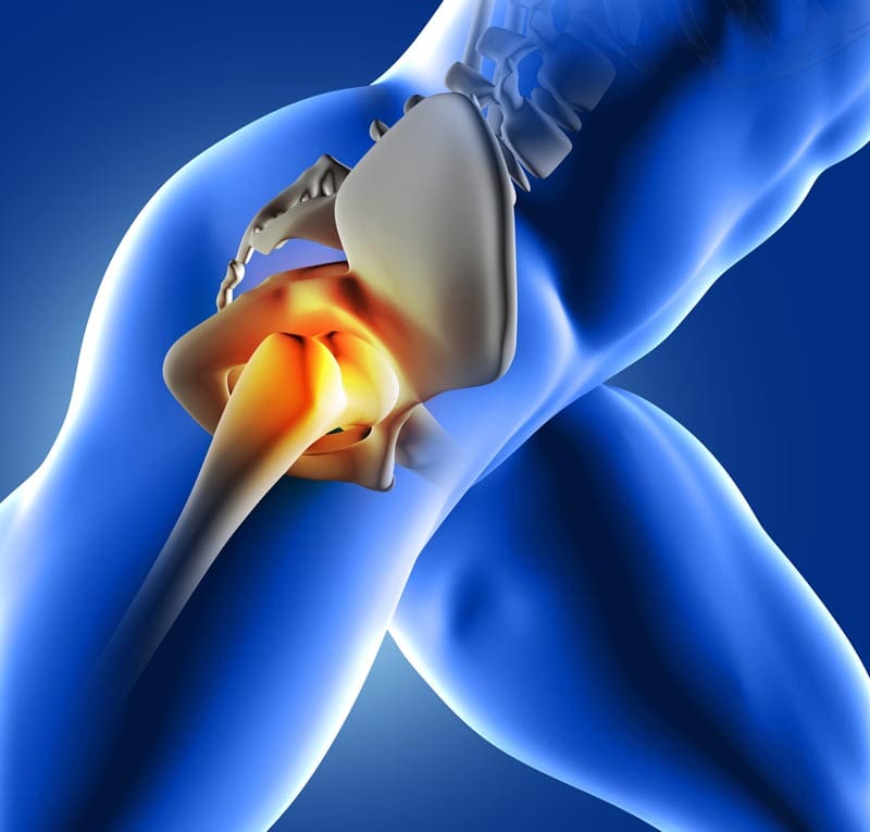
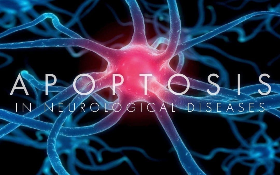
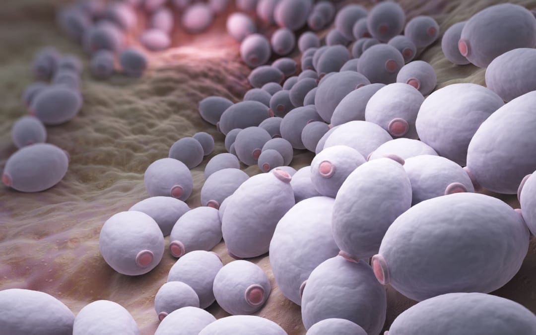
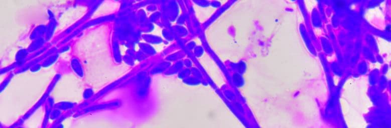 Small amounts aid in nutrient digestion and absorption.
However, overgrowth of candida can damage the intestinal lining and release toxic byproducts directly into the circulatory system.
If not addressed Candida overgrowth can turn chronic and lead to:
Small amounts aid in nutrient digestion and absorption.
However, overgrowth of candida can damage the intestinal lining and release toxic byproducts directly into the circulatory system.
If not addressed Candida overgrowth can turn chronic and lead to:
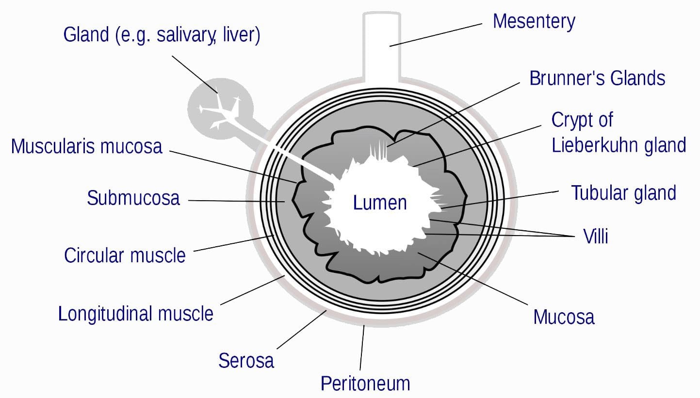


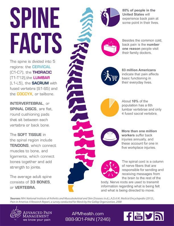
 These adjustments trigger the release of endorphins, which cause instant pain relief.
Our hips are made durable, but with age and normal wear and tear, this cartilage wears down, which can lead to injury.
This also includes the:
These adjustments trigger the release of endorphins, which cause instant pain relief.
Our hips are made durable, but with age and normal wear and tear, this cartilage wears down, which can lead to injury.
This also includes the:


