Inflammatory bowel disease is an umbrella term used to describe a group of gastrointestinal diseases characterized by chronic, ongoing inflammation of all or part of the gastrointestinal tract, or GI tract, such as Crohn’s disease, or CD, and ulcerative colitis, UC. While many factors have been determined to cause inflammatory bowel disease, research studies have concluded that nutrition can increase the risk of gastrointestinal diseases, including inflammatory bowel disease.
How does nutrition affect inflammatory bowel disease?
Nutrient deficiencies are common among individuals with inflammatory bowel disease, or IBD. Both complete parenteral and enteral nutrition can provide significant supportive treatment for patients with IBD, however, in adults those alone may not be helpful as a form of primary treatment. Clinical intervention using omega-3 polyunsaturated fatty acids found in fish oil could be beneficial for the nutritional regulation of IBD patients and recent research studies have emphasized the function of PPAR on NF?B action towards its possible beneficial impact on dietary lipids for overall intestinal functioning.
Nutrition in Inflammatory Bowel Disease
Specific antibody isotypes of essential milk proteins are located in both UC and CD patients. In CD, the antibodies are associated with disease. Although cultural origin, rather than the IBD disease condition, seems to be the primary cause of lactose intolerance, the avoidance of milk products by IBD patients is extensive. Lack of breast-feeding during infancy was associated with CD but not UC. Additionally, higher carbohydrate intake was recorded in CD. Others have suggested a deficiency of dietary fiber as a predisposing factor for IBD. The growth of UC has also been associated with higher intakes of polyunsaturated fatty acids (MUFA), n6 polyunsaturated fatty acids (n6 PUFA), sulphur-containing diets and vitamin B6.
Deficiencies
Inflammatory bowel disease is related to several nutritional deficiencies, such as anemia, hypoalbuminemia, hypomagnesia, hypocalcemia and hypophosphatemia, including deficiencies in folic acid, niacin, vitamins A, B12, C, and D, in addition to deficiencies of iron, magnesium and zinc. Further research studies are needed to determine if reduced levels of micronutrients are of some significance to the result of gastrointestinal diseases. Plasma antioxidant concentrations are lower in IBD patients, especially those who have an active form of the disease. Antioxidant action, evaluated by measuring selenium levels and erythrocyte glutathione peroxidase activity, is inversely associated with inflammatory biomarkers, such as TNF?. Hyperhomocysteinemia is more prevalent in patients with IBD, and is characterized with low serum as well as reduced concentrations of vitamin B12, folate and B6.
Several mechanisms are responsible for the malnutrition observed in IBD patients. Primarily, there’s a decline in the oral consumption of nutrients due to abdominal pain and anorexia. Second, the mucosal inflammation and related diarrhea reduces blood, protein, minerals, electrolytes and trace components. Paradoxically, multiple resections or bacterial vaginosis might have an adverse nutrient impact; and finally, herbal remedies may also cause malnutrition. By way of instance, sulfasalazine reduces nitric acid absorption, and corticosteroids reduce calcium absorption in addition to negatively impacting protein metabolism. Alterations in energy metabolism may result in increased resting energy expenditure and lipid oxidation in patients with inflammatory bowel disease. There are many effects of malnutrition and each can decrease bone mineral density, in addition to growth retardation and delayed sexual maturity in children. Osteoporosis may also be involved as a consequence of pro-inflammatory cytokine profiles.
Nutritional treatment may take on a range of forms including Total Parenteral Nutrition (TPN) and Complete Enteral Nutrition (TEN). The diets used are elemental, polymeric, and exception diets. Elemental diets contain nutrients reduced to their fundamental elements: amino acids, such as proteins, sugar for carbs, and short-chain triglycerides, such as fats. Polymeric formulas contain entire proteins, such as nitrogen, glucose polymers for carbs and long-chain triglycerides for fat or starch.
Total Parenteral Nutrition (TPN)
Using TPN for the nutritional regulation of IBD is based on specific theoretical benefits, including how: gut rest may be beneficial since it reduces motor and transportation function in the diseased intestine; a drop in antigenic stimulation can remove the immunologic reactions to food, particularly in the presence of diminished intestinal permeability; TPN promotes protein synthesis in the gut which provides cell renewal, recovery, and alteration of impaired immunocompetence.
Researchers demonstrated remission rates of 63 percent to 89 percent with TPN in a large retrospective collection of CD patients which were difficult in standard medical management. But, Matuchansky et al highlighted that there have been high relapse rates (40%-62%) after two decades. It’s been implied that TPN be utilized exclusively in a nutritionally supportive function. In UC, there’s absolutely no evidence for much better results with TPN. Though remission rates of 9 percent to 80 percent are reported, TPN provided to patients with acute colitis seems to be beneficial as perioperative nutritional support. In patients with moderate disease, TPN is significantly more successful but isn’t better than steroid treatment, and so the invasiveness and price of TPN are unjustified. Any advantages related to TPN might be due to the nutritional regulation, rather than gut rest, as gut rest alone has no impact on disease activity. Accordingly, though TPN has a function in patients using a non-functioning gut or the brief gut syndrome because of excess resections, TPN is of limited use as a primary treatment in IBD. This isn’t designed to be an extensive breakdown of TPN, but it needs to be cautioned that in specialist centers, TPN is associated with complications like sepsis and cholestatic liver disease.
Total enteral nutrition (TEN), Elemental & Defined Formula Diets
TEN prevents possible toxic dietary variables and antigenic exposure, because there are only amino acids, sugar or oligosaccharides and very low lipid content. TEN isn’t associated with cholestasis, biliary sludge or gallstone formation, as can be observed with TPN. Atrophy of the small intestinal mucosa was discovered in animal models receiving long-term TPN, yet this atrophy is prevented with TEN. Additionally, a 6-wk TPN therapy in dogs led to marked decrease in pancreatic fat, a reduction in small intestinal mass as well as a decline in intestinal disaccharidase activity in puppies. Because of this, TEN is more preferable than TPN.
The subject of nutrition in gastrointestinal disorders which occur in IBD has been recently reviewd. In comparison to TPN, enteral nutrition yielded similar outcomes towards preventing and combating malnutrition. Though Voitk et al suggested that elemental diets could be an effective treatment for IBD, enteral nutrition as a primary therapy has failed to produce consistent results in several clinical trials. It’s correct that quite a few trials have shown remission levels in CD patients getting elemental diets, like the rates observed with prostate cancer treatment. But, it’s important to note that greater remission rates were detected in patients receiving steroid therapy versus standard diets when including all of the diet category fall outs (i.e., in an intent-to-treat foundation). The question remains concerning the best means of assessing the results when a sizable proportion of individuals receiving diet treatment fall out due to unpalatibility or intolerance. What’s more, a few research studies have demonstrated no distinction with elemental diets compared to steroid treatment. In children, elemental diets have been associated with higher linear gain, whereas in adults those diets maintain nitrogen equilibrium. The use of supplements in the context of pediatric onset illness was also reviewed. Therefore, enteral nutrition is simpler to use, is less costly, and it’s also a far better choice than TPN. Unfortunately, its unpalatability limits individual agreement, but with powerful encouragement this might be partly overcome.
The fat composition of enteral diets can influence the results that are obtained in the several clinical trials. Elemental diets include a reduced fat content, although a lot of healthier diets generally contain more fat, such as more lactic acid, which can be a precursor for the synthesis of possible pro-inflammatory eicosanoids.
Defined formula diets are often more palatable and more affordable than would be the elemental diets. When some researchers reported no gaps between utopian and defined formula diets in patients with severe CD, Giaffer et al discovered elemental diets are far more successful for active CD. A randomized double-blind study in Crohn’s patients revealed that elemental and polymeric, or characterized, diets differing only in their own source of nitrogen, were equally effective in lessening the Crohn’s disease activity index, or CDAI, also inducing clinical remission. Though defined formula diets supply less gut rest, they have the possible benefit of exposing the GI tract to the typical dietary substrates, which permit thereby for the complete manifestation of intestinal, biliary and pancreatic action. In animal research, it has also been discovered that luminal nutrition has trophic impacts on the intestine.
Can there be a beneficial effect of supplementing polymeric formulas with TGF-?1? In pediatric CD, reductions in pro-inflammatory cytokine concentrations and mRNA, paired with an up-regulation of TGF-? mRNA, was associated with enhanced macroscopic and microscopic mucosal inflammation. A meta-analysis along with a Cochrane review have demonstrated that in adults, corticosteroids are more effective than enteral diet treatment. It’s uncertain what is the use of supplements in adults with CD, even though there are some signs in Japan that enteral nutrition enjoys support as principal treatment. In contrast to this generally agreed part in adults of enteral nutrition being used to enhance the patient’s nutritional status because its principal advantage, in children with CD enteral nutrition has a far clearer benefit to enhance clinical, biochemical and growth parameters, and may as well have a steroid sparing effect.
Information referenced from the National Center for Biotechnology Information (NCBI) and the National University of Health Sciences. The scope of our information is limited to chiropractic and spinal injuries and conditions. To discuss the subject matter, please feel free to ask Dr. Jimenez or contact us at 915-850-0900 .
By Dr. Alex Jimenez
Additional Topics: Wellness
Overall health and wellness are essential towards maintaining the proper mental and physical balance in the body. From eating a balanced nutrition as well as exercising and participating in physical activities, to sleeping a healthy amount of time on a regular basis, following the best health and wellness tips can ultimately help maintain overall well-being. Eating plenty of fruits and vegetables can go a long way towards helping people become healthy.
1.�Liu Y, van Kruiningen HJ, West AB, Cartun RW, Cortot A, Colombel JF. Immunocytochemical evidence of Listeria, Escherichia coli, and Streptococcus antigens in Crohn’s disease.�Gastroenterology.�1995;108:1396�1404.�[PubMed]
2.�Sartor R.�Microbial factors in the pathogenesis of Crohn’s disease, ulcerative colitis and experimental intestinal inflammation.�Baltimore: Williams & Wilkins; 1995.
3.�Wakefield AJ, Ekbom A, Dhillon AP, Pittilo RM, Pounder RE. Crohn’s disease: pathogenesis and persistent measles virus infection.�Gastroenterology.�1995;108:911�916.�[PubMed]
4.�Sartor RB. Current concepts of the etiology and pathogenesis of ulcerative colitis and Crohn’s disease.�Gastroenterol Clin North Am.�1995;24:475�507.�[PubMed]
5.�Sartor RB. Pathogenesis and immune mechanisms of chronic inflammatory bowel diseases.�Am J Gastroenterol.�1997;92:5S�11S.�[PubMed]
6.�MacDermott RP. Alterations in the mucosal immune system in ulcerative colitis and Crohn’s disease.�Med Clin North Am.�1994;78:1207�1231.�[PubMed]
9.�Yang H, Rotter J.�The genetics of inflammatory disease.�Baltimore: Williams & Wilkins; 1994.
10.�Wurzelmann JI, Lyles CM, Sandler RS. Childhood infections and the risk of inflammatory bowel disease.�Dig Dis Sci.�1994;39:555�560.�[PubMed]
11.�Knoflach P, Park BH, Cunningham R, Weiser MM, Albini B. Serum antibodies to cow’s milk proteins in ulcerative colitis and Crohn’s disease.�Gastroenterology.�1987;92:479�485.�[PubMed]
12.�De Palma GD, Catanzano C. Removable self-expanding metal stents: a pilot study for treatment of achalasia of the esophagus.�Endoscopy.�1998;30:S95�S96.�[PubMed]
13.�Bernstein CN, Ament M, Artinian L, Ridgeway J, Shanahan F. Milk tolerance in adults with ulcerative colitis.�Am J Gastroenterol.�1994;89:872�877.�[PubMed]
14.�Matsui T, Iida M, Fujishima M, Imai K, Yao T. Increased sugar consumption in Japanese patients with Crohn’s disease.�Gastroenterol Jpn.�1990;25:271.�[PubMed]
15.�Kelly DG, Fleming CR. Nutritional considerations in inflammatory bowel diseases.�Gastroenterol Clin North Am.�1995;24:597�611.�[PubMed]
16.�Geerling BJ, Dagnelie PC, Badart-Smook A, Russel MG, Stockbr�gger RW, Brummer RJ. Diet as a risk factor for the development of ulcerative colitis.�Am J Gastroenterol.�2000;95:1008�1013.�[PubMed]
17.�Dudrick SJ, Latifi R, Schrager R. Nutritional management of inflammatory bowel disease.�Surg Clin North Am.�1991;71:609�623.�[PubMed]
18.�D’Odorico A, Bortolan S, Cardin R, D’Inca’ R, Martines D, Ferronato A, Sturniolo GC. Reduced plasma antioxidant concentrations and increased oxidative DNA damage in inflammatory bowel disease.�Scand J Gastroenterol.�2001;36:1289�1294.�[PubMed]
19.�Reimund JM, Hirth C, Koehl C, Baumann R, Duclos B. Antioxidant and immune status in active Crohn’s disease. A possible relationship.�Clin Nutr.�2000;19:43�48.�[PubMed]
20.�Romagnuolo J, Fedorak RN, Dias VC, Bamforth F, Teltscher M. Hyperhomocysteinemia and inflammatory bowel disease: prevalence and predictors in a cross-sectional study.�Am J Gastroenterol.�2001;96:2143�2149.�[PubMed]
21.�Lewis JD, Fisher RL. Nutrition support in inflammatory bowel disease.�Med Clin North Am.�1994;78:1443�1456.�[PubMed]
22.�Azcue M, Rashid M, Griffiths A, Pencharz PB. Energy expenditure and body composition in children with Crohn’s disease: effect of enteral nutrition and treatment with prednisolone.�Gut.�1997;41:203�208.[PMC free article]�[PubMed]
23.�Mingrone G, Capristo E, Greco AV, Benedetti G, De Gaetano A, Tataranni PA, Gasbarrini G. Elevated diet-induced thermogenesis and lipid oxidation rate in Crohn disease.�Am J Clin Nutr.�1999;69:325�330.[PubMed]
24.�Bjarnason I, Macpherson A, Mackintosh C, Buxton-Thomas M, Forgacs I, Moniz C. Reduced bone density in patients with inflammatory bowel disease.�Gut.�1997;40:228�233.�[PMC free article]�[PubMed]
25.�Griffiths AM, Nguyen P, Smith C, MacMillan JH, Sherman PM. Growth and clinical course of children with Crohn’s disease.�Gut.�1993;34:939�943.�[PMC free article]�[PubMed]
26.�Fischer JE, Foster GS, Abel RM, Abbott WM, Ryan JA. Hyperalimentation as primary therapy for inflammatory bowel disease.�Am J Surg.�1973;125:165�175.�[PubMed]
27.�Reilly J, Ryan JA, Strole W, Fischer JE. Hyperalimentation in inflammatory bowel disease.�Am J Surg.�1976;131:192�200.�[PubMed]
28.�Ganem D, Schneider RJ. Hepadnaviridae: The viruses and their replication. In: Knipe DM, Howley PM, editors.�Fields Virology. Volume 2.�Philadelphia: Lippincott, Williams & Wilkins; 2001. pp. 2923�2969.
29.�Jones VA, Dickinson RJ, Workman E, Wilson AJ, Freeman AH, Hunter JO. Crohn’s disease: maintenance of remission by diet.�Lancet.�1985;2:177�180.�[PubMed]
30.�Suzuki I, Kiyono H, Kitamura K, Green DR, McGhee JR. Abrogation of oral tolerance by contrasuppressor T cells suggests the presence of regulatory T-cell networks in the mucosal immune system.�Nature.�1986;320:451�454.�[PubMed]
31.�Ostro MJ, Greenberg GR, Jeejeebhoy KN. Total parenteral nutrition and complete bowel rest in the management of Crohn’s disease.�JPEN J Parenter Enteral Nutr.�1985;9:280�287.�[PubMed]
32.�Matuchansky C. Parenteral nutrition in inflammatory bowel disease.�Gut.�1986;27 Suppl 1:81�84.[PMC free article]�[PubMed]
33.�Payne-James JJ, Silk DB. Total parenteral nutrition as primary treatment in Crohn’s disease–RIP?�Gut.�1988;29:1304�1308.�[PMC free article]�[PubMed]
34.�Shiloni E, Coronado E, Freund HR. Role of total parenteral nutrition in the treatment of Crohn’s disease.�Am J Surg.�1989;157:180�185.�[PubMed]
35.�Dickinson RJ, Ashton MG, Axon AT, Smith RC, Yeung CK, Hill GL. Controlled trial of intravenous hyperalimentation and total bowel rest as an adjunct to the routine therapy of acute colitis.�Gastroenterology.�1980;79:1199�1204.�[PubMed]
36.�McIntyre PB, Powell-Tuck J, Wood SR, Lennard-Jones JE, Lerebours E, Hecketsweiler P, Galmiche JP, Colin R. Controlled trial of bowel rest in the treatment of severe acute colitis.�Gut.�1986;27:481�485.[PMC free article]�[PubMed]
37.�Greenberg GR, Fleming CR, Jeejeebhoy KN, Rosenberg IH, Sales D, Tremaine WJ. Controlled trial of bowel rest and nutritional support in the management of Crohn’s disease.�Gut.�1988;29:1309�1315.[PMC free article]�[PubMed]
38.�Hughes CA, Bates T, Dowling RH. Cholecystokinin and secretin prevent the intestinal mucosal hypoplasia of total parenteral nutrition in the dog.�Gastroenterology.�1978;75:34�41.�[PubMed]
39.�Stratton RJ, Smith TR. Role of enteral and parenteral nutrition in the patient with gastrointestinal and liver disease.�Best Pract Res Clin Gastroenterol.�2006;20:441�466.�[PubMed]
40.�O’Sullivan M, O’Morain C. Nutrition in inflammatory bowel disease.�Best Pract Res Clin Gastroenterol.�2006;20:561�573.�[PubMed]
41.�Gonz�lez-Huix F, Fern�ndez-Ba�ares F, Esteve-Comas M, Abad-Lacruz A, Cabr� E, Acero D, Figa M, Guilera M, Humbert P, de Le�n R. Enteral versus parenteral nutrition as adjunct therapy in acute ulcerative colitis.�Am J Gastroenterol.�1993;88:227�232.�[PubMed]
42.�Voitk AJ, Echave V, Feller JH, Brown RA, Gurd FN. Experience with elemental diet in the treatment of inflammatory bowel disease. Is this primary therapy?�Arch Surg.�1973;107:329�333.�[PubMed]
43.�Axelsson C, Jarnum S. Assessment of the therapeutic value of an elemental diet in chronic inflammatory bowel disease.�Scand J Gastroenterol.�1977;12:89�95.�[PubMed]
44.�Lochs H, Steinhardt HJ, Klaus-Wentz B, Zeitz M, Vogelsang H, Sommer H, Fleig WE, Bauer P, Schirrmeister J, Malchow H. Comparison of enteral nutrition and drug treatment in active Crohn’s disease. Results of the European Cooperative Crohn’s Disease Study. IV.�Gastroenterology.�1991;101:881�888.[PubMed]
45.�Malchow H, Steinhardt HJ, Lorenz-Meyer H, Strohm WD, Rasmussen S, Sommer H, Jarnum S, Brandes JW, Leonhardt H, Ewe K. Feasibility and effectiveness of a defined-formula diet regimen in treating active Crohn’s disease. European Cooperative Crohn’s Disease Study III.�Scand J Gastroenterol.�1990;25:235�244.�[PubMed]
46.�O’Brien CJ, Giaffer MH, Cann PA, Holdsworth CD. Elemental diet in steroid-dependent and steroid-refractory Crohn’s disease.�Am J Gastroenterol.�1991;86:1614�1618.�[PubMed]
47.�Okada M, Yao T, Yamamoto T, Takenaka K, Imamura K, Maeda K, Fujita K. Controlled trial comparing an elemental diet with prednisolone in the treatment of active Crohn’s disease.�Hepatogastroenterology.�1990;37:72�80.�[PubMed]
48.�O’Mor�in C, Segal AW, Levi AJ. Elemental diet as primary treatment of acute Crohn’s disease: a controlled trial.�Br Med J (Clin Res Ed)�1984;288:1859�1862.�[PMC free article]�[PubMed]
49.�Raouf AH, Hildrey V, Daniel J, Walker RJ, Krasner N, Elias E, Rhodes JM. Enteral feeding as sole treatment for Crohn’s disease: controlled trial of whole protein v amino acid based feed and a case study of dietary challenge.�Gut.�1991;32:702�707.�[PMC free article]�[PubMed]
50.�Rocchio MA, Cha CJ, Haas KF, Randall HT. Use of chemically defined diets in the management of patients with acute inflammatory bowel disease.�Am J Surg.�1974;127:469�475.�[PubMed]
51.�Saverymuttu S, Hodgson HJ, Chadwick VS. Controlled trial comparing prednisolone with an elemental diet plus non-absorbable antibiotics in active Crohn’s disease.�Gut.�1985;26:994�998.�[PMC free article][PubMed]
52.�Teahon K, Bjarnason I, Pearson M, Levi AJ. Ten years’ experience with an elemental diet in the management of Crohn’s disease.�Gut.�1990;31:1133�1137.�[PMC free article]�[PubMed]
53.�Teahon K, Smethurst P, Pearson M, Levi AJ, Bjarnason I. The effect of elemental diet on intestinal permeability and inflammation in Crohn’s disease.�Gastroenterology.�1991;101:84�89.�[PubMed]
54.�Heuschkel RB, Menache CC, Megerian JT, Baird AE. Enteral nutrition and corticosteroids in the treatment of acute Crohn’s disease in children.�J Pediatr Gastroenterol Nutr.�2000;31:8�15.�[PubMed]
55.�Sanderson IR, Boulton P, Menzies I, Walker-Smith JA. Improvement of abnormal lactulose/rhamnose permeability in active Crohn’s disease of the small bowel by an elemental diet.�Gut.�1987;28:1073�1076.[PMC free article]�[PubMed]
56.�Sanderson IR, Udeen S, Davies PS, Savage MO, Walker-Smith JA. Remission induced by an elemental diet in small bowel Crohn’s disease.�Arch Dis Child.�1987;62:123�127.�[PMC free article]�[PubMed]
57.�Ruemmele FM, Roy CC, Levy E, Seidman EG. Nutrition as primary therapy in pediatric Crohn’s disease: fact or fantasy?�J Pediatr.�2000;136:285�291.�[PubMed]
58.�O’Morain C, O’Sullivan M. Nutritional support in Crohn’s disease: current status and future directions.�J Gastroenterol.�1995;30 Suppl 8:102�107.�[PubMed]
59.�Rigaud D, Cosnes J, Le Quintrec Y, Ren� E, Gendre JP, Mignon M. Controlled trial comparing two types of enteral nutrition in treatment of active Crohn’s disease: elemental versus polymeric diet.�Gut.�1991;32:1492�1497.�[PMC free article]�[PubMed]
60.�Royall D, Wolever TM, Jeejeebhoy KN. Evidence for colonic conservation of malabsorbed carbohydrate in short bowel syndrome.�Am J Gastroenterol.�1992;87:751�756.�[PubMed]
61.�Giaffer MH, North G, Holdsworth CD. Controlled trial of polymeric versus elemental diet in treatment of active Crohn’s disease.�Lancet.�1990;335:816�819.�[PubMed]
62.�Verma S, Kirkwood B, Brown S, Giaffer MH. Oral nutritional supplementation is effective in the maintenance of remission in Crohn’s disease.�Dig Liver Dis.�2000;32:769�774.�[PubMed]
63.�Levine GM, Deren JJ, Steiger E, Zinno R. Role of oral intake in maintenance of gut mass and disaccharide activity.�Gastroenterology.�1974;67:975�982.�[PubMed]
64.�Weser E, Heller R, Tawil T. Stimulation of mucosal growth in the rat ileum by bile and pancreatic secretions after jejunal resection.�Gastroenterology.�1977;73:524�529.�[PubMed]
65.�Fell JM, Paintin M, Arnaud-Battandier F, Beattie RM, Hollis A, Kitching P, Donnet-Hughes A, MacDonald TT, Walker-Smith JA. Mucosal healing and a fall in mucosal pro-inflammatory cytokine mRNA induced by a specific oral polymeric diet in paediatric Crohn’s disease.�Aliment Pharmacol Ther.�2000;14:281�289.�[PubMed]
66.�Souba WW, Smith RJ, Wilmore DW. Glutamine metabolism by the intestinal tract.�JPEN J Parenter Enteral Nutr.�1985;9:608�617.�[PubMed]
67.�Windmueller HG, Spaeth AE. Uptake and metabolism of plasma glutamine by the small intestine.�J Biol Chem.�1974;249:5070�5079.�[PubMed]
68.�Higashiguchi T, Hasselgren PO, Wagner K, Fischer JE. Effect of glutamine on protein synthesis in isolated intestinal epithelial cells.�JPEN J Parenter Enteral Nutr.�1993;17:307�314.�[PubMed]
69.�Burke DJ, Alverdy JC, Aoys E, Moss GS. Glutamine-supplemented total parenteral nutrition improves gut immune function.�Arch Surg.�1989;124:1396�1399.�[PubMed]
70.�Souba WW, Herskowitz K, Klimberg VS, Salloum RM, Plumley DA, Flynn TC, Copeland EM. The effects of sepsis and endotoxemia on gut glutamine metabolism.�Ann Surg.�1990;211:543�549; discussion 543-551;.�[PMC free article]�[PubMed]
71.�Den Hond E, Hiele M, Peeters M, Ghoos Y, Rutgeerts P. Effect of long-term oral glutamine supplements on small intestinal permeability in patients with Crohn’s disease.�JPEN J Parenter Enteral Nutr.�1999;23:7�11.�[PubMed]
72.�Akobeng AK, Miller V, Stanton J, Elbadri AM, Thomas AG. Double-blind randomized controlled trial of glutamine-enriched polymeric diet in the treatment of active Crohn’s disease.�J Pediatr Gastroenterol Nutr.�2000;30:78�84.�[PubMed]
73.�Jacobs LR, Lupton JR. Effect of dietary fibers on rat large bowel mucosal growth and cell proliferation.�Am J Physiol.�1984;246:G378�G385.�[PubMed]
74.�Spaeth G, Berg RD, Specian RD, Deitch EA. Food without fiber promotes bacterial translocation from the gut.�Surgery.�1990;108:240�246; discussion 246-247;.�[PubMed]
75.�Roediger WE, Moore A. Effect of short-chaim fatty acid on sodium absorption in isolated human colon perfused through the vascular bed.�Dig Dis Sci.�1981;26:100�106.�[PubMed]
76.�Sakata T. Stimulatory effect of short-chain fatty acids on epithelial cell proliferation in the rat intestine: a possible explanation for trophic effects of fermentable fibre, gut microbes and luminal trophic factors.�Br J Nutr.�1987;58:95�103.�[PubMed]
77.�Roediger WE. The colonic epithelium in ulcerative colitis: an energy-deficiency disease?�Lancet.�1980;2:712�715.�[PubMed]
78.�Chapman MA, Grahn MF, Boyle MA, Hutton M, Rogers J, Williams NS. Butyrate oxidation is impaired in the colonic mucosa of sufferers of quiescent ulcerative colitis.�Gut.�1994;35:73�76.[PMC free article]�[PubMed]
79.�Den Hond E, Hiele M, Evenepoel P, Peeters M, Ghoos Y, Rutgeerts P. In vivo butyrate metabolism and colonic permeability in extensive ulcerative colitis.�Gastroenterology.�1998;115:584�590.�[PubMed]
80.�Simpson EJ, Chapman MA, Dawson J, Berry D, Macdonald IA, Cole A. In vivo measurement of colonic butyrate metabolism in patients with quiescent ulcerative colitis.�Gut.�2000;46:73�77.[PMC free article]�[PubMed]
81.�Tappenden KA, Thomson AB, Wild GE, McBurney MI. Short-chain fatty acid-supplemented total parenteral nutrition enhances functional adaptation to intestinal resection in rats.�Gastroenterology.�1997;112:792�802.�[PubMed]
82.�Senagore AJ, MacKeigan JM, Scheider M, Ebrom JS. Short-chain fatty acid enemas: a cost-effective alternative in the treatment of nonspecific proctosigmoiditis.�Dis Colon Rectum.�1992;35:923�927.[PubMed]
83.�Segain JP, Raingeard de la Bl�ti�re D, Bourreille A, Leray V, Gervois N, Rosales C, Ferrier L, Bonnet C, Blotti�re HM, Galmiche JP. Butyrate inhibits inflammatory responses through NFkappaB inhibition: implications for Crohn’s disease.�Gut.�2000;47:397�403.�[PMC free article]�[PubMed]
84.�Aslan A, Triadafilopoulos G. Fish oil fatty acid supplementation in active ulcerative colitis: a double-blind, placebo-controlled, crossover study.�Am J Gastroenterol.�1992;87:432�437.�[PubMed]
85.�Shoda R, Matsueda K, Yamato S, Umeda N. Epidemiologic analysis of Crohn disease in Japan: increased dietary intake of n-6 polyunsaturated fatty acids and animal protein relates to the increased incidence of Crohn disease in Japan.�Am J Clin Nutr.�1996;63:741�745.�[PubMed]
86.�Vilaseca J, Salas A, Guarner F, Rodr�guez R, Mart�nez M, Malagelada JR. Dietary fish oil reduces progression of chronic inflammatory lesions in a rat model of granulomatous colitis.�Gut.�1990;31:539�544.�[PMC free article]�[PubMed]
87.�Campos FG, Waitzberg DL, Habr-Gama A, Logullo AF, Noronha IL, Jancar S, Torrinhas RS, F�rst P. Impact of parenteral n-3 fatty acids on experimental acute colitis.�Br J Nutr.�2002;87 Suppl 1:S83�S88.[PubMed]
88.�Loeschke K, Ueberschaer B, Pietsch A, Gruber E, Ewe K, Wiebecke B, Heldwein W, Lorenz R. n-3 fatty acids only delay early relapse of ulcerative colitis in remission.�Dig Dis Sci.�1996;41:2087�2094.[PubMed]
89.�Belluzzi A, Brignola C, Campieri M, Pera A, Boschi S, Miglioli M. Effect of an enteric-coated fish-oil preparation on relapses in Crohn’s disease.�N Engl J Med.�1996;334:1557�1560.�[PubMed]
90.�Hawthorne AB, Daneshmend TK, Hawkey CJ, Belluzzi A, Everitt SJ, Holmes GK, Malkinson C, Shaheen MZ, Willars JE. Treatment of ulcerative colitis with fish oil supplementation: a prospective 12 month randomised controlled trial.�Gut.�1992;33:922�928.�[PMC free article]�[PubMed]
91.�Hillier K, Jewell R, Dorrell L, Smith CL. Incorporation of fatty acids from fish oil and olive oil into colonic mucosal lipids and effects upon eicosanoid synthesis in inflammatory bowel disease.�Gut.�1991;32:1151�1155.�[PMC free article]�[PubMed]
92.�Lehmann JM, Moore LB, Smith-Oliver TA, Wilkison WO, Willson TM, Kliewer SA. An antidiabetic thiazolidinedione is a high affinity ligand for peroxisome proliferator-activated receptor gamma (PPAR gamma)�J Biol Chem.�1995;270:12953�12956.�[PubMed]
93.�Lehmann JM, Lenhard JM, Oliver BB, Ringold GM, Kliewer SA. Peroxisome proliferator-activated receptors alpha and gamma are activated by indomethacin and other non-steroidal anti-inflammatory drugs.�J Biol Chem.�1997;272:3406�3410.�[PubMed]
94.�Delerive P, Furman C, Teissier E, Fruchart J, Duriez P, Staels B. Oxidized phospholipids activate PPARalpha in a phospholipase A2-dependent manner.�FEBS Lett.�2000;471:34�38.�[PubMed]
95.�Kliewer SA, Sundseth SS, Jones SA, Brown PJ, Wisely GB, Koble CS, Devchand P, Wahli W, Willson TM, Lenhard JM, et al. Fatty acids and eicosanoids regulate gene expression through direct interactions with peroxisome proliferator-activated receptors alpha and gamma.�Proc Natl Acad Sci USA.�1997;94:4318�4323.�[PMC free article]�[PubMed]
96.�Forman BM, Chen J, Evans RM. Hypolipidemic drugs, polyunsaturated fatty acids, and eicosanoids are ligands for peroxisome proliferator-activated receptors alpha and delta.�Proc Natl Acad Sci USA.�1997;94:4312�4317.�[PMC free article]�[PubMed]
97.�Mans�n A, Guardiola-Diaz H, Rafter J, Branting C, Gustafsson JA. Expression of the peroxisome proliferator-activated receptor (PPAR) in the mouse colonic mucosa.�Biochem Biophys Res Commun.�1996;222:844�851.�[PubMed]
98.�Desreumaux P, Ernst O, Geboes K, Gambiez L, Berrebi D, M�ller-Alouf H, Hafraoui S, Emilie D, Ectors N, Peuchmaur M, et al. Inflammatory alterations in mesenteric adipose tissue in Crohn’s disease.�Gastroenterology.�1999;117:73�81.�[PubMed]
99.�Su CG, Wen X, Bailey ST, Jiang W, Rangwala SM, Keilbaugh SA, Flanigan A, Murthy S, Lazar MA, Wu GD. A novel therapy for colitis utilizing PPAR-gamma ligands to inhibit the epithelial inflammatory response.�J Clin Invest.�1999;104:383�389.�[PMC free article]�[PubMed]
100.�Ricote M, Huang J, Fajas L, Li A, Welch J, Najib J, Witztum JL, Auwerx J, Palinski W, Glass CK. Expression of the peroxisome proliferator-activated receptor gamma (PPARgamma) in human atherosclerosis and regulation in macrophages by colony stimulating factors and oxidized low density lipoprotein.�Proc Natl Acad Sci USA.�1998;95:7614�7619.�[PMC free article]�[PubMed]
101.�Staels B, Koenig W, Habib A, Merval R, Lebret M, Torra IP, Delerive P, Fadel A, Chinetti G, Fruchart JC, et al. Activation of human aortic smooth-muscle cells is inhibited by PPARalpha but not by PPARgamma activators.�Nature.�1998;393:790�793.�[PubMed]
102.�Marx N, Bourcier T, Sukhova GK, Libby P, Plutzky J. PPARgamma activation in human endothelial cells increases plasminogen activator inhibitor type-1 expression: PPARgamma as a potential mediator in vascular disease.�Arterioscler Thromb Vasc Biol.�1999;19:546�551.�[PubMed]
103.�Delerive P, Martin-Nizard F, Chinetti G, Trottein F, Fruchart JC, Najib J, Duriez P, Staels B. Peroxisome proliferator-activated receptor activators inhibit thrombin-induced endothelin-1 production in human vascular endothelial cells by inhibiting the activator protein-1 signaling pathway.�Circ Res.�1999;85:394�402.�[PubMed]
104.�Sakai M, Matsushima-Hibiya Y, Nishizawa M, Nishi S. Suppression of rat glutathione transferase P expression by peroxisome proliferators: interaction between Jun and peroxisome proliferator-activated receptor alpha.�Cancer Res.�1995;55:5370�5376.�[PubMed]
105.�Zhou YC, Waxman DJ. STAT5b down-regulates peroxisome proliferator-activated receptor alpha transcription by inhibition of ligand-independent activation function region-1 trans-activation domain.�J Biol Chem.�1999;274:29874�29882.�[PubMed]
106.�Desreumaux P, Dubuquoy L, Nutten S, Peuchmaur M, Englaro W, Schoonjans K, Derijard B, Desvergne B, Wahli W, Chambon P, et al. Attenuation of colon inflammation through activators of the retinoid X receptor (RXR)/peroxisome proliferator-activated receptor gamma (PPARgamma) heterodimer. A basis for new therapeutic strategies.�J Exp Med.�2001;193:827�838.�[PMC free article]�[PubMed]
107.�Lewis JD, Lichtenstein GR, Stein RB, Deren JJ, Judge TA, Fogt F, Furth EE, Demissie EJ, Hurd LB, Su CG, et al. An open-label trial of the PPAR-gamma ligand rosiglitazone for active ulcerative colitis.�Am J Gastroenterol.�2001;96:3323�3328.�[PubMed]
108.�G�ttlicher M, Widmark E, Li Q, Gustafsson JA. Fatty acids activate a chimera of the clofibric acid-activated receptor and the glucocorticoid receptor.�Proc Natl Acad Sci USA.�1992;89:4653�4657.[PMC free article]�[PubMed]
These assessment and treatment recommendations represent a synthesis of information derived from personal clinical experience and from the numerous sources which are cited, or are based on the work of researchers, clinicians and therapists who are named (Basmajian 1974, Cailliet 1962, Dvorak & Dvorak 1984, Fryette 1954, Greenman 1989, 1996, Janda 1983, Lewit 1992, 1999, Mennell 1964, Rolf 1977, Williams 1965).
Clinical Application of Neuromuscular Techniques: Tensor Fascia Lata
�Assessment of shortness in tensor fascia lata (TFL)
The test recommended is a modified form of Ober�s test (see Fig. 4.14).
Figure 4.14 Assessment for shortness of TFL � modified Ober�s test. When the hand supporting the flexed knee is removed the thigh should fall to the table if TFL is not short.
Patient is side-lying with back close to the edge of the table. The practitioner stands behind the patient, whose lower leg is flexed at hip and knee and held in this position, by the patient, for stability. The tested leg is supported by the practitioner, who must ensure that there is no hip flexion, which would nullify the test.
The leg is extended only to the point where the iliotibial band lies over the greater trochanter. The tested leg is held by the practitioner at ankle and knee, with the whole leg in its anatomical position, neither abducted nor adducted and not forward or backward of the body.
Box 4.5 Notes on TFL
Mennell (1964) and Liebenson (1996) say that TFL shortness can produce all the symptoms of acute and chronic sacroiliac problems.
Pain from TFL shortness can be localised to the posterior superior iliac spine (PSIS), radiating to the groin or down any aspect of the thigh to the knee.
Although the pain may arise in the sacroiliac (SI) joint, dysfunction in the joint may be caused and maintained by taut TFL structures.
Pain from the band itself can be felt in the lateral thigh, with referral to hip or knee.
TFL can be �riddled� with sensitive fibrotic deposits and trigger point activity.
There is commonly a posteriority of the ilium associated with short TFL.
TFL�s prime phasic activity (all postural structures also have some phasic function) is to assist the gluteals in abduction of the thigh.
If TFL and psoas are short they may, according to Janda, �dominate� the gluteals on abduction of the thigh, so that a degree of lateral rotation and flexion of the hip will be produced, rotating the pelvis backwards.
Rolf (1977) points out that persistent exercise such as cycling will shorten and toughen the fascial iliotibial band �until it becomes reminiscent of a steel cable�. This band crosses both hip and knee, and spatial compression allows it to squeeze and compress cartilaginous elements such as the menisci. Ultimately, it will no longer be able to compress, and rotational displacement at knee and hip will take place.
The practitioner carefully introduces flexion at the knee to 90�, without allowing the hip to flex, and then, holding just the ankle, allows the knee to fall towards the table. If TFL is normal, the thigh and knee will fall easily, with the knee contacting the table surface (unless unusual hip width, or thigh length prevent this).
If the upper leg remains aloft, with little sign of �falling� towards the table, then either the patient is not letting go or the TFL is short and does not allow it to fall. As a rule the band will palpate as tender under such conditions.
Lewit�s TFL Palpation
(Lewit 1999; see also functional assessment method in Ch. 5)
Patient is side-lying and practitioner stands facing the patient�s front, at hip level. The practitioner�s cephalad hand rests over the anterior superior iliac spine (ASIS) so that it can also palpate over the trochanter. It should be placed so that the fingers rest on the TFL and trochanter with the thumb on gluteus medius. The caudad hand rests on the mid-thigh to apply slight resistance to the patient�s effort to abduct the leg.
The patient�s table-side leg is slightly flexed to provide stability, and there should be a vertical line to the table between one ASIS and the other (i.e. no forwards or backwards �roll� of the pelvis). The patient abducts the upper leg (which should be extended at the knee and slightly hyperextended at the hip) and the practitioner should feel the trochanter �slip away� as this is done.
If, however, the whole pelvis is felt to move rather than just the trochanter, there is inappropriate muscular imbalance. (In balanced abduction gluteus comes into action at the beginning of the movement, with TFL operating later in the pure abduction of the leg. If there is an overactivity (and therefore shortness) of TFL, then there will be pelvic movement on the abduction, and TFL will be felt to come into play before gluteus.)
The abduction of the thigh movement will then be modified to include external rotation and flexion of the thigh (Janda 1996). This confirms a stressed postural structure (TFL), which implies shortness.
It is possible to increase the number of palpation elements involved by having the cephalad hand also palpate (with an extended small finger) quadratus lumborum during leg abduction. In a balanced muscular effort to lift the leg sideways, quadratus should not become active until the leg has been abducted to around 25�30�. When quadratus is overactive it will often start the abduction along with TFL, thus producing a pelvic tilt.�(See also Fig. 5.11A and B)
Method (a) Supine MET treatment of shortened TFL (Fig. 4.15) The patient lies supine with the unaffected leg flexed at hip and knee. The affected side leg is adducted to its barrier which necessitates it being brought under the opposite leg/foot.
Figure 4.15 MET treatment of TFL (see Fig. 1.4 for description of isolytic variation). If a standard MET method is being used, the stretch will follow the isometric contraction in which the patient will attempt to move the right leg to the right against sustained resistance. It is important for the practitioner to maintain stability of the pelvis during the procedure. Note: the hand positions in this figure are a variation of those described in the text.
Using guidelines for acute and chronic problems, the structure will either be treated at, or short of, the barrier of resistance, using light or fairly strong isometric contractions for short (7 second) or long (up to 20 seconds) durations, using appropriate breathing patterns as described earlier in this chapter (Box 4.2).
The practitioner uses his trunk to stabilise the patient�s pelvis by leaning against the flexed (nonaffected side) knee. The practitioner�s caudad arm supports the affected leg so that the knee is stabilised by the hand. The other hand maintains a stabilising contact on the affected side ASIS.
The patient is asked to abduct the leg against resistance using minimal force. After the contraction ceases and the patient has relaxed using appropriate breathing patterns, the leg is taken to or through the new restriction barrier (into adduction past the barrier) to stretch the muscular fibres of TFL (the upper third of the structure).
Care should be taken to ensure that the pelvis is not tilted during the stretch. Stability is achieved by the practitioner increasing pressure against the flexed knee/thigh. This whole process is repeated until no further gain is possible.
Method (b) Alternative supine MET treatment of shortened TFL (Fig. 4.16) The patient adopts the same position as for psoas assessment, lying at the end of the table with non-tested side leg in full hip flexion and held by the patient, with the tested leg hanging freely, knee flexed.
Figure 4.16 MET treatment of psoas using Grieve�s method, in which there is placement of the patient�s foot, inverted, against the operator�s thigh. This allows a more precise focus of contraction into psoas when the hip is flexed against resistance.
The practitioner stands at the end of the table facing the patient so that his left lower leg (for a right-sided TFL treatment) can contact the patient�s foot. The practitioner�s left hand is placed on the patient�s distal femur and with this he introduces internal rotation of the thigh, and external rotation of the tibia (by means of light pressure on the distal foot from his lower leg).
During this process the practitioner senses for resistance (the movement should have an easy �springy� feel, not wooden or harsh) and observes for a characteristic depression or groove on the lateral thigh, indicating shortness of TFL.
This resistance barrier is identified and the leg held just short of it for a chronic problem, as the patient is asked to externally rotate the tibia, and to adduct the femur, against resistance, for 7�10 seconds. Following this the practitioner eases the leg into a greater degree of internal hip rotation and external tibial rotation, and holds this stretch for 10�30 seconds.
Method (c) Isolytic variation If an isolytic contraction is introduced in order to stretch actively the interface between elastic and non-elastic tissues, then there is a need to stabilise the pelvis more efficiently, either by use of wide straps or another pair of hands holding the ASIS downwards towards the table during the stretch.
The procedure consists of the patient attempting to abduct the leg as the practitioner overcomes the muscular effort, forcing the leg into adduction. The contraction/stretch should be rapid (2�3 seconds at most to complete). Repeat several times.
Method (d) Side-lying MET treatment of TFL The patient lies on the affected TFL side with the upper leg flexed at hip and knee and resting forward of the affected leg. The practitioner stands behind patient and uses caudad hand and arm to raise the affected leg (which is on the table) while stabilising the pelvis with the cephalad hand, or uses both hands to raise the affected leg into slight adduction (appropriate if strapping used to hold pelvis to table).
The patient contracts the muscle against resistance by trying to take the leg into abduction (towards the table) using breathing assistance as appropriate (see notes on breathing, Box 4.2). After the effort, on an exhalation, the practitioner lifts the leg into adduction beyond the barrier to stretch the interface between elastic and non-elastic tissues. Repeat as appropriate or modify to use as an isolytic contraction by stretching the structure past the barrier during the contraction.
Additional TFL Methods
Mennell has described superb soft tissue stretching techniques for releasing TFL. These involve a series of snapping actions applied by thumbs to the anterior fibres with patient side-lying, followed by a series of heel of hand thrusts across the long axis of the posterior TFL fibres.
Additional release of TFL contractions is possible by use of elbow or heel of hand �stripping� of the structure, neuromuscular deep tissue approaches (using thumb or a rubber-tipped T-bar) applied to the upper fibres and those around the knee, and specific deep tissue release methods. Most of these are distinctly uncomfortable and all require expert tuition.
Self-Treatment and Maintenance
The patient lies on her side, on a bed or table, with the affected leg uppermost and hanging over the edge (lower leg comfortably flexed). The patient may then introduce an isometric contraction by slightly lifting the hanging leg a few centimeters, and holding this position for 10 seconds, before slowly releasing and allowing gravity to take the leg towards the floor, so introducing a greater degree of stretch.
This is held for up to 30 seconds and the process is then repeated several times in order to achieve the maximum available stretch in the tight soft tissues. The counterforce in this isometric exercise is gravity.
Dr. Alex Jimenez offers an additional assessment and treatment of the hip flexors as a part of a referenced clinical application of neuromuscular techniques by Leon Chaitow and Judith Walker DeLany. The scope of our information is limited to chiropractic and spinal injuries and conditions. To discuss the subject matter, please feel free to ask Dr. Jimenez or contact us at 915-850-0900 .
By Dr. Alex Jimenez
Additional Topics: Wellness
Overall health and wellness are essential towards maintaining the proper mental and physical balance in the body. From eating a balanced nutrition as well as exercising and participating in physical activities, to sleeping a healthy amount of time on a regular basis, following the best health and wellness tips can ultimately help maintain overall well-being. Eating plenty of fruits and vegetables can go a long way towards helping people become healthy.
Suffer Sciatica: Are you experiencing pain along one side of your body from your lower back down through your hip and the back of your leg? If so, you could be suffering from a condition called sciatica.
According to the Mayo Clinic, sciatica can best be described as “most commonly occurring when a herniated disk or a bone spur on the spine compresses part of the nerve. This causes inflammation, pain and often some numbness in the affected leg.”
A variety of issues weigh in on an individual’s likelihood of ending up with sciatica. Most of them deal with increased pressure on the spine.
Suffer Sciatica: Causes
Obesity: carrying too much weight is instrumental in bringing on a number of health related issues. Extra pounds overload the spine, causing damage that results in sciatica.
Improper Lifting: Individuals who frequently twist the bodies and lift heavy loads are more likely to suffer from sciatica. Certain jobs that require these movements are a key cause of the condition.
Sedentary Lifestyle. A person’s job does not have to involve lifting to be responsible for this condition. Sitting for extended periods without stretching or standing puts excess pressure on the spine and can cause sciatica.
Too Many Birthdays. Getting older can affect all of our body’s joints and bones in a negative manner, especially if we never committed to an exercise routing. An individual’s back often deteriorates with age, causing bone spurs and herniated disks that sometimes result in sciatica.
Treatment options for sciatica are varied, and the choice depends on the severity of the condition.
Pain Medication: A common and easy way to treat sciatica is with drug therapy. Anti-inflammatory drugs are frequently used to reduce�the inflammation around the nerve, which is a big contributor of the pain. Over-the-counter pain medicines, as well as codeine, may also help with pain management.
Acupuncture. Alternative therapies like acupuncture have shown positive results in the treatment of sciatica. If a drug-free treatment option appeals to you, find an experienced acupuncturist in your area and talk to them about treatment options.
Strengthening Exercises. A consistent exercise program strengthens your muscles and helps the body function effectively. Ask your doctor which exercises assist the body with bouncing back from sciatica.
Supplements. Supplying the body with vital vitamins and minerals assists in overall health in general, including improvement from sciatica. Daily doses of supplements such as calcium, magnesium, St. John’s Wort, and Vitamin B12 have shown to treat sciatica effectively.
Chiropractic Care. Chiropractors understand all things spine-related, and work with the body as a whole to help it heal itself. Chiropractic treatment for sciatica works to align the spine and reduce the stress to the lower back. Treatment helps alleviate the underlying causes of the condition, and shows positive results in a short amount of time.
Cortisone Injections. Most of the time, sciatica can be treated by the less invasive measures mentioned above. However, severe bouts of sciatica may require a shot of cortisone directly into the inflamed area. Individuals generally choose this option when other treatments have garnered no relief.
Dealing with sciatica is painful and irritating, as the condition often sidelines the sufferer from daily activities. By knowing the treatment options that are effective in combating both the underlying causes and the pain of sciatica, sufferers can begin a regimen that will help them get back on their feet, pain-free in the shortest period of time possible and no longer have to suffer.
If you are suffering from sciatica and would like to talk to an experienced chiropractor about how to treat the condition, contact us today.
Sciatica
This article is copyrighted by Blogging Chiros LLC for its Doctor of Chiropractic members and may not be copied or duplicated in any manner including printed or electronic media, regardless of whether for a fee or gratis without the prior written permission of Blogging Chiros, LLC.
It has currently been accepted that the interaction between environmental factors, and that of certain genes, can influence the destructive immune response characterized in many autoimmune diseases. As a matter of fact, approximately less than 10 percent of those people with a higher genetic susceptibility to disease may actually develop autoimmunity. This implies a solid environmental cause behind the beginning of the autoimmune process. Environmental factors have also been believed to likely affect the results of the process as well as the rate of development of autoimmune diseases. One theory is that intestinal luminal antigens absorbed through the gut might be involved in the pathogenesis of autoimmune diseases. The intestinal epithelium is the largest mucosal surface in the human body and it provides a connection between the external environment and the mammalian host.
What environmental factors cause autoimmune diseases?
Healthy, mature intestinal mucosa with its absolute tight junctions, or TJs, is the most significant barrier for the passage of macromolecules, as seen on Figure 1. In a physiological state, quantitatively small but immunologically active antigens can cross the mucosal barrier. These antigens are absorbed through the mucosa via two practical paths. The massive collection of absorbed proteins, amounting to about 90 percent, cross the intestinal barrier throughout the transcellular pathway followed by lysosomal degradation which converts the proteins into smaller, non-immunogenic peptides. The remaining proteins are then carried as entire proteins, causing antigen-specific immune responses in the body. This occurrence utilizes the Microfold (M) cell pathway or the paracellular pathway, which requires a subtle but complex balance of intercellular TJs that can result in antigenic tolerance.
Figure 1
After the integrity of the intestinal barrier are compromised, best known as TJ disassembly, an immune response to environmental antigens that spanned the gut mucosa can grow, leading to autoimmune diseases or allergies. The cells that play a vital part in this immune response lie in close proximity to the intestinal epithelial barrier. Another critical component for this immune response is the human leukocyte antigen, or HLA, system. HLA class I and II genes encode the antigen presenting cell (APC) glycoprotein receptors that present antigens to T cells in the intestinal mucosa. Susceptibility to up to 50 diseases, such as celiac disease, or CD, and type 1 diabetes, or T1D, has been associated with certain HLA class I or class II alleles. A typical denominator of these diseases is the occurrence of numerous preexisting conditions which can lead to autoimmunity. The first is a hereditary susceptibility for the host immune system to recognize, and potentially misinterpret, an environmental antigen introduced within the gastrointestinal tract, or GI tract. Second, the host needs to be exposed to the antigen. Finally, the antigen needs to be introduced into the gastrointestinal mucosal immune system, following its M-cell passage or paracellular passage, usually blocked by TJ competency, from the intestinal lumen to acquire the intestine submucosa. In most instances, higher intestinal permeability precedes disease and triggers an abnormality in antigen delivery which triggers an immune response, ultimately causing autoimmunity. Researchers have therefore hypothesized that genes, environment, and decreased intestinal barrier function are all critical to develop autoimmune diseases, especially CD and T1D.
Gliadin as an Environmental Factor of Autoimmune Diseases
Celiac Disease
Gluten is a well-known environmental factor that triggers celiac disease. It is the gliadin fraction of wheat germ and equal alcohol-soluble proteins in distinct grains, known as prolamins, which are connected to the growth of intestinal damage. A standard characteristic of the prolamins of wheat, rye, and barley is a greater content of glutamine (>30%) and proline (>15%), whereas the non-toxic prolamins of rice and corn have decreased glutamine and proline content. However, the environmental factor that influenced the development CD is complex and unknown. Some aspects of gluten consumption might help determine the risk of CD incidence, particularly in: the amount of gluten intake, the higher the amount, the larger the risk; the caliber of consumed gluten, a few grains contain more hazardous epitopes than others; and the pattern/timing of infant feeding. Recent research studies suggest that the pattern of infant nutrition might have a very important role on the development of the CD as well as that of other autoimmune diseases. Breastfeeding is believed to delay or reduce the possibility of developing CD. The positive effects of breast milk may be attributed to its influence on the microbial colonization procedure for the own newborn’s intestine. The genus Bifidobacterium is predominant in the feces of breast-fed infants, while a larger variety of bacterial groups, including Bacteroides, Streptococcus, Clostridium, etc., are found in the fecal microbiota of all formula-fed infants. Changes in the composition of the intestinal microbiota also occur as a consequence of the following changes from breastfeeding or formula feeding to weaning and even the introduction of solid food. Alterations in the intestinal balance between favorable and possibly harmful bacteria have also been associated with allergy symptoms, type 1 diabetes and inflammatory bowel diseases, among others.
Type 1 Diabetes
It is believed that genetically predisposed individuals develop T1D after encountering one or more environmental factors of the disease. Fast improvements could be made in disease prevention and treatment if these environmental factors were identified. Amongst the others, gliadin has only been the subject of a series of research studies that aim at establishing its part in the pathogenesis of type 1 diabetes. Early introduction of gliadin-containing cereals were reported to raise the prospect of islet cell autoimmunity in humans. Gliadin-specific, lamina propria-derived T cells play an important role in the pathogenesis of CD. The same HLA class II haplotype, DQ (? 1 * 0501, �1 * 0201), that can be connected with gliadin peptides in CD is also one of two HLA class II haplotypes inherited most frequently by people with T1D. There are also signs of immunological activity in the intestine of T1D patients: jejunal specimens from T1D patients have been found to consist of much higher doses of interferon gamma (IFN?)- and tumor necrosis factor-alpha (TNF-?) positive cells in contrast to people with healthy controls, suggesting an inflammatory response. Still another study found substantially increased manifestation of HLA-DR and HLA-DP molecules on intestinal villi of jejunal specimens from T1D patients in comparison with specimens from healthy controls. Recent evidence confirmed these findings by assessing the mucosal immune response to gliadin in the jejunum of patients with T1D. Small intestinal biopsies from children with T1D were cultured with gliadin and evaluated for epithelial infiltration and lamina propria T-cell activation. The caliber of intraepithelial CD3+ cells and of lamina propria CD25+ mononuclear cells has been higher in jejunal biopsies from T1D patients versus control subjects. In the patients’ biopsies cultured with enzymatically treated gliadin, there was epithelial infiltration by CD3 cells, a more significant growth in lamina propria CD25+ and CD80+ cells, enhanced manifestation of lamina propria cells favorable into ligand and receptor molecules ?4/?7 and ICAM 1, along with enhanced expression of CD54 and crypt HLA-DR. Also, ?4 positive T cells have been recovered in the pancreatic islets of an T1D person, providing an immediate connection between gliadin-activated T cells and destruction of pancreatic islet cells.
Findings from research studies using non-obese diabetic, or NOD, mice in addition to the BioBreeding diabetes-prone, or BBDP, rats have also implicated wheat gliadin as a nutritional supplement diabetogen. In BBDP rats, gliadin vulnerability is accompanied by increased intestinal permeability, and changes in gut microbiota composition, as seen on Figure 2., presumably allow food antigens to grow in contact with all the underlying lamina propria. Feeding NOD mice and BBDP rats a gluten free hydrolyzed casein diet resulted in a delay and decline in T1D development. Interestingly, these T1D animal models additionally demonstrated the moment of exposure to wheat proteins is quite important to the development of T1D. Delaying the vulnerability of diabetogenic wheat proteins by prolonging the breastfeeding period decreased T1D expansion from the BBDP rats. What is more, exposing neonatal rats or mice to diabetogenic wheat components or bacterial antigens diminished T1D incidence, which is perhaps due to the induction of immunological tolerance.
Figure 2
Rats that were fed corn protein-based diets developed T1D and demonstrated a moderate celiac-like enteropathy. Mesenteric lymph nodes, or MLNs, which drain the gut, are the substantial inductive site where dietary antigens are famous in the gut-associated connective tissue. The authors described an increase in the expression ratio of T-bet:Gata3, master transcription factors for Th1 and Th2 cytokines, respectively, in the MLN by wheat-fed BBDP rats compared to this by BBDR rats, mainly due to diminished Gata3 expression. Also, CD3+CD4+IFN?+ T cells were prevalent in the MLN of wheat-fed BBDP rats, but remained at control levels in BBDP rats fed with a diabetes-retardant wheat-free diet. BioBreeding diabetes-prone MLN cells increased quickly in response to wheat protein antigens in a particular, dose-dependent manner, and 93 percent of cells were CD3+CD4+ T cells. This proliferation was connected using a minimum proportion of CD4+CD25+ T cells and a greater proportion of dendritic cells in the MLN of BBDP rats. These results suggest that, before insulitis is established, the MLNs of wheat-fed BBDP rats contain a remarkably large proportion of Th1 cells that rapidly increased particularly in response to wheat protein antigens. Collectively, these research studies suggest a deranged mucosal immune response to gliadin in T1D and a direct connection between gliadin-induced stimulation of gut mucosal T cells and abuse of pancreatic islet cells, as seen on Figure 2.
Link between Gliadin, Zonulin & Increased Intestinal Permeability in Autoimmune Diseases
Researchers have generated enough evidence to support that gliadin can induce increased intestinal permeability by releasing preformed zonulin. Intestinal cell lines exposed to gliadin released zonulin from the cell medium with subsequent zonulin binding to the cell surface, rearrangement of the cell cytoskeleton, loss of occludin-ZO1 protein interaction, and increased monolayer permeability. Pre-treatment with all of the zonulin antagonist AT1001 blocked these alterations without affecting zonulin release. When exposed to luminal gliadin, intestinal biopsies from patients with celiac disease in remission expressed a continuous luminal zonulin discharge and increase in intestinal permeability. On the contrary, biopsies from non-CD patients showed a limited, transient zonulin release, which was paralleled by a decline in intestinal permeability that had not reached the level of permeability found in celiac disease cells. As a matter of fact, when gliadin was added to the basolateral side of cell lines or intestinal biopsies, no zonulin release was detected. The latter finding indicates that gliadin interacts using an intestinal luminal receptor, which encouraged researchers to comprehend this issue. In vitro experiments revealed specific colocalization of gliadin along with the chemokine receptor CXCR3 expressed in human and mouse intestinal epithelium and lamina propria. Gliadin vulnerability led to a tangible establishment of CXCR3 and MyD88. Ex vivo experiments revealed that gliadin exposure to intestinal segments from wild-type mice increased zonulin terminal and intestinal permeability, whereas CXCR3 intestinal segments failed to respond to gliadin. The increased intestinal permeability appeared cause a specific impact for gliadin, because the subsequent CXCR3 ligand, IP-10, did not affect intestinal barrier function. Based on these figures, researchers suggested that gliadin contrasts to CXCR3 additionally lead to stimulation of the zonulin pathway and improved intestinal permeability in a MyD88-dependent manner.
Conclusive Remarks
The classical paradigm of the pathogenesis of autoimmune diseases involving certain receptor makeup and exposure to environmental factors was contested with the addition of a third component, the decrease of intestinal barrier function. Genetic predisposition, miscommunication between innate and adaptive immunity, exposure to environmental factors and loss in intestinal barrier function secondary to the breakdown of intercellular tight junctions, or TJs, seem to be vital components in the pathogenesis of autoimmune disorders. Both in CD and T1D gliadin may play a role in inducing loss of intestinal barrier function or inducing the gastrointestinal response in genetically predisposed individuals. This new hypothesis suggests that after the digestive process is triggered, it is not auto-perpetuating, but rather, it might be balanced or reversed by preventing the continuous interaction between genes and the environment. Since TJ dysfunction allows this interaction, new treatment procedures targeted at re-establishing the intestinal barrier function supply innovative, unexplored procedures for caring for autoimmune diseases. Information referenced from the National Center for Biotechnology Information (NCBI) and the National University of Health Sciences. The scope of our information is limited to chiropractic and spinal injuries and conditions. To discuss the subject matter, please feel free to ask Dr. Jimenez or contact us at 915-850-0900 .
By Dr. Alex Jimenez
Additional Topics: Wellness
Overall health and wellness are essential towards maintaining the proper mental and physical balance in the body. From eating a balanced nutrition as well as exercising and participating in physical activities, to sleeping a healthy amount of time on a regular basis, following the best health and wellness tips can ultimately help maintain overall well-being. Eating plenty of fruits and vegetables can go a long way towards helping people become healthy.
1.�Fasano A.�Tight Junctions.�CRC Press, Inc.; Boca Raton, FL: 2001. Pathological and therapeutic implications of macromolecule passage through the tight junction; pp. 697�722.
2.�Mowat AM. Anatomical basis of tolerance and immunity to intestinal antigens.�Nat. Rev. Immunol.�2003;3:331�341.�[PubMed]
3.�Fasano A. Intestinal zonulin: open sesame!�Gut.�2001;49:159�162.�[PMC free article]�[PubMed]
4.�Brandtzaeg P, Halstensen TS, Kett K, et al. Immunobiology and immunopathology of human gut mucosa: humoral immunity and intraepithelial lymphocytes.�Gastroenterol.�1989;97:1562�1584.�[PubMed]
5.�Brandtzaeg P. Overview of the mucosal immune system.�Curr. Top. Microbiol. Immunol.�1989;146:13�25.�[PubMed]
6.�Bjorkman PJ, Saper MA, Samraoui B, et al. Structure of the human class I histocompatibility antigen, HLA-A2.�Nature.�1987;329:506�512.�[PubMed]
7.�Bjorkman PJ, Saper MA, Samraoui B, et al. The foreign antigen binding site and T cell recognition regions of class I histocompatibility antigens.�Nature.�1987;329:512�518.�[PubMed]
8.�Cuvelier C, Mielants H, De Vos M, et al. Major histocompatibility complex class II antigen (HLA-DR) expression by ileal epithelial cells in patients with seronegative spondylarthropathy.�Gut.�1990;31:545�549.[PMC free article]�[PubMed]
9.�Wendling D. Role of the intestine in the physiopathology of inflammatory rheumatism.�Rev. Rhum. Mal. Osteoartic.�1992;59:389�392.�[PubMed]
10.�Bjarnson I, Williams P, Smethurst P, et al. Effect of non-steroidal anti-inflammatory drugs and prostaglandins on the permeability of the human small intestine.�Gut.�1986;27:1292�1297.[PMC free article]�[PubMed]
11.�Bjarnason I, Peters TJ, Levi AJ. Intestinal permeability: clinical correlates.�Dig. Dis.�1986;4:83�92.[PubMed]
12.�Pratesi R, Gandolfi L, Garcia SG, et al. Prevalence of coeliac disease: unexplained age-related variation in the same population.�Scand. J. Gastroenterol.�2003;38:747�50.�[PubMed]
13.�Fasano A, Berti I, Gerarduzzi T, et al. Prevalence of celiac disease in at-risk and not-at-risk groups in the United States: a large multicenter study.�Arch. Int. Med.�2003;163:286�292.�[PubMed]
14.�Nistico L, Fagnani C, Coto I, et al. Concordance, disease progression, and heritability of coeliac disease in Italian twins.�Gut.�2006;55:803�808.�[PMC free article]�[PubMed]
15.�Louka AS, Sollid LM. HLA in coeliac disease: unravelling the complex genetics of a complex disorder.�Tissue Antigens.�2003;61:105�117.�[PubMed]
16.�Vader W, Stepniak D, Kooy Y, et al. The HLA-DQ2 gene dose effect in celiac disease is directly related to the magnitude and breadth of gluten-specific T cell responses.�Proc. Natl. Acad. Sci. USA.�2003;100:12390�12395.�[PMC free article]�[PubMed]
17.�Monsuur AJ, Bakker PI, Alidazeh BZ, et al. Myosin IXB variant increases the risk of celiac disease and points toward a primary intestinal barrier defect.�Nat. Gen.�2005;37:1341�1344.�[PubMed]
18.�Wapenaar MC, Monsuur AJ, van Bodegraven AA, et al. Associations with tight junction genes PARD3 and MAGI2 in Dutch patients point to a common barrier defect for coeliac disease and ulcerative colitis.�Gut.�2008;57:463�467.�[PubMed]
19.�Kelly MA, Rayner ML, Mijovic CH, et al. Molecular aspects of type 1 diabetes.�Mol. Pathol.�2003;56:1�10.�[PMC free article]�[PubMed]
20.�Santiago JL, Martinez A, Nunez C, et al. Association of MYO9B haplotype with type 1 diabetes.�Hum. Immunol.�2008;69:112�115.�[PubMed]
21.�Sollid LM. Breast milk against celiac disease.�Gut.�2002;51:767�768.�[PMC free article]�[PubMed]
22.�Gr�nlund M-M, Arvilommi H, Kero P, et al. Importance of intestinal colonization in the maturation of humoral immunity in early infancy: a prospective follow up study of healthy infants aged 0�6 months.�Arch. Dis. Child. Fetal. Neon.�2000;83:F186�F192.�[PMC free article]�[PubMed]
23.�Kirjavainen PV, Arvola T, Salminen SJ, et al. Aberrant composition of gut microbiota of allergic infants: a target of bifidobacterial therapy at weaning?�Gut.�2002;51:51�55.�[PMC free article]�[PubMed]
24.�Sartor RB. Therapeutic manipulation of the enteric microflora in inflammatory bowel diseases: antibiotics, probiotics, and prebiotics.�Gastroenterol.�2004;126:1620�1633.�[PubMed]
25.�Lefebvre DE, Powell KL, Strom A, et al. Dietary proteins as environmental modifiers of type 1 diabetes mellitus.�Annu. Rev. Nutr.�2006;26:175�202.�[PubMed]
26.�Ziegler AG, Schmid S, Huber D, et al. Early infant feeding and risk of developing type 1 diabetes-associated autoantibodies.�JAMA.�2003;290:1721�1728.�[PubMed]
27.�Norris JM, Barriga K, Klingensmith G, et al. Timing of initial cereal exposure in infancy and risk of islet autoimmunity.�JAMA.�2003;290:1713�1720.�[PubMed]
28.�Lundin KEA, Scott H, Hansen T, et al. Gliadin-specific, HLA-DQ (?180501,�1 * 0201) restricted T cells isolated from the small intestinal mucosa of celiac patients.�J. Exp. Med.�1993;178:187�196.[PMC free article]�[PubMed]
29.�Agardh D, Nilsson A, Tuomi T, et al. Prediction of silent celiac disease at diagnosis of childhood type 1 diabetes by tissue transglutaminase autoantibodies and HLA.�Pediatric Diab.�2001;2:58�65.�[PubMed]
30.�Westerholm-Ormio M, Vaarala O, Pihkala P, et al. Imunologic activity in the small intestinal mucosa of pediatric patients with type 1 diabetes.�Diabetes.�2003;52:2287�2295.�[PubMed]
31.�Savilahti E, Ormala T, Saukkonen U, et al. Jejuna of patients with insulin-dependent diabetes mellitus (IDDM) show signs of immune activation.�Clin. Exp. Immunol.�1999;116:70�77.�[PMC free article][PubMed]
32.�Auricchio R, Paparo F, Maglio M, et al. In vitro deranged intestinal immune response to gliadin in type 1 diabetes.�Diabetes.�2004;53:1680�1683.�[PubMed]
33.�Hanninen A, Salmi M, Simell O, et al. Endothelial cell-binding properties of lymphocytes infiltrated into human diabetic pancreas: Implications for pathogenesis in IDDM.�Diabetes.�2003;42:1656�1662.[PubMed]
34.�Chakir H, Lefebvre DE, Wang H, et al. Wheat protein-induced proinflammatory T helper 1 bias in mesenteric lymph nodes of young diabetes-prone rats.�Diabetologia.�2005;48:1576�1584.�[PubMed]
35.�Scott FW, Cloutier HE, Kleeman R, et al. Potential mechanisms by which certain foods promote or inhibit the development of spontaneous diabetes in BB rats. Dose, timing, early effect on islet area, and switch in infiltrate from Th1 to Th2 cells.�Diabetes.�1997;46:589�598.�[PubMed]
36.�Funda DP, Kaas A, Taskalova-Hogenova H, et al. Gluten-free but also gluten-enriched (gluten+) diet prevent diabetes in NOD mice; the gluten enigma in type 1 diabetes.�Diab. Metab. Res. Rev.�2008;24:59�63.�[PubMed]
37.�Meddings JB, Jarand J, Urbanski SJ, et al. Increased gastrointestinal permeability is an early lesion in the spontaneously diabetic BB rat.�Am. J. Physiol.�1999;276:G951�957.�[PubMed]
38.�Visser J, Brugman S, Klatter F, et al. Short-term dietary adjustment with a hydrolyzed casein-based diet postpones diabetes development in the diabetes-prone BB rat.�Metabolism.�2003;52:333�337.�[PubMed]
39.�Brugman S, Klatter F, Visser J, et al. Neonatal oral administration of DiaPep277, combined with hydrolysed casein diet, protects against Type 1 diabetes in BB-DP rats. An experimental study.�Diabetologia.�2004;47:1331�1333.�[PubMed]
40.�Brugman S, Klatter F, Visser J, et al. Antibiotic treatment partially protects against type 1 diabetes in the Bio-Breeding diabetes-prone rat. Is the gut flora involved in the development of type 1 diabetes?�Diabetologia.�2006;49:2105�2108.�[PubMed]
41.�Visser J, Groen H, Klatter F, et al. The diabetes prone BB rat model of IDDM shows duration of breastfeeding to influence Type 1 diabetes development later in life.�Diabetologia.�2003;46:1711�1713.[PubMed]
42.�Scott FW, Rowsell P, Wang GS, et al. Oral exposure to diabetes-promoting food or immunomodulators in neonates alters gut cytokines and diabetes.�Diabetes.�2002;51:73�78.�[PubMed]
43.�Fasano A, Fiorentini C, Donelli G, et al. Zonula occludens toxin modulates tight junctions through protein kinase C-dependent actin reorganization,�in vitro.�J. Clin. Invest.�1995;96:710�720.[PMC free article]�[PubMed]
44.�Fasano A, Uzzau S, Fiore C, et al. The enterotoxic effect of zonula occludens toxin (Zot) on rabbit small intestine involves the paracellular pathway.�Gastroenterol.�1997;112:839�846.�[PubMed]
45.�Marcial MA, Carlson SL, Madara JL. Partitioning of paracellular conductance along the ileal crypt-villus axis: a hypothesis based on structural analysis with detailed consideration of tight junction structure-function relationships.�J. Membr. Biol.�1984;80:59�70.�[PubMed]
46.�Uzzau S, Lu R, Wang W, et al. Purification and preliminary characterization of the zonula occludens toxin receptor from human (CaCo2) and murine (IEC6) intestinal cell lines.�FEMS Microbiol. Lett.�2001;194:1�5.�[PubMed]
47.�Wang W, Uzzau S, Goldblum SE, et al. Human zonulin, a potential modulator of intestinal tight junctions.�J. Cell Sci.�2000;113:4435�4440.�[PubMed]
48.�Fasano A, Baudry B, Pumplin DW, et al.�Vibrio cholerae�produces a second enterotoxin, which affects intestinal tight junctions.�Proc. Natl. Acad. Sci. USA.�1991;88:5242�5246.�[PMC free article]�[PubMed]
49.�Baudry B, Fasano A, Ketley JM, et al. Cloning of a gene (zot) encoding a new toxin produced by�Vibrio cholerae.�Infect. Immun.�1992;60:428�434.�[PMC free article]�[PubMed]
50.�Di Pierro M, Lu R, Uzzau S, et al. Zonula occludens toxin structure-function analysis. Identification of the fragment biologically active on tight junctions and of the zonulin receptor binding domain.�J. Biol. Chem.�2001;276:19160�19165.�[PubMed]
51.�El Asmar R, Panigrahi P, Bamford P, et al. Host-dependent activation of the zonulin system is involved in the impairment of the gut barrier function following bacterial colonization.�Gastroenterol.�2002;123:1607�1615.
52.�Fasano A, Not T, Wang W, et al. Zonulin, a newly discovered modulator of intestinal permeability, and its expression in coeliac disease.�Lancet.�2000;358:1518�1519.�[PubMed]
53.�Watts T, Berti I, Sapone A, et al. Role of the intestinal tight junction modulator zonulin in the pathogenesis of type-I diabetes in BB diabetic prone rats.�Proc. Natl. Acad. Sci. USA.�2005;102:2916�2921.�[PMC free article]�[PubMed]
54.�Sapone A, de Magistris L, Pietzak M, et al. Zonulin upregulation is associated with increased gut permeability in subjects with type 1 diabetes and their relatives.�Diabetes.�2006;55:1443�1449.�[PubMed]
55.�Clemente MG, Virgiliis S, Kang JS, et al. Early effects of gliadin on enterocyte intracellular signalling involved in intestinal barrier function.�Gut.�2003;52:218�223.�[PMC free article]�[PubMed]
56.�Drago S, El Asmar R, De Pierro M, et al. Gliadin, zonulin and gut permeability: Effects on celiac and non-celiac intestinal mucosa and intestinal cell lines.�Scand. J. Gastroenterol.�2006;41:408�419.�[PubMed]
57.�Lammers KM, Lu R, Brownley J, et al. Gliadin induces an increase in intestinal permeability and zonulin release by binding to the chemokine receptor CXCR3.�Gastroenterol.�2008;135:194�204.[PMC free article]�[PubMed]
58.�Barbeau WE, Bassaganya-Riera J, Hontecillas R. Putting the pieces of the puzzle together � a series of hypotheses on the etiology and pathogenesis of type 1 diabetes.�Med. Hypotheses.�2007;68:607�619.[PubMed]
Expectant Mothers: Pregnancy is an exciting, precious time in a woman’s life, full of new experiences. Unfortunately, the baby’s development brings about bodily changes that often wreak havoc on the back and joints, and end up causing pain. These issues also frequently cause issues during delivery, and increase the time it takes for the body to recover post-pregnancy.
Expectant moms benefit from chiropractic care in a number of ways. Here are five key ways chiropractic care helps alleviate the toll pregnancy takes on a woman’s body.
#1: Expectant Mothers: Chiropractic Keeps The Spine In Alignment.
Pregnancy adds significant additional weight to a woman’s body in a short amount of time. This change bears on the spine, frequently pulling it out of alignment.
When this happens, the pain can be quite severe. Chiropractic care during pregnancy works to keep the spine in alignment and all supporting tendons working optimally, to be better prepared and able to adequately support the extra weight.
#2: Chiropractic Reduces Need For Pain Relievers.
Most times, individuals experiencing moderate pain pop a couple of over the counter pain relievers and think nothing of it. However, pregnant women strive to avoid medications when possible.
Chiropractic adjustments decrease the underlying issues that cause pain, so the patient relies less on medications. Experiencing less pain as well as eliminating the need for pain killers is a win-win situation for expectant mothers.
#3: Chiropractic Strengthens And Repairs Joints.
Pregnancy really beats up an expectant mothers joints. Chiropractic care for expectant mothers is a productive way to minimize the effect the large, protruding abdomen has on her hips, legs, and ankles.
Treating the body as a whole, chiropractic treatment works to strengthen the body and promotes healing of injured or strained areas.
#4: Chiropractic Helps Achieve Pelvic Alignment.
An aligned pelvis is critical to the birthing process, and increases the chances of being able to give birth naturally. According to the American Pregnancy Association,
“When the pelvis is misaligned it may reduce the amount of room available for the developing baby. This restriction is called intrauterine constraint. A misaligned pelvis may also make it difficult for the baby to get into the best possible position for delivery. This can affect the mother�s ability to have a natural, non-invasive birth.”
An experienced chiropractor can effectively align the pelvis before delivery, so the mother is able to deliver with little incident.
#5: Chiropractic Increases The Body’s Ability To Bounce Back.
Let’s face it, every pregnant woman thinks “will I ever fit in the clingy red dress again?” The healthier and stronger a woman’s body is before and during pregnancy, the easier it is to get back into shape once the baby is born. Eating right and safely exercising are effective ways to accomplish this.
Chiropractic care is also a valuable component to fitness. Expectant mothers who choose chiropractic enjoy better posture, less pain, and increased mobility, especially late in the third trimester.
This allows them to maintain exercise routines and be active longer than those suffering from back pain and achy joints. After the delivery, it’s easier to get back into a fitness routine, and into that red dress, if the new mother’s joints, back, and hips are aligned and functioning properly.
Chiropractic care can serve to reduce pain and increase the overall heath of expectant mothers, letting her relax and focus on the more pleasant aspects of pregnancy. Expecting women who commit to chiropractic care can look forward to a stronger body, the chances of a smoother delivery, and an easier recovery after the baby comes.
Pregnancy & Chiropractic Care
This article is copyrighted by Blogging Chiros LLC for its Doctor of Chiropractic members and may not be copied or duplicated in any manner including printed or electronic media, regardless of whether for a fee or gratis without the prior written permission of Blogging Chiros, LLC.
Parents of children suffering from ear infections are all too familiar with this cry. Tugging at their ears, crying, and trouble sleeping are all signs of an ear infection in your little one.
Children are more susceptible to ear infections than adults because of their smaller Eustachian tubes, and their less effective immune systems. Unfortunately, chronic ear infections can quickly become a painful, ongoing problem.
Parents have a variety of options at their disposal to treat their child’s ear infections. Drops, antibiotics, and ventilation tubes placed by surgical procedure are all commonly used treatments for chronic infections. Chiropractic care is another option available and is rapidly gaining acceptance and popularity for the effective treatment of ear infections.
According to HealthyChild.com, ear infections are the reason for 35% of pediatrician visits, and ear pain is the number one reason a child visits a chiropractor. Children who suffer from recurring ear infections benefit from chiropractic care in several ways that are not available through other, more traditional forms of treatment.
Here are a few of the benefits chiropractic care offer for ear infections are:
Ear Infections: It Is Non-Invasive
Unlike the surgical procedure that is often used to combat ear infections by inserting tubes in the child’s ears, chiropractic adjustments provide a non-invasive form of treatment.
This treatment option works well because a little one’s spine may be moved out of alignment from the birthing process, or from any number of spills he or she makes learning to walk, run, or navigate stairs. An experienced chiropractor can map out a treatment plan that includes gentle adjustments of the child’s neck and spine. The goal is to decrease fluid build up within the ear canals that bring on ear infections and pain.
It Is A Painless Option Of Treatment
The last thing parents want in any medical treatment is to cause their child more pain. A negative experience at a doctor’s office can create a fear that lasts for years to come.
Children with ear infections have nothing to fear from visiting a chiropractor. During a chiropractic visit, the child will participate in adjustments that are pain free. The treatments will be able to reduce the pain in a short amount of time in many instances, and reduce the chance of dealing with another ear infection down the road.
It Treats The Underlying Cause
Other options of treatment may decrease the symptoms of an ear infection, but never get to the actual root issue. Chiropractic care strives to return the body in its entirety to its normal function.
This is the main building block concept for chiropractic in general. By re-aligning the spine, the body as a whole can be healthier and more resistant to infection and disease. Chiropractic care helps adjust the child’s body back to a normal state, thus providing a way for it to heal itself and fight off future ear (and other) infections better than simply using drops and antibiotics.
If your child is susceptible to ear infections, there are steps you can take to minimize the chances of an occurrence in the first place. Frequent hand washing to prevent the spread of germs and avoiding cigarette smoke are two simple ways to combat ear infections.
Chiropractic treatment continues to show promise in treating the underlying issues that cause a child to suffer from ear infections. If you are looking for a treatment that is pain free, not intimidating to the child, and an alternative to invasive surgery, enlisting the help of a chiropractor is a fantastic option.
So if your child is experiencing chronic ear infections, give us a call. Our Doctor of Chiropractic is here to help!
Doctors Of Chiropractic Ensure Patients Have Strong Posture
This article is copyrighted by Blogging Chiros LLC for its Doctor of Chiropractic members and may not be copied or duplicated in any manner including printed or electronic media, regardless of whether for a fee or gratis without the prior written permission of Blogging Chiros, LLC.
Muscle Energy Techniques: A revolution has taken place in manipulative therapy involving a movement away from high velocity/low amplitude thrusts (HVT � now commonly known as �mobilization with impulse� and characteristic of most chiropractic and, until recently, much osteopathic manipulation) towards gentler methods which take far more account of the soft tissue component (DiGiovanna 1991, Lewit 1999, Travell & Simons 1992).
Greenman (1996) states that: �Early [osteopathic] techniques did speak of muscle relaxation with soft tissue procedures, but specific manipulative approaches to muscle appear to be 20th century phenomena.� One such approach � which targets the soft tissues primarily, although it also makes a major contribution towards joint mobilization � has been termed muscle energy technique (MET) in osteopathic medicine. There are a variety of other terms used to describe this approach, the most general (and descriptively accurate) of which was that used by chiropractor Craig Liebenson (1989, 1990) when he described muscle energy techniques as �active muscular relaxation techniques�. Muscle energy techniques evolved out of osteopathic procedures developed by pioneer practitioners such as T. J. Ruddy (1961), who termed his approach �resistive duction�, and Fred Mitchell Snr (1967). As will become clear in this chapter, there also exists a commonality between Muscle energy techniques and various procedures used in orthopaedic and physiotherapy methodology, such as proprioceptive neuromuscular facilitation (PNF). Largely due to the work of experts in physical medicine such as Karel Lewit (1999), MET has evolved and been refined, and now crosses all interdisciplinary boundaries.
MET has as one of its objectives the induced relaxation of hypertonic musculature and, where�appropriate (see below), the subsequent stretching of the muscle. This objective is shared with a number of �stretching� systems, and it is necessary to examine and to compare the potential benefits and drawbacks of these various methods (see Box 1.1).
MET, as presented in this book, owes most of its development to osteopathic clinicians such as T. J. Ruddy (1961) and Fred Mitchell Snr (1967), with more recent refinements deriving from the work of people such as Karel Lewit (1986, 1999) and Vladimir Janda (1989) of the former Czechoslovakia, both of whose work will be referred to many times in this text.
T. J. Ruddy (1961)
In the 1940s and 50s, osteopathic physician T. J. Ruddy developed a treatment method involving patient-induced, rapid, pulsating contractions against resistance which he termed �rapid resistive duction�. It was in part this work which Fred Mitchell Snr used as the basis for the evolution of MET (along with PNF methodology, see Box 1.1). Ruddy�s method called for a series of rapid, low amplitude muscle contractions against resistance, at a rate a little faster than the pulse rate. This approach is now known as pulsed MET, rather than the tongue-twisting �Ruddy�s rapid resistive duction�.
As a rule, at least initially, these patient-directed pulsating contractions involve an effort towards the barrier, using antagonists to shortened structures. This approach can be applied in all areas where sustained contraction muscle energy technique procedures are appropriate, and is particularly useful for self-treatment, following instruction from a skilled practitioner. Ruddy suggests that the effects include improved local oxygenation, venous and lymphatic circulation, as well as a positive influence on both static and kinetic posture, because of the effects on proprioceptive and interoceptive afferent pathways.
Ruddy�s work formed part of the base on which Mitchell Snr and others constructed MET and aspects of its clinical application are described in Chapter 3.
Fred Mitchell Snr
No single individual was alone responsible for MET, but its inception into osteopathic work must be credited to F. L. Mitchell Snr, in 1958. Since then his son F. Mitchell Jnr (Mitchell et al 1979) and many others have evolved a highly sophisticated system of manipulative methods (F. Mitchell Jnr, tutorial on biomechanical procedures, American Academy of Osteopathy, 1976) in which the patient �uses his/her muscles, on request, from a precisely controlled position in a specific direction, against a distinctly executed counterforce�.
Philip Greenman
Professor of biomechanics Philip Greenman (1996) states that:
The function of any articulation of the body which can be moved by voluntary muscle action, either directly or indirectly, can be influenced by muscle energy procedures … . Muscle energy techniques can be used to lengthen a shortened, contractured or spastic muscle; to strengthen a physiologically weakened muscle or group of muscles; to reduce localized edema, to relieve passive congestion, and to mobilize an articulation with restricted mobility.
Sandra Yale
Osteopathic physician Sandra Yale (in DiGiovanna 1991) extols MET�s potential in even fragile and severely ill patients:
Muscle energy techniques are particularly effective in patients who have severe pain from acute somatic dysfunction, such as those with a whiplash injury from a car accident, or a patient with severe muscle spasm from a fall. MET methods are also an excellent treatment modality for hospitalized or bedridden patients. They can be used in older patients who may have severely restricted motion from arthritis, or who have brittle osteoporotic bones.
Edward Stiles
Among the key MET clinicians is Edward Stiles, who elaborates on the theme of the wide range of MET application (Stiles 1984a, 1984b). He states that:
Basic science data suggests the musculoskeletal system plays an important role in the function of other systems. Research indicates that segmentally related somatic and visceral structures may affect one another directly, via viscerosomatic and somaticovisceral reflex pathways. Somatic dysfunction may increase energy demands, and it can affect a wide variety of bodily processes; vasomotor control, nerve impulse patterns (in facilitation), axionic flow of neurotrophic proteins, venous and lymphatic circulation and ventilation. The impact of somatic dysfunction on various combinations of these functions may be associated with myriad symptoms and signs. A possibility which could account for some of the observed clinical effects of manipulation.
As to the methods of manipulation he now uses clinically, Stiles states that he employs muscle energy methods on about 80% of his patients, and functional techniques (such as strain/counterstrain) on 15�20%. He uses high velocity thrusts on very few cases. The most useful manipulative tool available is, he maintains, muscle energy techniques.
J. Goodridge and W. Kuchera
Modern osteopathic refinements of MET � for example the emphasis on very light contractions which has strongly influenced this text � owe much to physicians such as John Goodridge and William Kuchera, who consider that (Goodridge & Kuchera 1997):
Localization of force is more important than intensity. Localization depends on palpatory proprioceptive perception of movement (or resistance to movement) at or about a specific articulation … . Monitoring and confining forces to the muscle group or level of somatic dysfunction involved are important for achieving desirable changes. Poor results are most often due to improperly localized forces, often with excessive patient effort.
Early Sources Of Muscle Energy Techniques
MET emerged squarely out of osteopathic tradition, although a synchronous evolution of treatment methods, involving isometric contraction and stretching, was taking place independently in physical therapy, called PNF (see Box 1.1).
Fred Mitchell Snr (1958) quoted the words of the developer of osteopathy, Andrew Taylor Still: �The attempt to restore joint integrity before soothingly restoring muscle and ligamentous normality was putting the cart before the horse.� As stated earlier, Mitchell�s work drew on the methods developed by Ruddy; however, it is unclear whether Mitchell Snr, when he was refining MET methodology in the early 1950s, had any awareness of proprioceptive neuromuscular facilitation (PNF), a method which had been developed a few years earlier, in the late 1940s, in a physical therapy context (Knott & Voss 1968).
PNF method tended to stress the importance of rotational components in the function of joints and muscles, and employed these using resisted (isometric) forces, usually involving extremely strong contractions. Initially, the focus of PNF related to the strengthening of neurologically weakened muscles, with attention to the release of muscle spasticity following on from this, as well as to improving range of motion at intervertebral levels (Kabat 1959, Levine et al 1954) (see Box 1.1).
Postisometric Relaxation & Reciprocal Inhibition: Two Forms Of MET (Box 1.2)
A term much used in more recent developments of muscle energy techniques is postisometric relaxation (PIR), especially in relation to the work of Karel Lewit (1999). The term postisometric relaxation refers to the effect of the subsequent reduction in tone experienced by a muscle, or group of muscles, after brief periods during which an isometric contraction has been performed.
The terms proprioceptive neuromuscular facilitation (PNF) and postisometric relaxation (PIR) (the latent hypotonic state of a muscle following isometric activity) therefore represent variations on the same theme. A further variation involves the physiological response of the antagonists of a muscle which has been isometrically contracted � reciprocal inhibition (RI).
When a muscle is isometrically contracted, its antagonist will be inhibited, and will demonstrate reduced tone immediately following this. Thus the antagonist of a shortened muscle, or group of muscles, may be isometrically contracted in order to achieve a degree of ease and additional movement potential in the shortened tissues.
Sandra Yale (in DiGiovanna 1991) acknowledges that, apart from the well understood processes of reciprocal inhibition, the precise reasons for the effectiveness of MET remain unclear � although in achieving PIR the effect of a sustained contraction on the Golgi tendon organs seems pivotal, since their response to such a contraction seems to be to set the tendon and the muscle to a new length by inhibiting it (Moritan 1987). Other variations on this same theme include �hold�relax� and �contract�relax� techniques (see Box 1.1).
Lewit & Simons (1984) agree that while reciprocal inhibition is a factor in some forms of therapy related to postisometric relaxation techniques, it is not a factor in PIR itself, which is a phenomenon resulting from a neurological loop, probably involving the Golgi tendon organs (see Figs 1.1 and 1.2).
Liebenson (1996) discusses both the benefits of, and the mechanisms involved in, use of muscle energy techniques (which he terms �manual resistance techniques�, or MRT):
Two aspects to MRT [i.e. MET by another name] are their ability to relax an overactive muscle … and their ability to enhance stretch of a shortened muscle or its associated fascia when connective tissue or viscoelastic changes have occurred.
Two fundamental neurophysiological principles account for the neuromuscular inhibition that occurs during application of these techniques. The first is postcontraction inhibition [also known as postisometric relaxation, or PIR], which states that after a muscle is contracted, it is automatically in a relaxed state for a brief, latent, period. The second is reciprocal inhibition (RI) which states that when one muscle is contracted, its antagonist is automatically inhibited.
Liebenson suggests that there is evidence that the receptors responsible for PIR lie within the muscle and not in the skin or associated joints (Robinson 1982).
Where pain of an acute or chronic nature makes controlled contraction of the muscles involved difficult, the therapeutic use of the antagonists can patently be of value. Thus modern MET incorporates both postisometric relaxation and reciprocal inhibition methods, as well as aspects unique to itself, such as isokinetic techniques, described later.
A number of researchers, including Karel Lewit of Prague (Lewit 1999), have reported on the usefulness of aspects of MET in the treatment of trigger points, and this is seen by many to be an excellent method of treating these myofascial states, and of achieving the restoration of a situation where the muscle in which the trigger lies is once more capable of achieving its full resting length, with no evidence of shortening.
Travell & Simons (1992) mistakenly credited Lewit with developing MET, stating that �The concept of applying post-isometric relaxation in the treatment of myofascial pain was presented for the first time in a North American journal in 1984 [by Lewit]�. In fact Mitchell Snr had described the method some 25 years previously, a fact acknowledged by Lewit (Lewit & Simons 1984).
Key Points About Modern Muscle Energy Techniques
MET methods all employ variations on a basic theme. This primarily involves the use of the patient�s own muscular efforts in one of a number of ways, usually in association with the efforts of the therapist:
1. The operator�s force may exactly match the effort of the patient (so producing an isometric contraction) allowing no movement to occur � and producing as a result a physiological neurological response (via the Golgi tendon organs) involving a combination of:
� reciprocal inhibition of the antagonist(s) of the muscle(s) being contracted, as well as
� postisometric relaxation of the muscle(s) which are being contracted.
The operator�s force may overcome the effort of the patient, thus moving the area or joint in the direction opposite to that in which the patient is attempting to move it (this is an isotonic eccentric contraction, also known as an isolytic contraction).
The operator may partially match the effort of the patient, thus allowing, although slightly retarding, the patient�s effort (and so producing an isotonic concentric, isokinetic, contraction).
Other variables may be also introduced, for example involving:
l Whether the contraction should commence with the muscle or joint held at the resistance barrier or short of it � a factor decided largely on the basis of the degree of chronicity or acuteness of the tissues involved
How much effort the patient uses � say, 20% of strength, or more, or less
The length of time the effort is held � 7�10 seconds, or more, or less (Lewit (1999) favours 7� 10 seconds; Greenman (1989), Goodridge & Kuchera (1997) all favour 3�5 seconds)
Whether, instead of a single maintained contraction, to use a series of rapid, low amplitude contractions (Ruddy�s rhythmic resisted duction method, also known as pulsed muscle energy techniques)
The number of times the isometric contraction (or its variant) is repeated � three repetitions are thought to be optimal (Goodridge & Kuchera 1997)
The direction in which the effort is made � towards the resistance barrier or away from it, thus involving either the antagonists to the muscles or the actual muscles (agonists) which require �release� and subsequent stretching (these variations are also known as �direct� and �indirect� approaches, see p. 8)
Whether to incorporate a held breath and/or specific eye movements to enhance the effects of the contraction � desirable if possible, it is suggested (Goodridge & Kuchera 1997, Lewit 1999)
What sort of resistance is offered (for example by the operator, by gravity, by the patient, or by an immovable object)
Whether the patient�s effort is matched, overcome or not quite matched � a decision based on the precise needs of the tissues � to achieve relaxation, reduction in fibrosis or tonifying/ reeducation
Whether to take the muscle or joint to its new barrier following the contraction, or whether or not to stretch the area/muscle(s) beyond the barrier � this decision is based on the nature of�the problem being addressed (does it involve shortening? fibrosis?) and its degree of chronicity
Whether any subsequent (to a contraction) stretch is totally passive, or whether the patient should participate in the movement, the latter being thought by many to be desirable in order to reduce danger of stretch reflex activation (Mattes 1995)
Whether to utilize Muscle energy techniques alone, or in a sequence with other modalities such as the positional release methods of strain/counterstrain, or the ischaemic compression/inhibitory pressure techniques of neuromuscular technique (NMT) � such decisions will depend upon the type of problem being addressed, with myofascial trigger point treatment frequently benefiting from such combinations (see description of integrated neuromuscular inhibition (INIT), p. 197 (Chaitow 1993)).
Greenman summarises the requirements for the successful use of MET in osteopathic situations as �control, balance and localisation�. His suggested basic elements of MET include the following:
A patient/active muscle contraction, which
� commences from a controlled position
� is in a specific direction (towards or away from a restriction barrier)
The operator applies distinct counterforce (to meet, not meet, or to overcome the patient�s force)
The degree of effort is controlled (sufficient to obtain an effect but not great enough to induce trauma or difficulty in controlling the effort).
What is done subsequent to the contraction may involve any of a number of variables, as will be explained.
The essence of MET then is that it uses the energy of the patient, and that it may be employed in one or other of the manners described above with any combination of variables depending upon the particular needs of the case. Goodridge (one of the first osteopaths to train with Mitchell Snr in 1970) summarises as follows: �Good results [with MET] depend on accurate diagnosis, appropriate levels of force, and sufficient localization. Poor results are most often caused by inaccurate diagnosis, improperly localized forces, or forces that are too strong� (Goodridge & Kuchera 1997) (see also Box 1.3).
Using agonist or antagonist? (Box 1.4)
As mentioned, a critical consideration in MET, apart from degree of effort, duration and frequency of use, involves the direction in which the effort is made. This may be varied, so that the operator�s�force is directed towards overcoming the restrictive barrier (created by a shortened muscle, restricted joint, etc.); or indeed opposite forces may be used, in which the operator�s counter-effort is directed away from the barrier.
There is general consensus among the various osteopathic experts already quoted that the use of postisometric relaxation is more useful than reciprocal inhibition in normalizing hypertonic musculature. This, however, is not generally held to be the case by experts such as Lewit and Janda, who see specific roles for the reciprocal inhibition variation.
Osteopathic clinicians such as Stiles and Greenman believe that the muscle which requires stretching (the agonist) should be the main source of �energy� for the isometric contraction, and suggest that this achieves a more significant degree of relaxation, and so a more useful ability to subsequently stretch the muscle, than would be the case were the relaxation effect being achieved via use of the antagonist (i.e. using reciprocal inhibition).
Following on from an isometric contraction � whether agonist or antagonist is being used � there is a refractory, or latency, period of approximately 15 seconds during which there can be an easier (due to reduced tone) movement towards the new position (new resistance barrier) of a joint or muscle.
Variations On The Muscle Energy Techniques Theme
Liebenson (1989, 1990) describes three basic variations which are used by Lewit and Janda as well as by himself in a chiropractic rehabilitation setting.
Lewit�s (1999) modification of MET, which he calls postisometric relaxation, is directed towards relaxation of hypertonic muscle, especially if this relates to reflex contraction or the involvement of myofascial trigger points. Liebenson (1996) notes that �this is also a suitable method for joint mobilisation when a thrust is not desirable�.
Lewit�s postisometric relaxation method
(Lewit 1999)
The hypertonic muscle is taken, without force or �bounce�, to a length just short of pain, or to the point where resistance to movement is first noted (Fig. 1.3).
The patient gently contracts the affected hypertonic muscle away from the barrier (i.e. the agonist is used) for between 5 and 10 seconds, while the effort is resisted with an exactly equal counterforce. Lewit usually has the patient inhale during this effort.
This resistance involves the operator holding the contracting muscle in a direction which would stretch it, were resistance not being offered.
The degree of effort, in Lewit�s method, is minimal. The patient may be instructed to think in terms of using only 10 or 20% of his available strength, so that the manoeuvre is never allowed to develop into a contest of strength between the operator and the patient.
After the effort, the patient is asked to exhale and to let go completely, and only when this is achieved is the muscle taken to a new barrier with all slack removed but no stretch � to the extent that the relaxation of the hypertonic muscles will now allow.
Starting from this new barrier, the procedure is repeated two or three times.
In order to facilitate the process, especially where trunk and spinal muscles are involved, Lewit usually asks the patient to assist by looking with his eyes in the direction of the contraction during the contracting phase, and in the direction of stretch during the stretching phase of the procedure.
The key elements in this approach, as in most MET, involve precise positioning, as well as taking out slack and using the barrier as the starting and ending points of each contraction.
What Is Happening?
Karel Lewit, discussing MET methods (Lewit 1999), states that medullary inhibition is not capable of explaining their effectiveness. He considers that the predictable results obtained may relate to the following facts:
During resistance using minimal force (isometric contraction) only a very few fibers are active, the others being inhibited
During relaxation (in which the shortened musculature is taken gently to its new limit without stretching) the stretch reflex is avoided � a reflex which may be brought about even by passive and non-painful stretch (see Mattes� views p. 3).
He concludes that this method demonstrates the close connection between tension and pain, and between relaxation and analgesia.
The use of eye movements as part of the methodology is based on research by Gaymans (1980) which indicates, for example, that flexion is enhanced by the patient looking downwards, and extension by the patient looking upwards. Similarly, sidebending is facilitated by looking towards the side involved. These ideas are easily proved by self-experiment: an attempt to flex the spine while maintaining the eyes in an upwards (towards the forehead) looking direction will be found to be less successful than an attempt made to flex while looking downwards. These eye-direction aids are also useful in manipulation of the joints.
Effects of Muscle energy techniques
Lewit (1999) discusses the element of passive muscular stretch in MET and maintains that this factor does not always seem to be essential. In some areas, self-treatment, using gravity as the resistance factor, is effective, and such cases sometimes involve no element of stretch of the muscles in question. Stretching of muscles during MET, according to Lewit (1999), is only required when contracture due to fibrotic change has occurred, and is not necessary if there is simply a disturbance in function. He quotes results in one series of patients in his own clinic in which 351 painful muscle groups, or muscle attachments, were treated by MET (using postisometric�relaxation) in 244 patients. Analgesia was immediately achieved in 330 cases and there was no effect in only 21 cases. These are remarkable results by any standards.
Lewit suggests, as do many others, that trigger points and �fibrositic� changes in muscle will often disappear after MET contraction methods. He further suggests that referred local pain points, resulting from problems elsewhere, will also disappear more effectively than where local anaesthesia or needling (acupuncture) methods are employed.
Janda�s postfacilitation stretch method
Janda�s variation on this approach (Janda 1993), known as �postfacilitation stretch�, uses a different starting position for the contraction and also a far stronger isometric contraction than that suggested by Lewit and most osteopathic users of Muscle energy techniques:
The shortened muscle is placed in a mid-range position about halfway between a fully stretched and a fully relaxed state.
The patient contracts the muscle isometrically, using a maximum degree of effort for 5�10 seconds while the effort is resisted completely.
On release of the effort, a rapid stretch is made to a new barrier, without any �bounce�, and this is held for at least 10 seconds.
The patient relaxes for approximately 20 seconds and the procedure is repeated between three and five times more.
Some sensations of warmth and weakness may be anticipated for a short while following this more vigorous approach.
Reciprocal inhibition variation
This method, which forms a component of PNF methodology (see Box 1.1) and Muscle energy techniques, is mainly used in acute settings, where tissue damage or pain precludes the use of the more usual agonist contraction, and also commonly as an addition to such methods, often to conclude a series of stretches whatever other forms of MET have been used (Evjenth & Hamberg 1984):
The affected muscle is placed in a mid-range position.
The patient is asked to push firmly towards the restriction barrier and the operator either completely resists this effort (isometric) or allows a movement towards it (isotonic). Some degree of rotational or diagonal movement may be incorporated into the procedure.
On ceasing the effort, the patient inhales and exhales fully, at which time the muscle is passively lengthened.
Liebenson notes that �a resisted isotonic effort towards the barrier is an excellent way in which to facilitate afferent pathways at the conclusion of treatment with active muscular relaxation techniques or an adjustment (joint). This can help reprogram muscle and joint proprioceptors and thus re-educate movement patterns.� (See Box 1.2.)
Strengthening variation
Another major muscle energy variation is to use what has been called isokinetic contraction (also known as progressive resisted exercise). In this the patient starts with a weak effort but rapidly progresses to a maximal contraction of the affected muscle(s), introducing a degree of resistance to the operator�s effort to put the joint, or area, through a full range of motion. The use of isokinetic contraction is reported to be a most effective method of building strength, and to be superior to high repetition, lower resistance exercises (Blood 1980). It is also felt that a limited range of motion, with good muscle tone, is preferable (to the patient) to having a normal range with limited power. Thus the strengthening of weak musculature in areas of permanent limitation of mobility is seen as�an important contribution in which isokinetic contractions may assist.
Isokinetic contractions not only strengthen the fibres which are involved, but also have a training effect which enables them to operate in a more coordinated manner. There is often a very rapid increase in strength. Because of neuromuscular recruitment, there is a progressively stronger muscular effort as this method is repeated. Isokinetic contractions, and accompanying mobilisation of the region, should take no more than 4 seconds at each contraction in order to achieve maximum benefit with as little fatiguing as possible, either of the patient or the operator. Prolonged contractions should be avoided. The simplest, safest, and easiest-to-handle use of isokinetic methods involves small joints, such as those in the extremities. Spinal joints may be more difficult to mobilise while muscular resistance is being fully applied.
The options available in achieving increased strength via these methods therefore involve a choice between either a partially resisted isotonic contraction, or the overcoming of such a contraction, at the same time as the full range of movement is being introduced (note that both isotonic concentric and eccentric contractions will take place during the isokinetic movement of a joint). Both of these options should involve maximum contraction of the muscles by the patient. Home treatment of such conditions is possible, via self-treatment, as in other MET methods.
Isolytic Muscle Energy Techniques
Another application of the use of isotonic contraction occurs when a direct contraction is resisted and overcome by the operator (Fig. 1.4). This has been termed isolytic contraction, in that it involves the stretching, and sometimes the breaking down, of fibrotic tissue present in the affected muscles. Adhesions of this type are reduced by the application of force by the operator which is just greater than that being exerted by the patient. This procedure can be uncomfortable, and the patient should be advised of this. Limited degrees of effort are therefore called for at the outset of isolytic contractions. This is an isotonic eccentric contraction, in that the origins and insertions of the muscles involved will become further separated, despite the patient�s effort to approximate them. In order to achieve the greatest degree of stretch (in the condition of myofascial fibrosis, for example), it is necessary for the largest number of fibers possible to be involved in the isotonic contraction. Thus there is a contradiction in that, in order to achieve this large involvement, the degree of contraction should be a maximal one, and yet this is likely to produce pain, which is contraindicated. It may also, in many instances, be impossible for the operator to overcome.
This stretches the muscles which are contracting (TFL shown in example) thereby inducing a degree of controlled microtrauma, with the aim of increasing the elastic potential of shortened or fibrosed tissues.
The patient should be instructed to use about 20% of possible strength on the first contraction, which is resisted and overcome by the operator, in a contraction lasting 3�4 seconds. This is then repeated, but with an increased degree of effort on the part of the patient (assuming the first effort was relatively painless). This continuing increase in the amount of force employed in the contracting musculature may be continued until, hopefully, a maximum contraction effort is possible, again to be overcome by the operator. In some muscles, of course, this may require a heroic degree of effort on the part of the operator, and alternative methods are therefore desirable. Deep tissue techniques, such as neuromuscular technique, would seem to offer such an alternative. The isolytic manoeuvre should have as its ultimate aim a fully relaxed muscle, although this will not always be possible.
Why Fibrosis Occurs Naturally
An article in the Journal of the Royal Society of Medicine (Royal Society of Medicine 1983) discusses connective tissue changes:
Aging affects the function of connective tissue more obviously than almost any organ system. Collagen fibrils thicken, and the amounts of soluble polymer decrease. The connective tissue cells tend to decline in number, and die off. Cartilages become less elastic, and their complement of proteoglycans changes both quantitatively and qualitatively. The interesting question is how many of these processes are normal, that contribute blindly and automatically, beyond the point at which they are useful? Does prevention of aging, in connective tissues, simply imply inhibition of cross linking in collagen fibrils, and a slight stimulation of the production of chondroitin sulphate proteoglycan?
The effects of various soft tissue approaches such as NMT and Muscle energy techniques will impact directly on these tissues as well as on the circulation and drainage of the affected structures, which suggests that the ageing process can be influenced. Destruction of collagen fibrils, however, is a serious matter (for example when using isolytic stretches), and although the fibrous tissue may be replaced in the process of healing, scar-tissue formation is possible, and this makes repair inferior to the original tissues, both in functional and structural terms. An isolytic contraction has the ability to break down tight, shortened tissues and the replacement of these with superior material will depend, to a large extent, on the subsequent use of the area (exercise, etc.), as well as the nutritive status of the individual. Collagen formation is dependent on adequate vitamin C, and a plentiful supply of amino acids such as proline, hydroxyproline and arginine. Manipulation, aimed at the restoration of a degree of normality in connective tissues, should therefore take careful account of nutritional requirements.
The range of choices in stretching, irrespective of the form of prelude to this � strong or mild isometric contraction, starting at or short of the barrier � therefore covers the spectrum from all- passive to all-active, with many variables in between.
Putting It Together
Many may prefer to use the variations, as described above, within individual settings. The recommendation of this text, however, is that they should be �mixed and matched� so that elements of all of them may be used in any given setting, as appropriate. Lewit�s (1999) approach seems ideal for more acute and less chronic conditions, while Janda�s (1989) more vigorous methods seem�ideal for hardy patients with chronic muscle shortening.
Muscle energy techniques offers a spectrum of approaches which range from those involving hardly any active contraction at all, relying on the extreme gentleness of mild isometric contractions induced by breath-holding and eye movements only, all the way to the other extreme of full-blooded, total- strength contractions. Subsequent to isometric contractions � whether strong or mild � there is an equally sensitive range of choices, involving either energetic stretching or very gentle movement to a new restriction. We can see why Sandra Yale (in DiGiovanna 1991) speaks of the usefulness of MET in treating extremely ill patients.
Many patients present with a combination of recent dysfunction (acute in terms of time, if not in degree of pain or dysfunction) overlaid on chronic changes which have set the scene for their acute current problems. It seems perfectly appropriate to use methods which will deal gently with hypertonicity, and more vigorous methods which will help to resolve fibrotic change, in the same patient, at the same time, using different variations on the theme of MET. Other variables can be used which focus on joint restriction, or which utilise RI should conditions be too sensitive to allow PIR methods, or variations on Janda�s more vigorous stretch methods (see Box 1.1).
Discussion of common errors in application of Muscle energy techniques will help to clarify these thoughts.
Why Muscle Energy Techniques Might Be Ineffective At Times
Poor results from use of Muscle energy techniques may relate to an inability to localize muscular effort sufficiently, since unless local muscle tension is produced in the precise region of the soft tissue dysfunction, the method is likely to fail to achieve its objectives. Also, of course, underlying pathological changes may have taken place, in joints or elsewhere, which make such an approach of short-term value only, since such changes will ensure recurrence of muscular spasms, sometimes almost immediately.
MET will be ineffective, or cause irritation, if excessive force is used in either the contraction phase or the stretching phase.
The keys to successful application of Muscle energy techniques therefore lie in a precise focusing of muscular activity, with an appropriate degree of effort used in the isometric contraction, for an adequate length of time, followed by a safe movement through the previous restriction barrier, usually with patient assistance.
Use of variations such as stretching chronic fibrotic conditions following an isometric contraction and use of the integrated approach (INIT) mentioned earlier in this chapter represent two examples of further adaptations of Lewit�s basic approach which, as described above, is ideal for acute situations of spasm and pain.
To Stretch Or To Strengthen?
Marvin Solit (1963), a former pupil of Ida Rolf, describes a common error in application of Muscle energy techniques � treating the �wrong� muscles the �wrong� way:
As one looks at a patient�s protruding abdomen, one might think that the abdominal muscles are weak, and that treatment should be geared towards strengthening them. By palpating the abdomen, however, one would not feel flabby, atonic muscles which would be the evidence of weakness; rather, the muscles are tight, bunched and shortened. This should not be surprising because here is an example of muscle working overtime maintaining body equilibrium. In addition these muscles are supporting the sagging viscera, which normally would be supported by their individual ligaments. As�the abdominal muscles are freed and lengthened, there is a general elevation of the rib cage, which in turn elevates the head and neck.
Attention to tightening and hardening these supposedly weak muscles via exercise, observes Solit, results in no improvement in posture, and no reduction in the �pot-bellied� appearance. Rather, the effect is to further depress the thoracic structures, since the attachments of the abdominal muscles, superiorly, are largely onto the relatively mobile, and unstable, bones of the rib cage. Shortening these muscles simply achieves a degree of pull on these structures towards the stable pelvic attachments below.
The approach to this problem adopted by Rolfers is to free and loosen these overworked and only apparently weakened tissues. This allows for a return to some degree of normality, freeing the tethered thoracic structures, and thus correcting the postural imbalance. Attention to the shortened, tight musculature, which will also be inhibiting their antagonist muscles, should be the primary aim. Exercise is not suitable at the outset, before this primary goal is achieved.
The common tendency in some schools of therapy to encourage the strengthening of weakened muscle groups in order to normalise postural and functional problems is also discussed by Vladimir Janda (1978). He expresses the reasons why this approach is �putting the cart before the horse�: �In pathogenesis, as well as in treatment of muscle imbalance and back problems, tight muscles play a more important, and perhaps even primary, role in comparison to weak muscles� (Fig. 1.5). He continues with the following observation:
Clinical experience, and especially therapeutic results, support the assumption that (according to Sherrington�s law of reciprocal innervation) tight muscles act in an inhibitory way on their antagonists. Therefore, it does not seem reasonable to start with strengthening of the weakened muscles, as most exercise programmes do. It has been clinically proved that it is better to stretch tight muscles first. It is not exceptional that, after stretching of the tight muscles, the strength of the weakened antagonists improves spontaneously, sometimes immediately, sometimes within a few days, without any additional treatment.
This sound, well-reasoned, clinical and scientific observation, which directs our attention and efforts towards the stretching and normalizing of those tissues which have shortened and tightened, seems irrefutable, and this theme will be pursued further in Chapter 2.
Muscle energy techniques are designed to assist in this endeavor and, as discussed above, also provides an excellent method for assisting in the toning of weak musculature, should this still be required, after the stretching of the shortened antagonists, by use of isotonic methods.
Tendons
Aspects of the physiology of muscles and tendons are worthy of a degree of review, in so far as Muscle energy techniques and its effects are concerned (see also Box 1.5). The tone of muscle is largely the job of the Golgi tendon organs. These detect the load applied to the tendon, via muscular contraction. Reflex effects, in the appropriate muscles, are the result of this information being passed from the Golgi tendon organ back along the cord. The reflex is an inhibitory one, and thus differs from the muscle spindle stretch reflex. Sandler (1983) describes some of the processes involved:
When the tension on the muscles, and hence the tendon, becomes extreme, the inhibitory effect from the tendon organ can be so great that there is sudden relaxation of the entire muscle under stretch. This effect is called the lengthening reaction, and is probably a protective reaction to the force which, if unprotected, can tear the tendon from its bony attachments. Since the Golgi tendon organs, unlike the [muscle] spindles, are in series with the muscle fibres, they are stimulated by both passive and active contractions of the muscles.
Pointing out that muscles can either contract with constant length and varied tone (isometrically), or with constant tone and varied length (isotonically), he continues: �In the same way as the gamma efferent system operates as a feedback to control the length of muscle fibers, the tendon reflex serves as a reflex to control the muscle tone�.
The relevance of this to soft tissue techniques is explained as follows:
In terms of longitudinal soft tissue massage, these organs are very interesting indeed, and it is perhaps the reason why articulation of a joint, passively, to stretch the tendons that pass over the joint, is often as effective in relaxing the soft tissues as direct massage of the muscles themselves. Indeed, in some cases, where the muscle is actively in spasm, and is likely to object to being pummelled directly, articulation, muscle energy techniques, or functional balance techniques, that make use of the tendon organ reflexes, can be most effective.
The use of this knowledge in therapy is obvious and Sandler explains part of the effect of massage on muscle: �The [muscle] spindle and its reflex connections constitute a feedback device which can�operate to maintain constant muscle length, as in posture; if the muscle is stretched the spindle discharges increase, but if the muscle is shortened, without a change in the rate of gamma discharge, then the spindle discharge will decrease, and the muscle will relax.�
Sandler believes that massage techniques cause a decrease in the sensitivity of the gamma efferent, and thus increase the length of the muscle fibers rather than a further shortening of them; this produces the desired relaxation of the muscle. Muscle energy techniques provides for the ability to influence both the muscle spindles and also the Golgi tendon organs.
Joints & Muscle Energy Techniques
Bourdillon (1982) tells us that shortening of muscle seems to be a self-perpetuating phenomenon which results from an over-reaction of the gamma-neuron system. It seems that the muscle is incapable of returning to a normal resting length as long as this continues. While the effective length of the muscle is thus shortened, it is nevertheless capable of shortening further. The pain factor seems related to the muscle�s inability thereafter to be restored to its anatomically desirable length. The conclusion is that much joint restriction is a result of muscular tightness and shortening. The opposite may also apply where damage to the soft or hard tissues of a joint is a factor. In such cases the periarticular and osteophytic changes, all too apparent in degenerative conditions, are the major limiting factor in joint restrictions. In both situations, however, Muscle energy techniques may be useful, although more useful where muscle shortening is the primary factor.
The restriction which takes place as a result of tight, shortened muscles is usually accompanied by some degree of lengthening and weakening of the antagonists. A wide variety of possible permutations exists in any given condition involving muscular shortening which may be initiating, or be secondary to, joint dysfunction combined with weakness of antagonists. A combination of isometric and isotonic methods can effectively be employed to lengthen and stretch the shortened groups, and to strengthen and shorten the weak, overlong muscles.
Paul Williams (1965) stated a basic truth which is often neglected by the professions which deal with musculoskeletal dysfunction:
The health of any joint is dependent upon a balance in the strength of its opposing muscles. If for any reason a flexor group loses part, or all of its function, its opposing tensor group will draw the joint into a hyperextended position, with abnormal stress on the joint margins. This situation exists in the lumbar spine of modern man.
Lack of attention to the muscular component of joints in general, and spinal joints in particular, results in frequent inappropriate treatment of the joints thus affected. Correct understanding of the role of the supporting musculature would frequently lead to normalisation of these tissues, without the need for heroic manipulative efforts. Muscle energy techniques and other soft tissue approaches focus attention on these structures and offer the opportunity to correct both the weakened musculature and the shortened, often fibrotic, antagonists.
More recently, Norris (1999) has pointed out that:
The mixture of tightness and weakness seen in the muscle imbalance process alters body segment alignment and changes the equilibrium point of a joint. Normally the equal resting tone of the agonist and antagonist muscles allows the joint to take up a balanced position where the joint surfaces are evenly loaded and the inert tissues of the joint are not excessively stressed. However if the muscles on one side of a joint are tight and the opposing muscles relax, the joint will be pulled out of alignment towards the tight muscle(s).
Such alignment changes produce weight-bearing stresses on joint surfaces, and result also in shortened soft tissues chronically contracting over time. Additionally such imbalances result in reduced segmental control with chain reactions of compensation emerging (see Ch. 2).
Several studies will be detailed (Chs 5 and 8) showing the effectiveness of Muscle energy techniques application in diverse population groups, including a Polish study on the benefits of Muscle energy techniques in joints damaged by haemophilia, and a Swedish study on the effects of Muscle energy techniques in treating lumbar spine dysfunction, as well as an American/Czech study involving myofascial pain problems. In the main, the results indicate a universal role in providing resolution or relief of such problems by means of the application of safe and effective muscle energy techniques.
blank
References:
Anderson B 1984 Stretching. Shelter Publishing, Nolinas, California
Beaulieu J 1981 Developing a stretching program. Physician and Sports Medicine 9(11): 59�69
Blood S 1980 Treatment of the sprained ankle. Journal of the American Osteopathic Association
79(11): 689
Bourdillon J 1982 Spinal manipulation, 3rd edn. Heinemann, London
Chaitow L 1993 Integrated neuromuscular inhibition technique (INIT) in treatment of pain and
trigger points. British Journal of Osteopathy 13: 17�21
DiGiovanna E 1991 Osteopathic approach to diagnosis and treatment. Lippincott, Philadelphia
Evjenth O, Hamberg J 1984 Muscle stretching in manual therapy. Alfta, Sweden
Gaymans F 1980 Die Bedeuting der atemtypen fur mobilisation der werbelsaule maanuelle. Medizin
18: 96
Goodridge J P 1981 Muscle energy technique: definition, explanation, methods of procedure.
Journal of the American Osteopathic Association 81(4): 249�254
Goodridge J, Kuchera W 1997 Muscle energy treatment techniques. In: Ward R (ed) Foundations of
osteopathic medicine. Williams and Wilkins, Baltimore
Gray�s Anatomy 1973 Churchill Livingstone, Edinburgh
Greenman P 1989 Manual therapy. Williams and Wilkins, Baltimore
Greenman P 1996 Principles of manual medicine, 2nd edn. Williams and Wilkins, Baltimore
Grieve G P 1985 Mobilisation of the spine. Churchill Livingstone, Edinburgh, p 190
Jacobs A, Walls W 1997 Anatomy. In: Ward R (ed) Foundations of osteopathic medicine. Williams
and Wilkins, Baltimore
Janda V 1978 Muscles, central nervous regulation and back problems. In: Korr I (ed)
Neurobiological mechanisms in manipulative therapy. Plenum Press, New York
Janda V 1989 Muscle function testing. Butterworths, London
Janda V 1993 Presentation to Physical Medicine Research Foundation, Montreal, Oct 9�11Kabat H 1959 Studies of neuromuscular dysfunction. Kaiser Permanente Foundation Medical
Bulletin 8: 121�143
Knott M, Voss D 1968 Proprioceptive neuromuscular facilitation, 2nd edn. Harper and Row, New
York
Lederman E 1998 Fundamentals of manual therapy. Churchill Livingstone, Edinburgh
Levine M et al 1954 Relaxation of spasticity by physiological techniques. Archives of Physical
Medicine 35: 214�223
Lewit K 1986 Muscular patterns in thoraco-lumbar lesions. Manual Medicine 2: 105
Lewit K 1999 Manipulative therapy in rehabilitation of the motor system, 3rd edn. Butterworths,
London
Lewit K, Simons D 1984 Myofascial pain: relief by post isometric relaxation. Archives of Physical
Medical Rehabilitation 65: 452�456
Liebenson C 1989 Active muscular relaxation techniques (part 1). Journal of Manipulative and
Physiological Therapeutics 12(6): 446�451
Liebenson C 1990 Active muscular relaxation techniques (part 2). Journal of Manipulative and
Physiological Therapeutics 13(1): 2�6
Liebenson C (ed) 1996 Rehabilitation of the spine. Williams and Wilkins, Baltimore
McAtee R, Charland J 1999 Facilitated stretching, 2nd edn. Human Kinetics, Champaign, Illinois
Mattes A 1995 Flexibility � active and assisted stretching. Mattes, Sarasota
Mitchell F L Snr 1958 Structural pelvic function. Yearbook of the Academy of Osteopathy 1958,
Carmel, p 71 (expanded in references in 1967 yearbook)
Mitchell F L Snr 1967 Motion discordance. Yearbook of the Academy of Applied Osteopathy 1967,
Carmel, pp 1�5
Mitchell F Jnr, Moran P S, Pruzzo N 1979 An evaluation and treatment manual of osteopathic
muscle energy procedures. Valley Park, Illinois
Moritan T 1987 Activity of the motor unit during concentric and eccentric contractions. American
Journal of Physiology 66: 338�350
Norris C 1999 Functional load abdominal training (part 1). Journal of Bodywork and Movement
Therapies 3(3): 150�158
Robinson K 1982 Control of soleus motoneuron excitability during muscle stretch in man. Journal
of Neurology and Neurosurgical Psychiatry 45: 699
Royal Society of Medicine 1983 Connective tissues: the natural fibre reinforced composite material.
Journal of the Royal Society of Medicine 76
Ruddy T 1961 Osteopathic rhythmic resistive duction therapy. Yearbook of Academy of Applied
Osteopathy 1961, Indianapolis, p 58Sandler S 1983 Physiology of soft tissue massage. British Osteopathic Journal 15: 1�6
Schafer R 1987 Clinical biomechanics, 2nd edn. Williams and Wilkins, Baltimore
Solit M 1963 A study in structural dynamics. Yearbook of Academy of Applied Osteopathy 1963
Stiles E 1984a Patient Care May 15: 16�97
Stiles E 1984b Patient Care August 15: 117�164
Surburg P 1981 Neuromuscular facilitation techniques in sports medicine. Physician and Sports
Medicine 9(9): 115�127
Travell J, Simons D 1992 Myofascial pain and dysfunction, vol. 2, Williams and Wilkins, Baltimore
Voss D, Ionta M, Myers B 1985 Proprioceptive neuromuscular facilitation, 3rd edn. Harper and
Row, Philadelphia
Williams P 1965 The lumbo-sacral spine. McGraw Hill, New York
IFM's Find A Practitioner tool is the largest referral network in Functional Medicine, created to help patients locate Functional Medicine practitioners anywhere in the world. IFM Certified Practitioners are listed first in the search results, given their extensive education in Functional Medicine


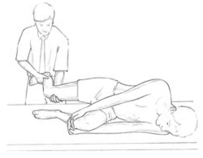
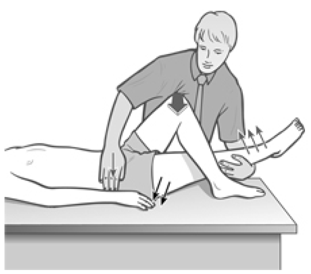
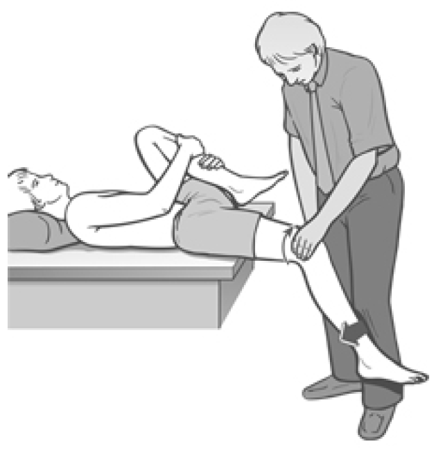


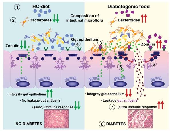

 #3: Chiropractic Strengthens And Repairs Joints.
 It Treats The Underlying Cause
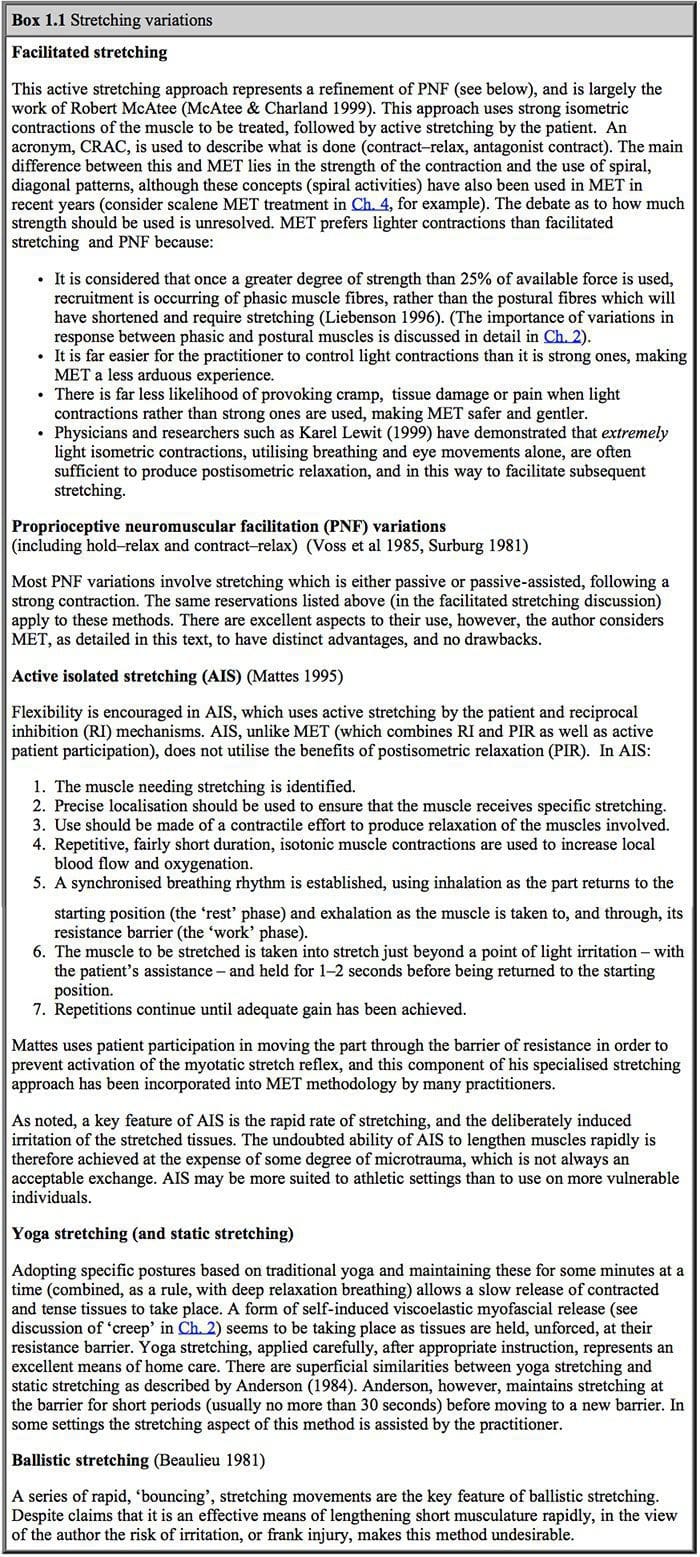 Edward Stiles

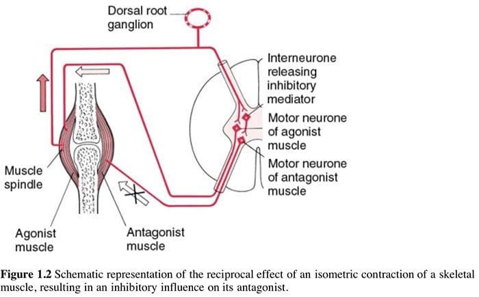 Liebenson (1996) discusses both the benefits of, and the mechanisms involved in, use of muscle energy techniques (which he terms �manual resistance techniques�, or MRT):
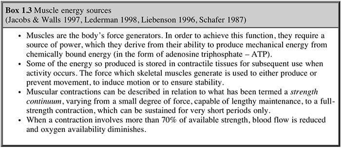 Using agonist or antagonist? (Box 1.4)
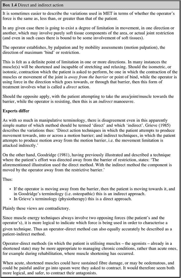
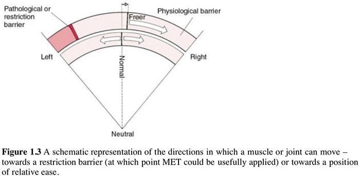 What Is Happening?
 This stretches the muscles which are contracting (TFL shown in example) thereby inducing a degree of controlled microtrauma, with the aim of increasing the elastic potential of shortened or fibrosed tissues.
 Muscle energy techniques are designed to assist in this endeavor and, as discussed above, also provides an excellent method for assisting in the toning of weak musculature, should this still be required, after the
 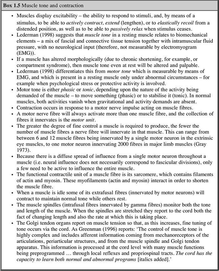 The relevance of this to soft tissue techniques is explained as follows:


