Chiropractic care is enjoying an upsurge in popularity as more people are veering away from invasive procedures and pharmaceuticals in favor of more natural treatment options for their pain. A recent Gallup poll for Palmer Chiropractic College confirms this. Of the people surveyed:
More than 35.5 million people said they sought chiropractic care within the 12 months (the Gallup survey was conducted from February 8, 2016, through March 11, 2016).
95% said chiropractic was effective in treating their condition.
89% said they would recommend chiropractic to friends and family.
97% said that they would likely see a chiropractor for neck or back pain.
88% said that chiropractic care is a good value for the money.
Non-Drug Treatments for Back Pain should be Sought First
In April 2017, the American College of Physicians published their updated guidelines for managing and treating low back pain. In it, they recommended seeking non-drug treatments which include the application of heat, exercise, stress reduction, and spinal manipulation before turning to medications. The purpose is to steer patients away from unnecessary medicating and toward healthier, more natural options as the first line of defense in managing low back pain.
Another study published in2017 in the Journal of the American Medical Association supported spinal manipulation as a preferred first treatment option over pharmaceuticals for patients with acute low back pain. While these recommendations are primarily in response to the opioid epidemic, it is also in response to the numerous studies that show chiropractic care is safe and effective for back pain management.
Chiropractic is a Healthier, Safer Option
The Centers for Disease Control (CDC) reports that opioid abuse and overdose has reach epidemic proportions. An estimated 91 Americans die every day from an opioid overdose. This includes opioids obtained by a prescription from a doctor.
Popular pain medications like oxycodone and hydrocodone have seen steadily increasing abuse, overdose, and death rates over the past two decades. In fact, deaths caused by an overdose of these drugs as well as methadone and others, have quadrupled since 1999.
These medications are highly addictive and have many unpleasant and even dangerous side effects. While most states are taking aggressive steps to curb the over-prescribing of these medications, patients are still finding ways around the prescribing guidelines and restrictions. This is a compelling reason for patients to seek natural, non-medicinal pain relief options first � and for their doctors to recommend them.
More People are Turning to Natural Remedies
Many patients are simply getting fed up with invasive treatments and numerous pills, causing them to turn to remedies that are more natural. One benefit of chiropractic and other natural remedies is that they rarely treats only the symptom.
Instead, it addresses the root of the problem to treat the cause. This approach has many benefits beyond being medication free and non-invasive. When the cause of the condition or problem is corrected, it can eliminate other troubling symptoms as well.
Chiropractic is a Healthier, Whole Body Option
A patient who sees their chiropractor for low back pain may find that after a few treatments their headaches, constipation, and digestive issues are also resolved. This is because chiropractic treats the body as a whole, unlike traditional medicine that tends to treat it in parts.
In the body, everything is connected, so a problem in one area could easily cause problems in other areas. By correcting the root of the problem, the patient receives more rounded healthcare.
Where traditional medicine will often opt to prescribe a pain pill for lower back pain, chiropractic care looks for the cause of the back pain and treats the pain symptom from the cause. The chiropractor may advise lifestyle changes, changes in diet, and even recommend supplements. With chronic health conditions like diabetes, heart disease, and obesity increasing at an alarming rate, chiropractic care offers a solution that treats the body as a whole in a more natural, safer way.
Sciatica is a set of symptoms characterized by discomfort and pain along the length of the sciatic nerve, which runs down the buttocks, hips, and thighs, into the feet and the legs. Also known as sciatic nerve pain, sciatica is brought on by the compression or impingement of the nerve through harms and/or ailments such as a herniated disc. The patients in the following video describe the way their quality of life has influenced. After getting chiropractic care with Dr. Alex Jimenez, chiropractor, patients talk how treatment has helped them achieve pain relief from their sciatica. Sciatic nerve pain is one of the most frequent health issues. The people highly recommend Dr. Alex Jimenez as the non-invasive pick for sciatica.
Nerve Pain Therapy
We are blessed to present to you�El Paso�s Premier Wellness & Injury Care Clinic.
As El Paso�s Chiropractic Rehabilitation Clinic & Integrated Medicine Center,�we passionately are focused on treating patients after frustrating injuries and chronic pain syndromes. We focus on improving your ability through flexibility, mobility and agility programs tailored for all age groups and disabilities.
We want you to live a life filled with more energy, positive attitude, better sleep, less pain, proper body weight and educated on how to maintain this way of life.
I assure you, I will only accept the best for you�
If you have enjoyed this video and we have helped you in any way, please feel free to subscribe and recommend�us.
Being involved in an automobile accident can influence the quality of life of an individual. Whiplash, pain, and neck pain, among other frequent automobile accident injuries, can severely limit a victim’s capacity to participate and engage in their regular activities. Dr. Alex Jimenez, a doctor of chiropractic, focuses on the identification, therapy, and prevention of a number of accidents and/or aggravated conditions associated with the musculoskeletal and nervous system, including auto accident injuries. Chiropractic care is an alternative treatment option which utilizes manual manipulations and adjustments, one of methods and other treatment procedures, to help treat many different health issues. Patients describe how Dr. Alex Jimenez has helped them find relief from their neck pain and back pain after an auto accident. Patients highly recommend Dr. Alex Jimenez as the non-surgical selection for automobile accident injuries, among other health problems such as whiplash.
Personal Chiropractic Care
We are blessed to present to you�El Paso�s Premier Wellness & Injury Care Clinic.
As El Paso�s Chiropractic Rehabilitation Clinic & Integrated Medicine Center,�we passionately are focused on treating patients after frustrating injuries and chronic pain syndromes. We focus on improving your ability through flexibility, mobility and agility programs tailored for all age groups and disabilities.
We want you to live a life filled with more energy, positive attitude, better sleep, less pain, proper body weight and educated on how to maintain this way of life.
I assure you, I will only accept the best for you�
If you have enjoyed this video and we have helped you in any way, please feel free to subscribe and recommend�us.
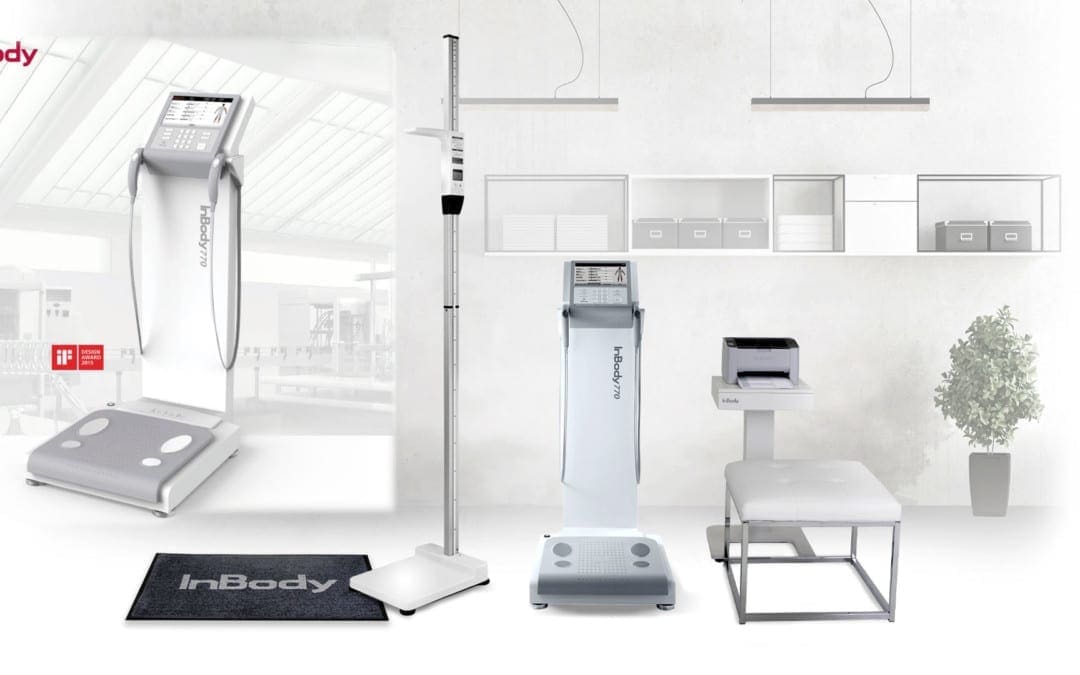
The InBody770 represents the apex of BIA technology. The InBody770 reports on body composition and also reports a complete breakdown of body water. There are 6 frequencies, which create a total of 30 impedance values. There is no other machine on the planet that can match the accuracy and dependability of the InBody770.
The InBody 770
Quick Tests
Measure fat mass, muscle mass, and body water levels in under a minute. No dunking, pinching, or discomfort. Stand on the device and hold the hand electrodes.
No Predicting
Just impedance is used to determine your body composition; No estimations like age and gender are needed or required to predict your composition.
Cloud Software
Automatically save all of the information from InBody to Lookin’Body Web, InBody’s cloud database management system. Easily view and manage customer’s results and see progress anytime.
Check Out The InBody 770 Result Sheet
To find out how to use this data in practice? Download the free guide here.
The knee is the largest, complicated joint in the human body. The knee joint is exposed to many different wellness issues. Knee injuries related to sports are a common issue that can affect playing time. Dr. Jimenez helps many athletes recover from knee injuries along with a variety of other sports-related injuries. Individuals share their experience of knee injury rehabilitation and treatment Dr. Jimenez has provided them. As an added benefit it has enhanced their�sports performance. Chiropractic rehabilitation is an alternative treatment option focusing on spinal adjustments and manual manipulations to deal with many different accidents and/or conditions related to the musculoskeletal and nervous system. Patients say Dr. Jimenez is the non-surgical, chiropractic choice.
Non-Surgical Injury Rehabilitation
We are blessed to present to you�El Paso�s Premier Wellness & Injury Care Clinic.
As El Paso�s Chiropractic Rehabilitation Clinic & Integrated Medicine Center,�we passionately are focused on treating patients after frustrating injuries and chronic pain syndromes. We focus on improving your ability through flexibility, mobility and agility programs tailored for all age groups and disabilities.
We want you to live a life filled with more energy, positive attitude, better sleep, less pain, proper body weight and educated on how to maintain this way of life.
I assure you, I will only accept the best for you�
If you have enjoyed this video and we have helped you in any way, please feel free to subscribe and recommend�us.
Painful symptoms may influence a person’s everyday physical actions. Patients with neck pain and spine pain caused by an assortment of health problems, including automobile accidents, have found tremendous relief from their symptoms with Dr. Alex Jimenez. Chiropractic care is a safe and effective, alternative treatment option which concentrates on the diagnosis, treatment, and prevention of injuries and/or conditions related to the musculoskeletal and nervous systems. Dr. Alex Jimenez has enhanced the quality of life of many patients by carefully restoring the integrity of their spine and improving debilitating symptoms. Patients highly recommend Dr. Alex Jimenez since the non-surgical selection for neck pain and spine pain, among other health problems, in El Paso, Tx.
Non-Surgical Care
�
We are blessed to present to you�El Paso�s Premier Wellness & Injury Care Clinic.
As El Paso�s Chiropractic Rehabilitation Clinic & Integrated Medicine Center,�we passionately are focused on treating patients after frustrating injuries and chronic pain syndromes. We focus on improving your ability through flexibility, mobility and agility programs tailored for all age groups and disabilities.
We want you to live a life filled with more energy, positive attitude, better sleep, less pain, proper body weight and educated on how to maintain this way of life.
I assure you, I will only accept the best for you�
If you have enjoyed this video and we have helped you in any way, please feel free to subscribe and recommend�us.
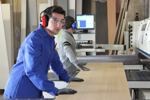
Assembly lines have long been the standard for factory workers. Henry Ford got the ball rolling on December 1, 1913, when he created the very first assembly line to mass produce a car. Workers stood for hours, doing the same tasks over and over. Although Ford took steps to reduce at least some of the damage, many factory workers still went home with aching backs and feet, migraines, fallen arches, and repetitive motion injuries.
Now, more than a hundred year later, some things have changed. According to the United States Department of Labor Bureau of Labor Statistics, there were 1,834,000 assembly line and fabrication jobs in 2014. Technology is better, and some tasks can be automated making some people�s jobs more manageable � and, unfortunately, eliminating some as well.
Despite the great strides in technology, there are still some things that haven�t changed all that much. The working conditions in many factories are often still not as healthy as they could be. Many workers are still required to stand for long periods of time and perform repetitive motions for hours without a break. This can lead to injuries, pain, and certain conditions that can cause immobility, inflexibility, and even disability. The good news is, chiropractic can help.
Working in a Standing Position Can Be Bad For Your Health
Many assembly line jobs require that the worker stand for long periods of time. While standing is a natural posture for humans and, by itself does not pose any real harm or health problem, working in a standing position every day isn�t good for you. It can lead to muscle fatigue, stiff shoulders and neck, swelling of the legs and feet, low back pain, varicose veins, fallen arches, and sore feet � to name a few.
Another problem with standing for extended or frequent periods of time without any breaks (such as walking or stretching) can cause the joints in the feet, knees, hips, and spine to become locked or immobilized temporarily. If the behavior continues, it can cause degenerative damage, leading to rheumatic diseases because the ligaments and tendons become damaged.
Other Assembly Line Related Health Problems
Barring traumatic injury due to an accident, working in a factory environment can cause problems with mobility, pain, and flexibility. The nature of the job places specific demands on the human body that can lead to certain types of injuries and health conditions, which include:
Repetitive motion injury � When a worker performs the same task that involves the same movements over and over, it can lead to certain types of injuries. Carpal tunnel is common repetitive motion injury.
Overexertion � Lifting, pulling, even standing can take a toll on the body, especially when it is done without adequate breaks. The person can get muscle fatigue, pulled muscles, and pulled tendons.
Body movement injuries � When the worker is continuously reaching, twisting, crawling, and bending, it can cause problems with the muscles and joints.
Chiropractic can Help Assembly Line Workers
Chiropractic care can help keep bodies flexible and help with range of motion. It is a very effective, non-invasive treatment for pain and can help with joint and muscular problems as well. Regular chiropractic treatments can help you better manage your body�s response to your work environment. It can also undo many of the ill effects that that type of work can cause.
You can enjoy more pain-free days without invasive surgeries or medications that leave you groggy, nauseous, or worse. When you sit down with your chiropractor, he or she will talk to you about your medical history as well as your current lifestyle. After a complete evaluation, you will be given a plan of action that may include lifestyle changes, dietary changes, and recommended supplements in addition to spinal manipulation. Chiropractic is all about whole body wellness, and that is what will help you perform better on your job and recover faster.
IFM's Find A Practitioner tool is the largest referral network in Functional Medicine, created to help patients locate Functional Medicine practitioners anywhere in the world. IFM Certified Practitioners are listed first in the search results, given their extensive education in Functional Medicine