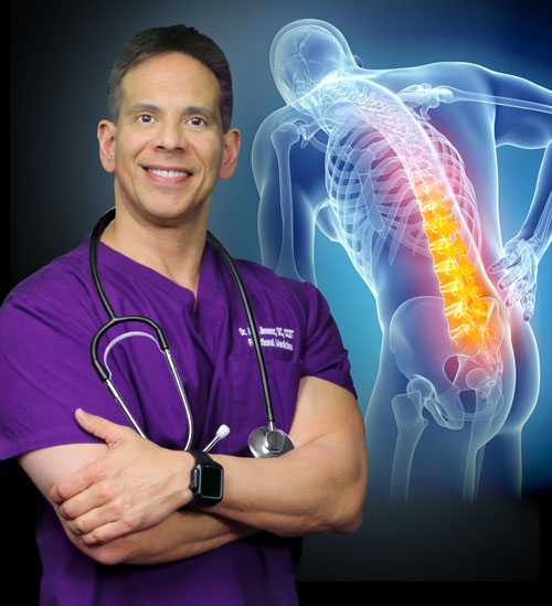
Patients involved in automobile accident injuries discuss how their symptoms affected their well-being. Through the utilization of chiropractic care, these patients describe how Dr. Alex Jimenez, brought them back to their original quality of life.
Chiropractic medicine focuses on the musculoskeletal system, which is what keeps the body in balance. A sudden impact to the body, such as from an auto accident throws the body out of whack. Chiropractic brings it all back together in harmony. These patients give their testimonies and praise Dr. Jimenez as the best injury chiropractor in El Paso TX.
El Paso Back Clinic
We are blessed to present to you�El Paso�s Premier Wellness & Injury Care Clinic.
As El Paso�s Chiropractic Rehabilitation Clinic & Integrated Medicine Center,�we passionately are focused on treating patients after frustrating injuries and chronic pain syndromes. We focus on improving your ability through flexibility, mobility and agility programs tailored for all age groups and disabilities.
We want you to live a life filled with more energy, positive attitude, better sleep, less pain, proper body weight and educated on how to maintain this way of life.
I assure you, I will only accept the best for you�
If you have enjoyed this video and we have helped you in any way, please feel free to subscribe and recommend�us.
Many people with health issues associated with genetic SNPs due to abnormal methylation cycles, such as MTHFR, can tremendously benefit from DNA methylation support. Patients demonstrating imbalances in methylation metabolites, including elevated homocysteine, decreased SAMe, increased SAH, and a low SAMe:SAH ratio, can also benefit from DNA methylation support. Any individual who has insufficient levels of methyl donors due to poor nutrient status or malabsorption, especially with folate and vitamin B12 deficiencies, stress, hormone imbalances, and even older individuals, can benefit from DNA methylation support. �
Potential Risks of Methylation Supplements
Doctors and functional medicine practitioners commonly utilize natural supplementation for methylation support, however, many other healthcare professionals still frequently utilize synthetic supplementation. According to research studies, using synthetic supplements can cause a variety of health issues. Potential risks of using synthetic folic acid (FA) with unknown mechanisms include an increased risk for allergic diseases and IBD in children of mothers with an increased folic acid intake, impaired natural killer cell activity, insulin resistance in children, embryonic loss and growth delay, and diabetic comorbidity, among other health issues. � Moreover, using unmetabolized folic acid (FA) for methylation support can cause potential genotoxicity, according to research studies. Furthermore, the use of DHF, an intermediate of the DHFR enzyme, can inhibit thymidylate synthase and it can also inhibit MTHF, known as a pseudo MTHFR deficiency. Many more doctors and functional medicine practitioners prefer to utilize natural supplementation over synthetic supplementation for methylation support, to prevent any health issues due to potential risks. The use of 5mTHF and methylcobalamin (vitamin B12) is commonly utilized in functional medicine to avoid folic acid problems. � However, although the use of 5mTHF and methylcobalamin (vitamin B12) bypasses MTHFR enzyme deficits, not enough research studies have been conducted regarding the long-term safety and effectiveness of high doses of 5mTHF or methyl-B12 for methylation support. It’s essential for patients to seek professional help from a qualified and experienced doctor and functional medicine practitioner to determine the best type of supplementation for methylation support. Supplements for methylation support are recommended alongside proper nutrition, fitness, and lifestyle habits to naturally promote DNA methylation status and activity. �
Folate/B12 and Autism: Possible Connection
A recent press release of preliminary evidence findings from the Johns Hopkins research study determined that in 1,391 mother-child pairs in a Boston Birth Cohort, the highest maternal levels of vitamin B12, more than 600 pmol/L, and folate, equal to 59 nmol/L, had an increased risk of causing ASD. The risks were ultimately higher when both were combined. The research study demonstrated no risk difference based on MTHFR genotype or homocysteine. The full-paper has not been published and further research studies are still required to determine the outcome measures of supplementation for methylation support for pregnant women and children. �
Promoting methylation support is an essential process towards maintaining overall health and wellness. Although several research studies have demonstrated potential risks for certain types of supplementation for methylation support, healthcare professionals can help determine the proper supplements to promote methylation support. The purpose of the following article is to discuss the potential risks of supplementation for methylation support. It’s fundamental to understand how nutrition, lifestyle habits, supplements, and medicines, can improve DNA methylation, health and wellness. – Dr. Alex Jimenez D.C., C.C.S.T. Insight
Smoothies and Juices for Methylation Support
�
While many healthcare professionals can recommend nutritional guidelines and lifestyle modifications to improve methylation support, there�are several options you can try yourself at home. As described above, methylation support supplementation should be determined by a healthcare professional. Smoothies and juices are a fast and easy way to include all the necessary nutrients you need for methylation support without any side-effects. The smoothies and juices below are part of the Methylation Diet Food Plan.
Sea Green Smoothie Servings: 1 Cook time: 5-10 minutes
1/2 cup cantaloupe, cubed
1/2 banana
1 handful of kale or spinach
1 handful of Swiss chard
1/4 avocado
2 teaspoons spirulina powder
1 cup of water
3 or more ice cubes
Blend all ingredients in a high-speed blender until completely smooth and enjoy!
1/2 cup blueberries (fresh or frozen, preferably wild)
1 medium carrot, roughly chopped
1 tablespoon ground flaxseed or chia seed
1 tablespoons almonds
Water (to desired consistency)
Ice cubes (optional, may omit if using frozen blueberries)
Blend all ingredients in a high-speed blender until smooth and creamy. Best served immediately!
Sweet and Spicy Juice Servings: 1 Cook time: 5-10 minutes
1 cup honeydew melons
3 cups spinach, rinsed
3 cups Swiss chard, rinsed
1 bunch cilantro (leaves and stems), rinsed
1-inch knob of ginger, rinsed, peeled and chopped
2-3 knobs whole turmeric root (optional), rinsed, peeled and chopped
Juice all ingredients in a high-quality juicer. Best served immediately!
Ginger Greens Juice Servings: 1 Cook time: 5-10 minutes
1 cup pineapple cubes
1 apple, sliced
1-inch knob of ginger, rinsed, peeled and chopped
3 cups kale, rinsed and roughly chopped or ripped
5 cups Swiss chard, rinsed and roughly chopped or ripped
Juice all ingredients in a high-quality juicer. Best served immediately!
Zesty Beet Juice Servings: 1 Cook time: 5-10 minutes
1 grapefruit, peeled and sliced
1 apple, washed and sliced
1 whole beet, and leaves if you have them, washed and sliced
1-inch knob of ginger, rinsed, peeled and chopped
Juice all ingredients in a high-quality juicer. Best served immediately!
Protein Power Smoothie Serving: 1 Cook time: 5 minutes
1 scoop protein powder
1 tablespoon ground flaxseed
1/2 banana
1 kiwi, peeled
1/2 teaspoon cinnamon
Pinch of cardamom
Non-dairy milk or water, enough to achieve desired consistency
Blend all ingredients in a high-powered blender until completely smooth. Best served immediately!
ProLon� Fasting Mimicking Diet
Balanced methylation support can be achieved through proper nutrition. The ProLon� fasting mimicking diet offers a 5-day meal program which has been individually packed and labeled to serve the foods you need for the FMD in precise quantities and combinations. The meal program is made up of ready-to-eat or easy-to-prepare, plant-based foods, including bars, soups, snacks, supplements, a drink concentrate, and teas. The products are scientifically formulated and great tasting. Before starting the ProLon� fasting mimicking diet, 5-day meal program, please make sure to talk to a healthcare professional to find out if the FMD is right for you. The ProLon� fasting mimicking diet can help promote methylation support, among a variety of other healthy benefits.
�
Many doctors and functional medicine practitioners can recommend nutritional advice and/or guidelines to help improve DNA methylation. Proper nutrition and lifestyle habits can ultimately help improve DNA methylation. Nutrient deficiencies can ultimately cause DNA methylation deficits which may cause a variety of health issues, according to research studies. The scope of our information is limited to chiropractic, musculoskeletal and nervous health issues as well as functional medicine articles, topics, and discussions. To further discuss the subject matter above, please feel free to ask Dr. Alex Jimenez or contact us at 915-850-0900�.
Curated by Dr. Alex Jimenez
Additional Topic Discussion:�Acute Back Pain
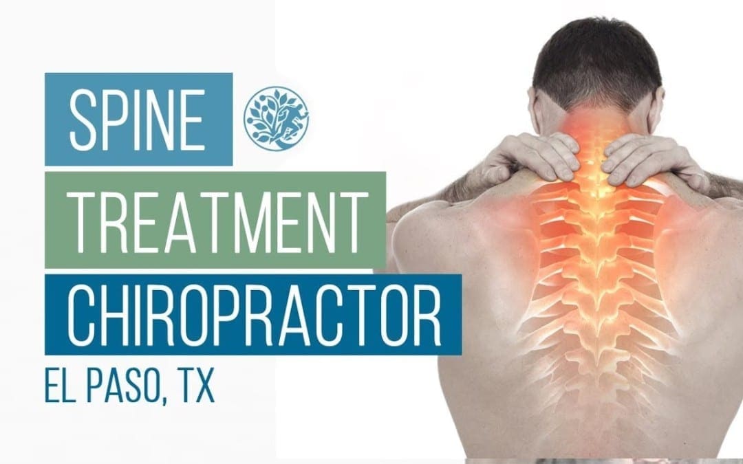
Back pain�is one of the most prevalent causes of disability and missed days at work worldwide. Back pain attributes to the second most common reason for doctor office visits, outnumbered only by upper-respiratory infections. Approximately 80 percent of the population will experience back pain at least once throughout their life. Your spine is a complex structure made up of bones, joints, ligaments, and muscles, among other soft tissues. Injuries and/or aggravated conditions, such as�herniated discs, can eventually lead to symptoms of back pain. Sports injuries or automobile accident injuries are often the most frequent cause of back pain, however, sometimes the simplest of movements can have painful results. Fortunately, alternative treatment options, such as chiropractic care, can help ease back pain through the use of spinal adjustments and manual manipulations, ultimately improving pain relief.
Formulas for Methylation Support
�
XYMOGEN�s Exclusive Professional Formulas are available through select licensed health care professionals. The internet sale and discounting of XYMOGEN formulas are strictly prohibited.
Proudly,�Dr. Alexander Jimenez makes XYMOGEN formulas available only to patients under our care.
Please call our office in order for us to assign a doctor consultation for immediate access.
If you are a patient of Injury Medical & Chiropractic�Clinic, you may inquire about XYMOGEN by calling 915-850-0900.
�
For your convenience and review of the XYMOGEN products please review the following link.*XYMOGEN-Catalog-Download
* All the above XYMOGEN policies remain strictly in force.
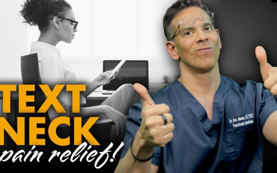
Weighing an average of 10 pounds, the human head is heavier than most people think. The head can put a great deal of pressure on the neck when it is placed in different positions for prolonged periods � especially regularly looking down at your phone.
The damage caused to the neck from time spent staring at mobile screens has been given the name �Text Neck�, and it is a growing problem among not only teens but for everyone.
Regularly Looking Down at Your Phone Is Cause For Neck Injury
Your body is well-designed to bear the weight of your head when you maintain good posture � but tilting your head down to look at your phone is not good posture. In fact, for every inch you move angle your head downward, you double the pressure on your spine.
Looking at your phone can put an extra ten or twenty pounds of pressure on your neck. That would be worth noting even if you only did it occasionally, but most people spend hours looking at their phones throughout the day. That amounts to hours of excessive pressure on the soft tissues that make up your neck � pressure that will inevitably lead to inflammation and discomfort if left unchecked.
According to this article featured in the Washington Post, the pressure you put on your neck by bending and staring at your phone is much like bending your finger back as far as it will go, and then keeping it in that position for approximately an hour. Day after day, such stress is bound to lead to complications.
Resulting Injuries from Text Neck
The strain put on the neck by text neck is enough to cause mild to severe injuries, including:
Sore muscles
Inflamed Tissues
Pinched nerves
Herniated discs
Elimination of the natural curve of the neck
These injuries can cause considerable pain and discomfort and may lead to further health complications. They can lead to neck and back pain that can last for years.
Tips to Avoid Text Neck
Smartphones offer numerous benefits and opportunities for enjoyment, so it is unlikely that most people will stop using them. Luckily, there are things you can do to protect yourself, including:
Work Your Eyes, Not Your Neck
One of the simplest ways to avoid text neck is to look down with your eyes instead of tilting your head down. While it may not be practical to always use this technique, it is certainly useful in many circumstances. Your eyes can tilt down with little effort and can allow you to lessen the tilt of your neck as you use your phone.
Strength Train your Neck & Shoulder Muscles
You will inevitably do some head tilting as you use your phone throughout the day. Strengthening the muscles that support your head is one way to protect your delicate neck tissues and maintain mobility. Simple exercises like turning your head each way repeatedly and using your hands to provide resistance can make your neck much stronger. Your shoulders also provide a lot of support for your neck. Shoulder exercises can increase the stability of your neck as well.
Have an Awareness of Your Head Position
Just maintaining awareness of how your head is tilted as you use your phone can help you avoid excessive tilting. Practice looking at your smartphone with your head upright to remind yourself of what good posture feels like, and pay attention during the times you deviate from good posture.
Chiropractic Treatment
If you are experiencing neck pain from text neck, or from any other type of injury, chiropractic can help. Please contact us now to schedule your appointment and get some relief!
Health issues associated with methylation deficits have become a well-known clinical problem for many healthcare professionals and patients. Numerous research studies have demonstrated that methylation deficits can cause a wide array of associated health issues, including ADD/ADHD, addiction, allergies, Alzheimer�s disease, anxiety, asthma, atherosclerosis, autism, behavioral problems, bipolar disorder, cancers, chemical sensitivity, chronic fatigue, cleft palate, diabetes, dementia, depression, Down syndrome, hypertension, fertility problems, fibromyalgia, insomnia, multiple sclerosis, neuropathy, Parkinson�s disease, schizophrenia, and thyroid disease. �
What Causes DNA Methylation Deficits?
Many healthcare professionals and patients ask themselves, why do these methylation deficits occur? The most common cause of methylation deficits frequently involves nutrient deficiencies, such as folate/folic acid deficiencies and vitamin B12 deficiencies. As previously discussed by doctors and functional medicine practitioners, methylation deficits caused by nutrient deficiencies can occur due to inadequate food or drink intake and malabsorption. Moreover, the increased consumption of processed foods, as well as following a vegan diet, can also cause folate/folic acid deficiencies and vitamin B12 deficiencies, among other nutrient deficiencies. � Furthermore, competition for the utilization of methyl donors in a variety of bodily processes can also be a common cause of methylation deficits. By way of instance, SAMe is essential for DNA methylation and other bodily functions, however, stress, the utilization of several drugs and/or medications like L-Dopa, hormones, inflammation, detoxification, and nutrient metabolism can also cause methylation deficits. Niacin, Selenium, and Phosphatidylethanolamine are examples of nutrients which are metabolized through DNA methylation. Competition for the utilization of methyl donors can cause problems associated with methylation deficits. � Methylation inhibitors can also cause methylation deficits. When SAMe is produced, it’s then metabolized into SAH, a powerful DNA methylation inhibitor of SAMe-dependent methyltransferases which includes DNMTs. Numerous research studies have also demonstrated that an individual’s genotype can ultimately cause methylation deficits and other health issues. Healthcare professionals can evaluate SNPs, such as MTHFR, C677T, and A1298C, in patients to demonstrate enzyme status and activity. Finally, aging can also cause methylation deficits. The process of aging alongside the causes above can ultimately cause methylation deficits. �
How to Diagnose DNA Methylation Deficits
Healthcare professionals can diagnose methylation deficits in patients through nutrition physical exams. Nutrition physical exams can help guide healthcare professionals and patients alike to determine which deficiencies may be causing DNA methylation deficits. By way of instance, nutrient deficiencies in DHA may manifest as xeroderma, dermatitis, keratosis pilaris, sensory neuropathy, and/or poor wound healing. Nutrient deficiencies in Zinc may manifest loss of taste/smell, delayed wound healing, oral candidiasis, nail changes like leukonychia, koilonychia, Beau�s lines, and onychorrhexis. Nutrient deficiencies in Magnesium may manifest as muscle spasms including blepharospasm, tremor, cardiac arrhythmias and nail changes including onychorrhexis. Last but not least, nutrient deficiencies in Potassium may manifest muscle spasms like blepharospasm or tremors and cardiac arrhythmia. �
Promoting methylation support is an essential process towards maintaining overall health and wellness. DNA methylation is involved in a variety of bodily functions. Maintaining and regulating healthy methylation can help prevent a variety of health issues, including methylation deficits caused by nutrient deficiencies, among other problems. The purpose of the following article is to discuss the causes of methylation deficits. It’s fundamental to understand how nutrition, lifestyle habits, supplements, and even medicines, can improve DNA methylation as well as to promote overall health and wellness. – Dr. Alex Jimenez D.C., C.C.S.T. Insight
Smoothies and Juices for Methylation Support
�
While many healthcare professionals can recommend nutritional guidelines and lifestyle modifications to improve methylation support, there are several options you can try yourself at home. As described above, methylation support supplementation should be determined by a healthcare professional. Smoothies and juices are a fast and easy way to include all the necessary nutrients you need for methylation support without any side-effects. The smoothies and juices below are part of the Methylation Diet Food Plan.
Sea Green Smoothie Servings: 1 Cook time: 5-10 minutes
1/2 cup cantaloupe, cubed
1/2 banana
1 handful of kale or spinach
1 handful of Swiss chard
1/4 avocado
2 teaspoons spirulina powder
1 cup of water
3 or more ice cubes
Blend all ingredients in a high-speed blender until completely smooth and enjoy!
1/2 cup blueberries (fresh or frozen, preferably wild)
1 medium carrot, roughly chopped
1 tablespoon ground flaxseed or chia seed
1 tablespoons almonds
Water (to desired consistency)
Ice cubes (optional, may omit if using frozen blueberries)
Blend all ingredients in a high-speed blender until smooth and creamy. Best served immediately! � Sweet and Spicy Juice Servings: 1 Cook time: 5-10 minutes
1 cup honeydew melons
3 cups spinach, rinsed
3 cups Swiss chard, rinsed
1 bunch cilantro (leaves and stems), rinsed
1-inch knob of ginger, rinsed, peeled and chopped
2-3 knobs whole turmeric root (optional), rinsed, peeled and chopped
Juice all ingredients in a high-quality juicer. Best served immediately! � Ginger Greens Juice Servings: 1 Cook time: 5-10 minutes � 1 cup pineapple cubes � 1 apple, sliced � 1-inch knob of ginger, rinsed, peeled and chopped � 3 cups kale, rinsed and roughly chopped or ripped � 5 cups Swiss chard, rinsed and roughly chopped or ripped Juice all ingredients in a high-quality juicer. Best served immediately!
� Zesty Beet Juice Servings: 1 Cook time: 5-10 minutes
1 grapefruit, peeled and sliced
1 apple, washed and sliced
1 whole beet, and leaves if you have them, washed and sliced
1-inch knob of ginger, rinsed, peeled and chopped
Juice all ingredients in a high-quality juicer. Best served immediately!
� Protein Power Smoothie Serving: 1 Cook time: 5 minutes
1 scoop protein powder
1 tablespoon ground flaxseed
1/2 banana
1 kiwi, peeled
1/2 teaspoon cinnamon
Pinch of cardamom
Non-dairy milk or water, enough to achieve desired consistency
Blend all ingredients in a high-powered blender until completely smooth. Best served immediately!
ProLon� Fasting Mimicking Diet
Balanced methylation support can be achieved through proper nutrition. The ProLon� fasting mimicking diet offers a 5-day meal program which has been individually packed and labeled to serve the foods you need for the FMD in precise quantities and combinations. The meal program is made up of ready-to-eat or easy-to-prepare, plant-based foods, including bars, soups, snacks, supplements, a drink concentrate, and teas. The products are scientifically formulated and great tasting. Before starting the ProLon� fasting mimicking diet, 5-day meal program, please make sure to talk to a healthcare professional to find out if the FMD is right for you. The ProLon� fasting mimicking diet can help promote methylation support, among a variety of other healthy benefits. �
Many doctors and functional medicine practitioners can recommend nutritional advice and/or guidelines to help improve DNA methylation. Proper nutrition and lifestyle habits can ultimately help improve DNA methylation. Nutrient deficiencies can ultimately cause DNA methylation deficits which may cause a variety of health issues, according to research studies. The scope of our information is limited to chiropractic, musculoskeletal and nervous health issues as well as functional medicine articles, topics, and discussions. To further discuss the subject matter above, please feel free to ask Dr. Alex Jimenez or contact us at 915-850-0900�. �
Curated by Dr. Alex Jimenez �
Additional Topic Discussion:�Acute Back Pain
Back pain�is one of the most prevalent causes of disability and missed days at work worldwide. Back pain attributes to the second most common reason for doctor office visits, outnumbered only by upper-respiratory infections. Approximately 80 percent of the population will experience back pain at least once throughout their life. Your spine is a complex structure made up of bones, joints, ligaments, and muscles, among other soft tissues. Injuries and/or aggravated conditions, such as�herniated discs, can eventually lead to symptoms of back pain. Sports injuries or automobile accident injuries are often the most frequent cause of back pain, however, sometimes the simplest of movements can have painful results. Fortunately, alternative treatment options, such as chiropractic care, can help ease back pain through the use of spinal adjustments and manual manipulations, ultimately improving pain relief. �
Formulas for Methylation Support
�
XYMOGEN�s Exclusive Professional Formulas are available through select licensed health care professionals. The internet sale and discounting of XYMOGEN formulas are strictly prohibited. � Proudly,�Dr. Alexander Jimenez makes XYMOGEN formulas available only to patients under our care.
Please call our office in order for us to assign a doctor consultation for immediate access.
If you are a patient of Injury Medical & Chiropractic�Clinic, you may inquire about XYMOGEN by calling 915-850-0900. �
� For your convenience and review of the XYMOGEN products please review the following link.*XYMOGEN-Catalog-Download
* All the above XYMOGEN policies remain strictly in force.
Ms. Hermosillo participates in fitness classes and follows healthy lifestyle habits. She participates in a variety of physical activities to achieve tip-top health. As a result, however, Ms. Hermosillo began to experience neck pain and back pain.
The symptoms were beginning to affect her life both professionally and personally. Ms. Hermosillo heard about local 20+ year El Paso, Texas chiropractor, Dr. Alex Jimenez that brought unbelievable relief of her symptoms! El Paso Back Clinic follows a safe and effective treatment focusing on a variety of health issues associated with the musculoskeletal and nervous systems. Ms. Hermosillo recommends Dr. Jimenez for personalized spine therapy for neck pain, back pain, and other conditions.
El Paso Back Clinic
We are blessed to present to you�El Paso�s Premier Wellness & Injury Care Clinic.
As El Paso�s Chiropractic Rehabilitation Clinic & Integrated Medicine Center,�we passionately are focused on treating patients after frustrating injuries and chronic pain syndromes. We focus on improving your ability through flexibility, mobility and agility programs tailored for all age groups and disabilities.
We want you to live a life filled with more energy, positive attitude, better sleep, less pain, proper body weight and educated on how to maintain this way of life.
Only the best for you!
If you have enjoyed this video and we have helped you in any way, please feel free to subscribe and recommend�us.
Sitting for long periods is associated with early death � upsetting news for the millions of Americans who spend extensive time sitting as they work and as they relax after work. A recent study conducted at Columbia� University has found that prolonged sitting destroys your health. Regular exercise, although great for you, failed to significantly reduce the risks associated with prolonged sitting.
Fortunately, there are things you can do to minimize the time you spend sitting. Experts recommend taking a movement break every 30 minutes to protect your health.
The Sitting Study
The study used hip-mounted accelerometers to measure how much time study participants � nearly 8,000 individuals � spent in sedentary positions. Over four years researchers tracked participants and recorded the number of deaths among participants. The study found that increased sedentary time leads to an increased risk of death from any cause. The risk of death went up regardless of how old the participants were or how often they exercised.
The study found that participants who sat less than 30 minutes had a 55% lower risk of death in comparison to participants who sat for longer periods of time.
Why Does Sitting Ruin Your Health?
Finding out that sitting for extended periods so greatly increases your risk of death is startling for many people. The next question is, �Why?� Unfortunately, the exact reasons are far from clear. The study was not meant to uncover the reasons why, so there is limited information to explain what occurs in the body to increase the risk of death. It will take further studies to provide an answer.
Methods to Protect Your Health
Although the exact reasons why sitting increases the risk of mortality are not yet clear, the recommendation to take a break from sitting every 30 minutes does offer an actionable step to protect your health. There are several other ways you can limit your sitting, including:
Sit & Standing Desks
There are a number of different desks designed to be used from a standing position, including stationary standing desks and sit to stand desks � both manually operated and motorized. Standing desks allow you to work exactly as you would at a sitting desk, only while standing. They take some getting used to, but most people who try them out find that they are an easy way to minimize time spent sitting.
When you purchase a standing or sit to stand desk, it may take some time to build up your stamina to use the desk for extended periods of time. Try standing for short periods, then sitting, then standing again to avoid excessive fatigue.
Apps
A number of apps are available to help you track your activity and make gradual lifestyle adjustments to increase your activity levels. Apple�s Health app includes an Activity monitor and coach that collects data from your iPhone, Apple Watch and third-party apps to let you know how much you are moving and to remind you to move more. You can set reminders on your app to tell you to move � such as every 30 minutes � as a way to ensure you avoid sitting for prolonged periods of time.
Timers
The simplest way to avoid sitting for more than 30 minutes is to set a timer on your phone or use a separate timer, to go off after 30 minutes of sitting. Once the timer goes off, engage in brisk walking or some other activity to get your heart rate up for five minutes. Then you can go back to sitting � after you set your timer again.
Helping You Stay as Healthy as Possible
Our chiropractic team is dedicated to helping you enjoy a long and healthy life. Please contact us to schedule an appointment for a comprehensive exam and targeted health advice from an experienced chiropractor.
Mr. Manuel Lozano was involved in a car accident which resulted in back pain. The painful symptoms began to manifest, and Mr. Lozano struggled to participate in his everyday physical activities. That’s when Manuel Lozano found chiropractic care with Dr. Alex Jimenez. Manuel Lozano received chiropractic care with Dr. Jimenez and found tremendous pain relief from his symptoms. Manuel Lozano discusses how Dr. Jimenez taught him how to improve his overall quality of life. Manuel Lozano highly recommends Dr. Jimenez, a chiropractor in El Paso, TX, as the non-surgical choice for automobile accident injuries.
El Paso Back Clinic
We are blessed to present to you�El Paso�s Premier Wellness & Injury Care Clinic.
As El Paso�s Chiropractic Rehabilitation Clinic & Integrated Medicine Center,�we passionately are focused on treating patients after frustrating injuries and chronic pain syndromes. We focus on improving your ability through flexibility, mobility and agility programs tailored for all age groups and disabilities.
We want you to live a life filled with more energy, positive attitude, better sleep, less pain, proper body weight and educated on how to maintain this way of life.
I assure you, I will only accept the best for you�
If you have enjoyed this video and we have helped you in any way, please feel free to subscribe and recommend�us.
IFM's Find A Practitioner tool is the largest referral network in Functional Medicine, created to help patients locate Functional Medicine practitioners anywhere in the world. IFM Certified Practitioners are listed first in the search results, given their extensive education in Functional Medicine