If you are experiencing any of these problems or situations in your body, why don’t you try foods that are rich with antioxidants by using the ORAC.
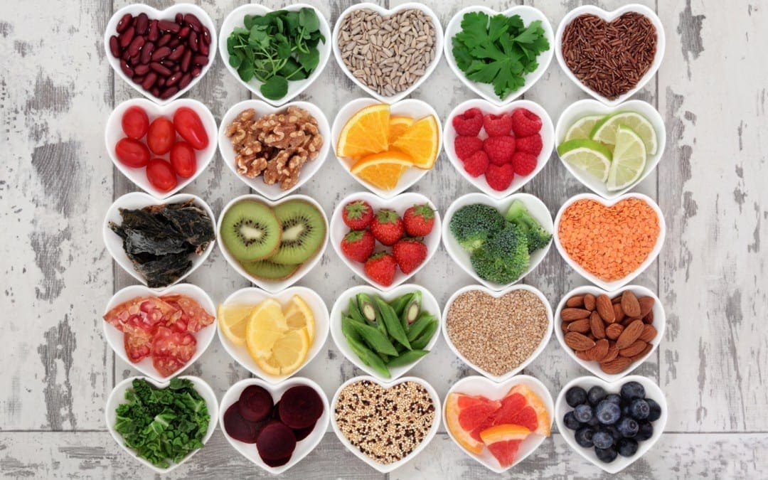
Throughout scientific literature, it has been well established at how necessary the daily vegetable and fruit intake is for anyone’s physiological health and wellbeing. Not many people understand the importance of consuming more vegetables and fruit in their diet. Many healthcare professionals will often prescribe patients fruits and vegetables as well as more exercising, although some do not often explain why they should eat more fruits and vegetables.
Fruits and vegetables are filled with antioxidants like polyphenols, phytochemicals, and nutrients that are measured by their ORAC value. ORAC or oxygen radical absorbance capacity is a measurement of any food, spice, or nutrient/substance�s ability to get rid of oxidative free radicals in a test tube. What is interesting is that free radicals and reactive oxidative species (ROS) are synonymous. What this means is that essentially free radicals are unstable molecules that are produced under a normal metabolic process.
Free radicals can take the electrons from other molecules that are nearby in the body, which will cause a chain reaction consisting of oxidative stress and damage. When the body has continuous oxidative damage from inflammation, it can be a significant culprit to many degenerative conditions and age-related diseases, harming the person.
Many people would consider any medical diagnosis of any illnesses or diseases to be part of “normal” aging. With any conditions, people will accept and adapt to it, however, it may come as a shock for many people to find out that �age-related diseases� are not inevitable but a sign of unhealthy aging. Studies found out that cultures living in the blue zones are the healthiest cultures that have the highest life expectancy around the world. With these healthy cultures, they are a testament to anyone that aging does not equal to disease and poor health.
With healthy aging, it is defined as the metabolic age of being “younger than” the biological and chronological age. A journal review mentioned how to reduce systemic inflammation is the key to longevity and can prevent the development of the chronic disease. Studies were researching how risk factors for all-cause mortality and cardiovascular disease incidences in elders that live in Northern Greece. They found out that drinking coffee and tea, consuming fruit in vast quantities, and exclusive usage of olive oil was associated with cardiovascular diseases.
For anyone that is living in the blue zone, their diets and lifestyle factors are the primary determinants for their health and are the essential factors of longevity. The diets from the blue zones are usually plant-based, high in fiber and loads of vegetables, while also incorporating locally-caught fish that are rich in omega-3 fatty acids, this diet is similar to the Mediterranean-style diet.
ORAC Nutrients
There are a few specific vitamins and dietary compounds in food that have some of the highest ORAC values. One of the vitamins is vitamin E subfractions, especially delta-and gamma tocotrienols, which have a higher ORAC value than alpha-tocopherol while respectively contributing to substantially more significant health benefits. The two foods that are very high on the ORAC charts are cherries and elderberries. These two are highly nutritious due to phytonutrient-rich components like anthocyanins and quercetin. For vegetables, leafy greens like spinach and lycopene possess a high antioxidant profile.
In early 2019, a study showed how the consumption of spinach could help modulate the metabolism of lipids in the liver, while also finding out that the hepatic accumulation of alpha-carotene, beta-carotene, and lutein was correlated with blood glucose and total serum cholesterol. The many other fruits and vegetables that have antioxidants components that can help damp oxidative stress in the body and are highly nutritious for anyone to eat.
With many high ORAC fruits and vegetables can be condensed and form into supplements inside capsules and powders. Research shows that powdered greens that contain fruits and vegetable blends have become very popular amongst the health and wellness community. It makes sense due to that these capsules are loaded with antioxidant-rich phytochemicals that support the individual’s overall health. Since densely packed fruits and vegetable powders are great tools, some individuals would argue that these powders are not adequate amounts for a person�s diet.
With antioxidant-rich green powders, they are helpful for older adults who have limited mobility. For anyone that cannot prepare their meals, the powders can be blended with water or added to smoothies for optimal nutrition for the body. Another benefit that green powders have is that they can help counteract the deleterious effects of process food intake. For anyone unwilling to change their dietary patterns if they are taking particular nutrient-depleting medicine, as well as anyone who smokes or drinks alcohol, will suffer from chronic diseases in their body.
Conclusion
With the abundance of other compounds and foods that have a high ORAC value, any foods that have these antioxidants can help the body by removing inflammation in the joints and the gut, combating free radicals, and getting rid of oxidative stress that will cause systemic inflammation. By using the ORAC value system, finding food that has high antioxidant properties are essential. Even incorporating more fruits and vegetables in the diet is necessary for a healthy, functional body. Some products are formulated to help counter the metabolic effects of stress and support the gastrointestinal system in the body.
The scope of our information is limited to chiropractic, musculoskeletal, and nervous health issues or functional medicine articles, topics, and discussions. We use functional health protocols to treat injuries or disorders of the musculoskeletal system. Our office has made a reasonable attempt to provide supportive citations and has identified the relevant research study or studies supporting our posts. We also make copies of supporting research studies available to the board and or the public upon request. To further discuss the subject matter above, please feel free to ask Dr. Alex Jimenez or contact us at 915-850-0900.
References:
Barclay, Eliza. �Eating To Break 100: Longevity Diet Tips From The Blue Zones.� NPR, NPR, 11 Apr. 2015, www.npr.org/sections/thesalt/2015/04/11/398325030/eating-to-break-100-longevity-diet-tips-from-the-blue-zones.
Elvira-Torales, Laura In�s, et al. �Ameliorative Effect of Spinach on Non-Alcoholic Fatty Liver Disease Induced in Rats by a High-Fat Diet.� International Journal of Molecular Sciences, MDPI, 3 Apr. 2019, www.ncbi.nlm.nih.gov/pubmed/30987167.
Hossain, Afzal, et al. �Enhancement of Antioxidant Quality of Green Leafy Vegetables upon Different Cooking Method.� Preventive Nutrition and Food Science, The Korean Society of Food Science and Nutrition, Sept. 2017, www.ncbi.nlm.nih.gov/pubmed/29043220.
Liu, Qing, et al. �Comparison of Antioxidant Activities of Different Grape Varieties.� Molecules (Basel, Switzerland), MDPI, 23 Sept. 2018, www.ncbi.nlm.nih.gov/pubmed/30249027.
Pisoschi, Aurelia Magdalena, et al. �Antioxidant Capacity Determination in Plants and Plant-Derived Products: A Review.� Oxidative Medicine and Cellular Longevity, Hindawi Publishing Corporation, 2016, www.ncbi.nlm.nih.gov/pubmed/28044094.
Team, DFH. �Cherries: Whats Not to Love?� Designs for Health, 5 Jan. 2018, blog.designsforhealth.com/cherries-whats-not-to-love.
Team, DFH. �Elevate the Immune System with Elderberry.� Designs for Health, 5 Jan. 2018, blog.designsforhealth.com/elevate-the-immune-system-with-elderberry-0.
Team, DFH. �Tocotrienols.� Designs for Health, 7 Dec. 2018, blog.designsforhealth.com/node/909.
Tsoupras, Alexandros, et al. �Inflammation, Not Cholesterol, Is a Cause of Chronic Disease.� Nutrients, MDPI, 12 May 2018, www.ncbi.nlm.nih.gov/pmc/articles/PMC5986484/.
Weil, Andrew. �ORAC: Scoring Antioxidants? – Dr. Weil.� DrWeil.com, 5 Aug. 2016, www.drweil.com/vitamins-supplements-herbs/vitamins/orac-scoring-antioxidants/.
Back pain during the holidays takes the joy out of everything.�All the�activities, events, and shopping can worsen spinal injuries or cause new ones.�And trying to power through the pain makes family traditions like tree-trimming, cooking, baking into a very cautionary experience.�When selecting a gift for friends or loved ones with back or neck pain they would appreciate something designed to help them feel better.
Although the massage devices and foam roller sets don�t work miracles they can ease the stress and discomfort that comes with living with pain.
There is a wealth of products that:
Support the back
Relieve muscle tension
Improve sleep quality
Affordable Back/Neck Gifts
Epsom salts –�A warm Epsom bath reduces muscle soreness, eases stress and soothes the skin. The salts’ magnesium helps the body reduce inflammation along with improving muscle and nerve function. The sulfates help the body absorb nutrients and flush body toxins.
Along with the health benefits, a bath is a nice way to relax after a long day. Gift sets contain a bag of salts, bath bombs, bubble bath, and other goodies.
Back massage pads – A good massage can relieve aches and pains. Massage pads knead the muscles while you�re watching TV, riding in a car, or resting.
Massagers have various settings that focus on various areas of the back and thighs with vibration and heat. Each massager is different and they have all kinds of modes. You might have to do a little research but the great thing is that they fit in a car/truck seat, office chair, or reclining chair.
Lumbar (Low Back) cushions – These pillows make sitting in a car,� truck or office chair, a whole lot more comfortable with low back support.
Travel cushions are made to take with you on road trips, work commutes, and are heavy-duty to help prevent slouching and ease low back pain caused by long sitting times.
Foam Rollers – Rollers are great for working out muscle soreness and stiffness. There are a variety of styles that combine foam roller benefits with massage ball portability, as well as, kits that have specialized rollers in different sizes, and shapes for different parts of the body.
Roll out the hips, upper back, calves, feet, and other parts at the house or on the road.
Indulgent Gifts
Massage as a gift – Massages help ease muscle tension, relieve aches and pains, and promote relaxation.
Gift a massage from a local chiropractor or physical therapist.
Look for licensed therapists that are specially trained in treating people with spine or neck pain.
Ergonomic chair – These chairs are made to support proper posture and reduce stress from long periods of sitting.
Consider a quality office chair. A good chair will have multiple adjustments so you can adjust the armrests, headrest, seat height, and depth. Ergonomic chairs provide more support than a standard office chair.
An infrared heat lamp – Heat is a solution that increases blood circulation and reduces muscle stiffness.
Heat lamps offer deep penetration that can be used at home for the larger areas of the body like the chest and back. A study found a group of patients that reported their pain levels decreased after six weeks of use.
Yoga classes – for the fitness people in your life gift a yoga class package.
The American College of Physicians Clinical guidelines recommends yoga as a first step in treating low back pain. As with any exercise program, they need to talk to their doctor before beginning yoga. Yoga Alliance and The International Association of Yoga Therapists (IAYT) are good places to find top qualified instructors.
The gift of relaxation, back support, and especially stress relief of family and friends will give you that fuzzy feeling all over, knowing that you’re helping someone out in a very real way!
Individuals who sit most of the day, like those working in a computer while sitting in an office chair, are also at high risk for non-accidental spine injury. Office ergonomics, or computer ergonomics, can help minimize the risk such as the dangers associated with prolonged sitting in an office chair, and carpal tunnel syndromes, such as lower back pain, neck strain, and leg pain.
According to the National Institute of Mental Health (NIH), approximately 20 percent of the population in the United States are diagnosed with a brain health issue every year, with depression and phobias being the most common types of diagnosable mental health issues. Moreover, the Centers for Disease Control and Prevention (CDC) reported that the suicide rate in the United States had reached 13 for every 100,000 people in 2014, which is the highest it had ever been since 1986. Scientists are starting to associate brain health issues with inflammation and its effects on the blood-brain barrier. �
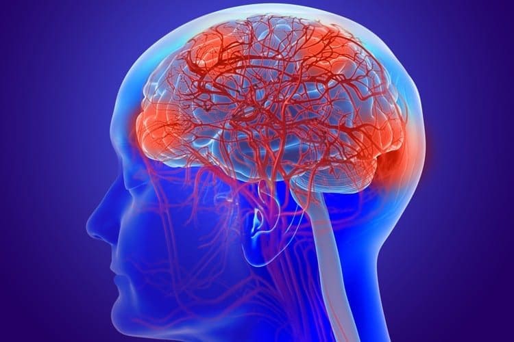
The blood-brain barrier (BBB) is a connection of blood vessels that protect the brain against harmful free radicals in the bloodstream. However, the blood-brain barrier is so effective at protecting the brain from these “harmful” components in the bloodstream, that it can ultimately even prevent drugs and/or medications from penetrating this security system to treat brain health issues. A research study published in Psychotherapy and Psychosomatics demonstrated that the effectiveness of antidepressants is only slightly more effective compared to placebos in the treatment of mental health issues. �
What Causes a Leaky Brain?
Scientists continue to analyze ways to effectively penetrate the blood-brain barrier to treat brain health issues. Several research studies have also determined that inflammation may reduce the function of brain cells in the frontal lobe of people diagnosed with depression. Other scientists are starting to believe that antidepressants and medicines used to treat depression are ineffective because these don’t necessarily treat inflammation in the brain. When the blood-brain barrier is damaged or injured, harmful components can enter the brain through the bloodstream and cause neurodegenerative symptoms. �
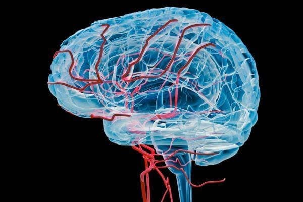
A “leaky brain” is a well-known term that is increasingly being used to describe blood-brain barrier permeability. A variety of blood tests, including those that measure the levels of the proteins occludin and zonulin, can be used to determine a leaky brain. Immunoglobulin levels may also be measured. Scientists also measure the levels of a molecule, known as microRNA-155, which increases with inflammation. MicroRNAs (miRNAs) play the fundamental role of regulating immune reactions, with miR-155 as a biomarker for inflammation in the brain due to a leaky blood-brain barrier. According to various research studies, this molecule can cause small gaps to develop in the BBB which can ultimately cause inflammation and lead to a leaky brain. �
Several different research studies have also discussed how inflammation on the blood-brain barrier can eventually cause a leaky brain. Meanwhile, other research studies have demonstrated a link between inflammation and a variety of psychiatric disorders. Scientists also demonstrated that pro-inflammatory cytokines can increase and cause increased blood-brain barrier permeability. Many harmful components can also affect the structure of the mitochondria and the blood-brain barrier. Microglial cells in the brain may also trigger and activate the release of molecules that can further affect the BBB. �
Further evidence has also associated blood-brain barrier dysfunction with a leaky gut. Scientists have suggested treating an underlying leaky gut to help treat a leaky brain. According to research studies, intestinal permeability, or a �leaky gut�, may ultimately be associated with blood-brain barrier permeability. Bacteria, small molecules, and toxins in the blood are commonly found in celiac disease, a well-known problem caused by gluten sensitivity or intolerance. Although true celiac disease is considered to be rare, a leaky gut associated with celiac disease and brain health issues are considered to be more common. �
One research study discusses the connection between the gut microbiome, inflammation, and the integrity of the blood-brain barrier. The scientists of a different research study discussed how a variety of treatments used to help improve the biodiversity of the gut microbiome, including a healthy diet and lifestyle modifications, fecal microbiota transplantation, prebiotics, and probiotics, have demonstrated to support the function of the gut-brain axis. Scientists believe it will be possible to use the gut microbiome to improve brain and mental health issues as well as to prevent further complications. �
Too much inflammation may cause a variety of brain and mental health issues associated with blood-brain barrier permeability. Because many research studies have suggested the connection between a leaky gut and a leaky brain, maintaining a healthy gut microbiome may be an effective treatment for brain and mental health. Although the brain is protected by the blood-brain barrier, this security system can frequently prevent drugs and/or medications from being able to effectively treat many brain and mental health issues. Scientists have started working towards developing successful ways to allow treatments to penetrate the blood-brain barrier.� – Dr. Alex Jimenez D.C., C.C.S.T. Insight
As previously mentioned, the National Institute of Mental Health (NIH) states that about 20 percent of Americans are diagnosed with a mental health issue every year, where depression and phobias are considered to be the most common types of diagnosable brain health issues. Furthermore, the Centers for Disease Control and Prevention (CDC) recorded that the suicide rate in the United States reached 13 for every 100,000 people in 2014, which is the highest it has ever been since 1986. Scientists associate mental health issues with brain inflammation and how it causes a “leaky” blood-brain barrier. �
The blood-brain barrier (BBB) is a group of blood vessels that protect the brain against “harmful” components in the bloodstream. However, because the blood-brain barrier can be so effective at protecting the brain from these harmful free radicals in the bloodstream, it can ultimately prevent drugs and/or medications from successfully penetrating the BBB to treat mental health issues. Research studies published in Psychotherapy and Psychosomatics determined that the effectiveness of certain medicines can only be slightly more effective, compared to placebos, in the treatment of brain health issues. �
The scope of our information is limited to chiropractic, musculoskeletal, and nervous health issues or functional medicine articles, topics, and discussions. We use functional health protocols to treat injuries or disorders of the musculoskeletal system. Our office has made a reasonable attempt to provide supportive citations and has identified the relevant research study or studies supporting our posts. We also make copies of supporting research studies available to the board and or the public upon request. To further discuss the subject matter above, please feel free to ask Dr. Alex Jimenez or contact us at 915-850-0900.�
Curated by Dr. Alex Jimenez �
References:
Figeley, Melanie. �Do You Have A Leaky Brain?� Biotics NW Inc., Biotics NW Inc., 15 Jan. 2019, www.bioticsnw.com/blogs/news/do-you-have-a-leaky-brain.
Neurotransmitter Assessment Form
The following Neurotransmitter Assessment Form can be filled out and presented to Dr. Alex Jimenez. The following symptoms listed on this form are not intended to be utilized as a diagnosis of any type of disease, condition, or any other type of health issue. �
Additional Topic Discussion: Chronic Pain
Sudden pain is a natural response of the nervous system which helps to demonstrate possible injury. By way of instance, pain signals travel from an injured region through the nerves and spinal cord to the brain. Pain is generally less severe as the injury heals, however, chronic pain is different than the average type of pain. With chronic pain, the human body will continue sending pain signals to the brain, regardless if the injury has healed. Chronic pain can last for several weeks to even several years. Chronic pain can tremendously affect a patient’s mobility and it can reduce flexibility, strength, and endurance. �
Neural Zoomer Plus for Neurological Disease
�
Dr. Alex Jimenez utilizes a series of tests to help evaluate neurological diseases. The Neural ZoomerTM Plus is an array of neurological autoantibodies which offers specific antibody-to-antigen recognition. The Vibrant Neural ZoomerTM Plus is designed to assess an individual�s reactivity to 48 neurological antigens with connections to a variety of neurologically related diseases. The Vibrant Neural ZoomerTM Plus aims to reduce neurological conditions by empowering patients and physicians with a vital resource for early risk detection and an enhanced focus on personalized primary prevention. �
Food Sensitivity for the IgG & IgA Immune Response
�
Dr. Alex Jimenez utilizes a series of tests to help evaluate health issues associated with food sensitivities. The Food Sensitivity ZoomerTM is an array of 180 commonly consumed food antigens that offers very specific antibody-to-antigen recognition. This panel measures an individual�s IgG and IgA sensitivity to food antigens. Being able to test IgA antibodies provides additional information to foods that may be causing mucosal damage. Additionally, this test is ideal for patients who might be suffering from delayed reactions to certain foods. Utilizing an antibody-based food sensitivity test can help prioritize the necessary foods to eliminate and create a customized diet plan around the patient�s specific needs. �
Gut Zoomer for Small Intestinal Bacterial Overgrowth (SIBO)
�
Dr. Alex Jimenez utilizes a series of tests to help evaluate gut health associated with small intestinal bacterial overgrowth (SIBO). The Vibrant Gut ZoomerTM offers a report that includes dietary recommendations and other natural supplementation like prebiotics, probiotics, and polyphenols. The gut microbiome is mainly found in the large intestine and it has more than 1000 species of bacteria that play a fundamental role in the human body, from shaping the immune system and affecting the metabolism of nutrients to strengthening the intestinal mucosal barrier (gut-barrier). It is essential to understand how the number of bacteria that symbiotically live in the human gastrointestinal (GI) tract influences gut health because imbalances in the gut microbiome may ultimately lead to gastrointestinal (GI) tract symptoms, skin conditions, autoimmune disorders, immune system imbalances, and multiple inflammatory disorders. �
Formulas for Methylation Support
� XYMOGEN�s Exclusive Professional Formulas are available through select licensed health care professionals. The internet sale and discounting of XYMOGEN formulas are strictly prohibited.
Proudly,�Dr. Alexander Jimenez makes XYMOGEN formulas available only to patients under our care.
Please call our office in order for us to assign a doctor consultation for immediate access.
If you are a patient of Injury Medical & Chiropractic�Clinic, you may inquire about XYMOGEN by calling 915-850-0900.
�
For your convenience and review of the XYMOGEN products please review the following link. *XYMOGEN-Catalog-Download �
* All of the above XYMOGEN policies remain strictly in force. �
Here are some tips that you can use throughout the year but are especially useful during the holidays. So give yourself the gift of spending happy holidays with less stress, neck and back pain. Christmas is a time of excitement, growth, family, friends mixed with stress.
Now there is good stress that we all use when we need it, but when everything just starts to close in all around you, that’s the bad kind of stress that we want to prevent and avoid. For people with chronic back or neck pain, the strain of getting your to-do list done can take the joy of the season right out and replace it with pain and misery. �Therefore plan ahead so you can get your holiday chores done without pain.
Five tips for making the holidays more enjoyable and comfortable
Tip 1 – Shop Simple
Back and leg pain, along with sciatica, can make walking at a shopping mall almost impossible. Instead of doing a lot of walking maybe pick up gift cards or go online. This way the family and friends get to pick whatever they want and you avoid lifting and carrying shopping bags. But, if you do buy personalized gifts, go online. Everything gets shipped straight to wherever you want.
Tip 2 – Plan Before Cooking
If you’re expecting a large crowd, prepare all you can ahead of time. Example: pies can be baked the day before.
Before you start cooking, look at the kitchen. Is there room for everyone to fit while helping, conversing, etc? If not, set up the space so people can flow easily around each other and get things done.
Use your counter-top to lean on every now and then for a few minutes and take the pressure off your back. Do not stay in any one position for an extended period. To help you not forget, set a timer to remind you to sit, rest, or stand. Also, let your guests help with serving and clean up so any stress on the body is reduced.
Tip 3 – Make Decorating a Group Activity
Don�t decorate by yourself. Especially when moving heavy objects. Get help so everyone is involved. This will prevent repetitive injuries when reaching or twisting and reduce stress to you and your family’s backs.
Tip 4: Make Time for Yourself
During the holidays we can get tempted to forget about our regular eating and exercise routines.
Stay focused, as you need strength and flexibility to be able to accomplish these tasks. So stretch out and continue your exercise regimen no matter how hectic it gets.
Stretching will help keep you limber and reduce the risk of a sprain or strain.
If there is no time for your regular exercise routine, don�t just drop it. Adapt and break up the exercises into little segments throughout the day.
Continue to eat the regular healthy meals you’re used to. Gaining weight will make back pain worse. Staying on track isn�t easy during the holidays. Take a look online for healthy strategies before the holiday parties begin.
Here are some examples:
Eat breakfast to prevent overeating later in the day.
At the party, fill up with low-calorie foods like vegetables, leafy green salads, and lean proteins.
Try to stay out of easy reach of nuts, chips or candies, cookies, etc.
If you’re going to have some high-calorie food have some, but don’t overdo it.
And if you�re going to indulge try something that doesn’t come around all the time, like a piece of your aunt�s pie, cookies, cake, ham, tamales, that you only get once a year.
Tip 5 – Look to the Future
Look ahead to the future and look up some simple ways to improve your back or neck pain. Setting some goals like adding a 5-minute walk break to your day that you could gradually increase to ten, fifteen minutes and so on. Signing up for a yoga/fitness class could be another idea. This could also be the year to replace your mattress. Whatever the case, take it all in, look at the options and figure out what works for you.
How to eliminate Back Pain naturally | (2020) Foot Levelers |El Paso, Tx
NCBI Resources
There are 3 major categories of stress: bodily, environmental and emotional.
Bodily stress: Caused by lack of sleep, disease, trauma or injury, and improper nutrition.
Environmental stress:�Caused by loud noises (sudden or sustained), pollution and world events, such as war and politics.
Emotional stress:�Caused by a variety of life events, such as moving homes, starting a new job and regular personal interactions. In contrast to the other two categories of stress, however, people can have some control over their emotional stress. Such can depend on the individual�s own attitude.
How often do you feel agitated, easily upset, and nervous between meals? How often do you have difficulty concentrating before eating? How often does your energy level drop in the afternoon? Inflammation associated with a leaky blood-brain barrier can cause a variety of brain health issues. Our brain is a complex organ that controls various structures and functions of our body, from our memory to our breathing, muscle movements, and hormones. �
Unfortunately, brain health issues are common in our society. According to the National Institute of Mental Health, approximately 20 percent of adults in the United States have a brain health issue and American adults spend about $113 billion on treatment every year. Additionally, these statistics don’t even include the treatment cost of autoimmune brain health issues, such as Alzheimer’s disease, Parkinson’s disease, multiple sclerosis, and autism. In the article below, we will discuss how inflammation is associated with a “leaky” blood-brain barrier and what you can do to improve a variety of brain health issues. �
“Leaky” Blood-Brain Barriers and Inflammation
Scientists have previously discussed how leaky gut syndrome, a well-known digestive health issue that affects the lining of the gastrointestinal (GI) tract, can be associated with various health issues. However, current research studies have shown that leaky gut syndrome may also be associated with a leaky blood-brain barrier or the breakdown of the blood-brain barrier.�The gut-brain connection ultimately suggests that a “leaky gut” may cause a “leaky brain”. �
Biomarkers using the proteins occludin and zonulin can help determine the presence of leaky gut syndrome and a leaky blood-brain barrier. Scientists have demonstrated that increased antibodies against occludin and zonulin are one way to determine leaky brain syndrome. Scientists have also demonstrated that a molecule known microRNA-155 can increase with inflammation. MicroRNA-155 can ultimately cause microscopic gaps to develop in the blood-brain barrier that permitted passage to “harmful” components. This permeability can trigger the brain’s immune system and cause brain inflammation. �
Various research studies from the cytokine model of cognitive function evaluated how brain inflammation and a leaky blood-brain barrier may be associated with cases of anxiety, depression, brain fog, and autoimmune brain health issues. By way of instance, scientists evaluated how inflammation can decrease the activation of brain cells in the frontal lobe of people with depression. In cases of brain inflammation, using drugs and/or medications, such as antidepressants, are frequently ineffective because they aren’t treating the underlying brain inflammation. If a patient suffers from anxiety, depression, brain fog, or autoimmune brain health issues, many healthcare professionals may suggest the presence of a leaky brain or a leaky blood-brain barrier. �
8 Steps to Improve a Leaky Blood-Brain Barrier
Inflammation and a leaky blood-brain barrier may ultimately cause a variety of bran health issues if it’s misdiagnosed and left untreated. If you suspect you may have anxiety, depression, brain fog, or autoimmune brain health issues due to inflammation associated with a leaky blood-brain barrier, it’s important to talk to a healthcare professional to receive proper diagnosis and treatment. Fortunately, there are steps you can take to improve a leaky blood-brain barrier:� �
Evaluate your blood-brain barrier by conducting labs for blood-brain barrier proteins, which can show BBB permeability, occludin and zonulin, which can show the antibodies against these proteins, homocysteine, which shows blood-brain barrier damage or injury through the increased levels of this amino acid, and brain agers like inflammation, which can accelerate the aging of the brain. High blood sugar may also show BBB permeability.
Determine a “leaky” blood-brain barrier by conducting microbiome labs. A “leaky gut” can cause a “leaky brain”. Understanding the gut-brain axis can help determine the presence of any underlying brain health issues. Bacterial imbalances and yeast overgrowths can cause neurological symptoms. By way of instance, anxiety and depression are associated with the bacteria known as Lactobacillus helveticus and Bifidobacterium longum.
Prevent yourself from eating foods, such as processed, refined and sugary foods as well as other toxins, that can cause a leaky blood-brain barrier.
Regulate stress for overall brain health. Research studies ultimately suggest that acute stress can increase the breakdown of the blood-brain barrier. Tai chi, yoga, and mindfulness meditation are several effective ways to help manage stress for the overall well-being of the human brain.
Take natural compounds, such as apigenin, baicalein, catechins, curcumin, luteolin, resveratrol, and rutin, that have all been suggested to reduce brain inflammation. However, because the correct dosage will be different for everyone, make sure to talk to a doctor before taking natural medicine.
Participate in exercise and physical activity that can help increase a brain-derived neurotrophic factor, BDNF, which helps promote brain health.
Reduce your consumption of alcohol. Alcohol can stress the brain and several research studies suggest that it can affect the blood-brain barrier.
Consider visiting a functional medicine practitioner to improve a leaky blood-brain barrier. While there’s no immediate fix for many chronic brain health issues, functional medicine customizes diagnostics and natural protocols based on your unique needs to help promote overall well-being.
The brain is protected by the blood-brain barrier, however, this security system can frequently prevent drugs and/or medications from being able to effectively treat brain health issues. Scientists have started working towards developing successful ways to allow treatments to penetrate the blood-brain barrier. Other research studies have demonstrated that the aging brain or neurodegeneration can cause the breakdown of the blood-brain barrier. However, research studies have demonstrated ways to restore and even reverse leaky blood-brain barriers and several brain health issues. Patients can take several steps to help improve a leaky blood-brain barrier. – Dr. Alex Jimenez D.C., C.C.S.T. Insight
How often do you feel agitated, easily upset, and nervous between meals? How often do you have difficulty concentrating before eating? How often does your energy level drop in the afternoon? Inflammation associated with a leaky blood-brain barrier can cause a variety of brain health issues. Our brain is a complex organ that controls various structures and functions of our body, from our memory to our breathing, muscle movements, and hormones. �
Unfortunately, brain health issues are common in our society. According to the National Institute of Mental Health, approximately 20 percent of adults in the United States have a brain health issue and American adults spend about $113 billion on treatment every year. Additionally, these statistics don’t even include the treatment cost of autoimmune brain health issues, such as Alzheimer’s disease, Parkinson’s disease, multiple sclerosis, and autism. In the article above, we discussed how inflammation is associated with a “leaky” blood-brain barrier and what you can do to improve a variety of brain health issues. �
The scope of our information is limited to chiropractic, musculoskeletal, and nervous health issues or functional medicine articles, topics, and discussions. We use functional health protocols to treat injuries or disorders of the musculoskeletal system. Our office has made a reasonable attempt to provide supportive citations and has identified the relevant research study or studies supporting our posts. We also make copies of supporting research studies available to the board and or the public upon request. To further discuss the subject matter above, please feel free to ask Dr. Alex Jimenez or contact us at 915-850-0900.�
Curated by Dr. Alex Jimenez �
References:
Cole, William. �Signs You Might Have A �Leaky Brain� + What To Do About It.� Mindbodygreen, Mindbodygreen, 22 July 2015, www.mindbodygreen.com/0-20800/signs-you-might-have-a-leaky-brain-what-to-do-about-it.html.
Neurotransmitter Assessment Form
The following Neurotransmitter Assessment Form can be filled out and presented to Dr. Alex Jimenez. The following symptoms listed on this form are not intended to be utilized as a diagnosis of any type of disease, condition, or any other type of health issue. �
Additional Topic Discussion: Chronic Pain
Sudden pain is a natural response of the nervous system which helps to demonstrate possible injury. By way of instance, pain signals travel from an injured region through the nerves and spinal cord to the brain. Pain is generally less severe as the injury heals, however, chronic pain is different than the average type of pain. With chronic pain, the human body will continue sending pain signals to the brain, regardless if the injury has healed. Chronic pain can last for several weeks to even several years. Chronic pain can tremendously affect a patient’s mobility and it can reduce flexibility, strength, and endurance. �
Neural Zoomer Plus for Neurological Disease
Dr. Alex Jimenez utilizes a series of tests to help evaluate neurological diseases. The Neural ZoomerTM Plus is an array of neurological autoantibodies which offers specific antibody-to-antigen recognition. The Vibrant Neural ZoomerTM Plus is designed to assess an individual�s reactivity to 48 neurological antigens with connections to a variety of neurologically related diseases. The Vibrant Neural ZoomerTM Plus aims to reduce neurological conditions by empowering patients and physicians with a vital resource for early risk detection and an enhanced focus on personalized primary prevention. �
Food Sensitivity for the IgG & IgA Immune Response
Dr. Alex Jimenez utilizes a series of tests to help evaluate health issues associated with food sensitivities. The Food Sensitivity ZoomerTM is an array of 180 commonly consumed food antigens that offers very specific antibody-to-antigen recognition. This panel measures an individual�s IgG and IgA sensitivity to food antigens. Being able to test IgA antibodies provides additional information to foods that may be causing mucosal damage. Additionally, this test is ideal for patients who might be suffering from delayed reactions to certain foods. Utilizing an antibody-based food sensitivity test can help prioritize the necessary foods to eliminate and create a customized diet plan around the patient�s specific needs. �
Gut Zoomer for Small Intestinal Bacterial Overgrowth (SIBO)
Dr. Alex Jimenez utilizes a series of tests to help evaluate gut health associated with small intestinal bacterial overgrowth (SIBO). The Vibrant Gut ZoomerTM offers a report that includes dietary recommendations and other natural supplementation like prebiotics, probiotics, and polyphenols. The gut microbiome is mainly found in the large intestine and it has more than 1000 species of bacteria that play a fundamental role in the human body, from shaping the immune system and affecting the metabolism of nutrients to strengthening the intestinal mucosal barrier (gut-barrier). It is essential to understand how the number of bacteria that symbiotically live in the human gastrointestinal (GI) tract influences gut health because imbalances in the gut microbiome may ultimately lead to gastrointestinal (GI) tract symptoms, skin conditions, autoimmune disorders, immune system imbalances, and multiple inflammatory disorders. �
Formulas for Methylation Support
� XYMOGEN�s Exclusive Professional Formulas are available through select licensed health care professionals. The internet sale and discounting of XYMOGEN formulas are strictly prohibited.
Proudly,�Dr. Alexander Jimenez makes XYMOGEN formulas available only to patients under our care.
Please call our office in order for us to assign a doctor consultation for immediate access.
If you are a patient of Injury Medical & Chiropractic�Clinic, you may inquire about XYMOGEN by calling 915-850-0900.
�
�
For your convenience and review of the XYMOGEN products please review the following link. *XYMOGEN-Catalog-Download �
* All of the above XYMOGEN policies remain strictly in force. �
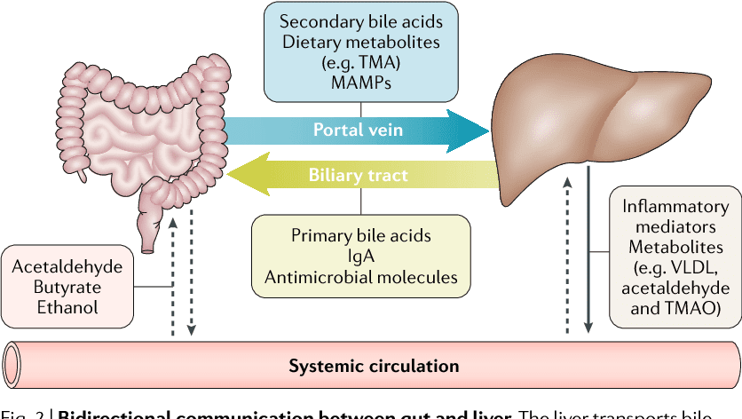
If you are experiencing any of these situations, then you might be experiencing some gut and liver issues in your body.
The role of the gut-liver axis in NAFLD (non-alcoholic fatty liver disease) has been examining probiotics and have found some new information on the gut microbiome and how probiotics work in NAFLD. The new information that future research found was quite interesting. It stated that there were about 26 major randomized controlled trials that used probiotics for NALFD that ranged between 20 to 200 individuals in four weeks to 1 year. The laboratory assessments included liver enzymes and anthropometric parameters in the body. Some of the studies added cardiovascular risk factors like C-reactive proteins and lipid profiles as markers for insulin resistance. Furthermore, most of the studies have used a probiotic formulation that includes multiple species, although a few were conducted by using a single strain.
What NAFLD does to the body is that it becomes a hepatic consequence of metabolic syndrome. This includes obesity, diabetes, and dyslipidemia. What is interesting about the connection between gut microbiota and NAFLD has been attracting a significant amount of attention in recent years. The data has shown that the gut microbiota can affect the hepatic lipid metabolism while also influencing the balance between pro/anti-inflammatory effectors in the liver.
Chronic Liver Diseases
Even though chronic liver disease is the primary cause of morbidity and mortality worldwide. Studies showed that gut dysbiosis was identified as an essential factor in the pathogenesis of the liver disease. The relationship between the gut microbiota and the liver is still not understood, but the dysfunction of leaky gut and an increased bacterial translocation into the liver. Another study showed that immense importance is a massive advancement in understanding the roles of the gut and liver microbiome that is driven by a high DNA sequencing and improving them.
There are many stages of liver disease that can happen, but when it comes to excessive alcohol in the liver. Studies show that excessive alcohol consumption is the leading cause of chronic liver disease worldwide. The stages of alcoholic liver disease are hepatic steatosis, steatohepatitis, and, ultimately, liver cirrhosis. One of the main characteristics of alcohol liver disease is that there is an increased gut permeability due to a direct toxic effect of alcohol on the epithelial cell in the gastrointestinal tract and a decreases expression of the tight-junction protein.
Probiotic Supplements
For probiotic supplementation, they have demonstrated a significant decrease in liver enzymes, which were compared to the placebo group. Studies found that probiotics were shown to have synergistic effects with metformin on liver enzymes for patients that have NASH. Any products that contained both prebiotics and probiotics can demonstrate a similar effect in the probiotic groups. In another study, it showed a reduction of intrahepatic fat that is measured by MRI, but the improvement in liver enzymes in the body did not reach any clinical significance. It is essential to know that liver enzymes can have a highly variable and do not always have a direct correlation with disease progression that they may encounter.
Research shows that there are five meta-analyses included and that they all demonstrated that probiotics and synbiotics have improved on AST and ALT levels in the body significantly. Surprisingly, several other studies have assisted probiotics by countering hepatic steatosis, fibrosis, and liver stiffness in the body. Ultrasound imaging can help assist these parameters and show some positive clinical outcomes with these two supplements.
When probiotics help restore the gastrointestinal barrier function in the body, they can eliminate the harmful bacteria that has interacted with the gastrointestinal system. Not only that, but probiotics can also be beneficial by modulating the immune system, reduce liver fats, and improve the liver enzymes as well. By using probiotics, they are most likely to be more productive by helping the body and preventing bacterial translocation in the gut, thus reducing the effects of the intestinal microbiota on the liver to prevent chronic illnesses from forming and causing havoc.
Conclusion
For individuals that have NAFLD, they will already establish the disease and required a higher nutrient intake demand than what can be obtained from any diets alone. So using dietary supplements should be considered to help reduce the NAFLD disease’s progression, thus improving the liver and its functions. The gut-liver axis is connected to the body since if anything happens to the liver like chronic diseases, it can affect the gut as well. Using probiotics to help the liver is essential to make sure that the liver is functioning correctly and that the body is being as healthy as possible. Some products are here to offer gastrointestinal and metabolic support while also supporting multiple aspects of the biliary system.
The scope of our information is limited to chiropractic, musculoskeletal, and nervous health issues or functional medicine articles, topics, and discussions. We use functional health protocols to treat injuries or disorders of the musculoskeletal system. Our office has made a reasonable attempt to provide supportive citations and has identified the relevant research study or studies supporting our posts. We also make copies of supporting research studies available to the board and or the public upon request. To further discuss the subject matter above, please feel free to ask Dr. Alex Jimenez or contact us at 915-850-0900.
References:
Jurgelewicz, Michael. �New Review Demonstrates the Role of the Gut Microbiome and Probiotics in Nonalcoholic Fatty Liver Disease.� Designs for Health, 25 Nov. 2019, blog.designsforhealth.com/node/1160.
Konturek, Peter Christopher, et al. �Gut?Liver Axis: How Do Gut Bacteria Influence the Liver?� Medical Sciences (Basel, Switzerland), MDPI, 17 Sept. 2018, www.ncbi.nlm.nih.gov/pmc/articles/PMC6165386/.
Tripathi, Anupriya, et al. �The Gut-Liver Axis and the Intersection with the Microbiome.� Nature Reviews. Gastroenterology & Hepatology, U.S. National Library of Medicine, July 2018, www.ncbi.nlm.nih.gov/pmc/articles/PMC6319369/.
Xie, Chencheng, and Dina Halegoua DeMarzio. “Role of Probiotics in Nonalcoholic Fatty Liver Disease: Does Gut Microbiota Matter?” MDPI, Multidisciplinary Digital Publishing Institute, 19 Nov. 2019, www.mdpi.com/2072-6643/11/11/2837.
There are all kinds of tools for all kinds of jobs. However, using the correct tool can mean the difference between a job well done and a job that got done but also generated injuries and pain.
Examples:
Long tools are best for when you need leverage, sparing the need for massive physical force.
Vice grips and clamps can grip/stabilize objects rather than trying to hold objects with your hands.
Tilt objects to avoid overbending the wrists.
Use a cart/dolly/arm straps to carry heavy loads.
Take some time to think about how to make the job easier on yourself and look up youtube tutorials to find innovative ways to do these jobs making it less stressful both mentally and physically.
The National Institute for Occupational Safety & Health guidelines hand tool use
Keep the wrists straight &� Avoid bending/rotating the wrists.
Instead bend the tool, not the wrist and there are a variety of bent-handle tools just for this reason. Using the handshake wrist position is a good way to approach a job, as it is a neutral position for the wrist.
Don’t stand still in one place for an extended time when using a heavy tool.
Instead, reduce the size and weight of the tool which will help avoid strain, keeping the elbows low and slightly bent.
Tools that place pressure on the base of the palm stress the soft tissues of the hands and fingers interrupting circulation and nerve function.
Instead, opt for one with finger grooves that fit the hand. Short-handles help by reducing stress on the soft tissues.
Don’t use tools that need a lot of grip force to use or hold.
Instead, use one with a grip that compresses like memory foam and shapes to the hand. This is far better than hard plastic.
Don’t use tools that need the fingers to grip.
Instead, use tools that utilize a full-hand power grip.
Do not use tools that have sharp-edged handles or areas where the hands could get pinched.
Instead, use tools that keep the hands/fingers safe.
Trigger-finger operational tools should be avoided as this can easily cause repetitive finger/hand/wrist injury from the constant on-off motion.
Instead, use tools with large switches that can be operated using all four fingers.
Excessive temperatures affect manual dexterity, therefore keep hands free from extreme heat and cold.
If possible, do a different job that’s away from the extreme weather and if not wear properly insulated work gloves.
Keep excessive vibration to a minimum. Excessive vibration can affect circulation.
Use tools with control�features that limit vibration to the extremities and whole body.
Wear gloves that fit. If they are too tight they will place extreme pressure on the hands. Loose-fitting gloves reduce grip strength and the ability to grip properly.
Instead have a selection that is designed for different jobs.
Safely Operating Tools that Cause Whole-Body Vibration��
There are power tools that vibrate no matter what and transmit vibration into the operator’s arms and hands, legs, and feet. Using a tool like this can cause a condition called white finger or Raynaud’s Phenomenon to present.
The symptoms include:
Aching in the wrists and muscles of the forearm
Tingling sensations
Numbness
Whiteness in the fingers from restricted circulation
This type of vibration from riveting tools, grinders, pneumatic hammers, drills, and chain saws will affect the whole body’s well being.
Suggestions to help reduce the risk of musculoskeletal disorders
Choose power tools with anti-vibration controls and handles coated with suppressant/cushioned material to help with vibration.
Maintenance power tools by making sure they are balanced, clean, and lubricated.
Use gloves designed to absorb or reduce vibration.
Ask for help if the job requires equipment or tools that vibrate.
Whether using a hand or power tool to get a job done, the whole body is involved. Executing proper posture and body mechanics, along with proper tool choice and how it is used is vital to injury prevention.
El Paso, TX Chiropractor Recommended
Chiropractic care can help keep bodies flexible and help with range of motion. It is a very effective, non-invasive treatment for pain and can help with joint and muscular problems as well. Regular chiropractic treatments can help you better manage your body�s response to your work environment. It can also undo many of the ill effects that that type of work can cause.
NCBI Resources
Standing for extended or frequent periods of time without any breaks (such as walking or stretching) can cause the joints in the feet, knees, hips, and spine to become locked or immobilized temporarily. If the behavior continues, it can cause degenerative damage, leading to rheumatic diseases because the ligaments and tendons become damaged.
IFM's Find A Practitioner tool is the largest referral network in Functional Medicine, created to help patients locate Functional Medicine practitioners anywhere in the world. IFM Certified Practitioners are listed first in the search results, given their extensive education in Functional Medicine