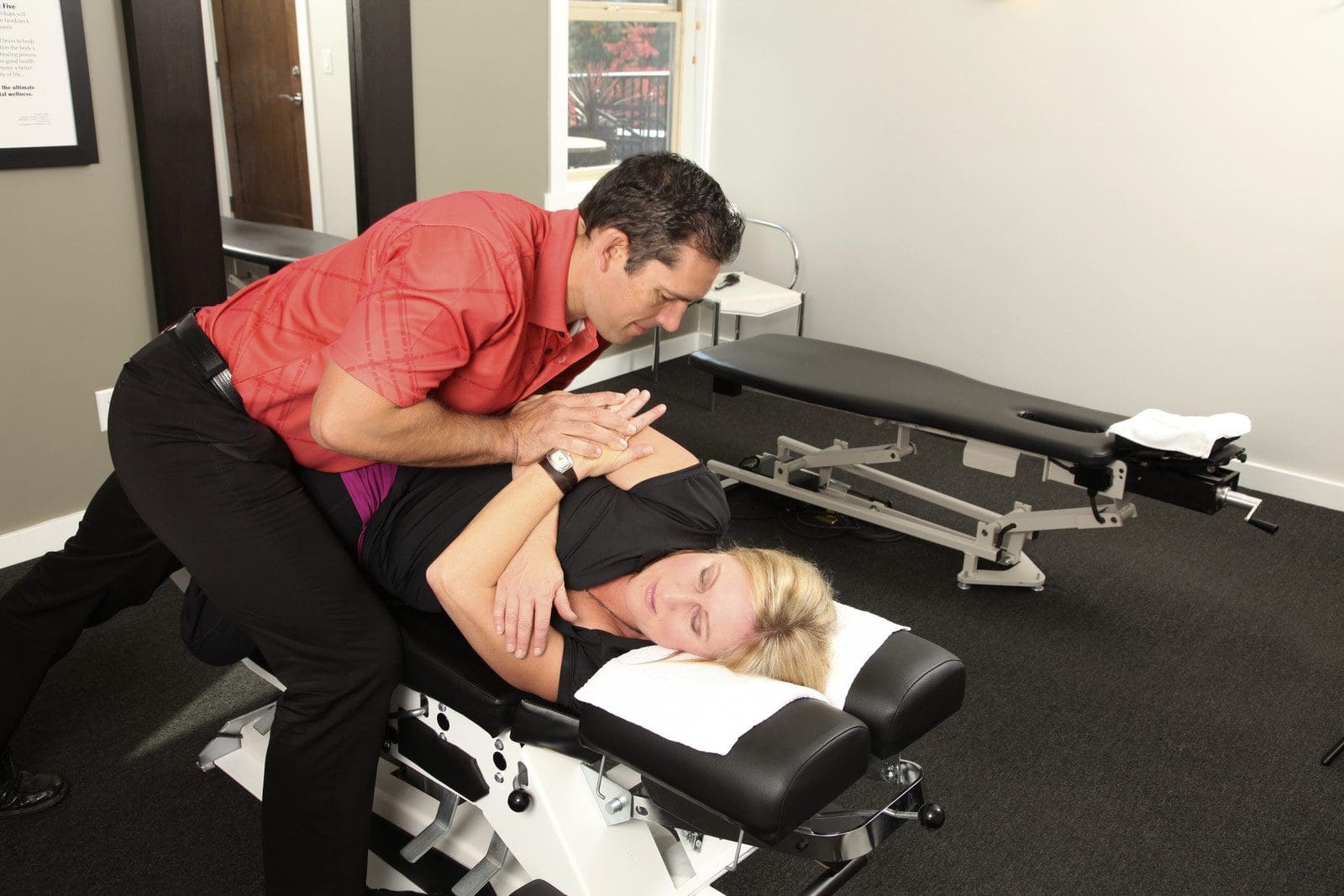
Neck pain caused by a whiplash injury definitely warrants a visit to a chiropractic whiplash specialist that can provide non-surgical treatment and pain relief.
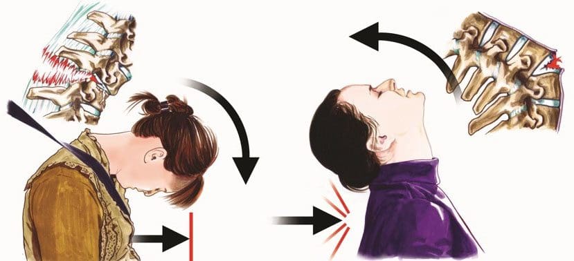
Whiplash is an injury to the neck muscles from a rapid forward and backward motion of the neck caused by trauma from a car accident, sports injury, slip and fall accident or even just turning one’s head but doing it with a fast whipping motion that causes the neck/spine muscles to become swollen and irritated. It can cause acute short-term neck pain and restricted movement.
How a Whiplash Injury Is Diagnosed
A chiropractor evaluates the spine in its entirety. If you go to a chiropractic clinic with neck pain following trauma. The chiropractor will examine the whole spine because the other areas of the spine could be affected and not just the neck region.
The chiropractor locates the areas where motion is restricted if there are any disc injuries, muscle spasms, and ligament injuries. They will first apply motion and staticpalpation diagnostic techniques where they feel and touch the various areas where the pain is present, as well as where there is no pain. A chiropractor will also feel for:
Tenderness
Tightness
How well the spinal joints move
They will also analyze the patient’s walk noting their posture and if there is possible spinal misalignment. This will help the chiropractor understand the patient’s body’s mechanics and what their spine is doing to compensate for the injury. This can mean:
Leaning to one side
Getting up in a very careful way so as to avoid pain
Hunching over
Only turning in one direction
In addition to the evaluation, they will also order an x-ray or an MRI to evaluate any deteriorating changes that could have existed before the whiplash injury. The images and physical and neurological evaluation resultsare compared to figure out and develop the best treatment plan.
Whiplash Treatment Stages
After a whiplash injury happens a chiropractor works to reduce neck inflammation with various therapies like:
Massage
Ultrasound
Light stretching
Soft manual therapy techniques
They may also recommend applying an ice pack on the neck and light neck support for a short time. As the inflammation and pain decrease the chiropractor will begin applying gentle spinal manipulation along with other techniques to restore the normal motion to the neck’s facet joints.
Chiropractic Whiplash Injury Treatment
A treatment plan depends on the severity of the whiplash injury. Some manipulation techniques used are:
Flexion-distraction technique
This is a gentle non-thrusting type of spinal manipulation that helps treat herniated discs. A whiplash injury can cause an aggravated bulging or herniated disc. If this happens a chiropractor uses a slow palm pump action on the disc rather than direct thrusting force.
Instrument-assisted manipulation
This technique also non-thrusting utilizes a hand-held instrument. A chiropractor generates force without thrusting directly into the spine. This therapy is great for older patients who may have degenerative joint syndrome.
Specific spinal manipulation
Spinal joints that are restricted or have abnormal motion are identified. Then the chiropractor restores motion to the joint with a gentle thrust. This stretches the soft tissue and stimulates the nervous system to bring back normal motion.
Along with these spinal therapies/techniques, a chiropractor also uses manual therapy to treat the soft tissues like the ligaments and muscles. Some examples of manual techniques are:
Instrument-assisted soft tissue therapy is where a chiropractor uses an instrument/s like the Graston technique, that gently treats any injured soft tissues. They will gently apply the instrument along the injured area with repeated strokes.
Manual joint stretching and resistance therapy is a form of manual therapy that uses the muscle’s own energy to create isometric contractions that help relax the muscles, and help lengthen the muscles.
Therapeutic massage is where a chiropractor or physical therapist performs massage to ease and relax muscle tension in the neck.
Trigger point therapy identifies specific tight painful points/areas of muscle by applying direct pressure with the hands or fingers on these points to alleviate the muscle tension.
Interferential electrical stimulation This technique uses low-frequency electrical current to stimulate the muscles and reduce inflammation.
Ultrasound increases blood circulation and helps reduce muscle spasms, stiffness, and pain. This happens by sending sound waves deep into the muscle’s tissue which generates low heat and increases circulation.
Therapeutic exercises to restore normal spinal motion and reduce whiplash symptoms.
Chiropractic medicine looks at the whole person and not just the symptoms. Neck pain is different for everyone, so chiropractors don�t just focus on the pain because the whiplash injury could have affected other areas that the patient doesn’t feel pain or anything.
But as the spine is a complex structure that works as a unit, a problem in one area can slowly or quickly start to create problems in other areas of the spine much like falling dominoes.
With these techniques, a chiropractor will help increase a patient’s daily activities back to normal as quickly as they can, depending on the severity of the injury. They will work as hard as they can to address any added spinal or nerve-related causes/injuries stemming from the original whiplash injury and treat them as well until normal movement is restored and there is no longer pain.
Remember that prevention is the key to optimal long-term health!
Our team has taken great pride in bringing our families and injured patients only clinically proved treatment protocols. �By teaching complete holistic wellness as a lifestyle, we also change not only our patient�s lives but their families as well.� We do this so that we may reach as many El Pasoans who need us, no matter the affordability issues.
El Paso, TX Chiropractic Neck Pain Treatment
NCBI Resources
Often,�people with whiplash don�t experience any effects until a day, or even two, after. The key is to stay ahead of the pain and take measures sooner rather than later to relieve it and keep it at bay. It also provides documentation should other issues arise, and you need the information for legal purposes.
If you are in an accident, especially if you get rear-ended, and experience whiplash, see a doctor that day ��even if you don�t feel much pain. The sooner you visit a chiropractic clinic, the sooner you can begin treatment should a problem develop.
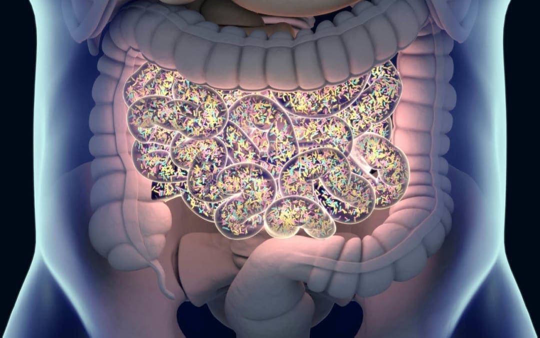
Do you feel irritable, nervous, shaky, or light-headed between meals? Do you have difficulty eating large meals in the morning? Do you feel fatigued after meals? Do you have sugar and sweet cravings after meals? Do you have an increased appetite?�If so, you may be experiencing early SIBO symptoms. �
SIBO, or small intestinal bacterial overgrowth, is a severe health issue that ultimately affects the small intestine in the digestive system. This gastrointestinal (GI) tract condition happens when the bacteria that generally grows in several different regions of the gut begin to grow in the small intestine. SIBO can commonly cause pain, discomfort, and diarrhea, among other symptoms. It can also cause malnutrition as bacteria utilize the human body�s nutrients.�
What are the Symptoms of SIBO?
Small intestinal bacterial overgrowth, or SIBO, is a serious condition that includes symptoms which can commonly affect the gut. These can include: �
pain or discomfort in the stomach
gas
bloating
constipation
diarrhea
cramps
indigestion
a general feeling of fullness
weight loss
What are the Causes of SIBO?
Small intestinal bacterial overgrowth (SIBO) is a severe health issue that is unfortunately not yet fully understood by researchers and healthcare professionals. According to research studies and clinical trials, however, this gastrointestinal, or GI, tract condition can ultimately happen when the small intestine is anatomically abnormal, due to pH changes in the small intestine, when the human body’s immune system isn’t functioning accordingly, or due to malfunctions in the muscular activity of the small intestine which can commonly cause food and bacteria to remain and not be removed from the organ. �
SIBO, or small intestinal bacterial overgrowth, is also commonly associated with a variety of health issues. These can involve the following, including: �
a stomach bug, known as viral gastroenteritis
celiac disease
Crohn�s disease
low stomach acid levels, known as hypochlorhydria
IBS or irritable bowel syndrome
gastroparesis
portal hypertension
nerve damage
cirrhosis
several gastric bypass procedures
surgical interventions which cause strictures or adhesions
What are the Risk Factors of SIBO?
Moreover, researchers and healthcare professionals have determined that an underlying chronic health issue and a previous surgery or surgical intervention that affects the gastrointestinal (GI) tract can be several risk factors of SIBO. Other wellness problems which can ultimately cause SIBO include: �
diabetes
scleroderma
hypothyroidism
Parkinson’s disease
HIV
narcotics or drugs/medications which slow down the digestive system
What is the Diagnosis for SIBO?
If you’ve experienced any of the SIBO symptoms mentioned above, see your doctor immediately. The doctor will ask the patient about their symptoms and medical history. The doctor will also perform a physical examination which may include palpating or gently feeling the patient’s abdomen. A qualified and experienced healthcare professional may also order additional blood, fecal, and/or any other tests to diagnose small intestinal bacterial overgrowth. �
A breath test is another common test utilized for the diagnosis of SIBO. Excess bacteria in the small intestine can cause the release of hydrogen and methane, two common gases which can be identified through a breath test. This test is non-invasive and can be performed in a doctor�s office. Before a breath test, the patient will need to fast overnight. During a breath test, the patient will first breathe into a tube. Then, the patient will take a specialized sweet drink provided by the doctor and they will breathe into several other tubes at regular intervals for 2 to 3 hours after taking the specialized sweet drink. �
If common tests for SIBO are inconclusive, the doctor may need to sample the fluid from the patient’s small intestine to see what bacteria is growing there. �
What is the Treatment for SIBO?
Common treatment approaches for SIBO, or small intestinal bacterial growth, can ultimately include a combination of antibiotics and diet modifications. �
Antibiotics
Treatment for SIBO first involves getting the bacteria in the digestive system under control. This is generally achieved by utilizing antibiotics, such as ciprofloxacin (Cipro), metronidazole (Flagyl), or rifaximin (Xifaxan). Further treatment for SIBO may also require intravenous (IV) therapy for nutrition and fluids if the serious gastrointestinal (GI) tract condition has ultimately caused malnutrition or dehydration, among a variety of other symptoms. �
Although antibiotics may help reduce the amount of bacteria in the small intestine, however, these will not always help address the underlying chronic health issues that caused the wellness problem in the first place. If the qualified and experienced healthcare professional determines that the patient’s SIBO is due to an underlying chronic health issue, the patient will also need to begin treatment for that wellness problem. Diet modifications may also help treat SIBO. �
Diet Modifications
Further research studies and clinical trials are still required to demonstrate if diet can cause small intestinal bacterial overgrowth (SIBO) but, many people with SIBO have reported feeling relief from their symptoms after diet modifications. Talk to your doctor before making any modifications to your diet. �
Furthermore, people with SIBO or other chronic health issues may only need to make small diet modifications to treat their symptoms. These can include: �
Eating a balanced and nutritious diet
Consuming minimal meals more often to prevent having too much food sit in the stomach
Avoid eating gluten products, if you have celiac disease or any other similar chronic health issues
The doctor may also recommend the patient to try an elemental diet to help treat SIBO. An elemental diet replaces food and drinks with several liquid formulas throughout an extended period of time. In one small-scale research study and clinical trial, approximately 80 percent of participants with SIBO had a normal breath test result following an elemental diet for 15 days. The researchers ultimately determined that an elemental diet may be a highly effective treatment approach for SIBO. However, further evidence is still needed. Talk to your doctor before starting an elemental diet and follow their instructions. �
Taking probiotics may also help restore the gut bacteria. A 2010 research study and clinical trial demonstrated that probiotic treatment can be more safe and effective for SIBO than taking antibiotics. However, a 2016 review determined that further evidence for the efficiency of probiotics in SIBO treatment was ultimately inconclusive. The best treatment approach for a patient with SIBO is to follow a qualified and experienced healthcare professional’s advice. �
SIBO, or small intestinal bacterial overgrowth, is a well-known and often severe health issue that generally occurs because of an underlying chronic condition or disease. Common symptoms may ultimately determine the presence of SIBO. In addition, if the patient has a chronic health issue, such as celiac disease or Crohn’s disease, they should talk to a doctor to develop a long-term treatment plan. SIBO, or small intestinal bacterial overgrowth is treatable. If left untreated, this gastrointestinal (GI) tract problem can also cause dehydration and malnutrition. Patients should contact a doctor immediately if they suspect they have SIBO so that they can begin treatment right away. – Dr. Alex Jimenez D.C., C.C.S.T. Insight
Neurotransmitter Assessment Form
�
The following Neurotransmitter Assessment Form can be filled out and presented to Dr. Alex Jimenez. The following symptoms listed on this form are not intended to be utilized as a diagnosis of any type of disease, condition, or any other type of health issue. �
The scope of our information is limited to chiropractic, musculoskeletal, and nervous health issues or functional medicine articles, topics, and discussions. We use functional health protocols to treat injuries or disorders of the musculoskeletal system. Our office has made a reasonable attempt to provide supportive citations and has identified the relevant research study or studies supporting our posts. We also make copies of supporting research studies available to the board and or the public upon request. To further discuss the subject matter above, please feel free to ask Dr. Alex Jimenez or contact us at 915-850-0900.�
Curated by Dr. Alex Jimenez �
References:
Madormo, Carrie. �Everything You Should Know About Small Intestinal Bacterial Overgrowth (SIBO).� Edited by Suzanne Falck, Healthline, Healthline, 14 June 2017, www.healthline.com/health/sibo#symptoms.
Additional Topic Discussion: Chronic Pain
Sudden pain is a natural response of the nervous system which helps to demonstrate possible injury. By way of instance, pain signals travel from an injured region through the nerves and spinal cord to the brain. Pain is generally less severe as the injury heals, however, chronic pain is different than the average type of pain. With chronic pain, the human body will continue sending pain signals to the brain, regardless if the injury has healed. Chronic pain can last for several weeks to even several years. Chronic pain can tremendously affect a patient’s mobility and it can reduce flexibility, strength, and endurance. �
Neural Zoomer Plus for Neurological Disease
Dr. Alex Jimenez utilizes a series of tests to help evaluate neurological diseases. The Neural ZoomerTM Plus is an array of neurological autoantibodies which offers specific antibody-to-antigen recognition. The Vibrant Neural ZoomerTM Plus is designed to assess an individual�s reactivity to 48 neurological antigens with connections to a variety of neurologically related diseases. The Vibrant Neural ZoomerTM Plus aims to reduce neurological conditions by empowering patients and physicians with a vital resource for early risk detection and an enhanced focus on personalized primary prevention. �
Food Sensitivity for the IgG & IgA Immune Response
Dr. Alex Jimenez utilizes a series of tests to help evaluate health issues associated with food sensitivities. The Food Sensitivity ZoomerTM is an array of 180 commonly consumed food antigens that offers very specific antibody-to-antigen recognition. This panel measures an individual�s IgG and IgA sensitivity to food antigens. Being able to test IgA antibodies provides additional information to foods that may be causing mucosal damage. Additionally, this test is ideal for patients who might be suffering from delayed reactions to certain foods. Utilizing an antibody-based food sensitivity test can help prioritize the necessary foods to eliminate and create a customized diet plan around the patient�s specific needs. �
Formulas for Methylation Support
XYMOGEN�s Exclusive Professional Formulas are available through select licensed health care professionals. The internet sale and discounting of XYMOGEN formulas are strictly prohibited.
Proudly,�Dr. Alexander Jimenez makes XYMOGEN formulas available only to patients under our care.
Please call our office in order for us to assign a doctor consultation for immediate access.
If you are a patient of Injury Medical & Chiropractic�Clinic, you may inquire about XYMOGEN by calling 915-850-0900.
�
For your convenience and review of the XYMOGEN products please review the following link. *XYMOGEN-Catalog-Download �
* All of the above XYMOGEN policies remain strictly in force. �
If you are experiencing any of these situations, then you might have experienced some trouble losing weight.
Trying to lose weight is harder than it looks. While the secret of losing weight is more accessible said than done, people are always trying to live healthier lives by exercising and eating right. Some people can maintain a healthy weight throughout their lives effortlessly; however, for others, it is a struggle that starts from when they were a kid, and it gets harder when they start growing up. There have been books on how to lose weight, and people gaining weight when they are middle-aged, it is shown around their mid-section of their bodies. Although when a person is trying to lose weight due to health reasons or wanting to get better, it can be a long, arduous journey.
There are many reasons why individuals are having trouble losing weight. It might be due to being older and that the body changes along with getting older as well. Here are some of the reasons why it is difficult to lose weight, the older a person gets.
Losing Muscle Mass
When a person age, their metabolism changes with them. When they are younger, their metabolism can make a person exercise with high intensity. As they get older, their metabolism changes, and they will slow down a bit when they are exercising. Not only that, but a person can lose their muscle mass when they reach the age of 30.
Studies have shown that the amount of lean muscle mass can naturally decline 3 to 8 percent per decade when a person hits 30, and it will be much harder when they are at the age of 60. This is due to sarcopenia. Sarcopenia is a condition that is characterized by losing skeletal muscle mass and functioning. This condition is progressive, and some of the risk factors include age, gender, and the level of physical activity a person is doing. Since the strength and muscle mass decline in older adults mostly, it can lead to acute and chronic diseases that can harm the body.
There is a way to combat muscle loss for anyone that wants to have lean muscles and to lose weight, is to add weight training to their exercise regime. Research shows that lifting weights is perfect for anyone to make sure that the body stays toned and muscular while also preventing a metabolic slowdown. Since male and females bodies are different, doing weight lifting will help the muscles look lean and toned for females, while for males, their muscles look more prominent and bulker, depending on the weights they are using and how many reps they are doing.
Getting Overly Stressed
As we get older, the more stress we can get. Stress is made up of the hormone cortisol, which is released into the body. It can also be in two categories, which are short term and long term. With short term stress, it is effortless to manage since a person can be worried about a project for school, getting a job interview, or worrying about the little things, the cortisol hormone is short term and can be easily managed.
When it is long-term stress, it can lead to chronic illnesses if it is in a person for far too long. The pressures of work, having too many obligations, or stressing out due to deadlines for projects are bad for a person to have since the cortisol levels are building up in the body. Even having a sedentary job can cause the body to develop stress and weight gain.
There are ways to reduce stress in the body like finding hobbies to enjoy, exercising, always help the body release the tension that is pent up, even having a self-care day can do many wonders to a person and their body. Trying to de-stress the body is excellent and beneficial to anyone because being stress-free is essential for losing weight.
Major Lifestyle Changes
When it comes to significant lifestyle changes, it can be any one of these changes that can cause a person to gain weight. It does not always happen from the inside, but it can happen to people when they enter their thirties. Some changes can include starting a family, trying to find time out of a hectic schedule, distractions, or homework. Whatever the reasons are, these lifestyle changes require much attention. When that happens, then the pounds start to creep in, causing weight gain.
Medical Condition
Sometimes when a person gains weight, it is due to a medical condition they might have, and it makes it harder for them to lose weight as well. These medical conditions include PCOS (polycystic ovarian syndrome), sleep apnea, and hypothyroidism. When these conditions affect the targeted body systems, it can cause many health problems to the body by causing it to dysfunction.
“There are many reasons why losing weight is hard for anyone. There is a wide variety of reasons like eating junk foods, not getting enough sleep, staying hydrated, or a hectic lifestyle. If we take the time to change one thing to maintain a healthy lifestyle, then the weight will slowly but surely go away.” -Dr. Alex Jimenez D.C., C.C.S.T. Insight
Conclusion
Losing weight is hard for anyone, and it can be easy for some while difficult for others. Since everyone has a different body structure, trying to lose weight is one of the more laborious tasks if an individual has problems trying to shed off the weight. By changing some of the habits that are causing the weight gain, it may be beneficial to not only the person but to their body. Some products can help the body’s metabolism and support the function of sugars and amino acids that help support even the gastrointestinal lining, the endocrine system, and help maintain the blood sugar levels.
The scope of our information is limited to chiropractic, musculoskeletal, and nervous health issues or functional medicine articles, topics, and discussions. We use functional health protocols to treat injuries or disorders of the musculoskeletal system. Our office has made a reasonable attempt to provide supportive citations and has identified the relevant research study or studies supporting our posts. We also make copies of supporting research studies available to the board and or the public upon request. To further discuss the subject matter above, please feel free to ask Dr. Alex Jimenez or contact us at 915-850-0900.
References:
Dray, Tammy. �Why Is It Harder to Lose Weight as You Get Older?� LIVESTRONG.COM, Leaf Group, 2019, www.livestrong.com/article/417064-why-is-it-harder-to-lose-weight-as-you-get-older/.
Gunnars, Kris. �20 Common Reasons Why You’re Not Losing Weight.� Healthline, 20 Aug. 2018, www.healthline.com/nutrition/20-reasons-you-are-not-losing-weight.
Lawler, Moira. �5 Reasons It’s Harder to Lose Weight With Age.� EverydayHealth.com, 27 June, 2019, www.everydayhealth.com/weight/weight-gain-and-aging.aspx.
Santilli, Valter, et al. �Clinical Definition of Sarcopenia.� Clinical Cases in Mineral and Bone Metabolism: the Official Journal of the Italian Society of Osteoporosis, Mineral Metabolism, and Skeletal Diseases, CIC Edizioni Internazionali, Sept. 2014, www.ncbi.nlm.nih.gov/pmc/articles/PMC4269139/.
Walston, Jeremy D. �Sarcopenia in Older Adults.� Current Opinion in Rheumatology, U.S. National Library of Medicine, Nov. 2012, www.ncbi.nlm.nih.gov/pmc/articles/PMC4066461/.
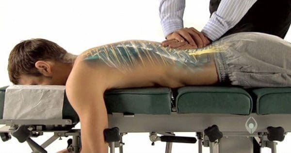
Chiropractic believes that optimal health comes from having a healthy nervous system, particularly a healthy spinal column.
Time, work, school, and everyday living means movement. And like anything that moves over and over again, at some point it’s going to get stuck or slip out of place. When the spine’s vertebrae become misaligned, pressure begins to press on the nerves that come out of the spinal cord. This is called a chiropractic subluxation and requires chiropractic realignment.
Chiropractors use a variety of techniques to mobilize the spinal discs so they can move freely along with returning them back�into place and in their proper positions.
These techniques are called spinal manipulations/adjustments.
During an adjustment, the vertebra is released or set free from the misaligned position and put back into the proper position in the spine’s column.
Adjustments allow the body to heal and maintain homeostasis.
Chiropractors are trained in a variety of adjustment techniques
Some are done by hand
Some use of specialized instruments
Ultrasound in physical therapy, treatment of shoulder muscles
Every patient is different, therefore a chiropractor will go through various treatment options with the patient, to choose the best technique and treatment plan for the patient’s condition.� Ask the chiropractor which technique they will be doing and how it works.
Adjustment Techniques
Chiropractors use�the following:
Toggle Drop
Using crossed hands a chiropractor presses down firmly on the areas that need adjusting and realignment. With a precise and quick thrust, the chiropractor adjusts the spine. This improves mobility in the vertebral joints.
Motion Palpation
This technique determines if the vertebrae are�freely�moving and with normal motion.
Lumbar Roll
The patient is on their side when the chiropractor performs a quick and precise thrust to the misaligned vertebra, that returns them to their proper position.
Treating Female Patient�s Spine
Release Work
Gentle pressure is applied with the fingertips which separate the vertebrae and opens them up.
Physiotherapist during an Achilles tendon treatment but the same technique applies to the spine
Table adjustment
Here a specialized table is used that drops incrementally with the adjustment. The chiropractor applies a quick thrust at the same time the table drops. This allows for lighter adjustments without twisting and turning that go with manual adjustments.
Instrument adjustments
This is considered the gentlest method of�spinal adjustment.
The patient lies on the table and the chiropractor uses an activator instrument to perform the adjustment/realignment/s.
TENS, Transcutaneous Electrical Nerve Stimulation in Physical Therapy. Therapist Positioning Electrodes onto Patient’s Lower Back
A chiropractic adjustment can be a great way to improve multiple areas of the body, along with improving overall health with non-invasive treatment. Dr. Jimenez has teamed with the top surgeons, clinical specialists, medical researchers and premiere rehabilitation�providers to bring El Paso the top clinical treatments to our community.��Providing high non-invasive protocols is our priority.
Severe *Sciatica”* Pain Relief | El Paso, Tx (2020)
NCBI Resources
The nervous system, which consists of the�(spine/nerves/brain)�is the central headquarters of the body.�Subluxation removal helps the body function at its optimum level. Chiropractic adjustments�reduce pressure on the nerves and ease the flow of communication and signals going back and forth.
Q: Recently diagnosed with a bulging disc at C5-C6, is there anything I can do at home to relieve my pain?
� El Paso, TX.
A: Great question because anyone that has woken up with a stiff neck can relate. This is the most common area of herniation in the neck. It is highly susceptible to regular wear and tear but poor neck posture, like looking down at a phone for too long and improper motion mechanics can also be a cause.
Taking care of yourself without having to go to the doctor is very important and it is critical to learn proper healing methods, and techniques.
Healthcare professionals will say that most of the healing takes place at home.
There are various ways to reduce pain at home, but remember that there is a point when symptoms do warrant professional medical attention.
Good news
Bulging and herniated discs typically heal within six months � and less than 10% ever need surgery.
Non-surgical treatment for cervical bulging discs is usually enough to relieve pain and the best part is that you can do many of these treatments yourself.
Under chiropractic/therapist supervision, these treatments can be utilized at home or with the assistance
Cervical pillow
A proper pillow will help improve the quality of sleep and also give the neck proper support and position to heal.
Herbal remedies
The natural approach to medications is also welcome. There are plenty of herbal supplements to help relieve pain. There is capsaicin cream, that you can apply and helps reduces pain.
Ice and heat therapy
Ice should be applied in a bag or towel for 15-20 minute intervals, two or three times daily. Heat treatment comes in the later stages of the therapy but follows the same on-off interval technique.
Over-the-Door Traction
This therapy is very effective in helping relieve pain and muscle spasms. Your head and neck are attached to a harness. This is connected to a rope that is connected to a pulley system that goes over a door at your home. Then while sitting, leaning back, or lying down you go through the exercise.
Motion exercises
These exercises help the neck recover its full range of motion. This prevents stiffness from setting in and also helps strengthen the muscles in the neck. A therapist or a chiropractor can show a variety of exercises depending on the area of injury/stiffness.
However, if the pain does not go away a physical therapist/chiropractor can be your best option. Passive physical therapy includes:
Ultrasound
T.E.N.S electrical nerve stimulation
Mild neck traction
These treatment options are non-invasive and effectively help manage pain. It’s always important to remember that if your pain continues or starts to become worse tell your doctor and get medical attention.
Chiropractic Alignment
If the pain continues, then you may be referred to a chiropractor for spinal alignment.
Chiropractors are experts at treating neck conditions.� Aligning the cervical segments of the neck is a must as the injury can return if the cervical discs are not in proper alignment or even worsen. Chiropractic treatment can give relief and also stop cervical spine degeneration.
*Neck* Pain Chiropractic Care El Paso, Texas
NCBI Resources
People have a 70% likelihood of developing neck pain lives and neck pain is an important issue affecting economic productivity in modern society. Neck pain is a work-related musculoskeletal disorder that can occur when a person works for a long time or at a high intensity. An increase in the number of individuals visiting hospitals with pain in the neck, upper extremities, and the head as a result of excessive tension. Home therapies can help reduce pain.
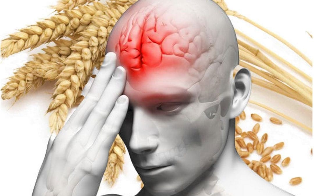
Many research studies have arguably analyzed how gluten can affect the nervous system. However, people with celiac disease and non-celiac gluten sensitivity have demonstrated a variety of symptoms, ranging from headaches and brain fog to autoimmune disease. Moreover, brain health issues, such as anxiety, depression, and migraines, among others, are also common symptoms in people with gluten sensitivity or intolerance.
Gluten ataxia, a severe autoimmune disorder, affects a small percentage of the population. Evidence suggests that brain health issues, such as schizophrenia and bipolar disorder, may also be affected by gluten. In the following article, we discuss several common gluten-related brain health issues.
Brain Fog, Headaches, Migraines, Insomnia, and ADHD
Many people with brain health issues like celiac disease as well as gluten sensitivity or intolerance understand the risks of consuming gluten. But, if they do eat gluten, many people report feeling that their brains “cloud up” and they feel less efficient, even clumsy. This brain health issue, known as brain fog, requires further research studies, however, it’s another common symptom associated with celiac disease and gluten sensitivity or intolerance.
Attention deficit-hyperactivity disorder (ADHD) is yet another common brain health issue in both adults and children. Headaches and migraines are also commonly reported as celiac disease symptoms and gluten sensitivity or intolerance symptoms. These symptoms may ultimately cause insomnia.
Anxiety and Depression
Research studies demonstrate that people with celiac disease experience anxiety and depression. People that don’t have celiac disease but who do have gluten sensitivity or intolerance also report experiencing anxiety and depression although the connection between the brain health issues is unknown. Researchers believe that gluten-related intestinal permeability, or leaky gut, may cause nutritional deficiencies that cause anxiety and depression.
However, that doesn’t necessarily explain why people with non-celiac gluten sensitivity or intolerance also experience anxiety and depression. Several gluten sensitivity or intolerance experts like New Zealand pediatrician Dr. Rodney Ford have hypothesized that gluten directly affects the brain and leads to the development of these brain health issues. Regardless, you’re far from being alone if you experience gluten-related anxiety and depression symptoms.
Schizophrenia and Bipolar Disorder
Many research studies suggest that gluten may be associated with two very severe brain health issues: schizophrenia and bipolar disorder. In schizophrenia, decades of research studies have shown that eliminating gluten from the diet of schizophrenics can help with the brain health issue. Research studies have ultimately demonstrated that a gluten-free diet can be beneficial for people with schizophrenia, but further research studies are needed.
In bipolar disorder, research studies have shown that people with celiac disease and gluten sensitivity or intolerance may experience the brain health issue. A research study on the levels of antibodies to gluten in the blood of people with bipolar disorder found increased levels during a manic episode.
Autoimmune Disease
When gluten consumption causes your own body to attack its own cells and tissues, you suffer from a gluten-related autoimmune disease. There are three common gluten-related autoimmune diseases: celiac disease, dermatitis herpetiformis, and gluten ataxia. In gluten ataxia, the immune system attacks the cerebellum, the region of the brain responsible for coordination. In many circumstances, the brain damage is irreversible, however, a strict gluten-free diet can help stop the progression of the autoimmune disease. Many people with gluten sensitivity or intolerance may also experience similar symptoms.
Celiac disease and gluten sensitivity or intolerance can ultimately lead to a wide variety of brain health issues and neurological diseases. However, in many circumstances, people can tremendously reduce or even resolve their gluten-related brain health issue symptoms by following a strict gluten-free diet.
Gluten intolerance or sensitivity is described as the human body’s inability to digest or break down the gluten protein found in wheat and a variety of other grains. This health issue can ultimately range from a mild or moderate intolerance or sensitivity to full-blown celiac disease, a severe autoimmune disorder related to gluten intolerance or sensitivity. Additionally, research studies have demonstrated that people with gluten intolerances or sensitivities may also develop brain health issues or neurological diseases. Talking to a naturopathic doctor or functional medicine practitioner can help determine if you have a gluten intolerance or sensitivity. Avoiding gluten altogether can ultimately help improve your overall health and wellness. – Dr. Alex Jimenez D.C., C.C.S.T. Insight
Neurotransmitter Assessment Form
[wp-embedder-pack width=”100%” height=”1050px” download=”all” download-text=”” attachment_id=”52657″ /]
The following Neurotransmitter Assessment Form can be filled out and presented to Dr. Alex Jimenez. Symptoms listed on this form are not intended to be utilized as a diagnosis of any type of disease, condition, or any other type of health issue.
The scope of our information is limited to chiropractic, musculoskeletal, and nervous health issues or functional medicine articles, topics, and discussions. We use functional health protocols to treat injuries or disorders of the musculoskeletal system. Our office has made a reasonable attempt to provide supportive citations and has identified the relevant research study or studies supporting our posts. We also make copies of supporting research studies available to the board and or the public upon request. To further discuss the subject matter above, please feel free to ask Dr. Alex Jimenez or contact us at 915-850-0900.�
Curated by Dr. Alex Jimenez
References:
Anderson, Jane. �How Gluten Can Have a Damaging Effect on Your Brain and Nerves.� Verywell Health, Verywell Health, 20 Nov. 2019, www.verywellhealth.com/gluten-related-neurological-symptoms-and-conditions-562317.
Additional Topic Discussion: Chronic Pain
Sudden pain is a natural response of the nervous system which helps to demonstrate possible injury. By way of instance, pain signals travel from an injured region through the nerves and spinal cord to the brain. Pain is generally less severe as the injury heals, however, chronic pain is different than the average type of pain. With chronic pain, the human body will continue sending pain signals to the brain, regardless if the injury has healed. Chronic pain can last for several weeks to even several years. Chronic pain can tremendously affect a patient’s mobility and it can reduce flexibility, strength, and endurance.
Neural Zoomer Plus for Neurological Disease
Dr. Alex Jimenez utilizes a series of tests to help evaluate neurological diseases. The Neural ZoomerTM Plus is an array of neurological autoantibodies which offers specific antibody-to-antigen recognition. The Vibrant Neural ZoomerTM Plus is designed to assess an individual�s reactivity to 48 neurological antigens with connections to a variety of neurologically related diseases. The Vibrant Neural ZoomerTM Plus aims to reduce neurological conditions by empowering patients and physicians with a vital resource for early risk detection and an enhanced focus on personalized primary prevention.
Food Sensitivity for the IgG & IgA Immune Response
Dr. Alex Jimenez utilizes a series of tests to help evaluate health issues associated with food sensitivities. The Food Sensitivity ZoomerTM is an array of 180 commonly consumed food antigens that offers very specific antibody-to-antigen recognition. This panel measures an individual�s IgG and IgA sensitivity to food antigens. Being able to test IgA antibodies provides additional information to foods that may be causing mucosal damage. Additionally, this test is ideal for patients who might be suffering from delayed reactions to certain foods. Utilizing an antibody-based food sensitivity test can help prioritize the necessary foods to eliminate and create a customized diet plan around the patient�s specific needs.
Formulas for Methylation Support
XYMOGEN�s Exclusive Professional Formulas are available through select licensed health care professionals. The internet sale and discounting of XYMOGEN formulas are strictly prohibited.
Proudly,�Dr. Alexander Jimenez makes XYMOGEN formulas available only to patients under our care.
Please call our office in order for us to assign a doctor consultation for immediate access.
If you are a patient of Injury Medical & Chiropractic�Clinic, you may inquire about XYMOGEN by calling 915-850-0900. For your convenience and review of the XYMOGEN products please review the following link. *XYMOGEN-Catalog-Download* All of the above XYMOGEN policies remain strictly in force.
Do you feel like grain consumption makes it difficult to focus or concentrate? Or does grain consumption make you feel like it leads to tiredness? Do you feel like grain consumption causes the development of any symptoms? Are you on a 100% gluten-free diet? Diet and environmental factors can affect brain health. Researchers and healthcare professionals have associated one specific component with neurological disease: gluten.
Brain health issues and neurological diseases have tremendously increased over the last several years. As a matter of fact, approximately 20 percent of adults in the United States have a diagnosable mental disorder and unfortunately, those statistics are expected to increase over the next few years. Depression is the most common cause of disability worldwide while anxiety affects more than 40 million Americans today. Moreover, Alzheimer’s disease is currently the sixth-leading cause of mortality in the United States.
A 2013 research study demonstrated that deaths associated with brain diseased have increased 66 percent in men and 92 percent in women since 1979. And, there’s one factor that all of these brain health issues and neurological diseases have in common: inflammation. Foods play a fundamental role in inflammation. There are many foods that will increase inflammation in the brain and body, arguably the biggest culprit is gluten.
How Does Gluten Affect the Brain?
While only one percent of Americans are diagnosed with celiac disease every year, there are probably many more under-diagnosed cases. As a matter of fact, only 10 percent of people with celiac disease show obvious symptoms. Research studies suggest that celiac disease can ultimately manifest as a neurological disease. However, celiac disease is a severe gluten sensitivity-autoimmune disorder, where there’s also approximately 1 in 20 people in the United States living with another health issue known as non-celiac gluten sensitivity.
Gluten has been demonstrated to increase levels of the protein zonulin in the gut which may ultimately lead to leaky gut syndrome. This gut permeability causes undigested food proteins and bacterial endotoxins to pass into the bloodstream, triggering an inflammatory-immune response in the body. � Increased zonulin levels in the gut have also been associated with increased zonulin levels in the brain. In other words, a leaky gut can lead to a leaky brain.
When the blood-brain barrier has been penetrated, the brain’s immune system, or the glial cells, become activated. The activated glial cells trigger inflammation in the brain. Gluten allows other foods to pass through the gut and brain lining.
A report in the American Journal of Clinical Nutrition discusses how there’s been a drastic change in our world throughout a considerably shortened period of time. Additionally, current food supply, soil depletion, and environmental toxins have all been barely introduced across human history. Approximately 99 percent of our genes developed before the production of agriculture, which is believed to have been about 10,000 years ago.
Researchers and healthcare professionals argue that diet and environmental factors are currently a mismatch for our genes. And, even more, recent refining, hybridization, and genetic modification of the grain supply have possibly only made matters much worse. Our genes are essentially living in a new world.
Wheat is not what it used to be. In our modern, toxic world, we have more varieties of unhealthy foods than the previous generations before us. It’s simply a matter of an individual’s own genetic interaction with gluten that determines the development of a brain health issue or neurological disease will occur.
What Can You Do to Improve Your Brain Health?
If you’ve been diagnosed with a brain health issue or neurological disease, here are several actions you can take to promote health and wellness:
Get gluten laboratory tests. Basic gluten lab tests generally only test for alpha-gliadin antibodies. This is only one of 24 varieties of wheat that your body may be sensitive or intolerant to. A wheat and gluten array will demonstrate different sensitivities or intolerances you may be having.
Get food reactivity laboratory tests. There are several other gluten-free proteins that can also mimic gluten. Or, you may also be having a separate food reactivity. What is generally healthy for one person may not necessarily be healthy for you or another person.
Get blood-brain barrier laboratory tests. Labs can evaluate blood-brain barrier permeability that causes brain health issues and neurological diseases.
Eat brain-boosting foods. Nourish your brain by eating a variety of brain-boosting foods, such as eggs and organ meats, among others.
Consider getting a functional medicine evaluation. Although being diagnosed with a brain health issue or neurological disease can be overwhelming, talking to a doctor and getting a functional medicine evaluation can ultimately help improve your overall health and wellness. Make sure to talk to a qualified and experienced doctor to find out if functional medicine is for you as well as to find out if you are sensitive or intolerant to gluten.
Gluten sensitivity or intolerance is the human body’s inability to break down or digest the gluten protein found in a variety of grains, including wheat. This health issue can ultimately range from a mild or moderate sensitivity or intolerance to full-blown celiac disease, a severe autoimmune disorder associated with gluten sensitivity or intolerance. In addition, research studies have demonstrated that people with gluten sensitivities or intolerances may also have brain health issues or neurological diseases. Talking to a naturopathic doctor or functional medicine practitioner can help determine if you have a gluten sensitivity or intolerance. Avoiding gluten can ultimately help improve your overall health and wellness. – Dr. Alex Jimenez D.C., C.C.S.T. Insight
Neurotransmitter Assessment Form
[wp-embedder-pack width=”100%” height=”1050px” download=”all” download-text=”” attachment_id=”52657″ /]
The following Neurotransmitter Assessment Form can be filled out and presented to Dr. Alex Jimenez. Symptoms listed on this form are not intended to be utilized as a diagnosis of any type of disease, condition, or any other type of health issue.
The scope of our information is limited to chiropractic, musculoskeletal, and nervous health issues or functional medicine articles, topics, and discussions. We use functional health protocols to treat injuries or disorders of the musculoskeletal system. Our office has made a reasonable attempt to provide supportive citations and has identified the relevant research study or studies supporting our posts. We also make copies of supporting research studies available to the board and or the public upon request. To further discuss the subject matter above, please feel free to ask Dr. Alex Jimenez or contact us at 915-850-0900.�
Curated by Dr. Alex Jimenez
References:
Cole, William. �What Gluten Can Do To Your Brain (Hint: It Isn’t Pretty).� Mindbodygreen, Mindbodygreen, 30 July 2015, www.mindbodygreen.com/0-20915/what-gluten-can-do-to-your-brain-hint-it-isnt-pretty.html.
Additional Topic Discussion: Chronic Pain
Sudden pain is a natural response of the nervous system which helps to demonstrate possible injury. By way of instance, pain signals travel from an injured region through the nerves and spinal cord to the brain. Pain is generally less severe as the injury heals, however, chronic pain is different than the average type of pain. With chronic pain, the human body will continue sending pain signals to the brain, regardless if the injury has healed. Chronic pain can last for several weeks to even several years. Chronic pain can tremendously affect a patient’s mobility and it can reduce flexibility, strength, and endurance.
Neural Zoomer Plus for Neurological Disease
Dr. Alex Jimenez utilizes a series of tests to help evaluate neurological diseases. The Neural ZoomerTM Plus is an array of neurological autoantibodies which offers specific antibody-to-antigen recognition. The Vibrant Neural ZoomerTM Plus is designed to assess an individual�s reactivity to 48 neurological antigens with connections to a variety of neurologically related diseases. The Vibrant Neural ZoomerTM Plus aims to reduce neurological conditions by empowering patients and physicians with a vital resource for early risk detection and an enhanced focus on personalized primary prevention.
Food Sensitivity for the IgG & IgA Immune Response
Dr. Alex Jimenez utilizes a series of tests to help evaluate health issues associated with food sensitivities. The Food Sensitivity ZoomerTM is an array of 180 commonly consumed food antigens that offers very specific antibody-to-antigen recognition. This panel measures an individual�s IgG and IgA sensitivity to food antigens. Being able to test IgA antibodies provides additional information to foods that may be causing mucosal damage. Additionally, this test is ideal for patients who might be suffering from delayed reactions to certain foods. Utilizing an antibody-based food sensitivity test can help prioritize the necessary foods to eliminate and create a customized diet plan around the patient�s specific needs.
Formulas for Methylation Support
XYMOGEN�s Exclusive Professional Formulas are available through select licensed health care professionals. The internet sale and discounting of XYMOGEN formulas are strictly prohibited.
Proudly,�Dr. Alexander Jimenez makes XYMOGEN formulas available only to patients under our care.
Please call our office in order for us to assign a doctor consultation for immediate access.
If you are a patient of Injury Medical & Chiropractic�Clinic, you may inquire about XYMOGEN by calling 915-850-0900. For your convenience and review of the XYMOGEN products please review the following link. *XYMOGEN-Catalog-Download* All of the above XYMOGEN policies remain strictly in force.
IFM's Find A Practitioner tool is the largest referral network in Functional Medicine, created to help patients locate Functional Medicine practitioners anywhere in the world. IFM Certified Practitioners are listed first in the search results, given their extensive education in Functional Medicine