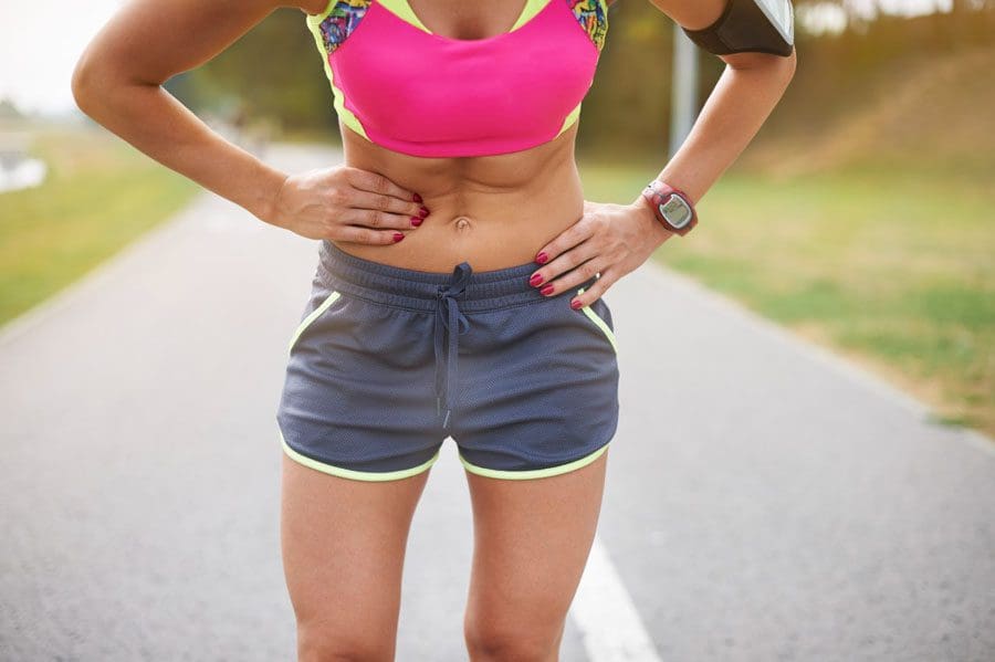
Children, teens, and adults participate in organized and recreational sports activities for fun, exercise, and social benefits. Individuals and parents are used to scrapes, bumps, bruises, sprains, and strains. However, internal abdominal injuries from the body colliding with another player or object are less common but dangerous. Abdominal injuries make up less than 4 percent of sports injuries but can be severe when they occur. These injuries are common in sports like wrestling, gymnastics, soccer, basketball, football, skiing, snowboarding, BMX freestyle, motocross, skateboarding, ice/field hockey, and lacrosse. Early symptoms are not always obvious or apparent and can be mild or seem to go in a different direction away from the abdominal region, which is why it is essential to know what to look for.
Internal Abdominal Injuries Athletes
There are about 3oo 000 abdominal sports-related injuries. Kids and young athletes risk injuring their abdominal organs because their abdominal wall is thinner and still in development. However, internal abdominal injuries to the stomach, small and large intestine, spleen, liver, and kidneys can and do happen in adults.
Injury Types
Sports-related internal abdominal injuries are considered rare, but studies suggest they are increasing. The most common sites include:
Liver
This causes pain in the upper right side of the abdomen.
The right lobe is the one that gets injured more often because it is bigger and presses against the ribcage.
A torn liver can cause severe bleeding.
Shock can develop from the bleeding, causing heart palpitations, rapid breathing, shortness of breath, and a pale, grey, and/or sweaty appearance.
The liver and spleen are the most commonly injured organs in sports. They are filled with blood and can get bruised, or ruptured, and can cause severe bleeding when torn or cut. Bleeding in the abdomen can irritate the diaphragm, which can cause pain in the shoulder. Sometimes shoulder pain is the only symptom making it difficult to diagnose and because bleeding can take time to develop, the symptoms might not present for several hours.
Spleen
This causes pain in the upper left side of the abdomen.
The spleen filters around 10% of the body’s blood supply every minute.
A torn spleen can cause rapid and life-threatening internal bleeding.
Kidneys
The kidneys can be injured by a blow/hit to the back or flank that causes bruising or laceration.
This injury can cause flank/side pain, blood in the urine, nausea, and/or vomiting.
Abdominals
A single organ or multiple organs can be injured.
This can be the pancreas, diaphragm, stomach, gallbladder, bladder, or intestines.
Bruising discoloration or bruising, particularly around the belly and flanks.
This can cause abdominal pain with movement that does not get better that could be accompanied by fever, nausea, or vomiting.
Running into an object, another player, or falling hard can cause bruising, laceration, or create a tear/opening of a bowel wall. Symptoms can be delayed days to weeks after the injury when inflammation or infection develops.
Recognizing Internal Injuries
Signs and symptoms to look for include:
Abdominal pain
Bruising around the abdominal area.
Tenderness over the injured area.
Rigid abdomen.
Left-arm and shoulder pain.
Right-sided abdominal pain and right shoulder pain.
Blood in the urine.
Cold, sweaty skin.
Nausea and vomiting.
Rapid heartbeat.
Low blood pressure.
Loss of consciousness.
Treatment
Chiropractic focuses on whole-body health and can help with abdominal injuries. The nervous and digestive systems are interconnected, meaning that damage could lead to viscerosomatic reflexeseven if not directly injured. If internal damage or bleeding has occurred, individuals will be referred to a specialist, surgeon, or another emergency medical professional. If internal damage is ruled out, a chiropractic treatment plan that includes adjustments, massage therapy, manual and mechanical decompression, exercises, stretches, and health coaching will help with tissue injuries and problems that are causing gastrointestinal distress.
Spinal Non-Surgical Decompression
References
Arumugam, Suresh, et al. “Frequency, causes and pattern of abdominal trauma: A 4-year descriptive analysis.” Journal of emergencies, trauma, and shock vol. 8,4 (2015): 193-8. doi:10.4103/0974-2700.166590
Barrett, Cassie, and Danny Smith. “Recognition and management of abdominal injuries at athletic events.” International journal of sports physical therapy vol. 7,4 (2012): 448-51.
Kucera, K. L., Currie, D. W., Wasserman, E. B., Kerr, Z. Y., Thomas, L. C., Paul, S., & Comstock, R. D. (2019). Incidence of Sport-Related Internal Organ Injuries Due to Direct-Contact Mechanisms Among High School and Collegiate Athletes Across 3 National Surveillance Systems. Journal of athletic training, 54(2), 152–164. https://doi.org/10.4085/1062-6050-271-17
Slentz, Cris A et al. “Effects of aerobic vs. resistance training on visceral and liver fat stores, liver enzymes, and insulin resistance by HOMA in overweight adults from STRRIDE AT/RT.” American journal of physiology. Endocrinology and metabolism vol. 301,5 (2011): E1033-9. doi:10.1152/ajpendo.00291.2011
Two surgeons discuss the diagnosis and treatment of acromioclavicular injuries in athletes. El Paso, TX. Chiropractor, Dr. Alexander Jimenez follows the discussion.
Acromioclavicular (AC) joint injuries most often occur in athletic young adults involved in collision sports, throwing sports, along with overhead activities like upper-extremity strength training. They account for 3% of all shoulder injuries and 40% of shoulder sports injuries. Athletes in their second and third decade of life are more often affected(1), and men are injured more commonly than women (5:1 to 10:1)(1,2).
Acromioclavicular dislocation was known as early as 400 BC by Hippocrates(3). He cautioned against mistaking it for glenohumeral (shoulder joint) dislocation and advocated treating with a compressive bandage in an attempt to hold the distal (outer) end of the clavicle in a diminished position. Almost 600 decades later Galen (129 AD) recognized his own acromioclavicular dislocation, which he sustained while wrestling(3). He left the tight bandage holding the clavicle down as it was too uneasy. In today’s era this injury is better known, but its treatment remains a source of fantastic controversy.
Anatomy
The acromioclavicular joint combines the collarbone to the shoulder blade and therefore links the arm to the axial skeleton. The articular surfaces are originally hyaline cartilage, which affects to fibrocartilage toward the end of adolescence. The average joint size is 9mm by 19mm(4). The acromioclavicular joint contains an intra-articular, fibrocartilaginous disc which may be complete or partial (meniscoid). This helps absorb forces in compression. There is marked variability in the plane of the joint.
Stabilizers
There is little inherent bony stability in the AC joint. Stability is provided by the dynamic stabilizers — namely, the anterior deltoid muscle arising from the clavicle and the trapezius muscle arising from the acromion.
Additionally, there are ligamentous stabilizers. The AC ligaments are divided into four — superior, inferior, anterior and posterior. The superior is most powerful and blends with muscles. The acromioclavicular ligaments contribute around two- thirds of the constraining force to superior and posterior displacement; however, with greater displacement the coracoclavicular ligaments contribute the major share of the resistance. The coracoclavicular ligament consists of the conoid and trapezoid. The conoid ligament is fan-shaped and resists forwards motion of the scapula, while the more powerful trapezoid ligament is level and resists backward movement. The coracoclavicular ligament helps bunch scapular and glenohumeral (shoulder joint) motion and the interspace averages 1.3 cm.
Mechanism Of Injury
The athlete who sustains an acromioclavicular injury commonly reports either one of two mechanisms of harm: direct or indirect.
Direct force: This is when the athlete falls onto the point of the shoulder, with the arm usually at the side and adducted. The force drives the acromion downwards and medially. Nielsen(5) found that 70 percent of acromioclavicular joint injuries are caused by an direct injury.
Indirect force: This is when the athlete falls onto an outstretched arm. The pressure is transmitted via the humeral head into the acromion, therefore the acromioclavicular ligament is disrupted and the coracoclavicular ligament is stretched.
On Examination
The athlete presents soon after the severe injury with his arm splinted to his side. The patient may state that the arm feels better using superiorly directed support on the arm. Most motions are limited secondary to pain near the top of the shoulder; the degree varies with the grade of sprain. The hallmark finding is localized swelling and tenderness over the acromioclavicular joint.
In dislocations, the outer part of the collarbone will appear superiorly displaced using a noticeable step deformity (in fact, it is the shoulder which sags beneath the clavicle). Occasionally, the deformity may only be apparent later, if first muscle spasm reduces acromioclavicular separation. Forced cross-body adduction (yanking the affected arm across the opposite shoulder) provokes discomfort. The clavicle can frequently be moved relative to the acromion.
Acromioclavicular Visualisation
The typical joint width measures 1-3mm. It’s regarded as abnormal if it is more than 7mm in men, and 6mm in women. Routine anteroposterior views of the shoulder reveal the glenohumeral jointnonetheless, that the acromioclavicular joint is over penetrated and so dark to interpret. Reduced exposure enhances visualization. The individual stands with both arms hanging unsupported, both acromioclavicular joints on one film. Weighted viewpoints (stress X-rays) are obtained with 10-15 lb weights not held but suspended from the individual’s wrists. They help differentiate type II-III injuries, but are of little clinical significance and therefore are no longer recommended in our practice.
Classification Of AC Separation
The importance of identifying the injury kind can’t be over emphasized because the treatment and prognosis hinge on an accurate diagnosis. The injuries are graded on the basis of that ligaments are injured and how badly they’re torn.
Allman (6) classified acromioclavicular sprains as grades I, II and III, representing respectively, no involvement, partial tearing, and total disruption of the coracoclavicular ligaments. More recently, Rockwood (1) has further classified the more severe injuries as standard III-VI.
The injuries are classified into six categories:
Type I This is the most common injury encountered. Only a mild force is needed to sustain such an injury. The acromioclavicular ligament is sprained with an intact coracoclavicular ligament. The acromioclavicular joint remains stable and symptoms resolve in seven to 10 days. This injury has an excellent prognosis.
Type II The coracoclavicular ligaments are sprained; however, the acromioclavicular ligaments are ruptured. Most players can return to their sport within three weeks. There is anecdotal evidence to suggest that steroid injections into the acromioclavicular joint speed up the resolution of symptoms, but this practice is not universal.
Type III The acromioclavicular joint capsule and coracoclavicular ligaments are completely disrupted. The coracoclavicular interspace is 25-100% greater than the normal shoulder.
Type IV This is a type III injury with avulsion of the coracoclavicular ligament from the clavicle, with the distal clavicle displaced posteriorly into or through the trapezius.
Type V This is type III but with exaggeration of the vertical displacement of the clavicle from the scapula-coracoclavicular interspace 100-300% greater than the normal side, with the clavicle in a subcutaneous position.
Type VI This is a rare injury. This is type III with inferior dislocation of the lateral end of the clavicle below the coracoid
Treatment
The treatment of acromioclavicular joint injuries varies based on the seriousness or grade of the injury.
Initial treatment: These can be quite painful injuries. Ice packs, anti-inflammatories plus a sling are utilized to immobilize the shoulder and then take the weight of the arm. As pain starts to subside, it is important to start moving the fingers, wrist and elbow to prevent shoulder stiffness. Next, it’s important to begin shoulder motion in order to stop shoulder stiffness.
Un-displaced injuries only require rest, ice, and then a slow return to activity over two to six weeks. Major dislocations require surgical stabilization in athletes if their dominant arm is involved, and if they participate in upper-limb sports
Type I & II: Ice pack, anti-inflammatory agents and a sling are used. Early motion based on symptoms is introduced. Pain usually subsides in about 10 days. Range-of-motion exercises and strength training to restore normal motion and strength are instituted as the patient�s symptoms permit. Some symptoms may be relieved by taping (taking stress off acromioclavicular joint). The length of time needed to regain full motion and function depends upon the severity or grade of the injury. The sport and the position played determine when a player can return to a sporting activity. A football player, who does not have to elevate his arm, can return sooner than a tennis or rugby player. When a patient returns to practice and competition in collision sports, protection of the acromioclavicular joint with special padding is important. A simple �doughnut� cut from foam or felt padding can provide effective protection. Special shoulder- injury pads, or off-the-shelf shoulder orthoses, can be used to protect the acromioclavicular joint after injury.
Some Type II injuries may develop late degenerative joint changes and will need a resection of the distal end of the clavicle for pain relief. It is important to note that after a resection of the distal end of the clavicle, particularly in a throwing athlete, there may be formation of heterotopic bone on the under surface of the clavicle which can cause a painful syndrome which presents like shoulder impingement.
Type III: The treatment of type III injury is less controversial than in past years. In the 1970s, most orthopaedic surgeons recommended surgery for type III acromioclavicular sprains(7). By 1991, most type III injuries were treated conservatively(8). This change in treatment philosophy was prompted by a series of retrospective studies(9). These showed no outcome differences between operative and nonoperative groups.
What’s more, the patients treated non-operatively returned to full activity (work or athletics) earlier than surgically treated groups(10, 11). The exceptions to this recommendation include people who perform repetitive, heavy lifting, people who operate with their arms above 90 degrees, and thin patients who have prominent lateral ends of the clavicles. These patients may benefit from surgical repair(12).
Any discussion about the management of acute injuries to the AC joint must deal with which of the many methods of surgical therapy described is the best for their situation, but whether surgery should be considered at all. Surgery is generally avoided in athletes participating in contact sports since they will often re-injure the shoulder later on.
Type IV-VI: Account for more than 10-15% of total acromioclavicular dislocations and should be managed surgically. Failure to reduce and fix these will lead to chronic pain and dysfunction.
Surgery
Surgical repair can be divided into anatomical or non- anatomical, or historically into four types:
? Coracoclavicular repairs (Bosworth screws(13), cerclage, Copeland and Kessel repair).
? Distal clavicular excision.
? Dynamic muscle transfers.
? Disadvantages of surgery are that there are risks of infection, a longer time to return to full function and continued pain in some cases.
For the individual with a chronic AC joint dislocation or subluxation that remains painful after three to six months of closed treatment and rehabilitation, surgery is indicated to improve functioning and comfort.
For sequelae of untreated type IV-VI, or painful type II and III injuries, the Weaver Dunn technique is advocated. This�entails removing the lateral 2cm of the clavicle and reattaching the acromial end of the coracoacromial ligament to the cut end of the clavicle, thus reducing the clavicle to a more anatomical position.
Postoperatively, the arm is supported in a sling for up to six weeks. Following the first two weeks, the patient is permitted to use the arm for daily activities at waist level. After six weeks, the sling or orthosis is discontinued, overhead actions are allowed, formal passive stretching is instituted, and light stretching using elastic straps is initiated. Stretching and strengthening are begun slowly and gradually. The athlete shouldn’t return to their sport without restriction until full strength and range of motion has been recovered. This usually occurs four to six months following operation.
Conclusion
AC joint injuries are an important source of pain at the shoulder area and have to be assessed carefully. The management of these injuries is nonoperative in the majority of cases. Type I and II injuries are treated symptomatically. The present trend in uncomplicated type III injuries are a non operative strategy. In the event the athlete develops following problems, a delayed reconstruction might be undertaken. In athletes involved in heavy lifting or prolonged overhead activities, surgery may be considered acutely. Type IV-VI injuries are generally treated operatively.
No matter what kind of treatment is chosen, the ultimate purpose is to restore painless function to the wounded AC joint so as to reunite the athlete safely and as quickly as possible back to their sport. It is possible in the vast majority of acromioclavicular joint injuries.
References
Reza Jenabzadeh and Fares Haddad
1. Rockwood CA Jr, Williams GR, Young CD. Injuries of the Acromioclavicular Joint. In CA Rockwood Jr, et al (eds), Fractures in Adults. Philadelphia: Lippincott-Raven, 1996; 1341-1431.
2. Dias JJ, Greg PJ. Acromioclavicular Joint Injuries in Sport: Recommendations for Treatment. Sports Medicine 1991; 11: 125-32.
3. Adams FL. The Genuine Works of Hippocrates (Vols 1,2). New York, William Wood 1886.
4. Bosworth BM. Complete Acromioclavicular Dislocation. N Eng J Med 2 41: 221-225,1949.
5. Nielsen WB. Injury to the Acromioclavicular Joint. J Bone Joint Surg 1963; 45B:434-9.
6. Allman FL Jr. Fractures and Ligamentous Injuries of the Clavicle and its Articulation. J Bone Joint Surg Am 1967;
49:774- 784.
7. Powers JA, Bach PJ: Acromioclavicular Separations: Closed or Open Treatment? Clin Orthop 1974; 104 (Oct): 213-223
8. Cox JS: Current Methods of Treatment of Acromioclavicular Joint Dislocations. Orthopaedics 1992; 15(9): 1041-1044
9. Clarke HD, Mc Cann PD: Acromioclavicular Joint Injuries. Orthop Clin North Am 2000; 31(2): 177-187
10. Press J, Zuckerman JD, Gallagher M, et al: Treatment of Grade III Acromioclavicular Separations: Operative versus
Nonoperative Management. Bull Hosp Jt Dis 1997;56(2):77-83
11. Galpin RD, Hawkins RJ, Grainger RW: A Comparative Analysis of Operative versus Nonoperative Treatment of Grade III Acromioclavicular Separations. Clin Orthop 1985; 193 (Mar): 150-155
12. Larsen E, Bjerg-Nielsen A, Christensen P: Conservative or Surgical Treatment of AC Dislocation: A Prospective, Controlled, Randomized Study. J Bone Joint Surg Am 1986;68(4):552-555
13. Bosworth BM. Complete Acromioclavicular Dislocation. N Engl. J. Med. 241: 221-225,1949.
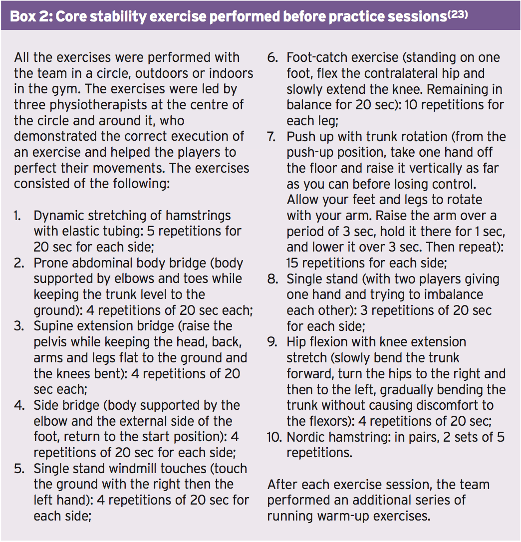
Core chiropractor, Dr. Alexander Jimenez continues from part I through the core stability routines.
Menu 6: Pulley, Standing
This menu challenges pelvic stability during unilateral standing upper body movements. The kinds of arm movements undertaken in many sports create strong rotational forces that have to be controlled by the trunk and pelvic muscles. The aim of these exercises, therefore, is to develop co-ordination and control of the pelvis.
Research has shown that unilateral exercises increase the recruitment of the core musculature. The core and pelvic muscles will all be using static contractions to hold the required postures, while the upper body muscles will be producing the limb movements. The resistance load on the arm is secondary to the stability challenge of the core. Overall this menu is intermediate.
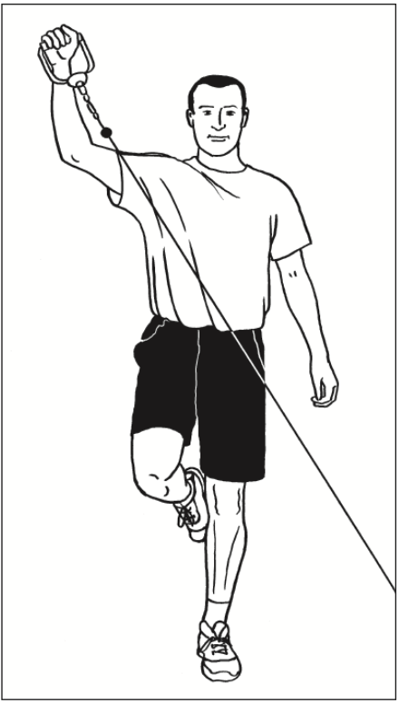
Rear Sling
Overview: The challenge of this exercise and its pair (see opposite) is to establish perfect pelvic alignment, while standing on one leg, against a rotational force from the upper body.
Technique: Stand on one leg to the side of the pulley column. Handle is attached at below-hip height. Grasp the handle with the hand on the opposite side (opposite to standing leg). Set perfect posture and pelvic alignment.
Brace the core and then pull the weight up and around the body, keeping the elbow straight, so that the arm rotates up
and out. Finish with hand above your head and out to the side slightly. The aim is to maintain perfect balance and pelvic
alignment as you raise and lower the arm diagonally. Reposition to repeat exercise for opposite leg/arm.
Perform 10 reps each side increasing to 20 reps; 2 to 3 sets.
Progression: Increase the weight.
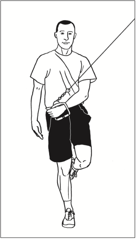
Front Sling
Overview: This is the natural opposite of the rear sling exercise. It involves a forward arm rotation, which must be controlled.
Technique: Stand on one leg to the side of pulley column. Handle is attached at above shoulder height. Grasp the handle with the arm nearest the column (opposite side to standing leg). Set perfect posture and pelvic alignment.
Brace your core; pull the weight down and around the body, keeping the elbow straight so that the arm rotates down and round. Finish with hand next to your hip across your body. The aim is to maintain perfect balance and pelvic alignment as you lower and raise the arm. Reposition to repeat with opposite leg/arm.
Perform 10 reps each side, increasing to 20 reps; 2 to 3 sets.
Progression: Increase the weight.
One Leg, One Arm Rowing
Overview: The challenge of this exercise is to maintain stability while standing on one leg and controlling against a pulling force from the upper body. The pelvis must stay fixed when the upper back and shoulder are pulling backwards.
Technique: Stand on one leg, facing the pulley column. Handle is attached at waist height. Grasp the handle with the opposite arm (same side as lifted leg). Your hand will be out directly in front of you in the start position. Set perfect posture and pelvic alignment, standing tall with shoulders back.
Brace your core; pull on the cable, leading with the elbow in a rowing movement Finish with hand by your side and elbow behind you. The aim is to maintain perfect balance and pelvic alignment as you perform the rowing movement. Reposition to repeat with opposite leg/arm.
Perform 10 reps each side; 2 to 3 sets.
Progression: Increase the weight.
Menu 7: Medicine Ball, Floor
The four exercises in this menu all involve throwing and catching the medicine ball while performing a trunk flexion or rotation movement. The action of throwing the ball during the muscle-shortening phase of each of the exercises increases the force production of the trunk muscles. The action of catching the ball at the start or during the muscle-lengthening phase of each exercise not only increases the force production but also the overall stability challenge.
The impact that the catch has on the upper limb has to be controlled by the trunk. You should be aiming to maintain good spine alignment and correct movement while making the catch. Only use a weight of medicine ball that will allow you to perform the exercises with good technique. If the ball is too heavy, you will sacrifice core stability, irrespective of your arm strength.
Overall these exercises are advanced. However they are also safe and effective for young athletes using light medicine balls to develop dynamic trunk movement and control.
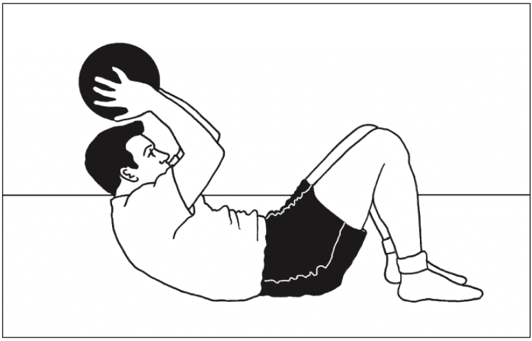
Sit Up & Throw
Overview: An advanced version of a sit-up exercise, in which the throwing action makes the crunch phase faster and the catching action adds load to the return phase.
Level: Advanced
Muscles targeted: Abdominals (Plus upper body)
Technique: You will need a partner to receive and pass the ball. Alternatively perform the exercise in front of a wall and use a medicine ball that will bounce back.
Start in the sit-up position (knees bent) with hands up ready�to receive the ball. Catch the ball and begin to lower back down. Do not collapse back down, control it with the abs and keep hands above the head as you lower down.
Once shoulders are touching the floor (keeping head up and eyes forward), reverse the movement. Throw the ball forward and crunch up at the same time. Follow the throwing action and complete the sit-up as fast as possible. Make sure you crunch as you throw so that the abs contribute to the force of the throw and help you sit up faster. Men should start with a 5kg ball; women with a 3kg ball.
Perform 10 to 20 reps; 2 to 3 sets
Progression: Progress to heavier ball once 3 sets of 20 reps is comfortable
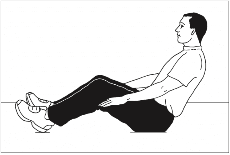
45-degree Sit, Catch and Pass
Overview: A very tough stability exercise that requires massive trunk musculature co-contraction to hold a good spine alignment against the impact of making the catch.
Technique: Sit up with knees bent and lean back at 45 degrees. Aim to hold a �lengthened� spine, with lumbar spine in neutral, shoulders back and neck long and relaxed. It takes a fair amount of control and strength endurance simply to hold this posture perfectly. Aim to get this right before progressing on to the catch and pass.
Raise hands in front of your face and receive a pass from a partner, around this height. As you catch the ball you must hold the long spine position. Do not flex the low back, or become round-shouldered. Gently throw the ball back. Men should start with a 3kg ball; women with a 2kg ball.
Complete a few passes, holding the position for 30 seconds. Perform 2 to 3 sets.
Progression: Raising the hands to above head height makes the stability challenge of the catch significantly harder. Catches made to either side of the head are also more challenging.
Sit & Twist Pass
Overview: A trunk rotation exercise involving catching and passing the medicine ball, which provides a challenge to the obliques to produce powerful rotation, but also pelvic stability, so that the sitting position is stable throughout the movement.
Level: Advanced
Muscles targeted: Abdominals, Obliques
Technique: Sit up with knees bent and lean back at 45 degrees. Aim to hold a �lengthened� spine, with lumbar spine in neutral, shoulders back and neck long and relaxed. Your feet, knees and hips should remain reasonably still throughout this exercise, the rotation coming from your waist and not your hips.
Hold hands to one side ready to receive the ball. Catch the ball to one side and absorb the catch by turning your shoulders further to that side. Reverse the rotation, turning back to the middle and release the ball. Continue rotating to the other side; receive the ball the other side and continue. Ensure you�can hold good posture throughout the movement, with a long spine and wide shoulders. Men should start with a 4 to 5kg ball; women with a 2 to 3kg ball.
Perform 10 to 20 reps.
Progression: Increase the weight of the ball once you can perform a set of 20 reps comfortably with perfect technique.
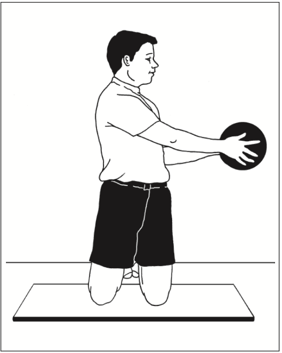
Kneeling Twist Pass
Overview: To perform the rotation movement in this position demands a greater range of motion, helping to develop strength through the full range of trunk rotation. It may also help to develop trunk rotation range of movement.
Level: Intermediate to advanced
Muscles targeted: Obliques
Technique: Kneel upright with good posture (lumbar spine in neutral, chest out, shoulders low). Start with the ball in hands and twist shoulders and head round as far as you can. Then, under control, twist around to the other side as far as possible, and hand the ball to partner. Turn back to the start position, receive the ball again and continue.
The aim of the movement is to rotate through the biggest shoulder turn you have. You can allow the hips to rotate a little with the shoulders, but not too much. You should feel a stretch in the side at the end of each twist.
As you gain greater flexibility and stability you will be able to�fix your pelvis square to the front and rotate through an increasingly full range of motion. Men should start with a 5 to 6kg ball; women with a 3 to 4kg ball.
Perform 10 reps then take the ball to the opposite side and repeat.
Menu 8: Medicine Ball, Standing
The aim of this menu is to perform trunk movements while standing on one leg. This is functional training for balance in sports and daily living activities. These exercises are advanced because of the requirements for lower limb balance and body movement awareness, which makes controlled performance of these trunk movements quite difficult. These moves also use the hip rotator and abductor muscles for control and stability.
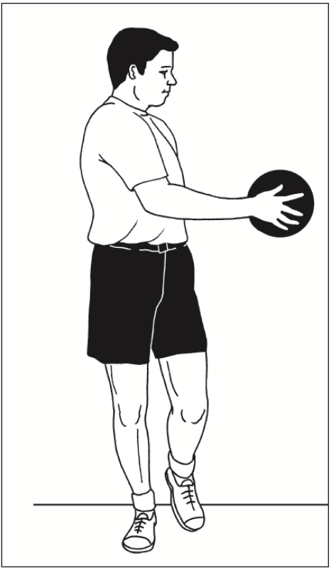
One-leg Twist Pass
Overview: A trunk rotation exercise performed on one leg. This requires good pelvic stability at the hip of the standing leg, for the trunk rotation to be dissociated from the pelvis.
Technique: Stand on one leg with hips facing square to the front. Hold medicine ball slightly out in front. Slowly twist from side to side. The rotation comes from the waist only,�head turning with the shoulders. Keep pelvis fixed square and knee in line with second toe throughout. Men should start with a 5 to 6 kg ball; women with a 3 to 4 kg ball.
Perform 10 slow reps; 2 to 3 sets. Repeat on other leg.
Progression: Swap the ball for a pulley machine and add resistance, once you have mastered the controlled balance on one leg.
One-leg Deadlifts with Rotation
Overview: An advanced exercise for the posterior chain of muscles, which includes rotation to challenge control of pelvis.
Level: Advanced
Muscles targeted: Erector spinae, Gluteals (max and med) Hamstrings, Piriformis
Technique: Stand on one leg. Flex the free leg a little at the knee to lift it off the floor, but do not flex or extend the hip of the free leg throughout the movement, in order to keep pelvis in control. Hold the ball in front of you.
Bend down, flexing at the knee and the hip. Lower down until the ball touches the floor by your foot, all the time keeping your arms straight and without reaching excessively with your upper back (ie, maintain a reasonably flat back). Stand back up, pushing down through the foot to use your gluteals correctly to extend the hips.
Alternate between touching the ball down on the inside and then the outside of the standing foot. This means you are internally or externally rotating the hip on alternate repetitions, challenging control of hip rotation. Keep the knee in line with�second toe as much as possible throughout. Men should use a 5kg ball; women use a 3kg ball.
Start with 5 slow controlled reps, 2 to 3 sets. Build up to 10 reps. Repeat on the opposite leg.
Progression: Increase the weight of the ball or use a dumb-bell as you get stronger.
One-leg Catch & Pass
Overview: The main aim of this exercise is to control the impact of the catch without losing balance or rotating excessively at the hips. It�s all about how effectively you can anticipate the impact and produce the required stiffness throughout the body to retain good posture and control. This is a very useful �reaction�-type stability exercise.
Level: Advanced
Muscles targeted: Everything
Technique: Stand on one leg with good posture (lumbar spine neutral, chest out, shoulders wide) and with hips square to the front. Hold hands up ready to catch. Receive catches anywhere within arm�s reach. Make sure the passes are varied in their placement. Aim to restrict movement to arms and/or turning your shoulders, keeping the pelvis and lower limb stable. Use a 2 to 3kg ball that is not too big, so it is easy to catch.
Start with 30 sec bouts of catch and pass on each leg; 2 to 3 sets.
Progression: Receive more forceful passes so the impact of the catch is greater.
Menu 9: Resistance-Based
Menu rationale
The aim of these three exercises is to progress the loading in order to build high-level trunk muscle strength. These exercises can be performed in the 5- to 10-repetition range with a suitably high weight for this number of reps. As you get stronger, you should prioritize an increase in weight rather than an increase in the number of reps. Overall, these exercises are very advanced.
Crunch with Weight
Overview: The standard isolated abdominal exercise with increased load.
Level: Advanced
Muscles targeted: Abdominals
Technique: Perform the crunch in the usual way: knees bent, low back flat, head up and looking forward. Curl the shoulders up and down using just the abdominals. The weight (medicine ball, dumb-bell or barbell weight plate) should be held above or behind the head. Arms are fixed, all they do is hold the weight in place. Do not use arms to move the weight relative to head as the crunch is performed. Keeping the elbows out helps to achieve this.
Perform 5 to10 reps; 2 to 3 sets.
Progression: Increase weight, maintaining the range of 5 to 10 reps per set.
Reverse Hypers
Overview: An excellent hip and back extension exercise to which it is very simple to add load.
Level: Advanced
Muscles targeted: Erector spinae, Gluteals
Technique: Lie on your front on a horizontal bench, with hips just off the end of the bench. Grasp bench legs firmly for support. Your legs should be straight with a dumb-bell between the ankles for resistance. Squeezing the gluteals, extend hips and lift legs and the dumb-bell off the floor. Stop when your back is slightly hyper-extended and hips are fully extended. Lower slowly until feet are just off the floor and continue.
Perform 8 to 10 reps; 2 to 3 sets.
Progression: Increase weight, maintaining the range of 8 to 10 reps per set.
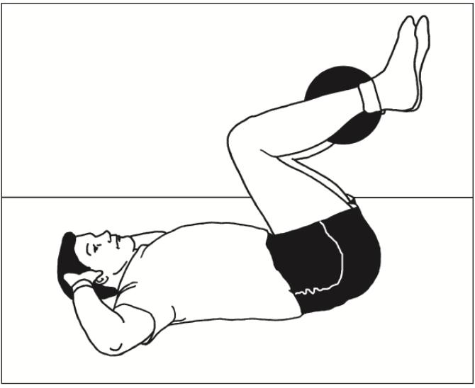
Reverse Crunch with Weight
Overview: This is a great exercise, as it requires good co- ordination and strength. Research shows that the obliques as well as the abdominals work very hard during this exercise, making it excellent value.
Level: Advanced
Muscles targeted: Abdominals, Obliques
Technique: Lie on back with hands behind head and elbows out to the sides. Knees should be bent and heels close to bum. Hold weight between your legs. Initiate the movement by curling the pelvis upwards (flattening the back into
the floor) and then continue to use the abs to pull the low back and pelvis off the floor. This is the bit that requires good co- ordination, as the temptation is to kick with the legs and pull the hips up with the hip flexors. Learn to focus on the abs before you add weight, as if you do this strictly it is very tough, especially for women (whose pelvises are relatively heavier).
Perform 5 to 10 reps; 2 to 3 sets.
Progression: Increase weight, maintaining the range of 5 to 10 reps per set.
Menu 10: Hanging Bar
Menu rationale
The aim of these three exercises is to work the abdominals as hard as possible with very advanced, gymnastic-style movements. Reasonable upper body strength is required for these exercises.
Hanging Leg Lifts
Overview: This exercise requires you to lift the full weight of your legs and (if possible) your pelvis, while hanging from a bar. Anyone who can perform these movements well through a good range of motion has achieved good strength.
Level: Advanced
Muscles targeted: Abdominals, Obliques, Hip flexors
Technique: Hang from a bar with arms straight. Lift knees, bringing them up as high as possible. At the top of the movement the knees should be near the chest and pelvis should be curled upwards (low back flexed). This extra curl of the pelvis ensures that the abdominals are working maximally. Do not kick legs up or swing the body excessively. Simply draw up knees, crunching as you lift. It is important to feel that the abdominals are doing the lion�s share of the work rather than the hip flexors or front of thigh muscles.
Perform 5 to 10 reps;, 2 to 3 sets.
Progression: Perform the same exercise with straight legs, lifting them up to 90 degrees in front of you, curling the pelvis at the top of the movement.
Windscreen Wipers
Overview: The ultimate ab-buster. Anyone who can do 10 reps of this exercise with good technique has a very strong core!
Level: Super advanced
Muscles targeted: Abdominals, Obliques, Hip flexors
Technique: Hang from bar with arms straight. Lift legs up in the air until feet are at approx head height. Maintaining the height of the lift, take the legs from side to side in an arc. The movement will look like a windscreen wiper, moving from side to side. Aim for at least 45 degrees of movement to each side.
Perform 5 to10 reps; 2 to 3 sets.
Progression: The straighter the legs, the harder the exercise. Increasing the range of movement to each side also makes it tougher.
Candlesticks
Overview: Another beauty! Lots of strength required to control this movement; only for the very strong.
Level: Super advanced
Muscles targeted: Abdominals, Obliques, Hip flexors
Technique: Lie flat and raise yourself up to a shoulder stand position, holding on to a bench/table leg/partner’s leg with your hands above your head. Establish a fully extended hip and leg position and then begin to lower your body down slowly to the floor. The body should move in an arc as a single unit (no sagging in the back, or bending at the hips or knees). Lower under control from vertical to just above horizontal.
Gripping firmly for stability, lift your body back up into shoulder stand, again keeping everything straight and aligned in a single unit.
Slow and controlled movement on the way down will help, and a maximal contraction of everything will get you back up.
For many athletes following any major endurance event they will return to their houses, to recover, celebrate, reflect and rebuild to their next career step. Some, like the athlete in this case study will need to now focus attention on delayed decisions concerning whether to go under the knife to sort out a chronic injury.�El Paso, TX’s Injury scientist, Dr. Alexander Jimenez takes a look at the study.
My client has been competing in triathlon for 10 or more years, although his career has included a range of serious injuries which have kept him from races for months on end. In the previous two to three decades, however, he’s enjoyed a sustained period of injury-free training and racing, and has climbed to the peak of the world rankings. But the emergence of hip pain has seen him once more return to the physio’s table.
The triathlete’s accident history highlights a common pattern among sportspeople: 2 tibial stress fractures, a femoral neck stress fracture and a serious ankle sprain — every one of these on his right side. The significant contributing element to the bone stress injuries is a 1.5cm leg-length gap (his right leg is shorter).
He’d first experienced comparable hip pain in 2004; it kept him from running for three months. At that time, nothing was detected on a bone scan or MRI, or so the pain went paralyzed. An intra-articular cortisone injection (CSI) elicited no improvement. The athlete remembers that he chose to train on his painful hip, never allowing the symptoms to settle. The nearest he ever came into an investigation was a hypothesis that he could have a little, undetected, labral lesion.
The present episode of hip pain began initially at night after a hard three-hour bicycle ride. Earlier this, however, he hadn’t cycled for five times. He described his initial symptom as a profound hip tightness (lateral and lateral), together with slight pain in his groin. He was able to continue to train however, was feeling that the hip tightness and pain following both cycling and running (swimming was symptom-free).
A week later his symptoms dramatically worsened when he flew from Australia to Singapore, on his way to a French high- altitude camp. As he got off the airplane, he felt deep hip pain as well as the tightness. As elite athletes tend to do, he coached anyway, running a tricky track session, which made the hip much worse: he was unable to ride or run without pain. He instantly started a course of anti- inflammatories.
I met him in Singapore and evaluated him in the airport, initially ruling out any prospect of a disease or systemic matter. He explained he had been feeling an ache during the night, lying in bed; on waking, the hip would be OK, but got worse the longer he walked.
On assessment, he had the following physical signs:
� walking with obvious limp
� pain on hopping (6/10)
�painful right hip quadrant/impingement test (full hip flexion/adduction)
� reduced right hip flexion (-10 degrees compared to left)
� reduced right hip internal rotation (-10 degrees compared to left)
� increased tone on palpation of TFL, adductors, hip flexors, gluteal, piriformis and deep rotators
� lumbar spine and SIJ were OK
� femoral shaft bone stress test was OK � leg length discrepancy (right side 1.5cm shorter)
� right innominate (pelvis) anteriorly rotated
� weakness in right hip abductors/extensors
� reduced calf endurance on right side (-5 reps)
� ankle dorsiflexion range of movement was OK
� reduced proprioception on right (single leg stance, eyes closed).
I thought the differential diagnoses were:
� femoral neck stress fracture
� labral tear, possibly with hip synovitis
� FAI (femoro-acetabular impingement), possibly with hip synovitis.
I initially treated the triathlete with soft- tissue techniques to reduce the tone around the hip joint. Trigger-point releases were performed on his TFL, adductors, gluteals, piriformis, deep rotators and iliopsoas.�This reduced his jump pain into 3/10. Manual long-leg grip further decreased the strain on hopping (2/10). He still had pain and stiffness on walking but it sensed “simpler. As he prepared to embark on his long run flight to Europe, I counseled him to not sit for too long and maintain his stylish as straight as possible to decrease any potential impingement from hip flexion.
Luckily, the hip didn’t get worse throughout the flight. On arrival at the French high-altitude training centre, we initiated a strategy of two swims and two intensive treatments a day, aiming at reducing muscle tone, restoring his range of hip movement and normal muscle control and stamina. We had been expecting that the problem was not a stress fracture, but just minor hip synovitis that could settle quickly. Following a week of conservative treatment, though, we were just able to keep his hop pain in 2/10, and that he still could not run 20 meters without any pain and limping.
In collaboration with medics, we flew to London to see a sports doctor and get MRI scans. The scans revealed no bone stress reaction, fracture or labral ripping — which was a big relief; however, it did show signs consistent with FAI (femoro-acetabular impingement). He had hip synovitis with a rectal lesion on his femur.
Hip injuries aren’t much reported among triathletes — in fact they are notably absent from reports on Olympic and Ironman triathlons, which mention knee, back, H/ Achilles, lower leg, ankle and shoulder as the most common accidents (1-3).
In this state, when the hip is in maximum flexion and internal rotation, the labrum and cartilage abut and impinge; damage to the articular cartilage and acetabular labrum results from this pathologic bony contact. The contact generally results in a structural abnormality of the femur (“camera impingement”) along with the acetabulum (“pincer impingement”) or a combination of both (“mixed impingement”). Over time, via repetitive micro-trauma, the aggravating motion hurts the hip cartilage or labrum (or both) during normal joint motion. This happens along the anterior femoral neck and the anterior–superior acetabular rim. FAI is a possible trigger of early hip joint degeneration (4).
Arthroscopic surgery is the direction of choice for FAI if symptoms do not settle; however as his next Competition was only three and a half a year off, surgery was not an option. Instead, over a five-day interval, the athlete had two cortisone (CSI) and local anesthetic injections into the hip joint (under ultrasound guidance) to settle the indicators.
Our aim was to grow the hip range of motion and extend the capsule to reduce any additional impingement, slowly returning to regular training. Following the competition, the athlete would then should see a hip arthroscopic surgeon to acquire a surgical opinion to the best option for long-term direction.
Injection Relief
After both shots my customer felt sore for five days. The initial CSI settled his pain on hopping to 1/10 and after seven days he managed to operate without symptoms. But minor hip stiffness and aching at the end of the day prevented him from progressing to optimal training, so that he then underwent a second steroid injection. This settled the hop pain into 0/10 and decreased the aching; so after five times he returned to mild cycling and after seven days he started running again, also.
The athlete admitted that, following the first shot, he had done more and gone tougher in training than directed, as he had felt “good. This mistake of “too much too soon — all too common in elite athletes — had led to excessive inflammation and aching in the hip nightly after training. After the next injection he returned to normal intensity slower and more gradually.
My client built his training up to regular levels by four months following the final injection (swimming five times per week, cycling four days and running six to seven days). He began with very easy cycling on a wind trainer for 30 minutes, building slowly to 90 minutes before cycling on the street. He cycled two days on and one day away and avoided hills to the first two weeks. He started jogging on the apartment for 15 minutes and slowly built up to 90 minutes after three weeks. He did not run hills or about the track; and as he ran only on every single day, he would diligently concentrate on technique.
From week six to week 11, my client remained on anti inflammatory medication and underwent two treatments a day.
The hands-on treatment continued to:
� increase hip range of movement
� stretch the hip capsule
� normalise pelvic symmetry and hip muscle tone
� improve muscle control and strength � improve proprioception
� ensure optimal biomechanics via video assessment (cycling and running).
Eleven weeks after he first felt his hip pain, the triathlete returned to racing; however he failed to finish the first race, partially because of minor hip stiffness but mainly due to “fitness. Fortunately there were not any prolonged symptoms after the race and a week after he successfully returned to competition, coming second in a really strong field. His very minor ongoing symptoms were handled with anti-inflammatory drugs and hands-on treatments.
If this athlete wants to pursue a long- term triathlon career up to the London Olympics, then he will now require surgery. The arthroscopic surgical technique initially assesses the cartilage and labral surfaces, debrides any abnormalities of the hip joint cartilage and hip labrum, removes the non-spherical segments of the femoral head�and any prominent sections of the anterior femoral neck and bony growths on the acetabular rim that may continue to contribute to hip joint impingement.�The alternative is early joint degeneration and onset of osteoarthritis.
References:
1. Wilk B et al: �The incidence of musculoskeletal injuries in an amateur triathlete racing club�. J Orthop Sports Phys
Ther 1995 Sep;22(3):108-12.
2. Collins K et al: �Overuse injuries in triathletes. A study of the 1986 Seafair Triathlon�. Am J Sports Med 1989 SepOct;17(5):675-80.
3. Korkia PK et al: �An epidemiological investigation of training and injury patterns in British triathletes�. Br J Sports Med 1994 Sep;28(3):191-6.
4. Ganz R. et al (2003): �Femoroacetabular impingement: a cause for osteoarthritis of the hip�. Clin Orthop Relat Res. 417:112�120. For more information see: www.hipfai.com
El Paso, TX. science based chiropractor, Dr. Alexander Jimenez looks at this uncommon problem � and how it can be treated.
The true incidence of obturator externus accidents is unknown, as frequently they may be misdiagnosed as hip joint pathology and/ or groin pathology as the website of symptoms as well as also the presenting objective signals may mimic other pathologies such as hip joint labrum pathology, anterior femoral triangle issues and perhaps even gluteal pathology.
Injury for this muscle gifts as a deep obscure groin/hip pain and functionally the muscle may still hide direct involvement as a pain generator since it is primarily a equilibrium muscle rather than a force-producing hip muscle.
This case study presents an unusual case of hip-related pain in a professional baseball player which also shown itself as an injury to the contralateral adductor longus.
The Player
As he was wrestled to the floor, his right hip was compelled at a rapid and loaded flexion/internal turning position. His first sensation was pain deep inside the anterior hip/groin area.
When he presented to the medical team with the accident, he complained of a profound catching sensation inside the hip joint location. It had been difficult to fully bend the hip and to also twist on the stationary limb (because he did whilst kicking a ball). His prior background consisted of a right-sided inguinal hernia repair five seasons before as well as a few gentle on again/off back osteitis pubis-type signs that would normally flare from the first period as his goal-kicking amounts have been increased. He was obviously a left- footed goal kicker.
On examination, he observed that the pain to become worse on passive flexion/internal rotation of the hip (hip walkway test). He was noticeably tight and irritated from the shallow TFL muscle, and also posteriorly across the greater trochanter around the insertion for the gluteals and deep hip rotators. He was also particularly high tone in the right iliopsoas muscle.
He was initially diagnosed clinically because of hip joint sprain due to the mechanism of harm being a pressured flexion/internal rotation type position that would always put pressure on the anterior hip joint capsule/labrum.
He was treated initially with deep iliopoas muscle sparks and hip joint mobilizations using a seat belt to gap the hip joint. He reacted reasonably well with the therapy and immediately felt more comfortable on a hip joint quadrant test. He was rested from coaching for 2 days and ran on the next day and played a match on the fourth day. But during the match, though his right hip did not create any pain, he’d notice pain on his left adductor source that was more pronounced during kicking.
Three days post-game he detected this ongoing left adductor origin pain and it was made worse by kicking again through training. An MRI was performed to Look at the left adductor origin and also the report noted:
Grade 1 left adductor longus strain deep in the
Grade 2 right obturator externus strain on its femoral attachment
Grade 1 right iliopsoas muscle strain in the MTJ.
The surprise finding on the MRI of a grade 2 obturator strain prompted the medical team to more formally assess the participant for ongoing hip joint disorder. The particular features to notice from this medical examination were:
Subjective
? A sensation of weakness and instability in the right hip whilst kicking with the left foot.
? No pain in the right hip with running, even with top-end speed. However, the left adductor longus was symptomatic on running and kicking.
Objective
? Pain on passive right hip internal rotation whilst in 90-degree hip flexion. This pain was deep anteriorly in the hip, almost presented as a groin problem.
? Some discomfort on resisted right hip flexion/external rotation deep inside the iliac fossa.
? Pain and weakness in the left adductor on adductor squeeze tests. These squeeze tests performed at 0/45/90 degrees of knee flexion with a pressure cuff between the knees. Usual pre-season scores measured 260/260/250. On current testing they measured 150/170/180. Pain was felt at the end of the squeeze.
? Discomfort with prone lie hip passive internal rotation. This pain was more focused around the right greater trochanter posteriorly.
Pathomechanics
It had been suspected that this player had endured a secondary injury to the left adductor longus (a muscle used a lot in goal-kicking) due to the inherent failure in bolstering the proper hip throughout the plant phase of the kick due to the inhibition of the right obturator externus, a muscle considered to be an important hip stabilizer and turning control muscle at the hip. With insufficient hip stabilization in kicking, the left hip was required to create more power to compensate for the unstable right hip to gain the length from the kick. Then the left adductor longus failed along with a strain injury led.
Management
The management of the matter initially centered on the two key features being the left-sided adductor strain and the right- sided obturator externus strain.
In the week following the accident, the player was sent to get a series of Actovegin shots to the left adductor longus. This was done according to protocol that was three injections every 48 hours — Monday/ Wednesday/Friday. In this five-day period the adductor longus was handled with deep tissue flush massage and gentle isometric adduction exercises at supine (chunk squeezes) in the three positions of examining — 0/45/90 levels of knee flexion — also as wall squat adductor squeezes in the same positions. The obturator externus was medicated with heavy tissue releases (obtained through the anterior groin region) and direct theraband strengthening of hip external rotation in sitting and in prone. Actovegin shots to the obturator externus are regarded as difficult because of problems with accessing this muscle through the superficial hip musculature.
The adductor exercises progressed into through array adduction with theraband resistance (equally with the left leg being the motion leg as well as the stability leg).
By 12 days post-injury it had been detected that the obturator externus strength had not improved and the player still had deep- seated right back pain pain. It was rationalised that perhaps the direct treatment to this muscle and also the direct open kinetic chain strengthening was possibly making the muscle texture worse. The choice was made to stop any direct hands-on therapy to the muscle and also to prevent any direct open kinetic chain strengthening. Instead the player lasted with bilateral theraband exercises of both hips into flexion and then abduction and expansion in addition to adduction. The avoidance of lead obturator externus soft tissue treatment and exercise appeared to improve the hip function immediately.
The participant started running 20 times post-injury and quickly progressed through running stages over a five-day period of conducting on alternate days. At this point the player’s adductor squeeze scores had improved to steps according to pre- season baselines. However, daily the player ran direct adductor strength operate using a Pilates reformer as a slider drill to immediately load into adduction in addition to hammering theraband adduction exercises in standing and in supine lying.
By 27 days post-injury the player managed to begin kicking, change in direction and rugby training. He played at 30 times post-injury with no ill effects.
Discussion
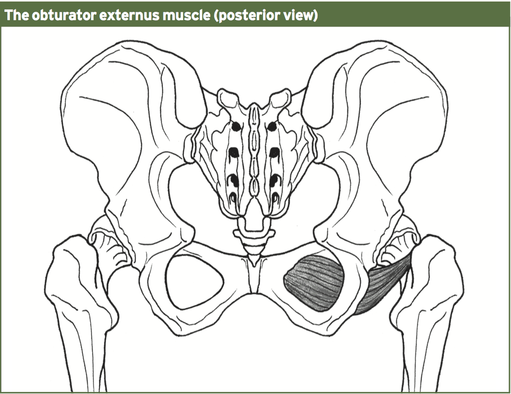
It arises immediately around the medial side of the obturator foramen, as well as the inferior ramus of the ischium; it also arises in the lateral two-thirds of this outer surface of the obturator membrane, and also in the tendinous arch which completes the canal to the passage of the obturator nerves and vessels.
The action of the muscle is to externally rotate the hip and also helps in hip adduction. It’s postulated to also work as a hip balance muscle in one legged stance along with the obturator internus, quadrutus femoris, piriformis and the gemelli muscles. In a practical activity such as kicking, the muscle acts to stabilize or hold the ball of the femur into the socket (acetabulum).
The incidence of harm to the obturator externus muscle is unknown because there are only a handful of case reports from the medical literature that highlight injuries for this muscle. Additionally, among the vexing issues is the difficulty in creating the correct clinical diagnosis based on the history and physical evaluation. MRI imaging is needed to correctly picture injuries to this muscle.
From the case study introduced, injury for the muscle was a direct result of forceful flexion/internal rotation mechanism to the hip joint. As the muscle primarily functions as a hip stabilizer during jogging, it is possible that a patient can mask symptoms during functioning as the muscle isn’t required to produce any hip skate for locomotion.
Nonetheless, in this event the muscle has a role in stability of the hip during kicking, and for that reason may have produced a poor pelvic/hip complicated during kicking that then led to an accident to the adductor longus on the other hand.
In addition, it seems that direct treatment to the muscle in the form of deep trigger point releases and also direct strengthening may actually delay healing in the muscle in case of injury. This may highlight the value of the muscle as a hip stabilizer instead of a legitimate torque manufacturer in hip rotation.
Most of us will experience it at some point — but how does it influence on athletic performance? Chiropractic injury specialist, Dr. Alexander Jimenez investigates.
Research postulates that 80 percent of the populace will undergo an acute onset of back pain at least once in their lifetimes. This adds a considerable financial burden not just on the medical system (physician consultations, prescribed drugs, physiotherapy) but also the financing of the workforce in lost employee hours and loss in productivity.
The types of lower back pain that an individual may experience include (but are not limited to):
1. Lumbar spine disc herniation with/ without sciatica
8. Inflammatory arthritis such as rheumatoid and anklyosing spondylitis
9. Facet joint sprains
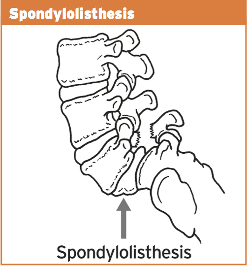
10. Bone injuries such as stress fractures, pars defects and spondylolisthesis.
The focus for this paper will be on the previous group — that the bone injuries. This may be simply postural (slow onset repetitive trauma) or related to sports; for instance, gymnastics.
The two demographic groups that tend to endure the most extension-related low back pain are:
1. People who endure all day, for instance, retailers, army, security guards etc.. Prolonged position will obviously force the pelvis to start to migrate to an anterior tilt management. This may begin to place compressive pressure on the facet joints of the spinal column as they also change towards an expansion position since they accompany the pelvic tilt.
2. Extension sports such as gymnastics, tennis, swimming, diving, football codes, volleyball, basketball, track and field, cricket fast bowlers. This is more pronounced in sports that involve extension/rotation.
Pathomechanics
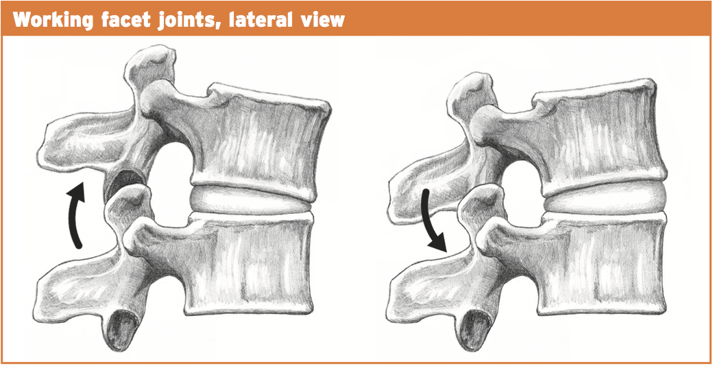
With normal extension of the lumbar spine (or backward bending), the facet joints begin to approximate each other and compress.�The articular processes of this facet above will abut the articular process of the facet below. This is a normal biomechanical movement. However, if the extension ranges are excessive, the procedures will impinge quite aggressively and damage to the cartilage surfaces within the facet joint can result. Sports such as gymnastics, functioning in tennis, and handling in American Soccer may all involve uncontrolled and excessive extension.
It would be unlikely that a bone stress response or even a stress fracture could be brought on by an isolated expansion injury. It would be more likely that a sudden forced extension injury may damage an already pre-existing bone strain reaction.
Similarly, if an individual stands daily and the pelvis migrates into lateral tilt, then the aspects will be placed under low load compression but for extensive intervals.
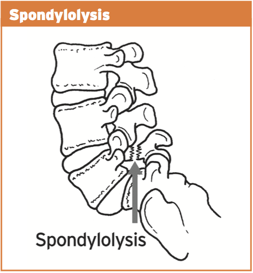
With ongoing uncontrolled loading, stress is then transferred from the facet joint to the bone below (pars interarticularis). This originally will manifest as a pressure reaction on the bone. This bone strain may advance to a stress fracture throughout the pars if uncorrected. This fracture is also referred to as a “pars flaw”, or spondylolysis.
It was initially considered that stress fractures of the pars was a congenital defect that introduced itself at the teenage years. However, it is now agreed that it is probably obtained through years of overuse into extension positions, especially in young sportspeople involved with expansion sports. What’s more, one-sided pars defects often occur more commonly in sport which also included a rotational component such as tennis serving or fast bowling in cricket.
The stress fracture can then advance to impact the opposite side, causing a bilateral strain fracture, with anxiety subsequently being transferred to the disk in between both levels.
Spondylolisthesis features bilateral pars defects which could possibly be a result of repetitive stress into the bilateral pars in extension athletics, but more likely it is an independent pathology that manifests in the early growing stages (9-14) as this pathology is often viewed in this age category. If they become symptomatic in later years because of involvement in expansion sports, it is exceedingly likely that the defects were there by a young age but presented asymptomatically. As a result of rapid growth spurts in teenage years and the high-volume training experienced by teenaged athletes, it is possible that these dormant spondylolisthesis then pose as ‘acute onset’ back pain in teenage years.
In summary, the progression of this bone stress reactions tends to follow the following continuum:
1. Facet joint irritation
2. Pars interarticularis stress response
3. Stress fracture to the pars
4. Pars defect (or spondylolysis)
5. Spondylolisthesis due to activity or more likely congenital and found later in teenage years due to participation in�extension sports.
The landmark publication related to spondylolysis and spondylolisthesis was presented by Wiltse et al (1976) and they classified these injuries as follows:
1. Type I: dysplastic � congenital abnormalities of L5 or the upper sacrum allow anterior displacement of L5 on the sacrum.
2. Type II: isthmic � a lesion in the pars interarticularis occurs. This is subclassified as
a. lytic, representing a fatigue fracture of the pars,
b. elongated but intact pars, and c. acute fracture.
3. Type III: degenerative � secondary to long-standing intersegmental instability with associated remodeling of the articular processes.
4. Type IV: traumatic � acute fractures in vertebral arch other than the pars.
5. Type V: pathological � due to generalized or focal bone disease affecting the vertebral arch.
The vast majority of spondylolysis and sponylolisthesis accidents are Type II — the isthmic variety.
For the purposes of this paper, we will refer to the above stages as the posterior arch bone stress injuries (PABSI).
Epidemiology
It is a lot more widespread at the L5 level (85-90 percent). It’s a high asymptomatic prevalence in the general population and is often found unintentionally on x ray imaging. Nonetheless, in athletes, particularly young athletes, it is a common reason for persistent low back pain. From the young athlete, the problem is often referred to as ‘active spondylolysis’.
Active spondylolysis is normal in virtually every gamenevertheless, sports such as gymnastics and diving and cricket pose a much greater danger due to the extension and turning character of the sport. The progression from an active spondylolysis into a non-union type spondylolisthesis has been associated with a greater prevalence of spinal disk degeneration.
Early detection through screening and imaging, therefore, will highlight those early at the bone stress phase and if caught early enough and managed, the progression to the larger and more complicated pathologies are avoided as a result of therapeutic capacity of the pars interarticularis in the early stages.
It is more common to find teens and young adults afflicted by PABSI. This will highlight the rapid growth of the spine through growth spurts that is also characterized by a delay in the motor control of the muscle system during this period. Furthermore, it’s thought that the neural arch actually gets stronger in the fourth decade hence possibly explaining the low incidence of bone stress reactions in mid ages.
The incidence of spondylolysis has been reported to be around 4-6% in the Caucasian population (Friedrikson et al 1984). The rates seem to be lower in females and also in African-American males. It has also been suggested that a link exists between pars defects and spina bifida occulta.
The incidence of spondylolysis seems to be higher in the young athletic population than in the general population. Studies in gymnasts, tennis, weightlifting, divers and wrestlers all show disproportionately high incidence of spondylolysis compared with the general population of age-matched subjects.
Tennis
The tennis serve generates excessive extension and rotation force. In addition, the forehand shot may also produce elevated levels of spinning/ extension. The more traditional forehand shot demanded a great deal of weight shift through the legs to the torso and arms. However, a more favorite forehand shot is to currently face the ball and also generate the force of this shot utilizing hip rotation and lumbar spine extension. This action does increase ball speed but also puts more extension and compressive loads on the spine potentially resulting in a greater degree of stress on the bone components.
Golf
The most likely skill component involved in golf that may cause a PABSI are the tee shot with a 1 wood when forcing for distance. The follow-through of this shot entails a significant quantity of spine rotation with maybe a level of spine expansion.
Cricket
Fast bowlers in cricket are the most susceptible to PABSI. This will occur on the opposite side to the bowling arm. As the front foot engages on plant stage, the pelvis abruptly stops moving but the spine and chest continue to proceed. With the wind-up of this bowling action (rotation), when coupled with expansion this can place large forces on the anterior arch of the thoracic. More than 50% of fast bowlers will create a pars stress fracture. Young players (up to 25) are most vulnerable. Cricket governments have implemented training and competition guidelines to avoid such injuries by restricting the number of meals in training/games.
Field Events
The more common field events to cause a PABSI would be high leap followed by javelin. Both these sports create enormous ranges of backbone extension and under significant load.
Contact Sports
Sports like NFL, rugby and AFL all require skill components that need backbone expansion under load.
Gymnastics/Dancers
It goes without saying that gymnastics and dancing involves a substantial amount of repetitive spine expansion, particularly backflips and arabesques. It has been suggested that nearly all Olympic degree gymnasts could have suffered from a pars defect. Many organizing bodies now put limits on the number of hours young gymnasts can instruct to prevent the repetitive loading on the spine.
Diving
Spine extension injuries occur mostly off the spring board and on water entrance.
Diagnosis Of PABSI In Athletes
Clinical investigation
These can pose as preventable injuries. Research shows that the incidence was emphasized from the general population that have nil indicators of back pain. But, individuals will typically complain of back ache that is deep and generally unilateral (one side). This may radiate into the buttock area. The most offending movements tend to be described as expansion moves or backward bending movements. This may be a slow progression of pain or might be initiated by one acute episode of back pain in a competitive extension motion.
On clinical examination:
1. Pain may be elicited with a one-leg extension/rotation test (standing on the leg on the affected side) � stork test.
2. Tenderness over the site of the fracture.
3. Postural faults such as excessive anterior tilt and/or pelvic asymmetry.
The one-legged hyperextension test (stork test) was suggested to be pathognomonic for busy spondylolysis. A negative evaluation was stated to effectively exclude the diagnosis of a bone stress-type injury, thus creating radiological investigations unnecessary.
But, Masci et al (2006) examined the connection between the one-legged hyperextension test and gold standard bone scintigraphy and MRI. They discovered that the one-legged hyperextension test was neither sensitive nor specific for active spondylolysis. Moreover, its negative predictive value was so poor. Thus, a negative test can’t exclude energetic spondylolysis as a possible cause.
Masci et al (2006) go on to indicate that the bad relationship between imaging and the one-legged test may be because of a number of factors. The extension test would be expected to move a significant extension force on to the lower back spine. In addition to putting substantial strain on the pars interarticularis, it might also stress different regions of the spinal column like facet joints as well as posterior lumbar disks, and this may subsequently induce pain in the existence of other pathology such as facet joint arthropathy and spinal disc disease. This will explain the poor specificity of the test. Conversely, the inadequate sensitivity of the test may be related to the subjective reporting of pain by issues performing the maneuvre, which may vary based on individual pain tolerance. Additionally, this evaluation can preferentially load the fifth cervical vertebra, and so bone stress located in the upper lumbar spine may not test positive.
Grade 1 spondylolisthesis are normally asymptomatic; nonetheless, grade 2+ lesions often present with leg pain, either with or without leg pain. On examination, a palpable slip could be evident.
Imaging
Clinical assessment of active spondylolysis and the more severe pars defects and spondylolisthesis can be notoriously non-specific; this is, not all patients suffering PABSI will present with favorable abstract features or positive signs on analyzing. Thus, radiological visualization is important for diagnosis. The imaging methods available in the diagnosis of bone stress injury are:
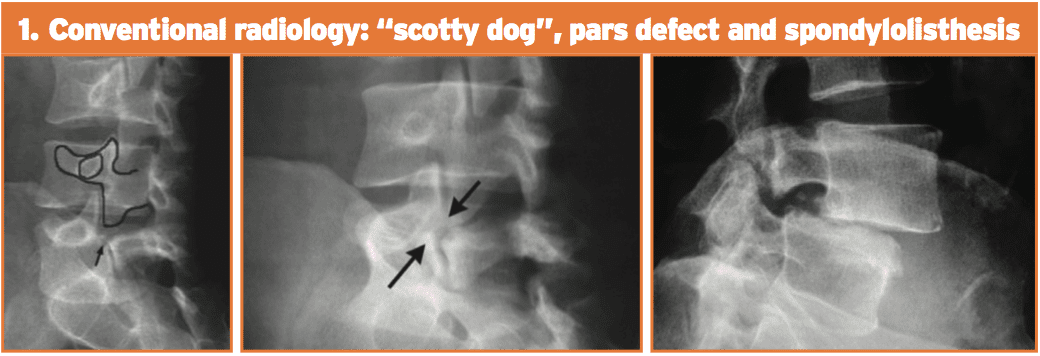
1. Conventional radiology. This test is not very sensitive but is highly unique. Its limits are partially because of the cognitive orientation of the pars defect. The oblique 45-degree films may show the timeless ‘Scotty Dog’ appearance. Spondylolisthesis can be looked at simply on a lateral movie x-ray.
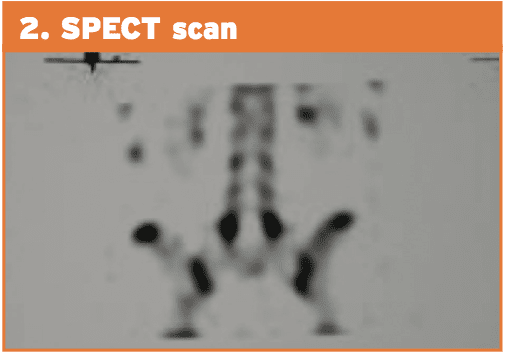
2. Planar bone scintigraphy (PBS) and single photon emission computed tomography (SPECT). SPECT enhances sensitivity in addition to specificity of PBS than straightforward radiographic study. Comparative research between PBS and conventional radiology have shown that scintigraphy is more sensitive. Patients with positive SPECT scan must then undergo a reverse gantry CT scan to assess whether the lesion is active or old.
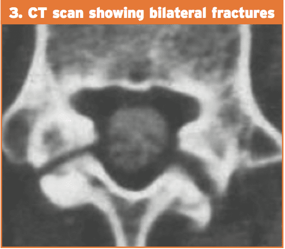
3. Computed tomography (CT). The CT scan is considered to be more sensitive than conventional radiology and with higher specificity than SPECT. Regardless of the type of cross-sectional image utilized, the CT scan provides information on the state of the flaw (intense fracture, unconsolidated flaw with geodes and sclerosis, pars in procedure for consolidation or repair). The “inverse gantry” perspective can evaluate this condition better. Repeat CT scan can be used to track progress and recovery of the pars defect.
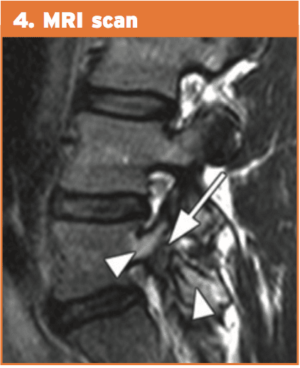
4. Magnetic resonance imaging (MRI). This technique shows pronounced changes in the signal in the amount of the pars. This is recognized as “stress response” and can be classified into five different degrees of action. MRI can be helpful for evaluating elements that stabilize isthmic lesions, for example intervertebral disc, common anterior ligament, and related lesions. The MRI isn’t as specific or sensitive as SPECT and CT combination.
Therefore, the current gold standards of investigation for athletes with low back pain are:
1. bone scintigraphy with single photon emission computed tomography (SPECT); if positive then
MRI has many advantages over bone scintigraphy, for instance, noninvasive nature of the imaging along with the absence of ionizing radiation. MRI changes in active spondylolysis include bone marrow edema, visualized as increased signal in the pars interarticularis on edema-sensitive sequences, and fracture, visualized as reduced signal in the pars interarticularis on T1 and T2 weighted sequences.
However, there is greater difficulty in detecting the changes of busy spondylolysis from MRI. Detecting pathology from MRI relies on the interpretation of distinct contrasts of signals compared with normal tissue. Unlike stress fractures in different parts of the body, the little region of the pars interarticularis may make detection of those changes harder.
However, unlike MRI, computed tomography has the capability to differentiate between acute and chronic fractures, and this differentiation might be an important determinant of fracture healing. Accordingly, in areas using pars interarticularis fractures discovered by MRI, it might nonetheless be necessary to execute thin computed tomography slices to determine whether or not a fracture is severe or chronic — an important factor in fracture resolution.
Exactly how effective are injury-prevention programs in soccer? El Paso, TX.s Scientific chiropractor Dr. Alexander Jimenez looks at the very latest evidence…
Football is the world’s most popular team sport. Injuries are a significant issue for both amateur and professional players. Indeed, previous research has estimated that soccer players are among the most injury-prone athletes having an estimated injury rate of 17-24 accidents per 1000 playing hours(1). Nineteen per cent of all sports injuries which exist in the Netherlands are because of soccer(two) and in Britain alone, the expense of therapy and time lost from work owing to football injuries is estimated to be approximately #1billion annually(1)!
In a landmark study, researchers followed two Champions League teams and gathered data on 2,229 players over seven seasons to examine the injury profile of muscular injuries in soccer players(3). They also analyzed the gamers’ training schedules and data out of their games to construct a detailed picture of the injury risks that were associated. The findings were as follows:
2,908 muscle injuries have been enrolled;
Normally, a player sustained 0.6 muscle injuries each season (equating to around 15 muscle injuries per season at a squad of 25 players);
Muscle injuries constituted 31 percent of all injuries and caused 27% of the total injury lack;
Ninety-two per cent of muscle injuries affected the four Big muscle groups of the lower limbs: hamstrings (37 percent), adductors (23 percent), quadriceps (19%), and calf muscles (13%);
Sixteen per cent of the muscular injuries were re-injuries; nonetheless, these re-injuries caused significantly longer absences than did the first injuries.
The prevalence of muscular injury increased with age.
The exact same group of researchers also carried out a follow-up study (published in 2013) in which they sought to establish the consequences of fixture congestion on injury rates among the gamers(4). Time-loss and exposure injuries were enrolled prospectively from 27 teams over 11 seasons. Matches were grouped based on the amount of recovery days before each match and the accident rates were compared between classes. The results showed that compared to a recovery interval of more or six days, muscular injury rates and overall injury rates were raised in league matches where players had had four or less recovery days.
Given the high levels of trauma among football players (a risk that is increased during periods of match congestion) and the financial pressure of the modern game, it’s perhaps unsurprising that medical team caring for players find that treatment of injuries in players is quite a frustrating and also a never-ending struggle. In addition, it clarifies why some players end up returning to contest prior to the injury has healed completely, setting the stage for injury recurrence, together with protracted absence of this participant from competition and training.
Injury Treatment
Treating injuries in football is both time- consuming and expensive, particularly at the elite level. And while there’s a large literature on the epidemiology of sports injuries, established protocols for treating muscular injuries and assessment criteria for imaging, and a number of clinical and functional tests that could assist the health staff in deciding the optimal point where an athlete can be safely returned to full participation(5,6), the current guidelines haven’t translated into a significant reduction in muscle injury levels in professional sports such as soccer.
To simplify things further, the evidence indicates a new injury often occurs within a couple of weeks after return to contest, and typically costs the player more lost playing time than the key injury(7). The most probable reasons for this observation are likely associated with bodily alterations following the first injury, such as muscle stiffness and/or fatigue, scar tissue formation, biomechanical alterations, neuromuscular inhibition, as well as inadequate treatment — for instance, overly aggressive or incomplete rehabilitation(8-10).
Injury-Prevention Programs
Even armed with knowledge that is up-to-date and the best technology is fraught with difficulty. Remembering the old adage that ‘an ounce of prevention is worth a pound of cure’, a alternative that is far better to attempt to prevent injuries from happening in the first place with an injury-prevention program. This is easier said than done. It is correct that there is an abundance of literature on the effectiveness of methods to avoid harm recurrence and muscle injury, such as enhancing flexibility eccentric and concentric exercises and drills. Despite this and apps like FIFA’s ‘The II’ (see Box 1), the incidence of muscle injuries generally, and the recurrence rate particularly, remains stubbornly high(11-16).
More recent studies indicate that in higher levels of functionality, there might not be much in the way of significant added benefits, while some early study appeared to give evidence for the efficacy of programs in football, as described in box 1. At a follow up to the study described above(18) and that was published this past year, the same group of investigators looked to see if an injury prevention program comprising 10 exercises designed to enhance stability, muscle strength, co-ordination, and versatility of the back, hip and leg muscles (FIFA’s ‘The II’) was effective concerning reducing injury levels and whether it offered any advantages in terms of reducing the related costs of following treatment for injuries that did occur(19).
From the analysis, 479 adult male amateur gamers aged 18-40 years have been split into two classes: the intervention group had been taught to do exercises focusing on core stability, bizarre training of thigh muscles, proprioceptive training, dynamic stabilization, and plyometrics with straight-leg orientation at every training session (2-3 sessions per week) through one season. The management team, meanwhile, continued their usual warm up.
As in the previous study, there were no significant differences in the percentage of players that are injured and injury rates between the two groups. What was intriguing was that in the intervention group, the price of injury treatment was 256 per participant. In the control group nonetheless treatment costs were twice at $606 per participant. The investigators commented that the cost savings in the intervention group may be the result of a rate of knee injuries, which have costs because of more lengthy rehabilitation periods and much more time lost at work compared to a number of different injuries.
Meanwhile, another study on an injury- prevention program (based on The II) in male amateur players had been printed in the end of last year(20). It discovered that (like the previous studies), an intervention program did not decrease the incidence of harm throughout the course of a season. However, such as the study, the players in the intervention group did incur less health care costs, although a justification for this finding wasn’t given. As if to validate the confusion surrounding the value of injury-prevention programs for football players, then a recently published systematic overview of all of the previous studies released thus far fought to achieve a definitive conclusion(21). Six studies involving a total of 6,099 participants met the inclusion criteria and the results of these were conflicting two of the six studies (among large and one of moderate quality) reported a decrease in injury rates that were actual. Four of the six research an ‘preventive effect’, even though the effect of a single study wasn’t statistically significant. Possible reasons for these contradictory findings might be subject choice (sex and level of ability), differences between the intervention programs implemented (content, training frequency and duration) and compliance with this application. What’s clear, however, is that studies investigating the type and seriousness of exercises within an injury-prevention program are still required to reduce the incidence of accidents in soccer efficiently.
Good News On Prevention
Since the review study cited previously(21) was printed, two quite newly published studies on injury-prevention apps in soccer seem to provide more encouraging news — for muscle injuries at least. In one, researchers studied elite players competing over two consecutive seasons, where the first (2008-2009) function as intervention period and the second, the management period (2009-2010)(22). In total, 26 (08/09) and 23 (09/10) elite male pro football players competing within the Scottish Premier League and European competition participated. The accident prevention training program was conducted twice weekly to the entirety of this season (58 avoidance sessions) and the results were compared with the control (no injury-prevention program) year.
On first inspection, the results were disappointing, showing an increase in the complete number of accidents within the intervention period (88 vs 72). But this was largely because of the greater quantity of contusion injuries sustained inside the intervention season (44) compared with control season (23). Assessing like for muscular injuries that were significantly fewer were observed during the intervention season, which had been even more impressive given the larger squad size at the intervention season.
Another newly published study by Italian scientists who researched the effect of a two-tiered injury-prevention program on initial injury and re-injury prevalence in 36 elite male football players also causes encouraging reading(23). During the season prior to that examined in the study, there had been 27 muscle injuries in the group, which accounted for 58.7 percent of the total injuries: 13 of these had occurred throughout practice and 14 during matches. The general incidence of muscular injuries was 5.6 injuries/1000 hours of training/playing exposure and the effect was 106.4 times absence/1000 hours exposure.
To try and decrease the speed of injury through the following season, the team doctor (also among the study’s authors) found an injury-prevention program, conducted 2-3 times per week. This consisted of two elements: a collection of core stability exercises conducted by the whole group prior to each practice session (see Box 2) along with an individualized injury prevention program, which has been started after assessment with kinesiologic and diagnostic tests. At the start of the year, every athlete underwent testing of leg flexibility using the Ober evaluation, Thomas evaluation and straight-leg-raising [SLR] test(24-26). The prone instability test(27) was completed to show spinal instability along with the stork test (28,29) to assess sacroiliac dysfunction. Quadriceps and hamstring strength were measured isokinetically and attention was directed in evaluation of immunity of gluteus medius’ power.
The injuries that happened based on MRI and clinical imaging findings were diagnosed by the medical team. An injury was defined as though it caused the participant to miss the next training session or match, and happened during a scheduled training session or match. An injured player was defined injured before the club medical staff cleared him for participation. Re-injuries were described as those that occurred as those that occurred at the same website no longer than three months following the player had returned to full involvement at early re-injuries and exactly the exact same site.
Results
Throughout the intervention season, a total of 64 injuries occurred — 36 (56 percent) during practice and 28 (44%) during matches. Of them, 20 were muscle injuries, accounting for 31.3 percent of the total injuries; 14 of which occurred during practice and 6 during games. In all, three re-injuries happened and (15 percent of overall muscle injuries) and there were not any premature re-injuries. In comparison with the preceding season with no intervention-program set up, there was a reduction in the number of times and muscle injuries . Specifically, whereas muscle injuries accounted for 31 percent of harms they accounted for 59% of all injuries. Significantly, the number of injuries per 1000 hours of training and playing time was reduced by over half of 5.6 to 2.5. Meanwhile, the number of days fell by nearly two-thirds 106 into 37. The investigators put the success of this intervention down to three key aspects:
An injury prevention program that comprised of core stability exercises similar to those in ‘The II’ program but which differed in its two-tiered arrangement (group and individual sessions), allowing for intense and special training. In contrast, the combined results in research into The II app are probably because of the non- special content and ineffective intensity.
The program’s continuity of commitment by the players to both the group and individual areas.
The addition of bizarre hamstring training in the group program (2 sets of 5 repetitions per week) combined with all the personalized application for players with a history of injury.
Using ice baths in the conclusion of every training session
The investigators cautioned that their study would have included a larger number of topics, but the data still showed a critical progress by the prior year over that. They also argued that by increasing the number of group and individual prevention training sessions, the outcomes could be enhanced.
Summary & Recommendations
Injury treatment in aggressive soccer is equally costly and time-consuming also given the pressures of the game, injury avoidance is more important than ever. But, despite extensive published literature on harm prevention strategies and initiatives such as FIFA’s ‘The II’, the injury rates in soccer remain high, especially in the higher levels.
The latest research indicates that while overall injury prevention programs such as The II might reduce the incidence of trauma in amateur gamers, especially by reducing the incidence of knee injury. However, they will probably not benefit professional players or level. Instead, combining a more individualized approach (using a far greater emphasis on particular exercises determined by kinesiologic and diagnostic testing) with team sessions seems to be desirable. Additionally, it is important that gamers are ‘on-board’ with almost any program and take part regularly (at least twice weekly) to achieve all the potential advantages.
References
1.Br J Sports Med. 2002;36:354-9.
2.Injury Prevention. 2011;17(2):1-5.
3.Am J Sports Med. 2011 Jun;39(6):1226-32
4.Br J Sports Med. 2013 Aug;47(12):743-7.
5. Knee Surg Sports Traumatol Arthrosc 2010; 18:1798-1803.
6.J Orthop Sports Phys Ther 2010; 40:67-81
7.Br J Sports Med 2005; 39:542-546
8. Sports Med 2004; 34:681-695
9. Am J Sports Med 2002; 30:199-203.
10. Sports Med 2012;42:209-226
11. Br J Sports Med 2012; 46:112-117.
12.Am J Sports Med 2004; 32(suppl 1):S5-S16.
13. Am J Sports Med 2010; 38:2051-2057.
14. Am J Sports Med 2010; 38:1147-1153.
15. Br J Sports Med 2006; 40:767-772
16. Am J Sports Med 2013; 41:327-335
17. Am J Sports Med 2002; 30(5):652-9
18.Br J Sports Med 2012 Dec;46(16):1114-8
19. J Physiother 2013 Mar; 59(1):15-23
20. Clin J Sport Med 2013 Nov; 23(6):500-1
21. Sports Med 2013 Apr; 43(4):257-65
22.J Strength Cond Res 2013 Dec; 27(12):3275-85
23. J Muscles, Ligaments and Tendons Journal 2013; 324 3 (4): 324-330
24. J Bone & Joint Surgery 1936; 18:105-110.
25. Phys Ther Sport 2007; 8:14-21.
26.J Orthopaedic and Sports Physical Therapy 1981; 2:117-133
27. Magee DJ. Orthopedic Physical Assessment. 3rd ed. Philadelphia, PA: W.B. Sauders Company; 1997.
28. Spine 2003; 28: 1593-1600
29. Clinical Biomechanics 2004; 19:456-464
IFM's Find A Practitioner tool is the largest referral network in Functional Medicine, created to help patients locate Functional Medicine practitioners anywhere in the world. IFM Certified Practitioners are listed first in the search results, given their extensive education in Functional Medicine