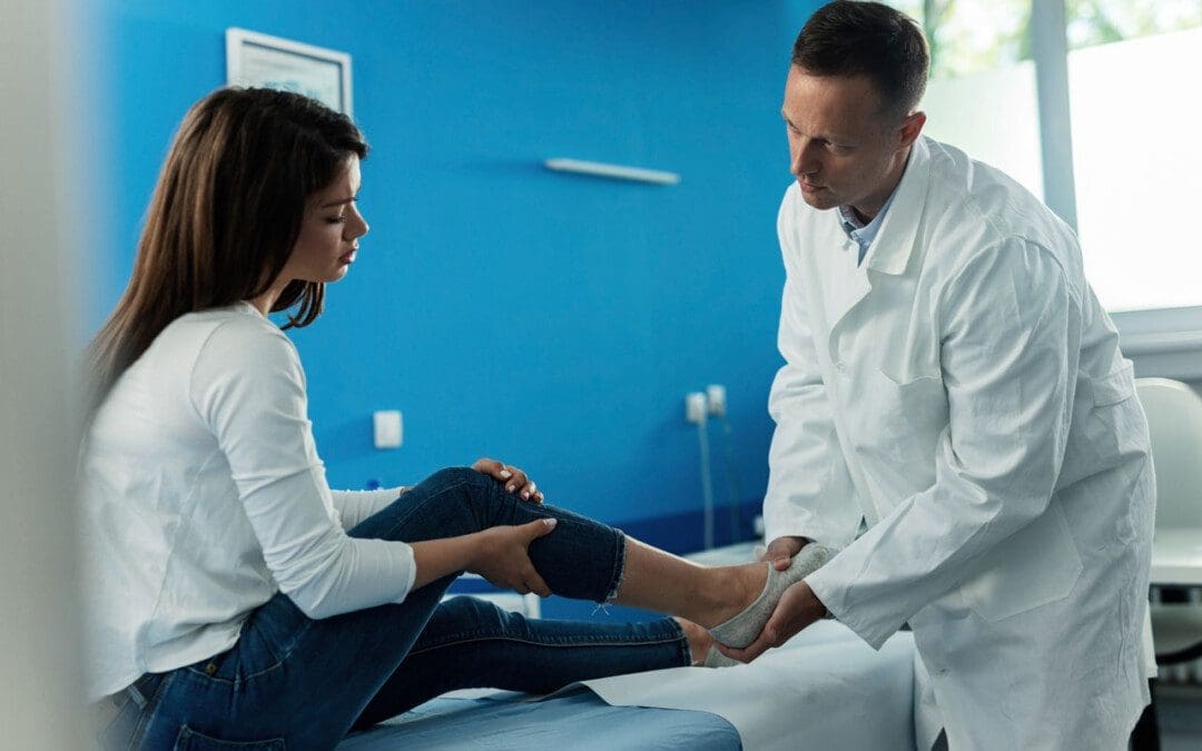
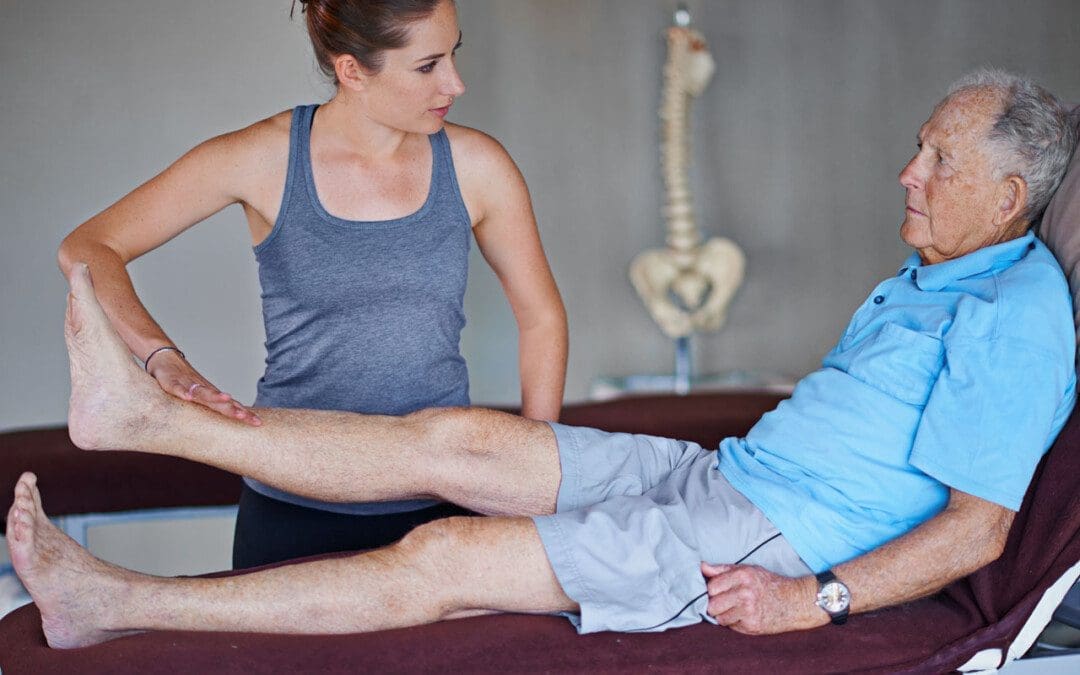
Individuals with plantar fasciitis may experience consistent flare-ups. Can knowing the causes help to find pain relief?
Plantar Fasciitis Flare-Up
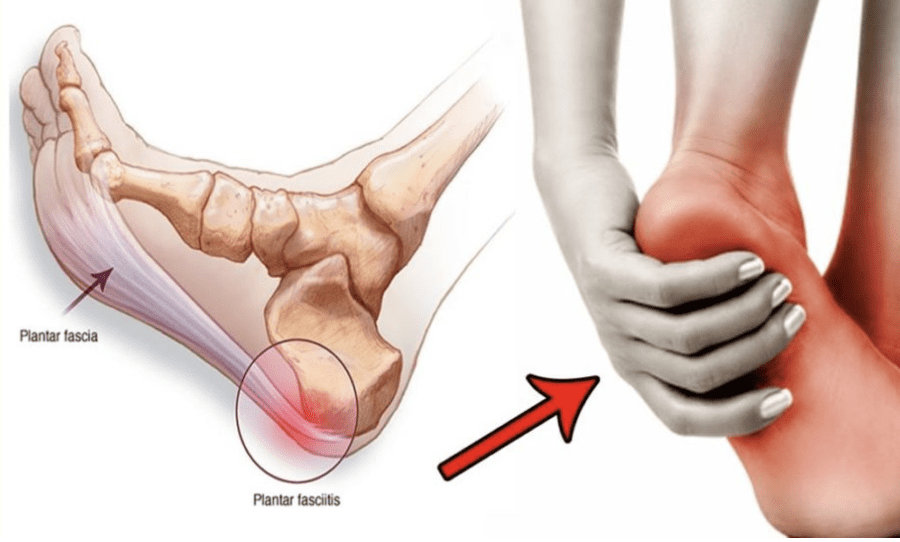
Plantar fasciitis is a common cause of heel and foot pain. The plantar fascia is a band of tissue that runs along the bottom of the foot and becomes inflamed. Certain factors can cause plantar fasciitis flare-ups, including:
Increased levels of physical activity.
Not stretching regularly.
Wearing shoes without proper support.
Weight gain.
Causes
A plantar fasciitis flare-up is often triggered by physical activity. (MedlinePlus. U.S. National Library of Medicine. 2022) It can also be brought on by underlying conditions, like increased body weight, arthritis, or the shape of the foot. (Johns Hopkins Medicine. 2023) Despite the root cause, there are activities and experiences that can contribute to and/or worsen the condition.
New Exercise Routine
Being highly physically active can exacerbate plantar fasciitis symptoms.
High heels, boots, or shoes that raise the heel above the toes.
Worn-out shoes like exercise workout shoes.
Not Stretching Properly or At All
Tight calves can increase pressure on the plantar fascia.
Stretching the calves, Achilles tendon/heel, and the bottom of the feet is highly recommended to help treat and prevent the condition. (Johns Hopkins Medicine. 2023)
Not stretching thoroughly or skipping stretches can worsen symptoms.
Individuals with plantar fasciitis are recommended to stretch before and after physical activities, exercise, before going to bed, and after waking up.
Working Through the Pain
Individuals may try to continue physical activities during a flare-up.
This is not recommended as doing so can cause more pain and worsen the condition.
When pain presents, it’s recommended to:
Stop all activities that strain the feet
Stay off the feet for at least a week.
Tearing the Plantar Fascia
The plantar fascia rarely tear completely from repeated stress known as a plantar fascia rupture.
Pascoe, S. C., & Mazzola, T. J. (2016). Acute Medial Plantar Fascia Tear. The Journal of orthopaedic and sports physical therapy, 46(6), 495. https://doi.org/10.2519/jospt.2016.0409
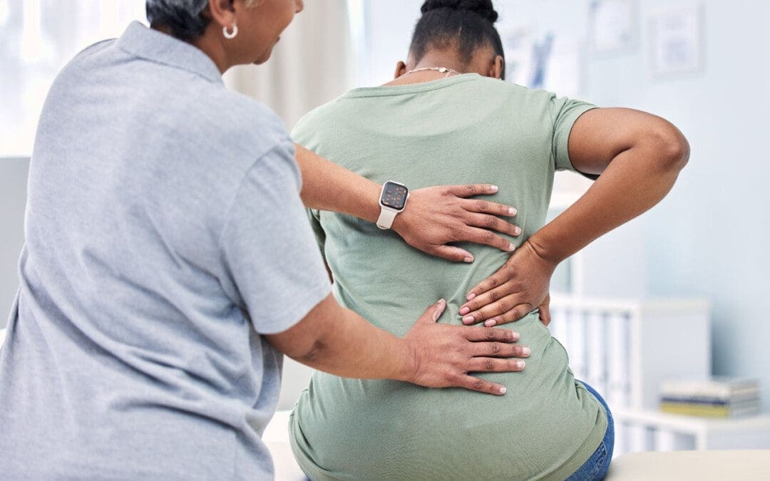
Individuals dealing with back pain problems could be suffering from a bulging disc. Could knowing the difference between slipped and herniated disc symptoms help with treatments and finding relief?
Bulging Disc Pain
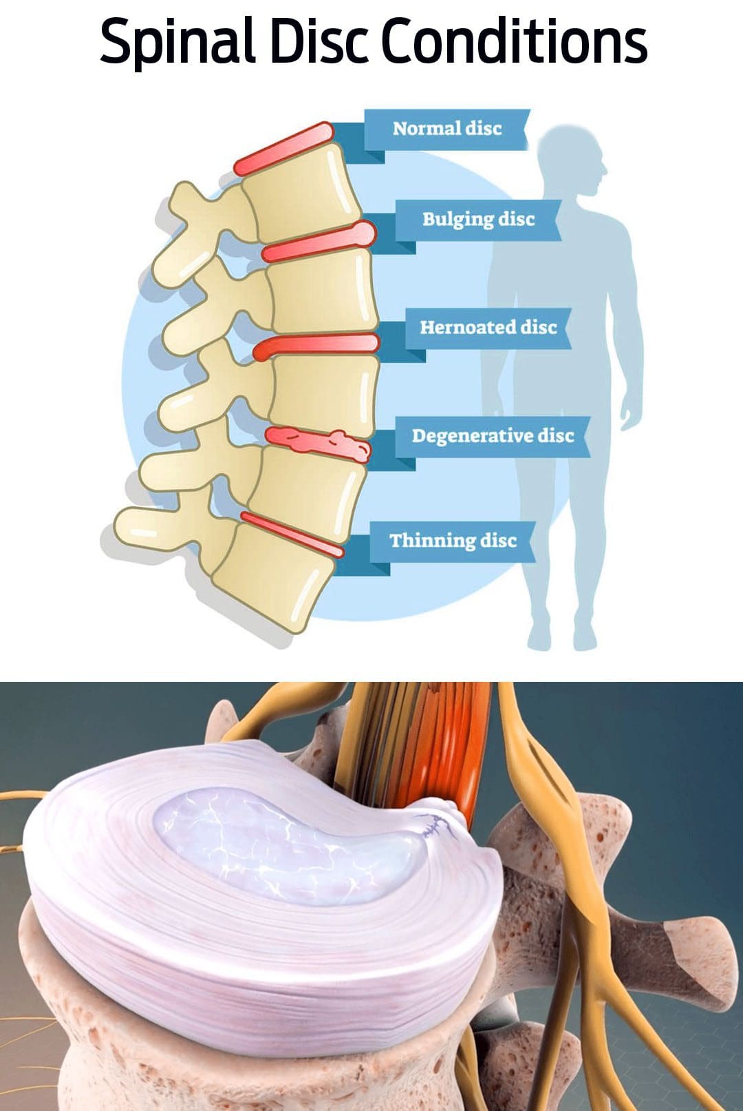
Back pain can become debilitating if not treated properly. A bulging disc is a common cause of cervical, thoracic, and lower back pain symptoms. It happens when one of the fluid-filled cushions between the vertebrae begins to shift out of place. Instead of being aligned with the edges, the disc bulges over. This begins to generate pressure on the nerves causing pain and inflammation.
Bulging discs are often caused by age, but repetitive movements and/or lifting heavy objects can contribute to the condition.
Symptoms can resolve on their own, but individuals are recommended to consult with a physical therapist and/or chiropractor to make sure the disc healed properly, otherwise, it can lead to worsening and/or further injuries.
Bulging Disc vs. Herniated Disc
Bulging and herniated discs cause pain symptoms.
They both can be linked to injuries and degenerative disc disease but are not the same condition. (Penn Medicine. 2018)
This is because the lower back is subject to all kinds of pressure and movement with daily activities, increasing the chances of pain and injuries.
The next most common place is the neck/cervical spine where there are constant movements making it prone to injury and pain symptoms.
Causes
Bulging discs are most often caused by body aging and normal wear and tear. As time goes on the intervertebral discs naturally degenerate, known as degenerative disc disease. This can cause the discs to pull downward, causing them to bulge from their placement. (Penn Medicine. 2018) Factors that can cause or worsen the condition include:
Practicing unhealthy postures.
Repetitive motions.
Lifting heavy objects
Spinal injuries.
Medical history of spinal or disc disease in the family.
Individuals with back pain that interferes with daily functions or has lasted longer than six weeks, should see a healthcare provider for a diagnosis. They will order a magnetic resonance imaging scan/MRI, which can show where a disc is protruding. (American Academy of Neurological Surgeons. 2023)
Rest
For bulging disc pain, resting the back is necessary. However,
Individuals diagnosed with peripheral neuropathy, or with small fiber neuropathy, can understanding symptoms and causes help with potential treatments?
Small Fiber Neuropathy
Small fiber neuropathy is a specific classification of neuropathy, as there are different types, which are nerve injury, damage, disease, and/or dysfunction. Symptoms can result in pain, loss of sensation, and digestive and urinary symptoms. Most cases of neuropathy like peripheral neuropathy involve small and large fibers. Common causes include long-term diabetes, nutritional deficiencies, alcohol consumption, and chemotherapy.
Small fiber neuropathy is diagnosed after diagnostic testing showing it is clear that the small nerve fibers are involved.
The small nerve fibers detect sensation, temperature, and pain and help regulate involuntary functions.
Isolated small-fiber neuropathy is rare, but research is ongoing on the type of nerve damage and potential treatments. (Stephen A. Johnson, et al., 2021)
Small fiber neuropathy is not specifically dangerous but is a sign/symptom of an underlying cause/condition that is damaging the body’s nerves.
Pain – symptoms can range from mild or moderate discomfort to severe distress and can happen at any time.
Loss of sensation.
Because the small nerve fibers help with digestion, blood pressure, and bladder control – symptoms of autonomic dysfunction can vary and can include:
Constipation, diarrhea, incontinence, urinary retention – the inability to completely drain the bladder.
If there is progressing nerve damage, the intensity of the pain can decrease, but the loss of normal sensation and autonomic symptoms can worsen. (Josef Finsterer, Fulvio A. Scorza. 2022)
Hypersensitivity to touch and pain sensations can cause pain without a trigger.
The loss of sensation can make individuals unable to accurately detect sensations of touch, temperature, and pain in affected areas, which can lead to various types of injuries.
Although more research is needed, certain disorders that were not considered neuropathies may have small fiber neuropathy components involved.
A study suggested that neurogenic rosacea, a skin condition, could have some elements of small fiber neuropathy. (Min Li, et al., 2023)
These small nerve fibers are distributed throughout the body including the tops of the fingers and toes, trunk, and internal organs.
These fibers are usually located in the superficial areas of the body, such as close to the skin’s surface. (Mohammad A. Khoshnoodi, et al., 2016)
The small nerve fibers that get damaged are involved in transmitting pain and temperature sensations.
Most nerves have a special type of insulation called myelin that protects them and increases the speed of nerve impulses.
Small nerve fibers may have a thin sheath, making them more susceptible to injury and damage at earlier stages of conditions and diseases. (Heidrun H. Krämer, et al., 2023)
Individuals At Risk
Most types of peripheral neuropathy cause damage to the small and large peripheral nerve fibers. Because of this, most neuropathies are a mix of small-fiber and large-fiber neuropathy. Common risk factors for mixed fiber neuropathy include: (Stephen A. Johnson, et al., 2021)
Diabetes
Nutritional deficiencies
Overconsumption of alcohol
Autoimmune disorders
Medication toxicity
Isolated small-fiber neuropathy is rare, but there are conditions that are known to contribute to the cause and include: (Stephen A. Johnson, et al., 2021)
Sjogren Syndrome
This autoimmune disorder causes dry eyes and mouth, dental problems, and joint pain.
It can also cause nerve damage throughout the body.
Fabry Disease
This condition causes a buildup of certain fats/lipids in the body that can lead to neurological effects.
Amyloidosis
This is a rare disorder that causes a buildup of proteins in the body.
The proteins can damage tissues like the heart or nerves.
Lewy Body Disease
This is a neurological disorder that causes dementia and impaired movement and can lead to nerve damage.
Lupus
This is an autoimmune disease that affects joints, skin, and sometimes nerve tissue.
Viral Infection
These infections typically cause a cold or gastrointestinal/GI upsetness.
Less often they can cause other effects like small fiber neuropathy.
These conditions have been seen to cause isolated small-fiber neuropathy or begin as small-fiber neuropathy before progressing to the large nerve fibers. They can also begin as a mixed neuropathy, with small and large fibers.
Progression
Often the damage progresses at a relatively moderate rate, leading to added symptoms within months or years. The fiber nerves that are affected by the underlying condition usually progressively deteriorate, regardless of where they are located. (Mohammad A. Khoshnoodi, et al., 2016) Medications can help alleviate damage to the peripheral nerves. For individuals that are diagnosed in the early stage, it is possible to stop the progression, and potentially prevent involvement of the large fibers.
Treatments
Treatment toward preventing the progression requires controlling the underlying medical condition with treatment options depending on the cause. Treatments that can help prevent the progression include:
Blood sugar control for individuals with diabetes.
Immune suppression for control of autoimmune diseases.
Plasmapheresis – blood is taken and the plasma is treated and returned or exchanged for the treatment of autoimmune diseases.
Symptom Treatment
Individuals can get treatment for the symptoms that will not reverse or cure the condition but can help with temporary relief. Symptomatic treatment can include: (Josef Finsterer, Fulvio A. Scorza. 2022)
Pain management can include medications and/or topical analgesics.
Physical therapy – stretching, massage, decompression, and adjustments to keep the body relaxed and flexible.
Rehabilitation to help improve coordination, which can be impaired by loss of sensation.
Medications to relieve GI symptoms.
Wearing specialized clothes such as neuropathy socks to help with foot pain symptoms.
Treatment and medical management of neuropathies usually involve a neurologist. A neurologist may prescribe medication to help alleviate pain symptoms and provide medical interventions like immunotherapy if there is concern that an autoimmune process could be the cause. Additionally, treatment could include the care of a physical medicine and rehabilitation physician or a physical therapy team to provide stretches and exercises to help strengthen the body and maintain mobility and flexibility.
Peripheral Neuropathy Myths & Facts
References
Johnson, S. A., Shouman, K., Shelly, S., Sandroni, P., Berini, S. E., Dyck, P. J. B., Hoffman, E. M., Mandrekar, J., Niu, Z., Lamb, C. J., Low, P. A., Singer, W., Mauermann, M. L., Mills, J., Dubey, D., Staff, N. P., & Klein, C. J. (2021). Small Fiber Neuropathy Incidence, Prevalence, Longitudinal Impairments, and Disability. Neurology, 97(22), e2236–e2247. https://doi.org/10.1212/WNL.0000000000012894
Finsterer, J., & Scorza, F. A. (2022). Small fiber neuropathy. Acta neurologica Scandinavica, 145(5), 493–503. https://doi.org/10.1111/ane.13591
Krämer, H. H., Bücker, P., Jeibmann, A., Richter, H., Rosenbohm, A., Jeske, J., Baka, P., Geber, C., Wassenberg, M., Fangerau, T., Karst, U., Schänzer, A., & van Thriel, C. (2023). Gadolinium contrast agents: dermal deposits and potential effects on epidermal small nerve fibers. Journal of neurology, 270(8), 3981–3991. https://doi.org/10.1007/s00415-023-11740-z
Li, M., Tao, M., Zhang, Y., Pan, R., Gu, D., & Xu, Y. (2023). Neurogenic rosacea could be a small fiber neuropathy. Frontiers in pain research (Lausanne, Switzerland), 4, 1122134. https://doi.org/10.3389/fpain.2023.1122134
Khoshnoodi, M. A., Truelove, S., Burakgazi, A., Hoke, A., Mammen, A. L., & Polydefkis, M. (2016). Longitudinal Assessment of Small Fiber Neuropathy: Evidence of a Non-Length-Dependent Distal Axonopathy. JAMA neurology, 73(6), 684–690. https://doi.org/10.1001/jamaneurol.2016.0057
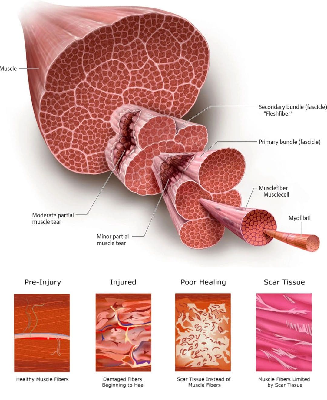
When individuals experience a neuromusculoskeletal injury strain, can following basic pulled muscle treatment protocols help in healing and a full recovery?
Pulled Muscle Treatment
A pulled muscle or muscle strain occurs when a muscle is stretched beyond its ability resulting in discomfort symptoms and mobility issues. Microscopic tears can occur within the muscle fibers potentially worsening the injury. This type of injury usually causes mild to severe pain, bruising, and immobility, and nerve injuries can develop as well. Common muscle strains include:
Pulled hamstrings
Groin strains
Pulled abdominal muscles
Calf strains
Pulled muscle treatment requires patience to promote proper healing and restoration of optimal function.
Individuals need to focus on the different stages of healing.
Gradually increase activity levels as the body allows to prevent stiffness and atrophy which can cause complications.
Symptoms
The usual symptoms of this type of injury include:
Pain
Limited mobility
Muscle spasms
Swelling
Bruising
Often individuals will feel a sudden grabbing or tearing sensation and are then unable to continue the activity.
Can limit the ability to perform certain activities.
May have moderate swelling and bruising.
Grade III
Severe injury that can cause significant pain.
Muscle spasms.
Swelling.
Significant bruising.
Basic Treatment Protocols
Most pulled muscle strain injuries heal with simple treatment. Following the right steps can ensure an expedited recovery. In the early stages after the injury, there is a balance between doing too much or not enough. The amount of activity an individual will be able to do, and the time required for recovery depends on the severity of the injury. Here are some guidelines in the right direction.
Rest
Rest is recommended for the early recovery stage.
Depending on the severity of the injury this could last from one to five days.
Immobilization is usually not necessary, and not moving at all can lead to muscle and joint stiffness.
To avoid injuries make sure the muscles are not over-exerted.
Gradually increase activity levels when starting an exercise program to build endurance.
Properly Warming Up
Warming up before taking on physical activities will help loosen the muscles and prevent injuries.
Beginning work or exercise with stiff muscles can lead to an increased chance of strain.
Studies have shown that temperature can influence the stiffness of a muscle. (K. W. Ranatunga. 2018)
Maintaining body and muscle warmth helps prevent injury and re-injury.
Injuries and Chiropractic: The Road To Recovery
References
Hospital for Special Surgery, Muscle Strain: What You Need to Know About Pulled Muscles.
Kary J. M. (2010). Diagnosis and management of quadriceps strains and contusions. Current reviews in musculoskeletal medicine, 3(1-4), 26–31. https://doi.org/10.1007/s12178-010-9064-5
Malanga, G. A., Yan, N., & Stark, J. (2015). Mechanisms and efficacy of heat and cold therapies for musculoskeletal injury. Postgraduate medicine, 127(1), 57–65. https://doi.org/10.1080/00325481.2015.992719
Mair, S. D., Seaber, A. V., Glisson, R. R., & Garrett, W. E., Jr (1996). The role of fatigue in susceptibility to acute muscle strain injury. The American journal of sports medicine, 24(2), 137–143. https://doi.org/10.1177/036354659602400203
Ranatunga K. W. (2018). Temperature Effects on Force and Actin⁻Myosin Interaction in Muscle: A Look Back on Some Experimental Findings. International journal of molecular sciences, 19(5), 1538. https://doi.org/10.3390/ijms19051538
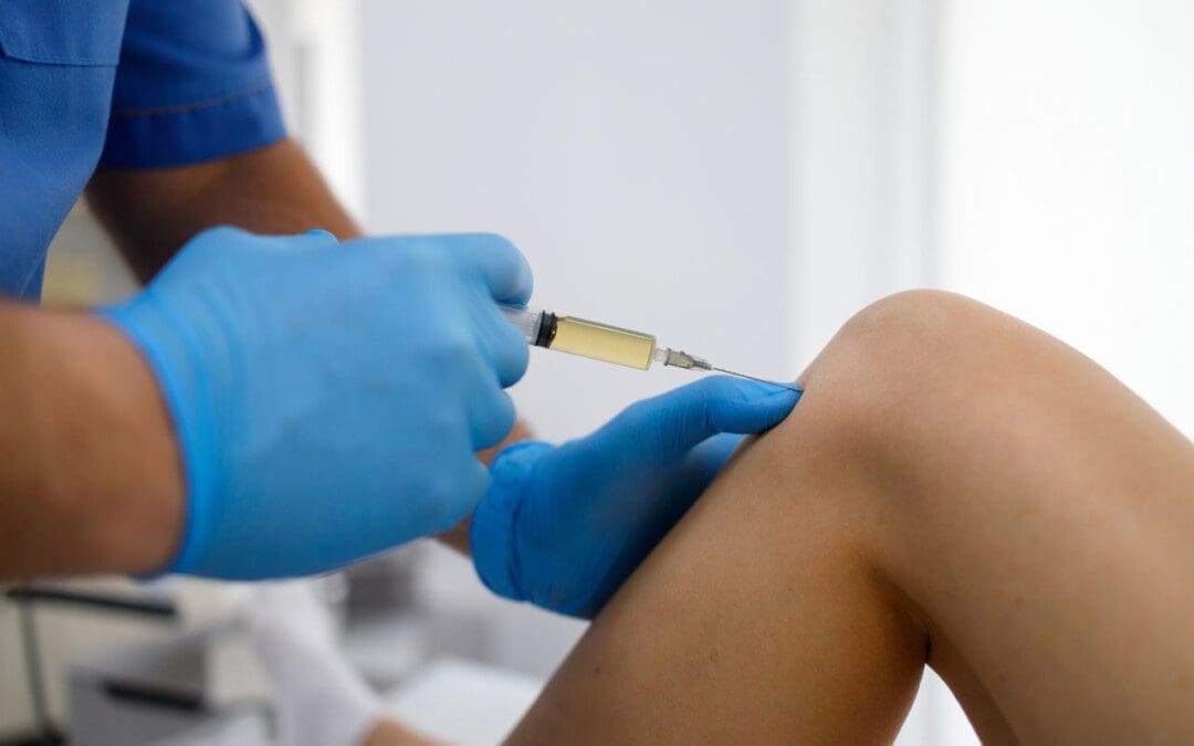
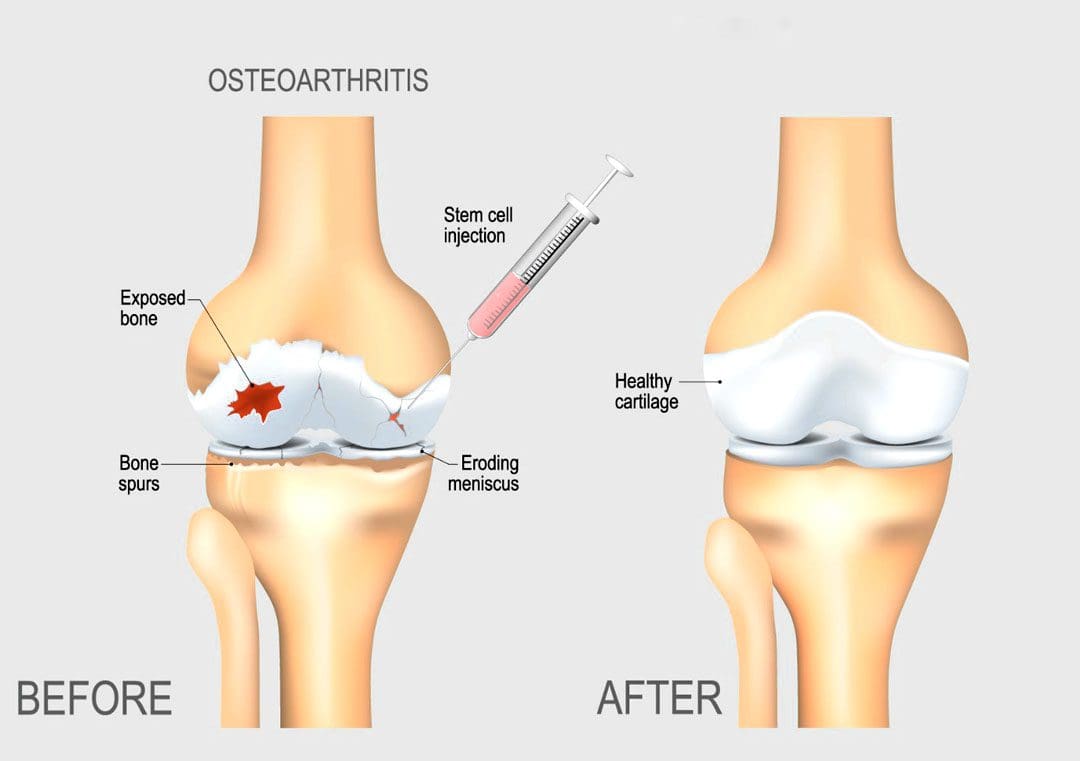
As the body ages, individuals want to stay active and maintain a healthy pain free lifestyle. Can regenerative cells for arthritis and cartilage damage be the future of neuromusculoskeletal medicine and joint healing?
Regenerative Cells For Arthritis and Cartilage Damage
Individuals want to continue to do the physical activities they love, which require healthy joints. Scientists are learning how to harness the abilities of regenerative cells to repair and regrow damaged and deteriorated cartilage. Current stem cell treatment of cartilage problems has not been shown to reverse the effects of arthritis and while studies show clinical improvement, further research is necessary. (Bryan M. Saltzman, et al., 2016)
Cartilage and How It Gets Damaged
Cartilage is a type of connective tissue. In the joints, there are a few types of cartilage. The most commonly referred to is the smooth lining known as articular or hyaline cartilage. This type forms a smooth layer of cushion on the end of a bone at the joint. (Rocky S. Tuan, et al., 2013)
The tissue is very strong and has the ability to compress and absorb energy.
It is very smooth allowing a joint to glide effortlessly through a limb’s range of motion.
When joint cartilage is damaged, the cushioning can wear down.
In traumatic injuries, a sudden force can cause the cartilage to break off and/or suffer damage, that exposes the underlying bone.
In osteoarthritis – degenerative or wear-and-tear arthritis, the smooth layer can wear down thin and unevenly.
Eventually, the cushion wears away, the joints become inflamed and swollen and movements become stiff and painful.
There are treatments for arthritis and cartilage damage, but these treatments are usually focused on relieving symptoms by smoothing down the damaged cartilage or replacing the joint surface with an artificial implant, like knee replacement or hip replacement surgeries. (Robert F. LaPrade, et al., 2016)
Regenerative Cells
Regenerative stem cells are special cells that have the ability to multiply and develop into different types of tissue. In an orthopedic surgery setting for joint problems, stem cells are obtained from adult stem cell primary sources which are bone marrow and fatty tissue. These cells have the ability to develop into cartilage cells, called chondrocytes. (Rocky S. Tuan, et al., 2013)
They also help by stimulating the body to reduce inflammation, stimulate cell repair, and improve blood circulation.
This process is caused by cellular signals and growth factors to stimulate the body to activate the healing processes.
Once stem cells have been obtained, they need to be delivered to the area of cartilage damage.
Cartilage is a complex tissue that is described as a scaffold structure that is composed of collagen, proteoglycans, water, and cells. (Rocky S. Tuan, et al., 2013)
To regenerate cartilage, the complex tissues must also be reconstructed.
There are studies on types of tissue scaffolds engineered to recreate a similar type of cartilage structure.
The stem cells can then be injected into the scaffold, in hopes of restoring a normal type of cartilage.
Non-Surgical Arthritis Treatments
Standard treatments such as cortisone shots or physical therapies work as well and provide benefits that could be utilized in combination with regenerative cells for arthritis and cartilage damage in the near future. Data takes time and therefore how this impacts the long-term health of a joint needs continued research in terms of tissue engineering and cell delivery to determine the best approach to help individuals.
Arthritis
References
LaPrade, R. F., Dragoo, J. L., Koh, J. L., Murray, I. R., Geeslin, A. G., & Chu, C. R. (2016). AAOS Research Symposium Updates and Consensus: Biologic Treatment of Orthopaedic Injuries. The Journal of the American Academy of Orthopaedic Surgeons, 24(7), e62–e78. https://doi.org/10.5435/JAAOS-D-16-00086
Saltzman, B. M., Kuhns, B. D., Weber, A. E., Yanke, A., & Nho, S. J. (2016). Stem Cells in Orthopedics: A Comprehensive Guide for the General Orthopedist. American journal of orthopedics (Belle Mead, N.J.), 45(5), 280–326.
Tuan, R. S., Chen, A. F., & Klatt, B. A. (2013). Cartilage regeneration. The Journal of the American Academy of Orthopaedic Surgeons, 21(5), 303–311. https://doi.org/10.5435/JAAOS-21-05-303
For individuals with stomach issues, can maintaining gut flora balance promote and improve gut health?
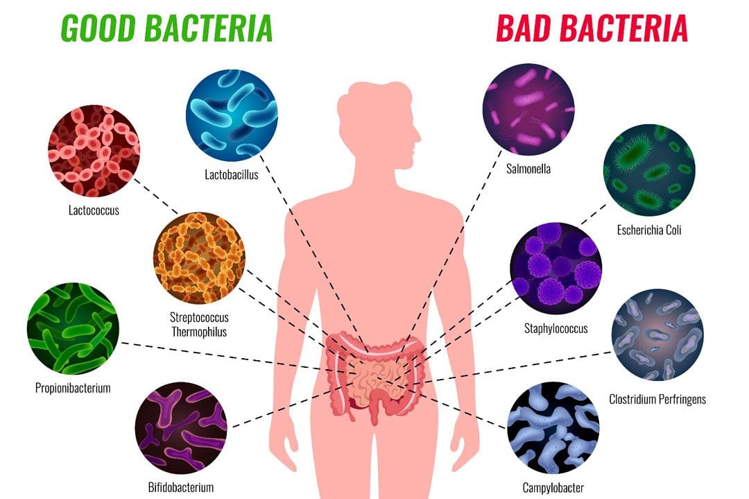
Gut Flora Balance
Maintaining gut flora balance is part of optimal digestive health. Gut microbiota, gut microbiome, or gut flora, are the microorganisms, including bacteria, archaea, fungi, and viruses that live in the digestive tract. The type and amount of bacteria present depend on their location in the body which could be the small intestine and colon. This is the storage housing for waste/stool, and the colon comprises hundreds of different types of bacteria, which have specific jobs and functions.
Unhealthy Flora
The more common pathogens are bacteria that can cause illness if left unchecked, including germs like streptococcus/strep throat or E. coli/urinary tract infections and diarrhea. Other common germs found in the colon include: (Elizabeth Thursby, Nathalie Juge. 2017)
Clostridioides Difficile
C. diff overgrowth can cause watery foul-smelling stools daily, and abdominal pain and tenderness.
Enterococcus Faecalis
Enterococcus faecalis is a cause of post-surgical abdominal and urinary tract infections.
Escherichia Coli
E. coli is the most common cause of diarrhea in adults.
This bacteria is present in almost every healthy adult’s colon.
Klebsiella
Klebsiella overgrowth is associated with a Western diet that consists of various meat and animal products.
Bacteroides
Bacteroide overgrowth is associated with colitis, which causes painful inflammation of the colon.
Healthy Flora
Healthy bacteria like Bifidobacteria and Lactobacillus, help maintain gut flora balance and keep the unhealthy bacteria in check. Without healthy flora, the entire colon can become overrun by bad flora, which can result in symptoms like diarrhea and/or illness. (Yu-Jie Zhang, et al., 2015) These protective, microscopic germs have important functions that include:
Assisting with vitamin synthesis – vitamins B and K in the small intestine.
Increases immune system function.
Maintaining regular bowel movements.
Maintaining a clean colon naturally without the need for colon cleansers.
Destroying the unhealthy bacteria.
Preventing unhealthy bacteria overgrowth.
Breaking up gas bubbles from food fermentation.
Bacterial Dismantling
Whether labeled as healthy bacteria or unhealthy, they are both single-celled organisms that can be destroyed quite easily. Sometimes, it is necessary, like when having to take antibiotics to kill a strep throat infection. However, the antibiotics also kill the beneficial bacteria, which can lead to compounding problems that can include: (Mi Young Yoon, Sang Sun Yoon. 2018)
Bowel irregularity – diarrhea and constipation.
Yeast overgrowth – can cause itching, burning around the anus and lead to vaginal and oral yeast infections.
Dysbiosis – the technical name for a lack of healthy bacteria or a bacterial imbalance.
Complications for individuals suffering from irritable bowel syndrome.
There are different ways to destroy bacteria including.
Prolonged diarrhea – can flush out the bad and good bacteria.
Stress
Completing a bowel prep, like those required for a colonoscopy.
Diagnosing Gut Flora Issues
Many times, problems with gut flora will correct themselves, and no action is required. However, individuals facing chronic bowel problems, like colitis or inflammatory bowel disease, may require medical intervention of their colon’s bacteria.
Comprehensive Digestive Stool Analysis/CDSA is a stool test that checks what type and amount of bacteria are present, nutrient absorption rates/digestion speed, and how well food is digested.
If there is a significant difference in the proportion of unhealthy versus beneficial bacteria, a healthcare provider may suggest taking a probiotic or a live microbial supplement to help repopulate and maintain gut flora balance.
Gut Dysfunction
References
Thursby, E., & Juge, N. (2017). Introduction to the human gut microbiota. The Biochemical journal, 474(11), 1823–1836. https://doi.org/10.1042/BCJ20160510
Zhang, Y. J., Li, S., Gan, R. Y., Zhou, T., Xu, D. P., & Li, H. B. (2015). Impacts of gut bacteria on human health and diseases. International journal of molecular sciences, 16(4), 7493–7519. https://doi.org/10.3390/ijms16047493
Yoon, M. Y., & Yoon, S. S. (2018). Disruption of the Gut Ecosystem by Antibiotics. Yonsei medical journal, 59(1), 4–12. https://doi.org/10.3349/ymj.2018.59.1.4
Quigley E. M. (2013). Gut bacteria in health and disease. Gastroenterology & hepatology, 9(9), 560–569.
Can combining chiropractic treatment with the common therapies of medication, exercise, and/or physical therapy help relieve sciatic endometriosis pain symptoms?
Sciatic Endometriosis
Sciatic endometriosis is a condition in which endometrial cells (tissue that resembles the lining of the uterus) grow outside of the uterine lining and compress the sciatic nerve. This places stress and pressure on the nerve causing back, pelvic, hip, and leg pain, especially before and during the menstrual cycle. It can also cause pain, irregular periods, and infertility. (The American College of Obstetricians and Gynecologists. 2021)
These areas of endometrial tissue growth are also known as lesions or implants.
Women with sciatic endometriosis often experience leg pain and weakness around the time of their menstrual cycle. (Lena Marie Seegers, et al., 2023)
Sciatic endometriosis can also cause pain when urinating, during a bowel movement, during sex, and fatigue, and irregular vaginal bleeding.
The abnormal growth may be caused by higher-than-normal levels of estrogen.
Researchers believe that endometriosis is related to retrograde menstruation, which causes menstrual blood to flow back into the pelvis instead of out through the vagina. (World Health Organization. 2023)
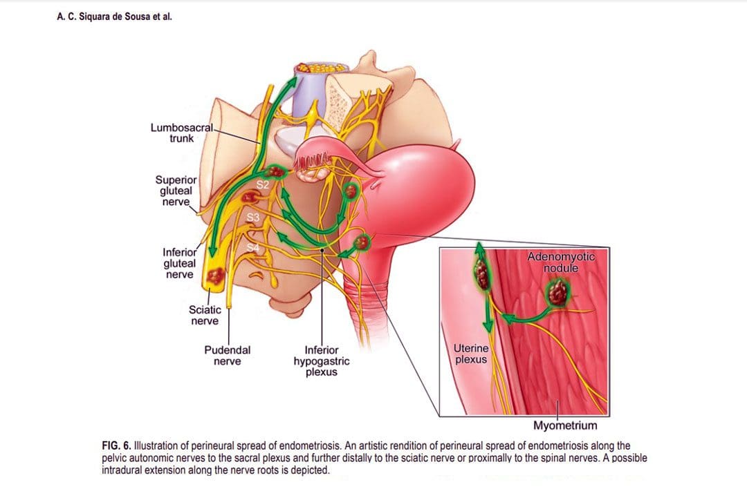
Sometimes, the cells grow in the area of the pelvis right above the sciatic nerve. (Adaiah Yahaya, et al., 2021)
The sciatic nerve is the longest nerve in the body and travels down the back of each leg. (Johns Hopkins Medicine. 2023)
When endometrial lesions place pressure on the sciatic nerve, they can cause irritation and inflammation leading to severe pelvic pain, which makes it harder to conceive. (Liang Yanchun, et al., 2019)
Symptoms
Some women with endometriosis experience no symptoms or misinterpret the symptoms as typical premenstrual syndrome/PMS signs. The most common signs and symptoms of sciatic endometriosis include:
Difficulty walking or standing.
Loss of sensation, muscle weakness, and reflex alteration.
Limping.
Balance problems.
Bloating and nausea.
Constipation or diarrhea before or after a period.
Painful, heavy, and/or irregular periods.
Bleeding between periods.
Pain during sex, urination, and bowel movements.
Pain in the stomach, pelvis, lower back, hips, and buttocks. (MedlinePlus. 2022)
Weakness, numbness, tingling, burning, or dull aching sensations in the back of one or both legs.
Endometriosis, including sciatic endometriosis, typically cannot be diagnosed with a pelvic examination or ultrasound by themselves. A healthcare provider may need to perform a biopsy using laparoscopy and discuss menstrual cycles, symptoms, and medical history.
The laparoscopy procedure involves making tiny incisions and taking a tissue sample with tools attached to a thin tube with a camera. (MedlinePlus. 2022)
Imaging tests, like magnetic resonance imaging/MRI, and computed tomography/CT scans, can help provide essential information about the location and size of any endometrial lesions. (The American College of Obstetricians and Gynecologists. 2021)
Treatment
Symptoms can sometimes be temporarily relieved with over-the-counter/OTC pain relievers. Depending on the condition and severity a healthcare provider may prescribe hormonal treatment to prevent new endometrial implants from growing. These can include:
Hormonal birth control.
Progestin – a synthetic form of progesterone.
Gonadotropin-releasing hormone – GnRH agonists.
If pain persists or worsens, individuals may need to undergo surgery to remove the tissue.
The American College of Obstetricians and Gynecologists. Endometriosis.
Seegers, L. M., DeFaria Yeh, D., Yonetsu, T., Sugiyama, T., Minami, Y., Soeda, T., Araki, M., Nakajima, A., Yuki, H., Kinoshita, D., Suzuki, K., Niida, T., Lee, H., McNulty, I., Nakamura, S., Kakuta, T., Fuster, V., & Jang, I. K. (2023). Sex Differences in Coronary Atherosclerotic Phenotype and Healing Pattern on Optical Coherence Tomography Imaging. Circulation. Cardiovascular imaging, 16(8), e015227. https://doi.org/10.1161/CIRCIMAGING.123.015227
World Health Organization. Endometriosis.
Yahaya, A., Chauhan, G., Idowu, A., Sumathi, V., Botchu, R., & Evans, S. (2021). Carcinoma arising within sciatic nerve endometriosis: a case report. Journal of surgical case reports, 2021(12), rjab512. https://doi.org/10.1093/jscr/rjab512
Johns Hopkins Medicine. Sciatica.
Yanchun, L., Yunhe, Z., Meng, X., Shuqin, C., Qingtang, Z., & Shuzhong, Y. (2019). Removal of an endometrioma passing through the left greater sciatic foramen using a concomitant laparoscopic and transgluteal approach: case report. BMC women’s health, 19(1), 95. https://doi.org/10.1186/s12905-019-0796-0
MedlinePlus. Endometriosis.
Center for Endometriosis Care. Sciatic endometriosis.
Chen, S., Xie, W., Strong, J. A., Jiang, J., & Zhang, J. M. (2016). Sciatic endometriosis induces mechanical hypersensitivity, segmental nerve damage, and robust local inflammation in rats. European journal of pain (London, England), 20(7), 1044–1057. https://doi.org/10.1002/ejp.827
Siquara de Sousa, A. C., Capek, S., Howe, B. M., Jentoft, M. E., Amrami, K. K., & Spinner, R. J. (2015). Magnetic resonance imaging evidence for perineural spread of endometriosis to the lumbosacral plexus: report of 2 cases. Neurosurgical focus, 39(3), E15. https://doi.org/10.3171/2015.6.FOCUS15208
IFM's Find A Practitioner tool is the largest referral network in Functional Medicine, created to help patients locate Functional Medicine practitioners anywhere in the world. IFM Certified Practitioners are listed first in the search results, given their extensive education in Functional Medicine