
A Comprehensive Look at the Thoracodorsal Nerve
Individuals experiencing pain symptoms like shooting, stabbing, or electrical sensations to the latissimus dorsi of the upper back could be caused by a nerve injury to the thoracodorsal nerve. Can knowing the anatomy and symptoms help healthcare providers develop an effective treatment plan?
Thoracodorsal Nerve
Also known as the middle subscapular nerve or the long subscapular nerve, it branches out from a part of the brachial plexus and provides motor innervation/function to the latissimus dorsi muscle.
Anatomy
The brachial plexus is a network of nerves that stem from the spinal cord in the neck. The nerves supply most of the sensation and movement of the arms and hands, with one on each side. Its five roots come from the spaces between the fifth through eighth cervical vertebrae and the first thoracic vertebra. From there, they form a larger structure, then divide, re-combine, and divide again to form smaller nerves and nerve structures as they travel down the armpit. Through the neck and chest, the nerves eventually join and form three cords that include:
- Lateral cord
- Medial cord
- Posterior cord
The posterior cord produces major and minor branches that include:
- Axillary nerve
- Radial nerve
The minor branches include:
- Superior subscapular nerve
- Inferior subscapular nerve
- Thoracodorsal nerve
Structure and Position
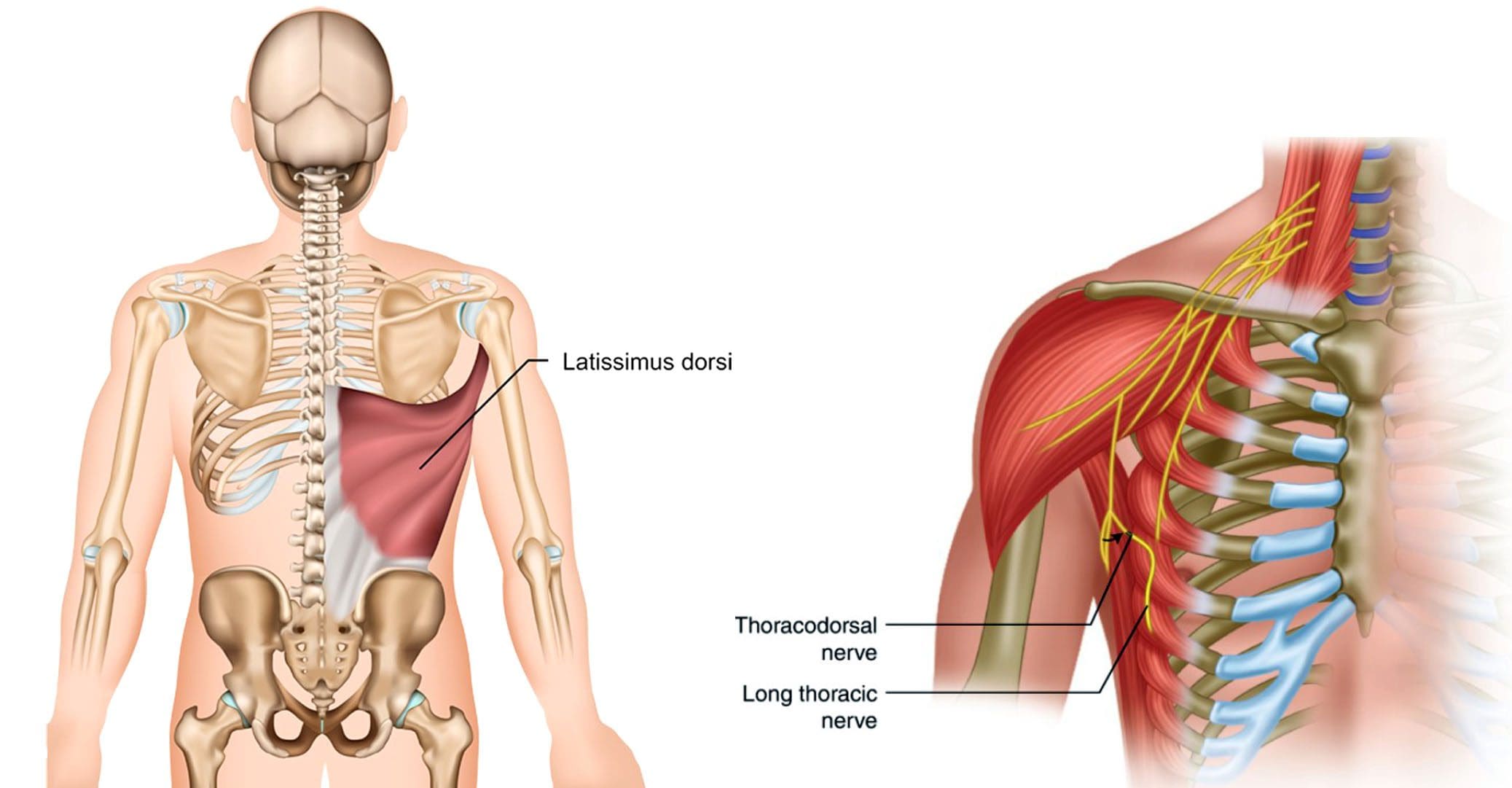
- The thoracodorsal nerve branches off the posterior cord in the armpit and travels down, following the subscapular artery, to the latissimus dorsi muscle.
- It connects to the upper arm, stretches across the back of the armpit, forming the axillary arch, and then expands into a large triangle that wraps around the ribs and the back.
- The thoracodorsal nerve lies deep in the latissimus dorsi, and the lower edge typically reaches close to the waist.
Variations
- There is a standard location and course of the thoracodorsal nerve, but individual nerves are not the same in everyone.
- The nerve typically branches off the posterior cord of the brachial plexus from three different points.
- However, different subtypes have been identified.
- The thoracodorsal nerve supplies the teres major muscle in about 13% of individuals. (Brianna Chu, Bruno Bordoni. 2023)
- The lats can have a rare anatomical variation known as a Langer’s arch, which is an extra part that connects to muscles or connective tissue of the upper arm beneath the common connecting point.
- In individuals with this abnormality, the thoracodorsal nerve supplies function/innervation) to the arch. (Ahmed M. Al Maksoud et al., 2015)
Function
The latissimus dorsi muscle cannot function without the thoracodorsal nerve. The muscle and nerve help:
- Stabilize the back.
- Pull the body weight up when climbing, swimming, or doing pull-ups.
- Assist with breathing by expanding the rib cage during inhalation and contracting when exhaling. (Encyclopaedia Britannica. 2023)
- Rotate the arm inward.
- Pull the arm toward the center of the body.
- Extend the shoulders by working with the teres major, teres minor, and posterior deltoid muscles.
- Bring down the shoulder girdle by arching the spine.
- To bend to the side by arching the spine.
- Tilt the pelvis forward.
Conditions
The thoracodorsal nerve can be injured anywhere along its path by trauma or disease. Symptoms of nerve damage can include: (U.S. National Library of Medicine: MedlinePlus. 2022)
- Pain that can be shooting, stabbing, or electrical sensations.
- Numbness, tingling.
- Weakness and loss of function in the associated muscles and body parts, including wrist and finger drop.
- Because of the nerve’s path through the armpit, doctors have to be cautious of the anatomical variants so they don’t inadvertently damage a nerve during breast cancer procedures, including axillary dissection.
- The procedure is performed to examine or remove lymph nodes and is used in staging breast cancer and in treatment.
- According to a study, 11% of individuals with axillary lymph node dissection suffered damage to the nerve. (Roser Belmonte et al., 2015)
Breast Reconstruction
- In breast reconstruction surgery, the lats can be used as a flap over the implant.
- Depending on the circumstances, the thoracodorsal nerve can be left intact or severed.
- The medical community has not agreed on which method has the best outcomes. (Sung-Tack Kwon et al., 2011)
- There is some evidence that leaving the nerve intact can cause the muscle to contract and dislocate the implant.
- An intact thoracodorsal nerve may also cause atrophy of the muscle, which can lead to shoulder and arm weakness.
Graft Uses
A portion of the thoracodorsal nerve is commonly used in nerve graft reconstruction to restore function after injury, which includes the following:
- Musculocutaneous nerve
- Accessory nerve
- Axillary nerve
- The nerve can also be used to restore nerve function to the triceps muscle in the arm.
Rehabilitation
If the thoracodorsal nerve is injured or damaged, treatments can include:
- Braces or splints.
- Physical therapy to improve range of motion, flexibility, and muscle strength.
- If there is compression, surgery may be required to alleviate the pressure.
Exploring Integrative Medicine
References
Chu B, Bordoni B. Anatomy, Thorax, Thoracodorsal Nerves. [Updated 2023 Jul 24]. In: StatPearls [Internet]. Treasure Island (FL): StatPearls Publishing; 2023 Jan-. Available from: https://www.ncbi.nlm.nih.gov/books/NBK539761/
Al Maksoud, A. M., Barsoum, A. K., & Moneer, M. M. (2015). Langer’s arch: a rare anomaly affects axillary lymphadenectomy. Journal of surgical case reports, 2015(12), rjv159. https://doi.org/10.1093/jscr/rjv159
Britannica, The Editors of Encyclopaedia. “latissimus dorsi“. Encyclopedia Britannica, 30 Nov. 2023, https://www.britannica.com/science/latissimus-dorsi. Accessed 2 January 2024.
U.S. National Library of Medicine: MedlinePlus. Peripheral neuropathy.
Belmonte, R., Monleon, S., Bofill, N., Alvarado, M. L., Espadaler, J., & Royo, I. (2015). Long thoracic nerve injury in breast cancer patients treated with axillary lymph node dissection. Supportive care in cancer : official journal of the Multinational Association of Supportive Care in Cancer, 23(1), 169–175. https://doi.org/10.1007/s00520-014-2338-5
Kwon, S. T., Chang, H., & Oh, M. (2011). Anatomic basis of interfascicular nerve splitting of innervated partial latissimus dorsi muscle flap. Journal of plastic, reconstructive & aesthetic surgery : JPRAS, 64(5), e109–e114. https://doi.org/10.1016/j.bjps.2010.12.008


