For individuals who like sweet foods but want to maintain health, how do honey and maple syrup compare nutritionally?
Honey and Maple Syrup
Cutting back on sugar intake is recommended for overall health, but that does not mean eliminating sweeteners completely for most. Honey and maple syrup can flavor various dishes, baked goods, beverages, and more. Because of their natural origins, honey and maple syrup can seem interchangeable as they are brown, thick, and sweet. Maple syrup and honey can be part of a healthy diet in moderation, sweetening everything from breakfast to dinnertime dishes. Depending on individual dietary goals or taste preferences, individuals may have a personal favorite, but neither is better than the other. The nutritional makeup of these two sweeteners is distinct, and there are recommended practices for when to include each in cooking and baking.
Nutrition
Honey is slightly higher in calories, carbohydrates, and sugar than maple syrup and somewhat lower in fat. Maple syrup comprises more micronutrients, such as calcium and manganese, but it also has more sodium than honey. Nutrition information for
For food-labeling purposes, honey and maple syrup have different serving sizes:
One tablespoon of honey
1/4 cup of maple syrup
Nutritional Comparison
Honey 1 tbsp. Maple syrup 1 tbsp.
Calories: 64 – 52
Fat: 0g – >1g
Sodium: >1mg – 2.4mg
Carbohydrates: 17.3g – 13.4g
Fiber: >1g – 0g
Sugars: 17.2g – 12.1g
Protein: >1g – >1g
Calcium: 1.26mg – 20.4mg
Manganese: 0.17mg – 0.582mg
Zinc: 0.046mg – 0.294mg
Riboflavin: 0.008mg – 0.254mg
Similarities
Honey and maple syrup are similar liquid sweeteners nutritionally. Both contain no substantial amount of fat or protein, with carbohydrates from sugar supplying all their calories. Both also contain some micronutrients and antioxidants. Research has revealed promising levels of antioxidants in honey (Schramm, D. D. et al., 2003) and maple syrup (Li, L., and Seeram, N. P. 2011) that could contribute to reducing oxidative stress, a key component in preventing some chronic diseases.
Differences
The nutrition differences between maple syrup and honey are more significant. Although a tablespoon of honey has only eight more calories than maple syrup, this can add up depending on the amount used. In a quarter cup, honey contains 32 more calories than maple syrup or 128 more calories in 1 cup. The carbohydrate count of maple syrup and honey is similar, but there is a difference of one teaspoon of added sugar. Honey and maple syrup are not identical in micronutrient levels. Honey contains small amounts of vitamins C and B, but individuals would have to consume a lot of honey to receive the benefits. In 100 grams, around five tablespoons of honey, individuals receive about 1% of the RDA for vitamin C. Maple syrup is a healthy source of manganese, riboflavin, and zinc.
Health Benefits
Honey Health Benefits
Honey’s antioxidants help clean cells of damaging free radicals. It can also be used as food and medicine during cold and flu season. Research has shown that a spoonful could be a more effective treatment for a cough than over-the-counter medications. (Oduwole, O. et al., 2018) Those with allergies may want to consider visiting a local beekeeper. While inconclusive, some studies (Asha’ari, Z. A. et al., 2013) have found that eating local raw honey can help improve seasonal allergy symptoms.
Maple Syrup Health Benefits
Maple syrup offers health benefits, particularly because it contains high amounts of manganese and riboflavin. Manganese is a trace mineral the body does not need very much of but does need to operate efficiently. It is essential to bone and tissue formation, blood clotting, proper immune response, and sexual function. Riboflavin helps create usable energy from food and could help prevent cancer and migraines. (National Institutes of Health Office of Dietary Supplements, 2022) Maple syrup also offers benefits for individuals with certain chronic health conditions. Its lower glycemic index makes it recommended for those with diabetes or other blood sugar conditions. Because of the type of sugar it contains, those with irritable bowel syndrome/IBS may be able to digest maple syrup better than honey. Maple syrup and honey both contain simple sugars; however, fructose in honey may not be digested as well in individuals with IBS who need a low FODMAP diet.
Preparation and Cooking
Honey and maple syrup have distinctive flavors, so the individual can choose where and when to use each. Maple syrup has a more earthy, woodsy taste, while honey has floral hints and a thicker texture. Honey does well in salad dressings and marinades, where it can cling to other foods, whereas maple syrup blends easily in baked goods. Both work well in various food preparations. Rules of thumb for using honey and maple syrup include:
Both can be used in place of sugar as long as the liquid in the recipe is adjusted.
For every cup of sugar replaced by honey or maple syrup, decrease other liquid ingredients by three to four tablespoons.
Scaling back a little is recommended if using either as a substitute for table sugar.
Both maple syrup and honey are sweeter than sugar; replacing sugar requires only three-fourths of either.
Lifestyle and Safety
Sometimes, when to use honey and maple syrup has nothing to do with nutrition or taste. Vegans, for example, often choose not to consume honey because it’s an insect by-product. A vegan lifestyle may prefer maple syrup, which is 100% plant-based. Parents of infants under one should keep honey off the menu. Honey can contain small amounts of the bacteria Clostridium botulinum, which can cause botulism poisoning in babies; for babies under 12 months old, maple syrup or other sweeteners are recommended in baked goods or cooked meals. However, while honey can be harvested almost anywhere honeybees live, maple syrup is native only to a small region of eastern North America, and obtaining the syrup may not be possible.
Injury Medical Chiropractic and Functional Medicine Clinic
Injury Medical Chiropractic and Functional Medicine Clinic providers use an integrated approach to create customized care plans for each patient and restore health and function to the body through nutrition and wellness, chiropractic adjustments, functional medicine, acupuncture, Electroacupuncture, and sports medicine protocols. If the individual needs other treatment, they will be referred to a clinic or physician best suited for them. Dr. Jimenez has teamed up with top surgeons, clinical specialists, medical researchers, nutritionists, and health coaches to provide the most effective clinical treatments.
Balancing Body and Metabolism
References
U.S. Department of Agriculture, FoodData Central. (2018). Syrups, maple. Retrieved from https://fdc.nal.usda.gov/fdc-app.html#/food-details/169661/nutrients
U.S. Department of Agriculture, FoodData Central. (2018). Honey. Retrieved from https://fdc.nal.usda.gov/fdc-app.html#/food-details/169640/nutrients
Schramm, D. D., Karim, M., Schrader, H. R., Holt, R. R., Cardetti, M., & Keen, C. L. (2003). Honey with high levels of antioxidants can provide protection to healthy human subjects. Journal of agricultural and food chemistry, 51(6), 1732–1735. https://doi.org/10.1021/jf025928k
Li, L., & Seeram, N. P. (2011). Further investigation into maple syrup yields 3 new lignans, a new phenylpropanoid, and 26 other phytochemicals. Journal of agricultural and food chemistry, 59(14), 7708–7716. https://doi.org/10.1021/jf2011613
Oduwole, O., Udoh, E. E., Oyo-Ita, A., & Meremikwu, M. M. (2018). Honey for acute cough in children. The Cochrane database of systematic reviews, 4(4), CD007094. https://doi.org/10.1002/14651858.CD007094.pub5
Asha’ari, Z. A., Ahmad, M. Z., Jihan, W. S., Che, C. M., & Leman, I. (2013). Ingestion of honey improves the symptoms of allergic rhinitis: evidence from a randomized placebo-controlled trial in the East coast of Peninsular Malaysia. Annals of Saudi medicine, 33(5), 469–475. https://doi.org/10.5144/0256-4947.2013.469
National Institutes of Health Office of Dietary Supplements. (2022). Riboflavin: Fact sheet for health professionals. Retrieved from https://ods.od.nih.gov/factsheets/Riboflavin-HealthProfessional/
Can Tabata training help burn more calories during and after workouts for individuals who do interval training?
Tabata Training
Also known as the Tabata Protocol, Tabata training can bring variety to workouts, burn more calories, and get more out of exercise time. This workout is a form of high-intensity interval training, or HIIT, designed to increase heart rate in the hard anaerobic zone for short periods. The intervals are short, the workouts are fast, and the body benefits. The recommendation is to add Tabata training once a week to see how the body responds.
Protocol Format
This HIIT workout is so effective because of the work-to-rest ratio. Individuals only get 10 seconds of rest between each 20-second session of exercise. That short interval is insufficient to recover fully, but it is great for building endurance and getting in shape. The format consists of:
Twenty seconds of a very high-intensity exercise – sprints, burpees, squat jumps, etc.
Tabata training can improve two of the body’s main energy systems. It targets the anaerobic energy system responsible for short, high-intensity exercises like sprints and the aerobic energy system for endurance exercises like long, slow running. In traditional interval training, moderate intensity and steady-state cardio target the aerobic system. Still, unless the individuals work way out of their comfort zone, they don’t necessarily improve the anaerobic system. (Astorino, T. A. et al., 2012) However, high-intensity interval training with a rest period shorter than the work period can target both systems, providing benefits for athletes and fitness enthusiasts. (Tabata, I. et al., 1996)
Safety Precautions
However, these workouts are not for everyone. Tabata training is advanced and best suited to athletes and experienced fitness enthusiasts. Beginners should start with lighter interval training and gradually work their way up to this level of intensity. Try 20 seconds on/10 seconds off with easier exercises like walking or low-impact moves like marching in place, step touches, or knee lifts.
Workout Tips
Individuals can do Tabata training with almost any activity or cardiovascular machine. This Tabata cardio workout includes a variety of bodyweight exercises that, if done at full intensity, will increase heart rate.
Warm-Up
Before trying this type of workout, ensure the body is thoroughly warmed up for at least 10 minutes.
Start Slow
Individuals new to this type of training start with 5 to 6 cycles of each exercise and increase rest to 20 or 30 seconds.
As the body gets used to the workout and builds stamina, gradually shorten the rest periods and increase the number of cycles.
Rest Between Sets
Individuals who do more than one Tabata set as many workouts call for rest for at least 60 seconds between sets.
Monitor Intensity Frequently
Intensity accumulates through each cycle, peaking at the end of the workout when muscles are tired, and form becomes sloppy, increasing the risk of injury.
Take Rest Days
The recommendation is that the workout be done no more than 1 to 2 times a week, with rest in between, to avoid overtraining and injury.
Listen to your body when doing any high-intensity exercise. High-intensity interval training is very taxing on the body, so it’s easy to overdo it if you’re not careful. If you feel too breathless, extend recovery times or take extra breaks. If you are in pain or discomfort, take a break, try different exercises, or stop for the day.
Injury Medical Chiropractic and Functional Medicine Clinic
Injury Medical Chiropractic and Functional Medicine Clinic treats patients’ injuries and chronic pain syndromes. We focus on improving ability through flexibility, mobility, and agility programs tailored to the individual. We use in-person and virtual health coaching and comprehensive care plans to ensure every patient’s personalized care and wellness outcomes. Our providers use an integrated approach to create customized care plans that include Functional Medicine, Acupuncture, Electro-Acupuncture, and Sports Medicine principles. Our goal is to relieve pain naturally by restoring health and function to the body. If he feels the individual needs other treatment, they will be referred to a clinic or physician best suited for them as Dr. Jimenez has teamed up with the top surgeons, clinical specialists, medical researchers, and premier rehabilitation providers to provide our community with the best clinical treatments.
Improving Athletic Performance Through Chiropractic
References
Emberts, T., Porcari, J., Dobers-Tein, S., Steffen, J., & Foster, C. (2013). Exercise intensity and energy expenditure of a tabata workout. Journal of sports science & medicine, 12(3), 612–613.
Tabata, I., Nishimura, K., Kouzaki, M., Hirai, Y., Ogita, F., Miyachi, M., & Yamamoto, K. (1996). Effects of moderate-intensity endurance and high-intensity intermittent training on anaerobic capacity and VO2max. Medicine and science in sports and exercise, 28(10), 1327–1330. https://doi.org/10.1097/00005768-199610000-00018
Astorino, T. A., Allen, R. P., Roberson, D. W., & Jurancich, M. (2012). Effect of high-intensity interval training on cardiovascular function, VO2max, and muscular force. Journal of strength and conditioning research, 26(1), 138–145. https://doi.org/10.1519/JSC.0b013e318218dd77
For individuals and athletes with a gluteal contusion with severe bruising, can a healthcare provider determine if there are any other injuries to underlying structures, including muscle or tendon tears?
Gluteal Contusion
A gluteal contusion is an injury, in this case, a bruise to the buttocks’ gluteal muscles caused by damage to muscle fibers and blood vessels. A buttock bruise is caused by direct bodily impact, typically from falls, automobile collisions, accidents, bumping into something, or being struck by an object or person. Like all bruises, a gluteal bruise most often results in pain and visible discoloration of the skin at the injury site, varying in severity from grade I to grade III, with higher-graded bruises requiring more time to heal. Most butt bruises can heal on their own with time and rest, but if bruising is severe, individuals may require physical therapy to restore full muscle function.
Symptoms
A contusion is a muscle injury that can affect the body’s skeletal muscles. A gluteal contusion can be painful, with a black and blue mark that changes color over time. Other symptoms may include: (Mount Sinai, 2024)
Tenderness to touch over the injury site
Increased pain with contraction of the glutes
Swelling
Discomfort with sitting
Causes
A contusion occurs from direct trauma and forceful impact on the gluteal muscles, causing damage to underlying blood vessels, muscle fibers, and sometimes bone, resulting in bleeding under the skin. (MedlinePlus, 2016) Direct impacts to the gluteal muscles that can cause a contusion include:
Falls
Car accidents
Direct hits to the buttocks from a piece of sports equipment or person.
Bumping into furniture, a door, or a counter.
Intramuscular injections into the gluteal muscles.
Individuals who take blood thinners or anticoagulant medication have an increased risk of bruising from direct contact injuries.
Diagnosis
A gluteal contusion is usually diagnosed through a physical examination and is generally straightforward to diagnose based on physical appearance, symptoms, and type of injury. Contusions can be graded based on the severity according to the following criteria (Fernandes, T. L. et al., 2015)
Grade I
An injury that affects only a small amount of muscle fibers, resulting in minimal pain, tenderness, and possible swelling.
Causes minimal or no loss of strength in the affected muscle or range of motion limitations.
Muscle use is typically unaffected.
Grade II
An injury that causes significant damage to muscle fibers, resulting in increased pain and impaired muscle contraction.
A small muscle defect can be felt to the touch.
Discoloration increases over the first few days after injury.
Grade III
An injury that involves extensive muscle fiber damage and bleeding across an entire area of a muscle that results in severe, and sometimes total, loss of muscle function.
Causes severe pain and significant discoloration of the skin.
When contusions are larger, deeper, and involve significant blood pooling and swelling, they are called hematomas.
If the bruising is severe, a diagnostic ultrasound, CT scan, or MRI may be used to determine whether any underlying structures are damaged.
Treatment
Contusions are generally mild injuries. Treatment typically involves rest to allow the muscles to heal from the bleeding and the bruising to dissipate.
Applying ice to the injury site can help relieve pain and inflammation.
If the bruising is severe, significant physical activity like sports, dancing, running, jumping, and weight lifting should be avoided until the muscles heal. (Mount Sinai, 2024)
With more severe bruising, contraction and stretching of the glutes are painful and can require longer healing and recovery time.
Physical therapy rehabilitation may be needed for more significant injuries to restore muscle function.
Prognosis
A mild injury usually heals on its own with time and rest. More significant injuries take longer to heal and may require physical therapy to build strength and range of motion if muscle function is affected.
Healing Time and Recovery
Healing and recovery times for gluteal contusions vary depending on the severity of the injury (Fernandes T. L. et al., 2015)
Grade I
Minor injuries that cause minimal discomfort typically heal fully in five days to two weeks.
Grade II
During the first two to three days, contusions develop, increasing discoloration under the skin, and complete healing can take two to three weeks.
Return to sport is typically resumed after a month.
Grade III
Contusions can take up to four to six weeks to heal, often requiring rehabilitation to restore muscle strength and range of motion.
Injury Medical Chiropractic and Functional Medicine Clinic
At Injury Medical Chiropractic and Functional Medicine Clinic, we passionately focus on treating patients’ injuries and chronic pain syndromes. We focus on improving ability through flexibility, mobility, and agility programs tailored to the individual. We use in-person and virtual health coaching and comprehensive care plans to ensure every patient’s personalized care and wellness outcomes. Our providers use an integrated approach to create personalized care plans that include Functional Medicine, Acupuncture, Electro-Acupuncture, and Sports Medicine principles. Our goal is to relieve pain naturally by restoring health and function to the body. If he feels the individual needs other treatment, they will be referred to a clinic or physician best suited for them as Dr. Jimenez has teamed up with the top surgeons, clinical specialists, medical researchers, and premier rehabilitation providers to provide our community with the best clinical treatments.
Building a Stronger Body
References
Mount Sinai. (2024). Bruise. https://www.mountsinai.org/health-library/injury/bruise
MedlinePlus. (2016). Bruises. Retrieved from https://medlineplus.gov/bruises.html
Fernandes, T. L., Pedrinelli, A., & Hernandez, A. J. (2015). MUSCLE INJURY – PHYSIOPATHOLOGY, DIAGNOSIS, TREATMENT AND CLINICAL PRESENTATION. Revista brasileira de ortopedia, 46(3), 247–255. https://doi.org/10.1016/S2255-4971(15)30190-7
Can the Oswestry Low Back Pain Disability Questionnaire help assess how low back pain impacts individuals’ ability to perform everyday tasks and activities and help physical therapists incorporate the outcome measure into an effective treatment plan?
Oswestry Disability Questionnaire
The Oswestry Disability Questionnaire, also known as the Oswestry Disability Index, provides objective data about an individual’s lower back pain. It determines the severity of the pain and how much it limits their daily activities. The questionnaire is a validated measure backed by research that can be used to justify the need for medical treatment. It includes questions regarding the symptoms and severity of low back pain and how these symptoms interfere with regular activities. Lower back pain can result from various causes (National Institute of Neurological Disorders and Stroke, 2020)
Arthritis, including inflammatory types of arthritis like psoriatic arthritis and ankylosing spondylitis.
Lumbar vertebrae compression fractures – usually from trauma or osteoporosis.
Low back surgery – including spinal fusions, discectomies, and laminectomies.
Spinal stenosis
Spondylolisthesis
Scoliosis
How The Questionnaire Works
The Oswestry Disability Questionnaire consists of 10 questions about the impact of lower back pain on daily life. The questions are divided into the following categories: (American Academy of Orthopedic Surgeons, N.D.)
Pain Intensity
How intense is the pain?
If painkillers are used, how much symptom relief do they provide?
Personal Care
Can the patient perform self-care activities like bathing and dressing when experiencing significant pain or limitations?
Whether physical assistance from another person is needed?
Lifting
Can the patient lift objects like weights with or without pain?
Can lifting be performed from the floor or a higher surface like a table if the objects are light, moderate, or heavy?
Walking
If and to what extent does the pain limit the patient’s walking distance and independence?
If an assistive device like a cane or crutches are needed?
Sitting
If so, how much pain limits the patient’s sitting tolerance?
Standing
If so, how much pain limits the patient’s standing tolerance?
Sleeping
If so, how much pain limits a patient’s sleeping duration?
Whether pain medication is needed to help the patient sleep comfortably?
Social Life
If and to what extent a patient’s social activities are limited because of pain symptoms?
Traveling
If so, to what extent does pain limit a patient’s ability to travel?
Employment and/or Homemaking Duties
Does pain limit a patient’s ability to perform job-related and/or household activities, including physically demanding and light duties?
Patients self-report the information and complete it on their own based on their understanding of the extent of their lower back pain and disability.
Each question can be scored between 0 and 5, with 0 indicating no limitations and 5 indicating complete disability.
The scores from all the questions are added together for a cumulative total score of 50 points.
Scores
The Oswestry Disability Questionnaire assesses how much a patient’s lower back pain limits daily activities. This information is used in clinical documentation for medical services. A higher score indicates a greater level of disability, according to the following scoring criteria:
0–4: No disability
5–14: Mild disability
15–24: Moderate disability
25–34: Severe disability
35–50: Completely disabled
Physical therapists must create individualized goals for each patient to develop a treatment plan and receive authorization from insurance companies. One of the most important aspects of a physical therapy goal is that it must be measurable. The Oswestry Disability Questionnaire provides a numerical score to track functional limitations and monitor the range of motion and strength testing. A baseline measurement is taken at the beginning of treatment, and progress is tracked in follow-up visits. A new score is used as a treatment goal. According to a study, the minimal clinically important difference (MCID) for the Oswestry Disability Questionnaire is 12.88. The MCID is the minimum score healthcare providers need to confirm a patient’s progress in function due to treatment. (Johnsen, L. G. et al., 2013)
By tracking changes in the total score before, during, and after treatment, healthcare providers can better assess whether treatment improves symptoms. A decrease in total score by 13 points or more would indicate that treatment is helping to improve a patient’s lower back pain and level of disability. Along with physical examination results, the patient’s score and the severity of symptoms can help healthcare providers determine an appropriate treatment plan.
No Disability
Treatment is unnecessary other than providing advice for lifting mechanics and general physical activity to maintain health.
Mild Disability
Conservative measures, such as physical therapy, exercise, hot or cold therapy, pain medication, and rest, are needed to help alleviate symptoms.
Moderate Disability
More aggressive intervention is needed, which can include extensive physical therapy services and pain management.
Severe Disability
Significant medical intervention is needed, including surgery, pain management, equipment like wheelchairs, and help from a caretaker.
Completely Disabled
Patients are either bedbound or have worsening symptoms, and a caretaker is needed to complete daily activities and self-care tasks.
Injury Medical Chiropractic and Functional Medicine Clinic
Improvements in range of motion, strength, and quality of movement and a decrease in total score can help show the treatment’s positive impact in managing lower back pain. A thorough medical exam and diagnostic tests, such as X-ray, MRI, or EMG, can help determine the underlying causes, discover the cause of the problem, and develop an effective treatment plan. Injury Medical Chiropractic and Functional Medicine Clinic works with primary healthcare providers and specialists to develop personalized treatment programs. Using an integrated approach to treating injuries and chronic pain syndromes to improve flexibility, mobility, and agility and help individuals return to normal activities. Our providers use Functional Medicine, Acupuncture, Electro-Acupuncture, and Sports Medicine principles. If other treatments are needed, Dr. Jimenez has teamed up with top surgeons, clinical specialists, medical researchers, and rehabilitation providers.
Optimizing Your Wellness
References
National Institute of Neurological Disorders and Stroke. (2020). Low Back Pain Fact Sheet. Retrieved from https://www.ninds.nih.gov/sites/default/files/migrate-documents/low_back_pain_20-ns-5161_march_2020_508c.pdf
American Academy of Orthopedic Surgeons. (N.D.). Oswestry Low Back Pain Disability Questionnaire. https://www.aaos.org/globalassets/quality-and-practice-resources/patient-reported-outcome-measures/spine/oswestry-2.pdf
Johnsen, L. G., Hellum, C., Nygaard, O. P., Storheim, K., Brox, J. I., Rossvoll, I., Leivseth, G., & Grotle, M. (2013). Comparison of the SF6D, the EQ5D, and the oswestry disability index in patients with chronic low back pain and degenerative disc disease. BMC musculoskeletal disorders, 14, 148. https://doi.org/10.1186/1471-2474-14-148
Individuals walking with a limp that results in pain could have an antalgic gait, an abnormal walking pattern commonly seen in emergency clinics and primary care offices. Can recognizing the symptoms help healthcare providers develop an effective treatment for the underlying cause?
Antalgic Gait
Limping and having an antalgic gait usually indicate a larger issue within the leg or lower back. It is the most common type of abnormal gait. There are various causes of antalgic gait, including acute injuries and gradually progressing medical conditions. The most common causes include osteoarthritis in one of the leg’s joints, lumbar radiculopathy, or an injury to a ligament or tendon. Paying attention to when the limping occurs, and any accompanying symptoms can help determine its origins.
Walking
When limping, the stance phase during walking is shorter than the swing phase. Individuals may widen their legs apart to provide a support base to compensate for the imbalance. In severe cases, an individual may swing their leg irregularly or take several side steps.
Causes and Symptoms
Antalgic gait can be caused by pain in any part of the lower extremity. Limping when walking may be a primary concern but is rarely the only complaint. Other associated symptoms may also be present, depending on the cause. These include:
Limited range of motion
Joint stiffness
Muscular weakness
Numbness and tingling
Pain
Swelling
Leg instability or buckling
Clicking or popping
Common Causes include:
Hip, knee, and/or Foot problems
When the hip, knee, ankle, or foot joints are injured or have some issue, walking can be painful and lead to a limp.
Sprains, Strains, or Soft-tissue Injuries
Sprains, strains, and soft-tissue injuries can result from acute injury or chronic, repetitive activities over time. (Pirker W. and Katzenschlager R. 2017) Sprains affect the body’s ligaments, while strains impact muscle tendons. However, sprains and strains occur when the impacted structure is overstretched or partially torn. The damage can lead to pain and antalgic gait. An injury to several other soft-tissue structures, including a bursa or fluid-filled sacs that reduce friction, meniscus, or fat pad, can also lead to limping. Symptoms typically include swelling, pain, and limited range of motion. More severe injuries can also make the leg feel unstable and cause it to give way when walking. Sometimes, bruising can also occur in the area of the injury. (American Academy of Orthopaedic Surgeons, 2020)
Osteoarthritis
Osteoarthritis occurs when the smooth, articular cartilage that lines the ends of bones begins to thin and deteriorate. This can alter the normal movement of a joint and lead to pain. Osteoarthritis symptoms gradually progress, affect individuals over 50, and worsen after periods of sedentary activity (Arthritis Foundation, Osteoarthritis, N.D.) Typically, it results in pain, stiffness, clicking, and occasionally swelling in the affected joint. These symptoms are usually worse in the morning and at the end of a long activity day. Moving around and warming the joint improves osteoarthritis symptoms. (Arthritis Foundation, Osteoarthritis, N.D.)
Lower Back Radiculopathy
Lumbar radiculopathy is when the nerve roots branching off the spine’s lower region become compressed or inflamed. This can occur because of disc issues like bulging, degeneration, herniation, bone spurring, or, rarely, a growth or tumor. (Johns Hopkins Medicine, 2024) Because these nerves control movement, sensation, and strength in the legs and feet, irritation in one can lead to limping. (Yokogawa N. et al., 2015) The antalgic gait from this condition frequently comes on suddenly and is commonly accompanied by back pain symptoms. This can include shooting pain and paresthesia in the leg. Depending on which nerve is involved, individuals may also experience muscular weakness in certain areas of the lower extremity. Sometimes, the affected leg feels like it will buckle while standing or walking. (Johns Hopkins Medicine, 2024)
Other causes include:
Broken bones
Tumors
Infections
Blood clots
Vascular issues
Treatment
Treatment for antalgic gait depends on the underlying cause but can include:
Rest, ice, and elevation are important for injuries. Individuals can control their initial symptoms by icing, elevating the leg, and resting from irritating activities.
Activity modifications
Antibiotics for infections
Pain relievers
Anti-inflammatories
Physical therapy is also frequently initiated to strengthen the core and alleviate walking symptoms.
A spinal injection or surgery can reduce the pressure on the nerve root if conservative interventions fail to improve antalgic gait patterns. (Johns Hopkins Medicine, 2024)
Crutches, canes, walkers, or assistive devices can reduce pressure traveling through an affected joint and improve overall walking quality. A study found that using a cane for two months helped reduce pain and improve function in individuals with knee osteoarthritis. (Fang M. A. et al., 2015)
Injury Medical Chiropractic and Functional Medicine Clinic
Though it can be tempting to ignore the limp and push through it, discussing the condition with a healthcare provider is important. A thorough medical exam and diagnostic tests, such as X-ray, MRI, or EMG, can help determine the underlying causes of a limp, help discover the cause of the problem, and help improve the quality of walking. Injury Medical Chiropractic and Functional Medicine Clinic works with primary healthcare providers and specialists to develop personalized treatment programs. Using an integrated approach to treating injuries and chronic pain syndromes to improve flexibility, mobility, and agility and help individuals return to normal activities. Dr. Jimenez has teamed up with top surgeons, clinical specialists, medical researchers, and rehabilitation providers if other treatments are needed.
Chiropractic and Integrative Healthcare
References
Pirker, W., & Katzenschlager, R. (2017). Gait disorders in adults and the elderly : A clinical guide. Wiener klinische Wochenschrift, 129(3-4), 81–95. https://doi.org/10.1007/s00508-016-1096-4
American Academy of Orthopaedic Surgeons. (2020). Sprains, strains, and other soft-tissue injuries. https://orthoinfo.aaos.org/en/diseases–conditions/sprains-strains-and-other-soft-tissue-injuries/
Yokogawa, N., Toribatake, Y., Murakami, H., Hayashi, H., Yoneyama, T., Watanabe, T., & Tsuchiya, H. (2015). Differences in Gait Characteristics of Patients with Lumbar Spinal Canal Stenosis (L4 Radiculopathy) and Those with Osteoarthritis of the Hip. PloS one, 10(4), e0124745. https://doi.org/10.1371/journal.pone.0124745
Fang, M. A., Heiney, C., Yentes, J. M., Harada, N. D., Masih, S., & Perell-Gerson, K. L. (2015). Effects of contralateral versus ipsilateral cane use on gait in people with knee osteoarthritis. PM & R : the journal of injury, function, and rehabilitation, 7(4), 400–406. https://doi.org/10.1016/j.pmrj.2014.09.018
Could stability running shoes help correct foot pronation for runners, athletes, and physically active individuals who tend to have foot pronation issues?
Stability Running Shoes
Stability is having firm, steady balance in the feet and ankles. Individuals with flat feet or feet that tend to pronate or turn inward can seriously compromise their running stability, potentially increasing their risk of injury and making running uncomfortable. Stability running shoes can help because they stabilize the feet and ankles that roll inward. They combine the right alignment, arch support, and cushioning, and this specialty footwear helps hold the feet and ankles steady and can help maintain a straight gait.
Pronation
Pronation occurs when the foot and/or ankle roll inward when running or walking. It’s a common issue, typically caused by pushing off the ground with a big toe and a second toe. Arches can be excessively strained when pronation or overpronation occurs, resulting in ankle or shin pain. Eventually, overpronation can even cause the feet to flatten. Many runners find that the instability of an inward-rolling foot makes them more prone to strained muscles or falls. However, a study determined that foot pronation was not associated with increased injury risk in novice runners wearing non-specialty shoes. (Nielsen R. O. et al., 2014) However, another study found that foot pronation contributed to joint loading or increased stress on the joints of the lower limbs after long-distance running. (Mei Q. et al., 2019) This extra stress could be a factor in the development of osteoarthritis.
Supination
In contrast to pronation, some runners experience the opposite problem of supination. Supination occurs when the ankle or foot rolls outward from the center, which can cause pain or injury as it increases the likelihood of ankle rolls or sprains.
Features
Stability running shoes with the right features can help stabilize the feet and ankles for safer, more efficient running. This could make a major difference in how individuals feel after a run. For example, those with weak ankles should look for ankle support shoes with motion control, arch support, and grippy traction. Stabilizing shoes offer the following structural supports.
Arch Support
The foot is less likely to turn inward with a firm, high arch support.
Midsole Cushioning
Like arch support, cushioning the entire mid-section of the foot helps hold it steady.
For example, in walking shoes for flat feet, extra supportive cushioning in the midfoot helps prevent further arch collapse.
Stability running shoes may advertise having bars, rails, or medial posts to help maintain balance and reduce pronation.
Heel Cups and Heel Counters
A deep heel cup sits under the heel, correctly aligning the foot and ankle.
Heel counters are hard plastic inserts that reinforce the back of a running shoe, increasing overall support and holding the foot in place.
Wider Base
A wider platform underneath the foot is another key to preventing the inward rolling of overpronation.
It’s a common feature of walking shoes for seniors that provide stability for balance issues.
Choose the Right Shoes
Selecting the best shoe for pronation issues does not have to be complex. At many athletic stores, customers start with an in-store gait, foot shape, and running style assessment. Overpronators should look for a shoe with at least some of the features listed, such as arch support, cushioning, heel cups, counters, or a wide base. For narrow or wide feet, seek out shoes made specifically for these issues. In any running shoe, comfort is the number one priority. The feet should feel firmly supported with no pinching, and the toe shoes should have plenty of wiggle room and be able to lace up without hassle.
Benefits
Stability running shoes may also improve performance. A well-cushioned, well-fitting stability shoe can enhance running comfort, making workouts more enjoyable. When running without pain, individuals are more likely to continue running long-term. A stability shoe that prevents overpronation can improve form, allowing faster and more efficient running. A study in the Journal of Orthopaedic and Sports Physical Therapy explored the potential of motion-control shoes, which are somewhat more stabilizing than stability shoes regarding running injuries. The authors concluded that these shoes may reduce the risk of injuries related to overpronation. (Willems T. M. et al., 2021) Another study compared stability shoes to neutral and motion-control shoes in female runners with various degrees of foot pronation. Those who ran in stability shoes missed the fewest days of training, an indicator that they experienced fewer injuries, but those who wore stability shoes reported more pain while running than those who wore neutral shoes. (Ryan M. B. et al., 2011)
Conclusion
Stability running shoes might be the solution for jogging pain and injuries. The only way to find out is to try them for yourself. Look for footwear with sturdy arch support, plenty of cushioning in the midsole, heel support, and a wide sole. At Injury Medical Chiropractic and Functional Medicine Clinic, we focus on what works for you and strive to create fitness and better the body through research methods and total wellness programs. These programs use the body’s ability to achieve improvement goals, and athletes can condition themselves to excel through proper fitness and nutrition. Our providers use an integrated approach to create personalized programs, including Functional Medicine, Acupuncture, Electro-Acupuncture, and Sports Medicine principles.
Correct Foot Pronation
References
Nielsen, R. O., Buist, I., Parner, E. T., Nohr, E. A., Sørensen, H., Lind, M., & Rasmussen, S. (2014). Foot pronation is not associated with increased injury risk in novice runners wearing a neutral shoe: a 1-year prospective cohort study. British journal of sports medicine, 48(6), 440–447. https://doi.org/10.1136/bjsports-2013-092202
Mei, Q., Gu, Y., Xiang, L., Baker, J. S., & Fernandez, J. (2019). Foot Pronation Contributes to Altered Lower Extremity Loading After Long Distance Running. Frontiers in physiology, 10, 573. https://doi.org/10.3389/fphys.2019.00573
Willems, T. M., Ley, C., Goetghebeur, E., Theisen, D., & Malisoux, L. (2021). Motion-Control Shoes Reduce the Risk of Pronation-Related Pathologies in Recreational Runners: A Secondary Analysis of a Randomized Controlled Trial. The Journal of orthopaedic and sports physical therapy, 51(3), 135–143. https://doi.org/10.2519/jospt.2021.9710
Ryan, M. B., Valiant, G. A., McDonald, K., & Taunton, J. E. (2011). The effect of three different levels of footwear stability on pain outcomes in women runners: a randomised control trial. British journal of sports medicine, 45(9), 715–721. https://doi.org/10.1136/bjsm.2009.069849
Individuals with inflammation, pain, and swelling on the tops of their feet or hands could be experiencing extensor tendonitis. What treatment options are available?
Extensor Tendonitis
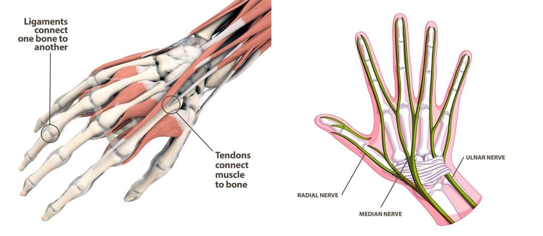
These tendons attach to muscles that straighten the fingers and lift the toes and the top of the foot. Extensor tendonitis is a condition characterized by inflammation of the tendons on the tops of the hands and feet. It often results from overuse of the muscles and from wearing tight shoes. Symptoms tend to worsen with activity and improve with rest. The condition does not usually require surgery; treatments include medications, home remedies/activity modification, and physical therapy.
Types
Tendonitis can develop in any tendon of the extensor muscles. These tendons are long, thin bands of tissue that can be felt on the tops of the hands and feet. The structures attach to muscles on one end and bones of the fingers and toes on the other. The extensor tendons in the hands include: (American Society for Surgery of the Hand, 2024)
The extensor digitorum communis straightens the index, middle, ring, and small fingers.
The extensor digiti minimi straightens the small finger.
The extensor indicis proprius straightens the index finger.
The extensor pollicis longus and extensor pollicis brevis muscles move the thumb into the thumb-up position.
The extensor digitorum longus and extensor digitorum brevis muscles lift the second, third, fourth, and fifth toes.
The extensor hallucis longus lifts the big toe.
Tendonitis vs Tendinopathy
Chronic tendon pain can lead to a condition known as tendinopathy.
Tendonitis results from inflammation.
Tendinopathy is the degeneration/micro-tearing of a tendon that occurs with long-term overuse.
Knowing which condition you are dealing with is important because it requires different treatments.
Symptoms
The primary symptom is pain in the affected tendon/s. Individuals may also experience swelling, and the skin can become red or warm to the touch. Extensor tendonitis can cause pain when using the affected muscles, moving the hand or foot in the opposite direction, and stretching the tendons. Usually, the pain worsens when using the affected muscles and improves with rest.
Causes
Extensor tendonitis in the hands usually results from overuse, which causes inflammation. However, it can also result from trauma, such as falling on the hand or an injury during physical or sports activities. Common activities include: (Hanson Z. C., and Lourie G. M. 2022)
Manual labor work
Typing
Computer mouse use
Weightlifting
Gymnastics
Playing a musical instrument
Extensor tendonitis in the foot can also result from overuse activities like running, especially uphill. However, it can also occur from wearing overly tight or tightly laced shoes for physical activities like running or dancing. Less common causes include: (Arthritis Foundation, N.D.)
Medical conditions like diabetes or arthritis
Medication side effects
Infection
Joint deformities
Treatment
Extensor tendonitis usually improves with conservative treatment, which includes self-care, activity modification, physical therapy, and medication.
Medications
Individuals can treat inflammation with non-steroidal anti-inflammatory drugs NSAIDs like:
Ibuprofen
Naproxen
Aspirin
Acetaminophen can help reduce pain.
In some cases, individuals may need prescription anti-inflammatory medications like corticosteroids or pain relievers for short-term use.
Self-Care and Activity Adjustments
Self-care includes:
Rest and avoid any activities that increase pain symptoms. If you can’t avoid them completely, take frequent breaks to allow the muscles to relax.
Apply ice to the hand or foot several times daily for up to 20 minutes.
Compression wraps should be applied on the foot or hand using an elastic bandage or soft splint to help support the injured tendons and reduce swelling.
Elevate the hand or foot if swollen above the heart level when resting.
Activity Adjustments
Modifying activities can help address the underlying cause/s.
Hand extensor tendonitis can develop from poor positioning.
Setting up an ergonomic workstation can help.
Consult a coach or trainer if the tendonitis is related to sports or exercise.
Individuals might need to adjust their technique or training schedule to decrease pressure on the tendons.
Physical Therapy
Physical therapy is an effective treatment. A therapy team can help determine the condition’s underlying cause and provide a personalized treatment program. Interventions can include:
Pain-reducing treatments like ultrasound, electrical stimulation, electroacupuncture, and laser therapy.
Extensor tendonitis can take weeks or even months to fully heal. Early diagnosis and determining the condition’s underlying cause rather than just treating symptoms are recommended for a faster and optimal recovery. Injury Medical Chiropractic and Functional Medicine Clinic works with primary healthcare providers and specialists to develop personalized treatment programs through an integrated approach to treating injuries and chronic pain syndromes, improving flexibility, mobility, and agility, relieving pain, and helping individuals return to normal activities. Dr. Jimenez has teamed up with top surgeons, clinical specialists, medical researchers, and rehabilitation providers if other treatments are needed.
Move Better, Live Better, with Chiropractic
References
American Society for Surgery of the Hand. (2024). Tendons. https://www.assh.org/handcare/safety/tendons#Finger%20Extensor
Hanson, Z. C., & Lourie, G. M. (2022). Middorsal Wrist Pain in the High-Level Athlete: Causes, Treatment, and Early Return to Play. Orthopaedic journal of sports medicine, 10(4), 23259671221088610. https://doi.org/10.1177/23259671221088610
Arthritis Foundation. Foundation, A. (N.D.). Tendinitis. https://www.arthritis.org/diseases/tendinitis
Bronner, S., Ojofeitimi, S., & Rose, D. (2008). Repair and rehabilitation of extensor hallucis longus and brevis tendon lacerations in a professional dancer. The Journal of orthopaedic and sports physical therapy, 38(6), 362–370. https://doi.org/10.2519/jospt.2008.2749
American Society for Surgery of the Hand. (2014). Extensor tendon injury. https://www.assh.org/handcare/condition/extensor-tendon-injury#:~:text=The%20tendon%20may%20take%20eight%20to%20twelve%20weeks,may%20include%20stitches%20%28for%20cuts%20in%20the%20tendon%29.
IFM's Find A Practitioner tool is the largest referral network in Functional Medicine, created to help patients locate Functional Medicine practitioners anywhere in the world. IFM Certified Practitioners are listed first in the search results, given their extensive education in Functional Medicine


 Extensor Tendonitis


