Functional Neurology: Obesity and Thyroid Dysfunction
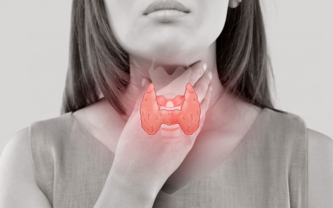
The endocrine system is made up of a collection of glands that release hormones that regulate a variety of bodily functions, including metabolism. The thyroid gland is a large, butterfly-shaped organ found in the center of the neck. The thyroid gland secretes three hormones, known as triiodothyronine (T3), thyroxine (T4), and calcitonin, in response to stimulation from the pituitary gland which secretes a compound, known as the thyroid-stimulating hormone (TSH). However, if the thyroid gland produces too little or too many hormones, it can ultimately cause a variety of health issues, including obesity. �
According to the American Association of Clinical Endocrinologists, approximately 27 million people in the United States have thyroid dysfunction. Healthcare professionals found a connection between diabetes and thyroid dysfunction. People with obesity and diabetes have an increased risk of developing thyroid dysfunction compared with the healthy population. Because the thyroid gland regulates metabolism, thyroid dysfunction can cause various metabolic health issues. Thyroid dysfunction can frequently occur due to iodine deficiency, autoimmune diseases, and surgery. �
How Thyroid Dysfunction Can Cause Obesity
Research studies demonstrated that thyroid dysfunction can ultimately lead to obesity. Understanding the connection between obesity and thyroid dysfunction can help reduce the risk of developing a variety of other health issues, including cancer. Thyroid dysfunction is associated with changes in body weight and composition, temperature, and energy. In a recent research study, 27,097 participants above 40 years of age with a body mass index (BMI) of at least 30.0 kg/m2, scientists found that thyroid dysfunction was associated with a higher BMI and an increased risk of developing obesity. �
Thyroid-stimulating hormone (TSH) levels are higher in people with obesity, according to research studies. Scientists also demonstrated that TSH levels appear to be closely associated with the degree of obesity and BMI. Surprisingly, research studies also found increased T3 levels in participants with obesity. Progressive fat accumulation was associated with an increase in TSH and T3 levels, regardless of insulin resistance and metabolic parameters. The ratio between T3 and T4 was also associated with both BMI and waist circumference in people with obesity, according to the research studies. �
Although people with obesity have increased TSH levels, their TSH receptors are frequently less expressed compared with healthy people. The reduced TSH receptor expression can ultimately cause thyroid dysfunction, further increasing TSH and T3 levels. Fortunately, healthcare professionals demonstrated that weight loss can help regulate thyroid function. Thyroid dysfunction can be reversed following weight loss due to diet and lifestyle modifications or surgery. Weight loss can also cause a considerable reduction in both TSH and T3. The decrease in T3 levels during weight loss can also increase energy. �
According to research studies, reduced T3 levels can make it difficult to maintain or promote weight loss. Evidence suggesting that TSH and T3 levels increase in people with obesity while TSH and T3 levels are reduced during weight loss supports the hypothesis that changes in thyroid function in people with obesity may be reversible through weight loss. However, it’s frequently challenging to identify participants with obesity who are only affected by mild thyroid dysfunction. Healthcare professionals should suspect thyroid dysfunction in people with obesity that also have increased TSH levels. �
Healthcare professionals determined that evaluating the thyroid gland using ultrasound may not necessarily help diagnose possible thyroid dysfunction in people with obesity. As a matter of fact, the moderate increase in TSH levels is frequently associated with an increase in thyroid volume and hypoechogenicity, or the reduced response of an organ using ultrasound, with an ultrasound pattern that suggests Hashimoto thyroiditis. Furthermore, the increased hypoechogenicity in people with obesity is associated with increased cytokines and other inflammatory markers produced by adipose tissue. �
The increased cytokines and inflammatory markers can ultimately increase TSH levels, increasing the size of the thyroid and leading to vasodilatation and increased thyroid vessel permeability with increased parenchymal inhibition of the thyroid gland which may be responsible for the hypoechogenicity with ultrasound. Average TSH was demonstrated to be higher in people with obesity compared with healthy people. It is essential to understand that�an ultrasound pattern suggesting Hashimoto thyroiditis may also suggest autoimmune diseases associated with thyroid dysfunction and obesity. �

The endocrine system is made up of a collection of glands, such as the thyroid gland, which release several different types of hormones that regulate a variety of bodily functions, including metabolism. The thyroid gland is a large, butterfly-shaped organ found in the center of the neck and it plays a fundamental role in the secretion of three hormones, including triiodothyronine (T3), thyroxine (T4), and calcitonin, following stimulation from the pituitary gland, which secretes a compound known as the thyroid-stimulating hormone (TSH). However, thyroid dysfunction can ultimately cause a variety of health issues, including obesity. According to the American Association of Clinical Endocrinologists, approximately 27 million people in the United States have thyroid dysfunction. Because the thyroid gland regulates metabolism, thyroid dysfunction can also cause various metabolic health issues. Thyroid dysfunction can frequently occur due to iodine deficiency, autoimmune diseases, and surgery, according to research studies. Scientists demonstrated a connection between thyroid dysfunction and obesity. – Dr. Alex Jimenez D.C., C.C.S.T. Insight
The endocrine system is made up of a collection of glands that release hormones that regulate a variety of bodily functions, including metabolism. The thyroid gland is a large, butterfly-shaped organ found in the center of the neck. The thyroid gland secretes three hormones, known as triiodothyronine (T3), thyroxine (T4), and calcitonin, in response to stimulation from the pituitary gland which secretes a compound, known as thyroid-stimulating hormone (TSH). However, if the thyroid gland produces too little or too many hormones, it can ultimately cause a variety of health issues, including obesity. �
According to the American Association of Clinical Endocrinologists, approximately 27 million people in the United States have thyroid dysfunction. Healthcare professionals found a connection between diabetes and thyroid dysfunction. People with obesity and diabetes have an increased risk of developing thyroid dysfunction compared with the healthy population. Because the thyroid gland regulates metabolism, thyroid dysfunction can cause various metabolic health issues. Thyroid dysfunction can frequently occur due to iodine deficiency, autoimmune diseases, and surgery. �
The scope of our information is limited to chiropractic, musculoskeletal, and nervous health issues or functional medicine articles, topics, and discussions. We use functional health protocols to treat injuries or disorders of the musculoskeletal system. Our office has made a reasonable attempt to provide supportive citations and has identified the relevant research study or studies supporting our posts. We also make copies of supporting research studies available to the board and or the public upon request. To further discuss the subject matter above, please feel free to ask Dr. Alex Jimenez or contact us at 915-850-0900.�
Curated by Dr. Alex Jimenez �
References:
- Doheny, Kathleen. �Does Underactive Thyroid Lead To Weight Gain-Or Vice-Versa?� EndocrineWeb, 16 Dec. 2019, www.endocrineweb.com/news/obesity/55275-does-underactive-thyroid-lead-weight-gain-vice-versa.
- Biondi, Bernadette. �Thyroid and Obesity: An Intriguing Relationship.� OUP Academic, Oxford University Press, 1 Aug. 2010, academic.oup.com/jcem/article/95/8/3614/2596481.
- Jacques, Jacqueline. �The Role of Your Thyroid in Metabolism and Weight Control.� Obesity Action Coalition, 2009, www.obesityaction.org/community/article-library/the-role-of-your-thyroid-in-metabolism-and-weight-control/.
�
Additional Topic Discussion: Chronic Pain
Sudden pain is a natural response of the nervous system which helps to demonstrate possible injury. By way of instance, pain signals travel from an injured region through the nerves and spinal cord to the brain. Pain is generally less severe as the injury heals, however, chronic pain is different than the average type of pain. With chronic pain, the human body will continue sending pain signals to the brain, regardless if the injury has healed. Chronic pain can last for several weeks to even several years. Chronic pain can tremendously affect a patient’s mobility and it can reduce flexibility, strength, and endurance. �
Neural Zoomer Plus for Neurological Disease
 Dr. Alex Jimenez utilizes a series of tests to help evaluate neurological diseases. The Neural ZoomerTM Plus is an array of neurological autoantibodies which offers specific antibody-to-antigen recognition. The Vibrant Neural ZoomerTM Plus is designed to assess an individual�s reactivity to 48 neurological antigens with connections to a variety of neurologically related diseases. The Vibrant Neural ZoomerTM Plus aims to reduce neurological conditions by empowering patients and physicians with a vital resource for early risk detection and an enhanced focus on personalized primary prevention. �
Food Sensitivity for the IgG & IgA Immune Response

Dr. Alex Jimenez utilizes a series of tests to help evaluate health issues associated with a variety of food sensitivities and intolerances. The Food Sensitivity ZoomerTM is an array of 180 commonly consumed food antigens that offers very specific antibody-to-antigen recognition. This panel measures an individual�s IgG and IgA sensitivity to food antigens. Being able to test IgA antibodies provides additional information to foods that may be causing mucosal damage. Additionally, this test is ideal for patients who might be suffering from delayed reactions to certain foods. Utilizing an antibody-based food sensitivity test can help prioritize the necessary foods to eliminate and create a customized diet plan around the patient�s specific needs. �
Gut Zoomer for Small Intestinal Bacterial Overgrowth (SIBO)

Dr. Alex Jimenez utilizes a series of tests to help evaluate gut health associated with small intestinal bacterial overgrowth (SIBO). The Vibrant Gut ZoomerTM offers a report that includes dietary recommendations and other natural supplementation like prebiotics, probiotics, and polyphenols. The gut microbiome is mainly found in the large intestine and it has more than 1000 species of bacteria that play a fundamental role in the human body, from shaping the immune system and affecting the metabolism of nutrients to strengthening the intestinal mucosal barrier (gut-barrier). It is essential to understand how the number of bacteria that symbiotically live in the human gastrointestinal (GI) tract influences gut health because imbalances in the gut microbiome may ultimately lead to gastrointestinal (GI) tract symptoms, skin conditions, autoimmune disorders, immune system imbalances, and multiple inflammatory disorders. �


Formulas for Methylation Support

XYMOGEN�s Exclusive Professional Formulas are available through select licensed health care professionals. The internet sale and discounting of XYMOGEN formulas are strictly prohibited.
Proudly,�Dr. Alexander Jimenez makes XYMOGEN formulas available only to patients under our care.
Please call our office in order for us to assign a doctor consultation for immediate access.
If you are a patient of Injury Medical & Chiropractic�Clinic, you may inquire about XYMOGEN by calling 915-850-0900.
![]() �
�
For your convenience and review of the XYMOGEN products please review the following link. *XYMOGEN-Catalog-Download �
* All of the above XYMOGEN policies remain strictly in force. �
� �
Modern Integrated Medicine
The National University of Health Sciences is an institution that offers a variety of rewarding professions to attendees. Students can practice their passion for helping other people achieve overall health and wellness through the institution’s mission. The National University of Health Sciences prepares students to become leaders in the forefront of modern integrated medicine, including chiropractic care. Students have an opportunity to gain unparalleled experience at the National University of Health Sciences to help restore the natural integrity of the patient and define the future of modern integrated medicine. �


 Obesity is a complex, multifactorial disease, and better understanding of the mechanisms underlying the interactions between lifestyle, environment, and genetics is critical for developing effective strategies for prevention and treatment [1].
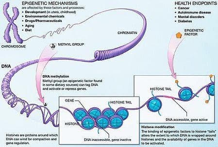
 Animal models provide unique opportunities for highly controlled studies that provide mechanistic insight into�the role of specific epigenetic marks, both as indicators of current metabolic status and as predictors of the future risk of obesity and metabolic disease. A particularly important aspect of animal studies is that they allow for the assessment of epigenetic changes within target tissues, including the liver and hypothalamus, which is much more difficult in humans. Moreover, the ability to harvest large quantities of fresh tissue makes it possible to assess multiple chromatin marks as well as DNA methylation. Some of these epigenetic modifications either alone or in combination may be responsive to environmental programming. In animal models, it is also possible to study multiple generations of offspring and thus enable differentiation between trans-generational and intergenerational transmission of obesity risk mediated by epigenetic memory of parental nutritional status, which cannot be easily distinguished in human studies. We use the former term for meiotic transmission of risk in the absence of continued exposure while the latter primarily entails direct transmission of risk through metabolic reprogramming of the fetus or gametes.
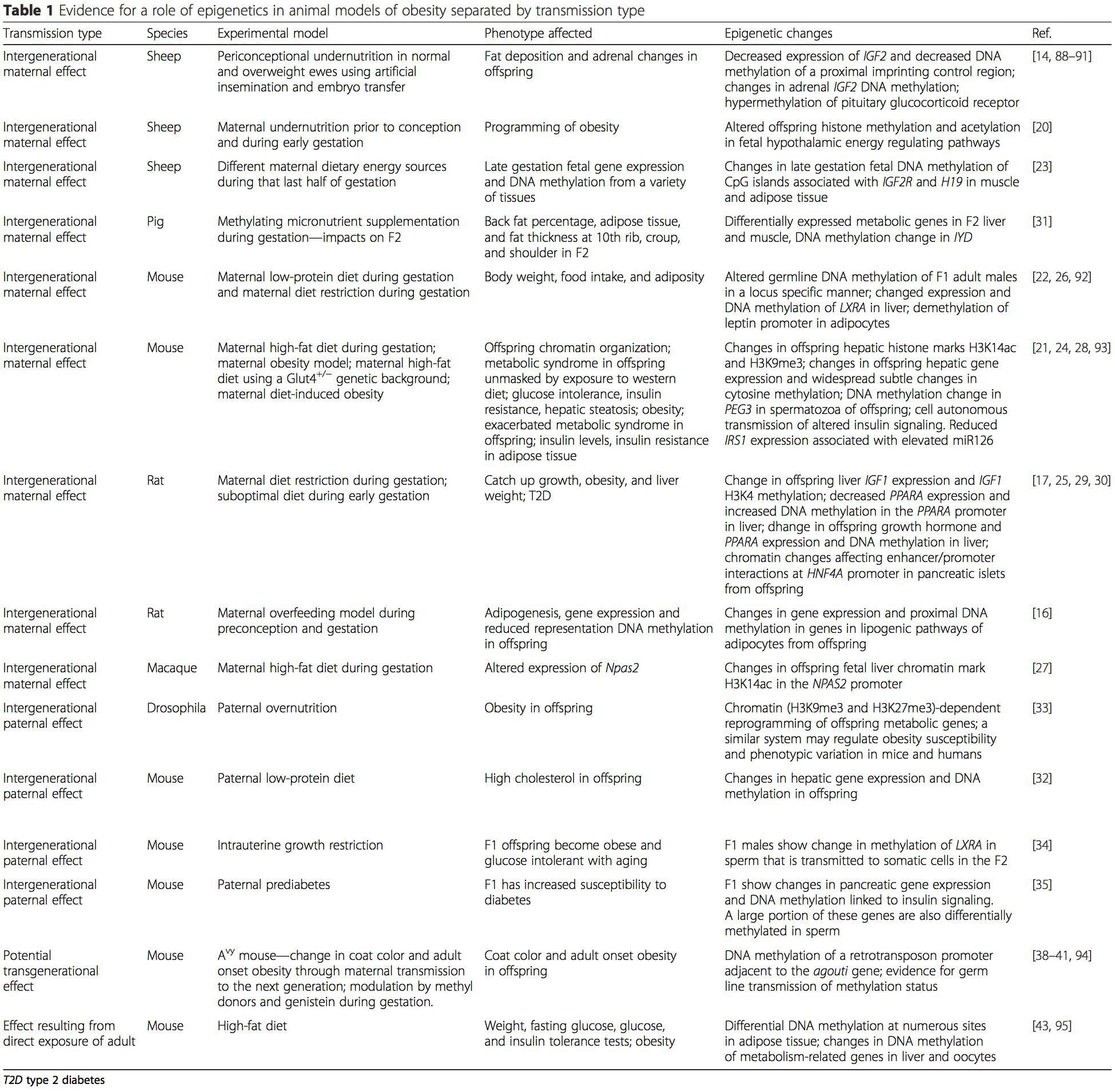 (i) Epigenetic Changes In Offspring Associated With Maternal Nutrition During Gestation
 Maternal nutritional supplementation, undernutrition, and over nutrition during pregnancy can alter fat deposition and energy homeostasis in offspring [11, 13�15, 19]. Associated with these effects in the offspring are changes in DNA methylation, histone post-translational modifications, and gene expression for several target genes,�especially genes regulating fatty acid metabolism and insulin signaling [16, 17, 20�30]. The diversity of animal models used in these studies and the common metabolic pathways impacted suggest an evolutionarily conserved adaptive response mediated by epigenetic modification. However, few of the specific identified genes and epigenetic changes have been cross-validated in related studies, and large-scale genome-wide investigations have typically not been applied. A major hindrance to comparison of these studies is the different develop mental windows subjected to nutritional challenge, which may cause considerably different outcomes. Proof that the epigenetic changes are causal rather than being associated with offspring phenotypic changes is also required. This will necessitate the identification of a parental nutritionally induced epigenetic �memory� response that precedes development of the altered phenotype in offspring.
 Emerging studies have demonstrated that paternal plane of nutrition can impact offspring fat deposition and epigenetic marks [31�34]. One recent investigation using mice has demonstrated that paternal pre-diabetes leads to increased susceptibility to diabetes in F1 offspring with associated changes in pancreatic gene expression and DNA methylation linked to insulin signaling [35]. Importantly, there was an overlap of these epigenetic changes in pancreatic islets and sperm suggesting germ line inheritance. However, most of these studies, although intriguing in their implications, are limited in the genomic scale of investigation and frequently show weak and somewhat transient epigenetic alterations associated with mild metabolic phenotypes in offspring.
 Stable transmission of epigenetic information across multiple generations is well described in plant systems and C. elegans, but its significance in mammals is still much debated [36, 37]. An epigenetic basis for grand- parental transmission of phenotypes in response to dietary exposures has been well established, including in livestock species [31]. The most influential studies demonstrating effects of epigenetic transmission impacting offspring phenotype have used the example of the viable yellow agouti (Avy) mouse [38]. In this mouse, an insertion of a retrotransposon upstream of the agouti gene causes its constitutive expression and consequent yellow coat color and adult onset obesity. Maternal transmission through the germ line results in DNA methylation�mediated silencing of agouti expression resulting in wild-type coat color and lean phenotype of the offspring [39, 40]. Importantly, subsequent studies in these mice demonstrated that maternal exposure to methyl donors causes a shift in coat color [41]. One study has reported transmission of a phenotype to the F3 generation and alterations in expression of large number of genes in response to protein restriction in F0 [42]; however, alterations in expression were highly variable and a direct link to epigenetic changes was not identified in this system.
 While many studies have identified diet-associated epigenetic changes in animal models using candidate site-specific regions, there have been few genome-wide analyses undertaken. A recent study focussed on determining the direct epigenetic impact of high-fat diets/ diet-induced obesity in adult mice using genome-wide gene expression and DNA methylation analyses [43]. This study identified 232 differentially methylated regions (DMRs) in adipocytes from control and high-fat fed mice. Importantly, the corresponding human regions for the murine DMRs were also differentially methylated in adipose tissue from a population of obese and lean humans, thereby highlighting the remarkable evolutionary conservation of these regions. This result emphasizes the likely importance of the identified DMRs in regulating energy homeostasis in mammals.
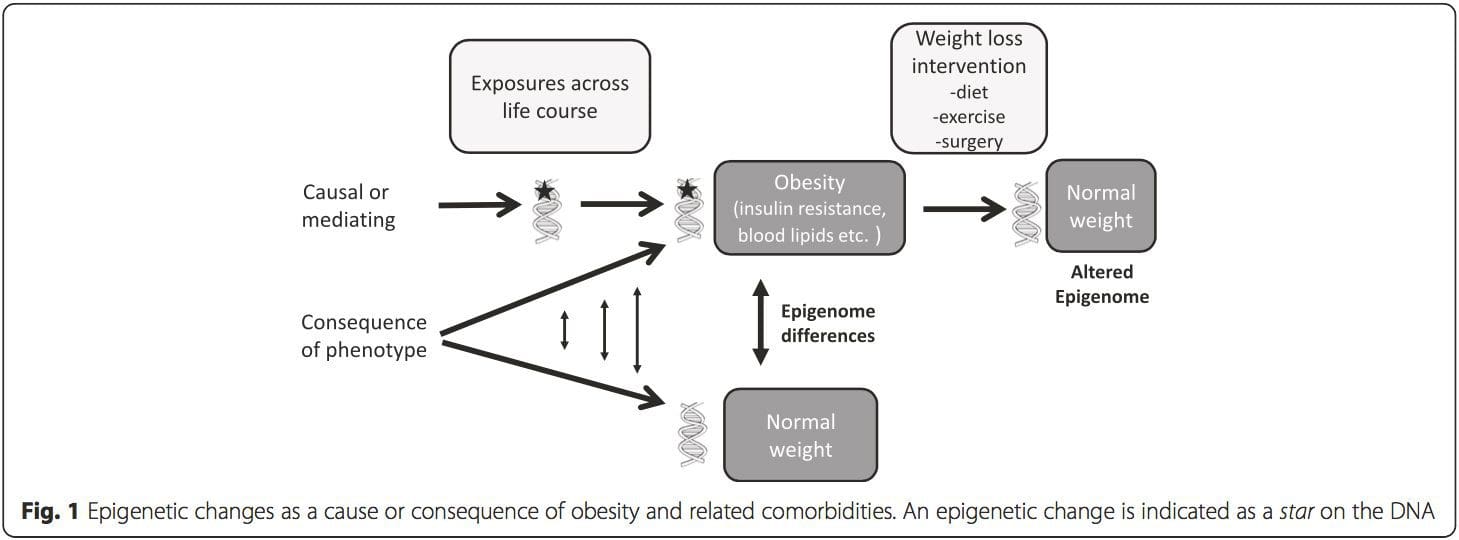 (i) Genetic association studies. Genetic polymorphisms that are associated with an increased risk of developing particular conditions are a priori linked to the causative genes. The presence of differential�methylation in such regions infers functional relevance of these epigenetic changes in controlling expression of the proximal gene(s). There are strong cis-acting genetic effects underpinning much epigenetic variation [7, 45], and in population-based studies, methods that use genetic surrogates to infer a causal or mediating role of epigenome differences have been applied [7, 46�48]. The use of familial genetic information can also lead to the identification of potentially causative candidate regions showing phenotype-related differential methylation [49].
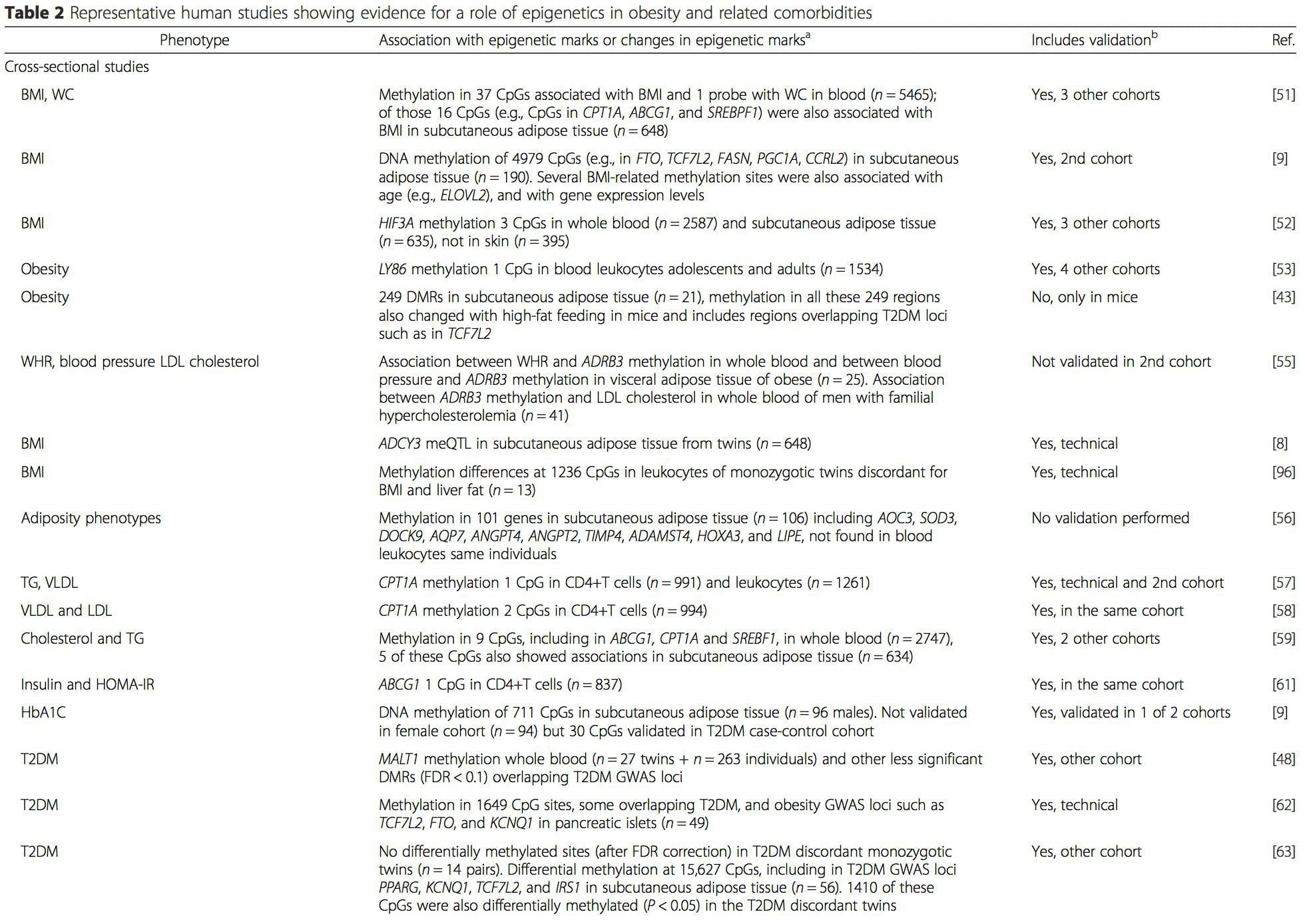
 From these studies, altered methylation of PGC1A, HIF3A, ABCG1, and CPT1A and the previously described RXRA [18] have emerged as biomarkers associated with, or perhaps predictive of, metabolic health that are also plausible candidates for a role in development of metabolic disease.
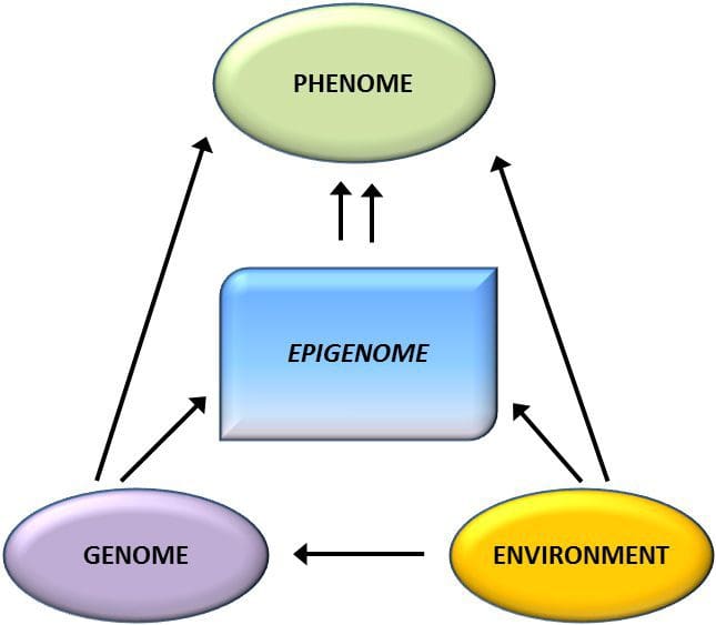 Epigenetic variation is highly influenced by the underlying genetic variation, with genotype estimated to explain ~20�40 % of the variation [6, 8]. Recently, a number of studies have begun to integrate methylome and genotype data to identify methylation quantitative trait loci (meQTL) associated with disease phenotypes. For instance, in adipose tissue, an meQTL overlapping�with a BMI genetic risk locus has been identified in an enhancer element upstream of ADCY3 [8]. Other studies have also identified overlaps between known obesity and T2DM risk loci and DMRs associated with obesity and T2DM [43, 48, 62]. Methylation of a number of such DMRs was also modulated by high-fat feeding in mice [43] and weight loss in humans [64]. These results identify an intriguing link between genetic variations linked with disease susceptibility and their association with regions of the genome that undergo epigenetic modifications in response to nutritional challenges, implying a causal relationship. The close connection between genetic and epigenetic variation may signify their essential roles in generating individual variation [65, 66]. However, while these findings suggest that DNA methylation may be a mediator of genetic effects, it is also important to consider that both genetic and epigenetic processes could act independently on the same genes. Twin studies [8, 63, 67] can provide important insights and indicate that inter-individual differences in levels of DNA methylation arise predominantly from non-shared environment and stochastic influences, minimally from shared environmental effects, but also with a significant impact of genetic variation.
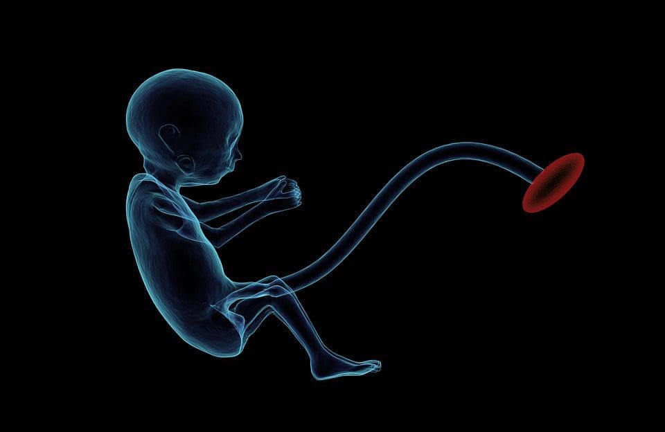 Prenatal environment: Two recently published studies made use of human populations that experienced �natural� variations in nutrient supply to study the impact of maternal nutrition before or during pregnancy on DNA methylation in the offspring [68, 69]. The first study used a Gambian mother-child cohort to show that both seasonal variations in maternal methyl donor intake during pregnancy and maternal pre-pregnancy BMI were associated with altered methylation in the infants [69]. The second study utilized adult offspring from the Dutch Hunger Winter cohort to investigate the effect of prenatal exposure to an acute period of severe maternal undernutrition on DNA methylation of genes involved in growth and metabolism in adulthood [68]. The results highlighted the importance of the timing of the exposure in its impact on the epigenome, since significant epigenetic effects were only identified in individuals exposed to famine during early gestation. Importantly, the epigenetic changes occurred in conjunction with increased BMI; however, it was not possible to establish in this study whether these changes were present earlier in life or a consequence of the higher BMI.
 Postnatal environment: The epigenome is established de novo during embryonic development, and therefore, the prenatal environment most likely has the most significant impact on the epigenome. However, it is now clear that changes do occur in the �mature� epigenome under the influence of a range of conditions, including aging, exposure to toxins, and dietary alterations. For example, changes in DNA methylation in numerous genes in skeletal muscle and PGC1A in adipose tissue have been demonstrated in response to a high-fat diet [75, 76]. Interventions to lose body fat mass have also been associated with changes in DNA methylation. Studies have reported that the DNA methylation profiles of adipose tissue [43, 64], peripheral blood mononuclear cells [77], and muscle tissue [78] in formerly obese patients become more similar to the profiles of lean subjects following weight loss. Weight loss surgery also partially reversed non-alcoholic fatty liver disease-associated methylation changes in liver [79] and in another study led to hypomethylation of multiple obesity candidate genes, with more pronounced effects in subcutaneous compared to omental (visceral) fat [64]. Accumulating evidence suggests that exercise interventions can also influence DNA methylation. Most of these studies have been conducted in lean individuals [80�82], but one exercise study in obese T2DM subjects also demonstrated changes in DNA methylation, including in genes involved in fatty acid and glucose transport [83]. Epigenetic changes also occur with aging, and recent data suggest a role of obesity in augmenting them [9, 84, 85]. Obesity accelerated the epigenetic age of liver tissue, but in contrast to the findings described above, this effect was not reversible after weight loss [84].
 DNA methylation changes associated with obesity or induced by diet or lifestyle interventions and weight loss are generally modest (<15 %), although this varies depending on the phenotype and tissue studied. For instance, changes greater than 20 % have been reported in adipose tissue after weight loss [64] and associations between HIF3A methylation and BMI in adipose tissue were more pronounced than in blood [52].
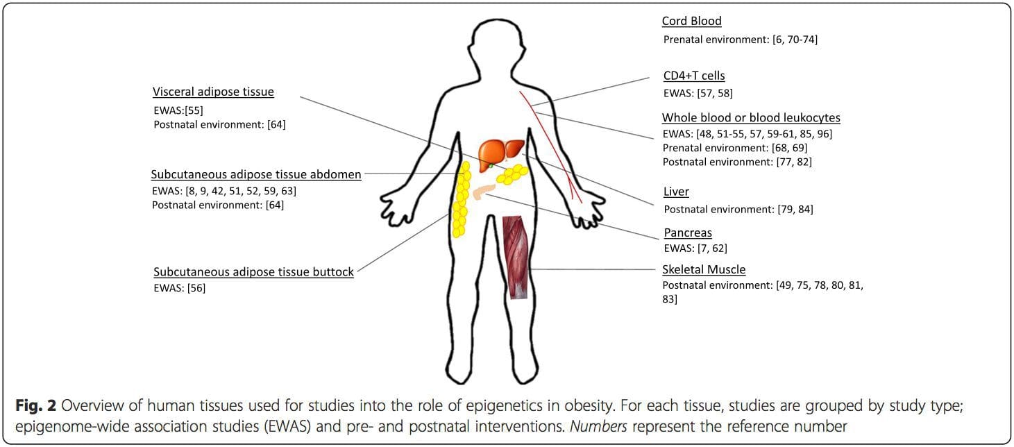 Conclusions


