Neural cell death can occur both during the development and throughout the pathophysiology of the nervous system. Two different types of cell death, known as necrosis and apoptosis, are involved in pathological neuronal loss, however, apoptosis is the process of programmed cell death during development. All types of cells will go through apoptosis. This mechanism controls neuronal growth where an excess of neurons is produced and only those which form connections with the target structures will receive enough survival factors. The remaining neurons will then ultimately go through death and removal. �
Apoptosis continues throughout life and it is the main process involved in the elimination of surplus, unwanted, damaged or aged cells. Dysregulation of apoptosis is demonstrated after damage or injury as well as in neurodegeneration and in tumorigenesis. Treatment approaches which influence the apoptotic pathway offer valuable therapeutic options in a wide variety of pathological states. The purpose of the article is to describe the significance of apoptosis in neurological diseases. �
What is Apoptosis?
Apoptosis is the well-conserved and highly controlled process of cell death involved in the removal of unnecessary, surplus, aged or damaged cells. Dysregulation of apoptosis can ultimately develop mutated cells which can result in malformations, autoimmune diseases, and even cancer. Abnormal apoptosis can also result in the elimination of healthy cells which can occur in health issues such as infection, hypoxic-ischaemic injury, neurodegenerative or neuromuscular diseases, and AIDS. �
Apoptosis is different from necrotic cell death. In necrosis, cell death is caused by an external factor and involves the early loss of tissue, damage to organs, and the leakage of cytoplasmic contents, leading to the recruitment of phagocytes which can cause an acute inflammatory reaction. In contrast, apoptosis is often considered cell suicide. According to research studies, cells which die due to apoptosis retain membrane and organelle structure and function until late in the process while still developing plasma membrane blebbing, reduced cytoplasmic volume, chromatin condensation, and nuclear fragmentation. �
In the final phases, cell fragments wrapped in plasma membrane pull away as apoptotic bodies which are then phagocytosed by healthy cells. The removal of cell debris also occurs in the absence of an inflammatory response, and this silent, quick, and efficient elimination of apoptotic cells mean that apoptosis can be difficult to find in cells. However, as many as 50 percent of the cells in developing adulthood may go through apoptosis where less than 1 percent of cells are apoptotic at any one time. �
Apoptosis in the Nervous System
Programmed cell death by apoptosis occurs in several developmental processes, such as body sculpting and removal of self-reacting resistant cells as well as sexual organ growth and gamete formation. The general principle of growth in multicellular organisms involves the development of excess numbers of cells, where the excess or unwanted cells are then removed by apoptosis through the development of functional organs. In the developing nervous system, apoptosis has been demonstrated to occur in neural tube formation and continues throughout terminal differentiation of the neural system. �
A growing number of neurotrophic factors, such as nerve growth factor family, including both the neurokines and development factors like insulin-like growth variables (IGF-I and IGF-II), encourage the survival of several types of neurons. Targeted disruption of genes encoding these factors or their receptors demonstrate that neurotrophic factors are significant for the development of specific neuronal populations. Neurotrophic factors function by binding to specific receptors in the cell membrane. Moreover, the effects of NGF offer a good illustration of the subtle command the system permits. �
The nerve growth factor receptor has high and low affinity components. It will function as a survival factor if it binds to the high-affinity trkA receptor but it will also cause apoptosis of retinal neurons or oligodendrocytes once it binds to the low-affinity receptor p75 in the absence of trkA. Nerve growth factor in the extracellular environment is consequently able to control neural development by both boosting the growth of several types of cells as well as the removal of other cells. �
In some cases, however, concentrated genetic disruption of neurotrophic factors or their receptors may leave the central nervous system seemingly unaffected, demonstrating that these variables can ultimately become biased. According to research studies, it has now become evident that the control of neuronal survival does not only depend on the supply of trophic molecules by the targets but also on activity, humoral factors, and trophic support from glia or glial cells. �
Furthermore, neurons don’t simply undergo programmed cell death during differentiation. Apoptosis appears to regulate cell numbers in systems as diverse as the disappearance of the germinal layer during the third trimester of pregnancy, the sexual differentiation of the medial preoptic nucleus where apoptosis is controlled by testosterone, lineages throughout the olfactory epithelium, oligodendrocyte development in the optic nerve, and the development of Schwann cells in the peripheral nervous system. Programmed cell death occurs in a variety of other processes in the developing nervous system. �
Apoptosis in Nervous System Injuries & Diseases
Although apoptosis is a fundamental process involved in the developing nervous system, apoptosis can ultimately be involved in a variety of nervous system injuries and diseases. In most cases, the connection between a specific mutation or trauma as well as the activation of apoptotic cascades remains evasive. An overview of a developing list of neurological diseases in which apoptosis has been implicated as a significant pathological mechanism is provided below. �
Neuronal Injury
Cerebral hypoxic-ischaemic injury is a cause of neurological injury and death. Magnetic resonance spectroscopy studies have demonstrated that transient hypoxia-ischemia contributes to a biphasic disturbance of cerebral energy metabolism. Related to the biphasic energy collapse, two waves of cell death appear to follow hypoxic-ischaemic injury in the developing brain. Immediate neuronal death is most likely due to necrosis resulting from the accumulation of calcium ions. �
Delayed cell death caused by hypoxic-ischemic injury appears to involve further mechanisms with increasing data which demonstrates that in the delayed phase, cell death occurs by apoptosis. The amount of apoptosis is directly associated with the magnitude of ATP depletion during hypoxia-ischemia. Apoptosis can occur in the brains of newborn babies following birth asphyxia and sudden intrauterine death. Apoptosis can also be notable in white matter injury in newborn babies. �
Apoptosis may continue for months after an hypoxic-ischaemic injury due to constant changes in cerebral energy metabolism in infants during the months after birth asphyxia. Following focal neural injury, apoptosis has been discovered in remote regions from the initial damage. After severe spinal cord injury in reptiles, apoptosis of oligodendrocytes occurs in distant degenerating fiber tracts and after forebrain injury in rats, apoptosis was demonstrated in the cerebellum. �
The apoptotic loss of oligodendrocytes could consequently be a potential source of secondary demyelination in paraplegia and in the chronic degeneration related to multiple sclerosis. Further research studies must be performed in order to provide further evidence on the role of apoptosis in this type of injury which begins from the report of which Bcl-2 expression boosts the growth and regeneration of retinal axons. Apoptosis in neuronal injury can be demonstrated in a variety of ways. �
Neural Cancers
A connection between apoptosis and the cell cycle is demonstrated in carcinogenesis where proto-oncogenes, such as c-fos, c-jun, and c-myc, can activate apoptosis and promote cell division while inactivation of the pro-apoptotic p53 tumor suppressor gene is a frequent mark of human neoplasia. By way of instance, in a number of gliomas, the reduction of wild p53 activity was connected to tumor progression, possibly leading to resistance to chemotherapy and radiotherapy. �
Although there have been reports of Bcl-2 overexpression in glioma cell lines, the correlation between the anti-apoptotic effect of this gene and malignancy is not yet clear. However, a homolog of Bcl-2, the brain associated apoptosis gene (BRAG-1), is found predominantly in the brain, and it is upregulated in human gliomas as a rearranged transcript. As demonstrated above, the process of apoptosis can also be significant in the development of neural cancers, according to research studies. �
Infectious Disease
Apoptosis may play a role in HIV encephalopathy. In the brain, the virus reproduces primarily in microglia which it enters through the CD4 receptor. Although the activation of microglia is believed to be the main reason for adrenal loss and demyelination, neurons die by apoptosis in HIV encephalopathies because of HIV mediated alterations in astrocyte function and aberrant stimulation of NMDA receptors or due to nitric oxide from the activation of inducible nitric oxide synthase. �
In subacute sclerosing panencephalitis, widespread apoptotic death was demonstrated to develop in the brain, although no correlation was observed between viral load, lymphocyte infiltration, and the number of apoptotic cells. DNA fragmentation indicative of apoptosis was detected in scrapie-infected sheep and mice brains, suggesting a function associated with cell death in spongiform encephalopathies. Apoptosis may also ultimately be involved in another infectious disease. �
Neurodegeneration
Spinal muscular atrophy is associated with mutations in the survival of motor neuron and neuronal apoptosis inhibitory protein (NAIP) enzymes. NAIP is closely related to the baculovirus inhibitor of apoptosis protein and inhibits apoptosis in many cell types. This implies that mutations in NAIP could deregulate apoptosis in spinal motor nerves, causing their death. Recent studies emphasize the importance of anti-apoptotic genes in cerebral protection which can rescue neurons. �
Apoptosis has also been implicated in retinal dystrophies such as retinitis pigmentosa. In this case, apoptosis results from mutations in the three photoreceptor genes, rhodopsin, peripherin, and the ?-subunit of cyclic guanosine monophosphate di esterase, resulting in photoreceptor degeneration. The absence of c-fos prevents apoptosis in those cells is unknown. Moreover, defined neurotrophins and growth factors injected intraocularly in animal models of retinal degeneration improve photoreceptor survival, suggesting that the apoptotic cascade can be obstructed by supplying exogenous survival signs. �
The mutation underlying Huntington’s disease is an expanded trinucleotide which is fundamental for normal development and can be regarded as a cell survival gene. Transgenic models demonstrated increased apoptosis in the neurons of an embryonic neuroectoderm. During apoptosis, caspase-3 (apopain) is improved by a gain of function associated with the triplet expansion. This is supported by the overexpression of specific trinucleotide repeats in transgenic mice. �
Most cerebellar ataxias are associated with neuronal loss. Ataxia-telangiectasia, caused by mutations in the ATM gene, is considered to have an apoptotic component. ATM shares extensive and significant homology with the DNA dependent protein kinases involved in DNA damage responses at different cell cycle checkpoints and is downregulated in most patients with ataxia-telangiectasia. The simple fact that inappropriate p53 mediated apoptosis is the major cause of death in ataxia-telangiectasia cells suggests that the mutation causes improper triggering of apoptosis by otherwise non-lethal DNA injury. �
From the familial form of amyotrophic lateral sclerosis gain of function, mutations in the gene encoding copper-zinc superoxide dismutase (sod-1) develop a dominant pro-apoptotic sign. Although cell harm by the accumulation of free radicals can trigger apoptosis, these mutants can induce apoptosis both in nerve cells in culture and in transgenic mice. Mental retardation in Down’s syndrome has also been associated with abnormal apoptosis. Although cortical neurons from fetal Down’s syndrome brains are different, they then degenerate and undergo apoptosis, according to research studies. �
Degeneration is blocked by treatment with free radical scavengers, suggesting that a defect in the metabolism of reactive oxygen species is the trigger for apoptosis. In Parkinson’s disease, the death of dopaminergic neurons in the substantia nigra was demonstrated to occur through apoptosis and may be obstructed by delivery of glial-derived neurotrophic factor. Alzheimer’s disease is associated with the progressive accumulation of ?-amyloid protein which is the fundamental component of neural plaques. The ?-amyloid peptide can cause neurons to undergo apoptosis in vitro research studies. �
Inherited Metabolic Disease
Furthermore, few data suggest that the acute encephalopathy associated with maple syrup urine disease is because of the induction of apoptosis by an accumulating metabolite of leucine, ?-keto isocaproic acid. This compound is a potent inducer of apoptosis in central nervous system glial cells and the result is significantly enhanced in the presence of leucine. Phenylalanine and leucine do not induce apoptosis in this system, suggesting that this result is ultimately unique. �
There are two ways in which a cell can die, necrosis and apoptosis. While necrosis occurs due to an external factor which harms the cell, apoptosis follows a controlled, predictable routine. Apoptosis is generally known as programmed cell death. Apoptosis, or programmed cell death, has many fundamental functions in the developing structures of the human body, however, research studies have demonstrated that abnormal apoptosis can be associated with the development of a variety of neurological diseases. – Dr. Alex Jimenez D.C., C.C.S.T. Insight
The purpose of the article above is to discuss the process of apoptosis, or cell death, in neurodegenerative diseases. Neurological diseases are associated with the brain, the spine, and the nerves. The scope of our information is limited to chiropractic, musculoskeletal and nervous health issues as well as functional medicine articles, topics, and discussions. To further discuss the subject matter above, please feel free to ask Dr. Alex Jimenez or contact us at 915-850-0900 . �
Curated by Dr. Alex Jimenez �
Additional Topic Discussion: Chronic Pain
Sudden pain is a natural response of the nervous system which helps to demonstrate possible injury. By way of instance, pain signals travel from an injured region through the nerves and spinal cord to the brain. Pain is generally less severe as the injury heals, however, chronic pain is different than the average type of pain. With chronic pain, the human body will continue sending pain signals to the brain, regardless if the injury has healed. Chronic pain can last for several weeks to even several years. Chronic pain can tremendously affect a patient’s mobility and it can reduce flexibility, strength, and endurance.
Formulas for Methylation Support
XYMOGEN�s Exclusive Professional Formulas are available through select licensed health care professionals. The internet sale and discounting of XYMOGEN formulas are strictly prohibited.
Proudly,�Dr. Alexander Jimenez makes XYMOGEN formulas available only to patients under our care.
Please call our office in order for us to assign a doctor consultation for immediate access.
If you are a patient of Injury Medical & Chiropractic�Clinic, you may inquire about XYMOGEN by calling 915-850-0900.
�
For your convenience and review of the XYMOGEN products please review the following link.*XYMOGEN-Catalog-Download �
* All of the above XYMOGEN policies remain strictly in force. �
For many years, most neuroscientists believed we were born with all the neurons we were ever going to carry in our brains. As children, we may develop new neurons to help create the pathways, known as neural circuits, which function as information highways between different regions of the brain. However, scientists believed that after a neural circuit was created, developing any new neurons could interrupt the flow of information and disable the brain’s communication system. �
Introduction to Brain Basics
In 1962, scientist Joseph Altman questioned this belief when he saw evidence of neurogenesis, or the birth of neurons, in a region of an adult rat’s brain known as the hippocampus. He then reported that newborn neurons migrated from their birthplace in the hippocampus to other regions of the brain. In 1979, another scientist, Michael Kaplan, proved Altman’s findings in the rat brain and in 1983, Kaplan found neural precursor cells in the forebrain of an adult monkey. �
In the early 1980s, a scientist attempting to explain how birds learn how to sing suggested that neuroscientists should once again analyze neurogenesis in the adult brain and start to determine how it can make sense. In several experiments, Fernando Nottebohm and his team revealed that the numbers of neurons in the forebrains of male canaries tremendously increased during the mating season. This was the same time in which the birds had to learn new songs to attract females. �
However, why did these bird’s brains create new neurons during such a vital time in learning? Nottebohm believed it was because new neurons helped keep new song patterns inside the neural tissues of the forebrain, or the region of the brain which regulates complex behaviors. These new neurons made learning possible. If birds developed new neurons to help them remember and learn new song patterns, Nottebohm believed that the brains of mammals may also be able to do the same. �
Elizabeth Gould discovered evidence of newborn neurons in a different region of the brain in monkeys. Fred Gage and Peter Eriksson also demonstrated that the adult human brain developed new neurons in a similar region. For several neuroscientists, neurogenesis in the adult brain is still an unproven theory. However, other neuroscientists believe that the evidence provides interesting possibilities associated with the role of adult-generated neurons in memory and learning. �
Architecture of the Neuron
The central nervous system, which includes the brain and the spinal cord, consists of two primary types of cells: the neurons and the glia. Glia outnumber neurons in several regions of the brain, however, neurons are the key structures in the brain. Neurons are information messengers. They utilize electrical impulses and chemical signals to transfer information between different regions of the brain and between the brain and the rest of the nervous system. Everything we think, feel, and do would be impossible without the utilization of neurons and the glial cells, known as astrocytes and oligodendrocytes. �
Neurons have three primary parts including a cell body and two extensions known as an axon and a dendrite. Within the cell body is a nucleus, which regulates the cell’s activities and holds the cell’s genetic material. The axon is characterized by a very long tail and it transfers messages from the cell. Dendrites are characterized similar to that of the branches of a tree and they receive messages from the cell. Neurons communicate with one another by sending chemicals, known as neurotransmitters, across a very small region, known as a synapse, found between the axons and the dendrites of adjacent neurons. � There are three types of neurons: �
Sensory neurons: Transfer information from the sense organs, such as the eyes and ears, to the brain.
Motor neurons: Manage voluntary muscle activity and transfer messages from nerve cells in the brain to muscles.
All other neurons are known as interneurons.
Scientists believe that neurons are the most varied type of cell in the human body. Within these three types of neurons are hundreds of different types of neurons, each with specific message-carrying abilities. The way these neurons communicate with one another by establishing connections is ultimately what makes people unique in how we think, feel, and act. �
Birth of the Neuron
The range to which new neurons are created in the brain has been a controversial topic among neuroscientists for many years. Meanwhile, although nearly all neurons are currently present in our brains by the time we’re born, there’s recent evidence to support that neurogenesis, or the scientific word utilized to describe the birth of neurons, is a lifelong procedure. Neurons are born in regions of the brain which are full of neural precursor cells, known as neural stem cells. These cells have the potential to develop all, if not all, of the different types of neurons and glia found in the brain. Neuroscientists have discovered how neural precursor cells function in the laboratory. Although this may not be exactly how these cells behave when they are in the brain, it gives us data about how they may function when they are in the brain’s environment. �
The science of stem cells is still very recent and could ultimately change with further discoveries, however, researchers have discovered enough evidence to support as well as to be able to demonstrate how neural stem cells create the other cells of the brain. Neuroscientists refer to this as a stem cell’s lineage and it is similar in principle to the concept of a family tree. �
Neural stem cells increase by dividing into two and creating two new stem cells, two early progenitor cells, or one of each. When a stem cell divides to create another stem cell, it is believed to self-renew. This new cell has the potential to make more stem cells. When a stem cell divides to create an early progenitor cell, it is said to differentiate. Differentiation is when a new cell is more technical in structure and function. An early progenitor cell doesn’t have the potential of a stem cell to create several different types of cells. It can only make cells within their distinct lineage. Early progenitor cells may self-renew or go in either of two ways. One type will develop astrocytes. The other type will develop neurons or oligodendrocytes. �
Migration of the Neuron
Once a neuron is born, it must go to the region of the brain where it will function. But, how does a neuron understand where to go? And, what helps it get there? Neuroscientists have determined that neurons utilize two different methods to travel: �
Several neurons migrate by following the long fibers of cells known as radial glia. These fibers extend from the inner layers to the outer layers of the brain. Neurons glide along the fibers until they reach their destination.
Neurons also travel by using chemical signals. Scientists have found special molecules on the surface of neurons, known as adhesion molecules, which bind with similar molecules on nearby glial cells or nerve axons. These chemical signals will also ultimately help guide the neuron to its final destination in the brain.
Not all neurons are successful in their journey. Scientists believe that only one-third of these neurons will reach their destination. Some cells die during the process of neuronal growth. Some neurons may also survive, but end up where they don’t belong. Mutations in the genes which regulate migration create regions of misplaced or abnormal neurons which can cause disorders, such as epilepsy. Scientists believe that schizophrenia is partially caused by misguided neurons. �
Differentiation of the Neuron
When a neuron reaches its destination, then it must begin to perform its initial function. This final measure of differentiation is one of the most misunderstood sections of neurogenesis. Neurons are in charge of the transfer and uptake of neurotransmitters, or chemicals which deliver information between cells. Depending on its location, a neuron may perform the role of a sensory neuron, a motor neuron, or an interneuron, sending and receiving specific neurotransmitters. �
In the developing brain, a neuron depends on molecular signals from other cells, including astrocytes, to determine its form and location, the type of transmitter it creates, and to which other neurons it can connect. These newborn cells establish neural circuits, or data pathways that connect from neuron to neuron, which is determined during adulthood. However, in the mature brain, neural circuits are already developed and neurons must find a way to fit in. As a new neuron settles in, it starts to look like enclosing cells. It then develops an axon and dendrites and begins to communicate with its neighbors. �
Death of the Neuron
Although neurons are the longest living cells within the human body, large numbers of them often die during migration and differentiation. The lives of some neurons can sometimes take unexpected turns. Several health issues associated with the brain, the spinal cord, and the nerves are the consequence of the unnatural deaths of neurons and supporting cells. �
In Parkinson’s disease, neurons which create the neurotransmitter dopamine die off at the basal ganglia, a region of the brain which controls body movements. This causes difficulty initiating movement.
In Huntington’s disease, a genetic mutation causes the over-production of a neurotransmitter known as glutamate, which kills neurons in the basal ganglia. As a result, individuals twist and writhe uncontrollably.
In Alzheimer’s disease, unusual proteins build up in and around neurons in the neocortex and hippocampus, sections of the brain which manage memory. When these neurons die, people lose their ability to remember and perform regular tasks. Physical damage to the brain and other regions of the central nervous system can also kill nerves.
Injury to the brain, or damage caused by a stroke, can kill nerves completely or gradually starve them of the oxygen and nutrients they need to survive. Spinal cord injury may disrupt communications between the brain and nerves when these lose their link to axons located under the site of injury. These neurons survive but they may lose their ability to communicate. �
Conclusion to Brain Basics
Scientists hope that by understanding more about the life and death of neurons, they could develop treatment options and perhaps even cures for brain diseases and disorders which ultimately affect the lives of many people in the United States. �
The most current research studies suggest that neural stem cells can generate many, if not all, of the several types of neurons located in the brain and the nervous system. Determining how to control these stem cells from the laboratory into specific types of neurons can develop a new supply of brain cells to replace the ones which have been damaged or died. �
Treatment approaches may also be created to take advantage of growth factors and other signaling mechanisms within the brain which tells precursor cells to make new neurons. This will make it easy to fix, reshape, and renew the brain from within. �
A neuron is characterized as a nerve cell which is considered to be the basic building block of the central nervous system. Neurons are similar to other cells in the human body, however, neurons are responsible for transferring and transmitting information throughout the human body. As previously mentioned above, there are also several different types of neurons which are in charge of a variety of functions. Understanding the life and death of neurons is essential to help understand the mechanisms of neurological diseases and hopefully their treatment and cure.� – Dr. Alex Jimenez D.C., C.C.S.T. Insight
The purpose of the article is to understand the life and death of neurons and how these relate with neurological diseases. Neurological diseases are associated with the brain, the spine, and the nerves. The scope of our information is limited to chiropractic, musculoskeletal and nervous health issues as well as functional medicine articles, topics, and discussions. To further discuss the subject matter above, please feel free to ask Dr. Alex Jimenez or contact us at 915-850-0900 . �
Curated by Dr. Alex Jimenez �
Additional Topic Discussion: Chronic Pain
Sudden pain is a natural response of the nervous system which helps to demonstrate possible injury. By way of instance, pain signals travel from an injured region through the nerves and spinal cord to the brain. Pain is generally less severe as the injury heals, however, chronic pain is different than the average type of pain. With chronic pain, the human body will continue sending pain signals to the brain, regardless if the injury has healed. Chronic pain can last for several weeks to even several years. Chronic pain can tremendously affect a patient’s mobility and it can reduce flexibility, strength, and endurance.
Formulas for Methylation Support
XYMOGEN�s Exclusive Professional Formulas are available through select licensed health care professionals. The internet sale and discounting of XYMOGEN formulas are strictly prohibited.
Proudly,�Dr. Alexander Jimenez makes XYMOGEN formulas available only to patients under our care.
Please call our office in order for us to assign a doctor consultation for immediate access.
If you are a patient of Injury Medical & Chiropractic�Clinic, you may inquire about XYMOGEN by calling 915-850-0900.
�
For your convenience and review of the XYMOGEN products please review the following link.*XYMOGEN-Catalog-Download �
* All of the above XYMOGEN policies remain strictly in force. �
Neurological diseases are characterized as health issues associated with the brain, the spine, and the nerves which connect them. Neurological disease is considered to be one of the most prevalent health issues with a high burden to the patients, their families, and society. However, there are now estimates of the burden of neurological diseases in the United States. �
Neurological Disease Prevalence and Costs
The most prevalent and costly neurological diseases, according to several recent research studies, include Alzheimer disease and other dementias, chronic low back pain, stroke, traumatic brain injury, migraine headaches, epilepsy, multiple sclerosis, spinal cord injury, and Parkinson’s disease. Many other neurological diseases were excluded due to their mixed etiologies. �
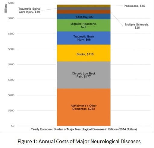
The most common neurological disorders described above cost the United States approximately $789 billion in 2014, which may increase as the elderly population increases between 2011 and 2050, according to a research study published in the Annals of Neurology. The research study demonstrates the price of the serious annual financial burden in the US and has been demonstrated as healthcare professionals have suggested budget reductions for federally-funded research studies. �
According to these demographic statistics, the American Neurological Association, or the ANA, commissioned a research study by former ANA marketing committee and public advocacy committee chair Clifton L. Gooch, MD, currently professor and chair of the Department of Neurology in the University of South Florida’s Morsani College of Medicine in Tampa. �
The research study, the Burden of Neurological Disease in the United States: A Summary Report and Call to Action, demonstrated the annual cost of the most prevalent neurological diseases, including Alzheimer’s disease and other dementias, chronic low back pain, stroke, traumatic brain injury, migraine headaches, epilepsy, multiple sclerosis, spinal cord injury, and Parkinson’s disease. Neurological disease ultimately affects an estimated 100 million people in the United States every year and, together with the costs of stroke and dementia alone, these are estimated to total over $600 billion by 2030. �
Funding for Neurology in the United States
The tremendous and sustained capital investments made in cardiovascular and cancer research studies beginning in the 1970s have considerably increased lifespan. Ironically, however, the number of older adults who have a higher chance of developing neurological diseases have increased, which has developed a growing outbreak among healthcare professionals. �
“Preliminary research studies, including those of cancer, focus considerable research study investment to the neurological diseases which are impacting the quality of life and mortality of more and more people in the United States every year,” stated Gooch, referring to the $1.8 billion in funding for cancer and neurology research approved by Congress in 2016. �
“We hope the findings of the report will serve as a wake-up call to Congress to improve much needed clinical and basic research funding necessary to discover treatments which can mitigate, and finally cure, the considerable amount of neurological diseases which have developed profound consequences in our patients as well as for the national economy.” �
“The future of funding for neurological research studies was an issue in 2012 when the ANA voted to support this particular research study,” stated ANA President Barbara G. Vickrey, MD, MPH. “With the reductions now being suggested to the NIH funding from the President of the United States, this has become of even greater concern today. As representatives of the scholars working to eradicate these health issues, we feel we must raise our collective perceptions, armed with the facts.” �
Annual Cost of Neurological Disease Overview
Researchers gathered the information from the research study through a complete review of the world literature among the most prevalent and costly neurological diseases in the United States. To be conservative, researchers focused on the prevalence and cost estimates they considered to be the most comprehensive and accurate, excluding neurological diseases, such as depression and chronic pain, which frequently have mixed etiologies beyond primary nervous system injury. �
“A complete accounting of all neurological diseases would considerably increase price tag estimates,” wrote the authors of the research study. Indirect and direct costs for the most common neurological diseases previously mentioned above, have been demonstrated in the research study and were estimated according to maintenance standards for each health issue. �
Alzheimer’s disease and other dementias accounted for $243 billion of their $789 billion total, while chronic lower back pain represented $177 billion, and stroke represented $110 billion.�As well as documenting the fiscal costs of neurological disease, Gooch and his USF colleagues ultimately recommend an action plan for reducing the burden of these health issues through infrastructure investment in neurological research and enhanced clinical management of neurological disorders. �
Many research studies have demonstrated how several of the most common neurological diseases pose a serious annual financial burden in the United States. The most prevalent and costly neurological health issues, such as Alzheimer’s disease and other dementias, chronic low back pain or sciatica, as well as stroke, among other common neurological diseases mentioned above, have been estimated to have an annual cost totalling $789 billion in 2014, according to research studies. These annual costs have also been demonstrated to considerable increase further over time.� – Dr. Alex Jimenez D.C., C.C.S.T. Insight
The purpose of the article is to demonstrate the annual cost of several of the most prevalent neurological diseases. Neurological diseases are associated with the brain, the spine, and the nerves. The scope of our information is limited to chiropractic, musculoskeletal and nervous health issues as well as functional medicine articles, topics, and discussions. To further discuss the subject matter above, please feel free to ask Dr. Alex Jimenez or contact us at 915-850-0900 . �
Curated by Dr. Alex Jimenez �
Additional Topic Discussion: Chronic Pain
Sudden pain is a natural response of the nervous system which helps to demonstrate possible injury. By way of instance, pain signals travel from an injured region through the nerves and spinal cord to the brain. Pain is generally less severe as the injury heals, however, chronic pain is different than the average type of pain. With chronic pain, the human body will continue sending pain signals to the brain, regardless if the injury has healed. Chronic pain can last for several weeks to even several years. Chronic pain can tremendously affect a patient’s mobility and it can reduce flexibility, strength, and endurance.
Formulas for Methylation Support
XYMOGEN�s Exclusive Professional Formulas are available through select licensed health care professionals. The internet sale and discounting of XYMOGEN formulas are strictly prohibited.
Proudly,�Dr. Alexander Jimenez makes XYMOGEN formulas available only to patients under our care.
Please call our office in order for us to assign a doctor consultation for immediate access.
If you are a patient of Injury Medical & Chiropractic�Clinic, you may inquire about XYMOGEN by calling 915-850-0900.
�
For your convenience and review of the XYMOGEN products please review the following link.*XYMOGEN-Catalog-Download �
* All of the above XYMOGEN policies remain strictly in force. �
IFM's Find A Practitioner tool is the largest referral network in Functional Medicine, created to help patients locate Functional Medicine practitioners anywhere in the world. IFM Certified Practitioners are listed first in the search results, given their extensive education in Functional Medicine