We often think of proteins are nutrients found in the food we eat and the main component of muscles, however, proteins are microscopic molecules located inside of cells which actually perform a variety of fundamental roles. The function of a protein depends on its shape, and when protein formation goes awry, the resulting misshapen proteins can cause numerous health issues, such as when proteins neglect their essential roles or when they form a sticky, clumpy clutter inside of cells. Protein formation is an error-prone procedure and mistakes along the way have been associated with neurological diseases. �
There are approximately 20,000 to over 100,000 unique types of proteins found inside a common human cell. Why so many? Proteins are the workhorses of the human cell. By way of instance, several of these proteins are structural, lending stiffness and rigidity to thin neurons or muscle tissues. Other proteins shuttle them to new places and bind to specific molecules and others catalyze responses. A property of proteins is possible through diversity and specificity in their role when they fold. �
Why Proteins Fold into a Functional Shape
A protein generally begins in the cell as a lengthy chain of about 300 building blocks known as amino acids. There are 22 different types of amino acids and their order decides what protein chain will fold onto itself. After folding, two types of structures will generally form. Several regions of the protein chain coil into slinky-like formations known as “alpha-helices,” while other regions fold into zigzag patterns known as “beta-sheets,” which resemble the folds of a paper fan. �
Both of these structures may interact to form complex structures. In one protein structure, many beta-sheets wrap themselves around to form a hollow tube. The tube is also generally short where the overall structure resembles snakes (alpha-helices) emerging out of a can (beta-sheet tubing ). Moreover, several other protein structures with descriptive names include the “beta-barrel,” that the”beta-propeller,” the”alpha/beta-horseshoe,” as well as the “jelly-roll fold”. �
These intricate structures allow proteins to perform their variety of roles in the cell. The “snakes in a can” protein, when embedded into a cell membrane, creates a tube which enables traffic in and out of cells. Other proteins form contours with pockets known as “active sites” which are perfectly shaped to bind to a certain molecule like a lock and key. By bending into different shapes, proteins can do different functions. To draw an analogy, all vehicles are made from steel, while a bus, dump truck, crane, or Zamboni are shaped to execute their very own tasks however races are won by the slick shape of a racecar. �
Why Protein Folding Sometimes Fails
Protein folding ultimately allows a protein to take a functional shape, however, it’s an intricate procedure which can sometimes fail. According to research studies, protein folding can go wrong due to three major reasons: �
A person may have a mutation which affects an amino acid in the protein chain, making it difficult for a specific protein to locate its favored fold or “native” state. This is how it is for mutations, such as those contributing to cystic fibrosis or sickle cell anemia. These mutations are found in the DNA sequence or “gene” which encodes one special protein. Therefore, these types of inherited mutations affect only that protein and its related function.
On the other hand, protein folding failure can be seen as an ongoing and much more general procedure which affects several proteins. When proteins are made, the structure which reads the instructions from DNA to produce the long chains of amino acids can make errors. Researchers estimate that the ribosome makes mistakes in as many as 1 in every 7 proteins. These mistakes can make the proteins which are resultantly inclined to continue to fold improperly.
Even though an amino acid chain does not have any mutations or mistakes, it may still not reach its own preferred folded shape because proteins don’t fold properly 100 percent of their time. Protein folding becomes much more difficult if the conditions in the cell change due to external factors such as temperature and acidity.
A collapse in protein folding can cause a variety of neurological diseases and researchers hypothesize that many health issues are associated with folding problems. There are two problems which exist in cells which don’t protein fold correctly. �
One type of problem, known as “loss of function,” results when not enough of a particular protein folds correctly, causing a lack of “specialized functions” necessary to perform a particularly important role. By way of instance, imagine a correctly folded protein is shaped to bind a toxin and split it into noxious compounds. Without enough of that protein accessible, the toxin will build-up to damaging levels. In another instance, a protein may be responsible for metabolizing sugar which can then be utilized by the cell for energy. The cell will grow due to lack of energy if not enough of this protein is accessible. The reason the cell becomes ill, in these cases, is because of a lack of one particular folded, functional protein. Cystic fibrosis, Tay-Sachs disease, Marfan syndrome, and some types of cancer are examples of health issues which result when one type of protein is unable to perform its role. Who knew that one type of protein out of thousands may be so significant? �
Proteins folding may also impact the overall health and wellness of the cell regardless of the utilization of the protein. When proteins fail to fold into their functional state, the consequent misfolded proteins could be contorted into shapes which are harmful to the crowded cell environment. Most proteins have sticky, “water-hating” amino acids which they bury deep inside their own core. Misfolded proteins utilize these parts on their exterior, such as a chocolate-covered candy which has been crushed to reveal a gooey center. These misfolded proteins commonly stick together to form clumps known as “aggregates.” Researchers discovered that the accumulation of misfolded proteins plays a fundamental role in several neurological diseases, including Alzheimer’s disease, Parkinson’s disease, Huntington’s disease, and Lou Gehrig’s (ALS) disease, however, researchers are still working to discover exactly how these misfolded molecules affect the well-being of the cells. �
One misfolded protein ultimately stands out from among the rest and it deserves particular attention. The “prion” protein in Creutzfeldt-Jakob disease, also known as mad cow disease, is an illustration of a misfolded protein gone rogue. This protein isn’t simply irreversibly misfolded, however, it also transforms other functional proteins into a similar twisted condition. �
How Cells Protect from Misfolded Proteins
Recent research studies have demonstrated that protein misfolding often occurs inside of cells. Fortunately, cells also have many systems in place and are accustomed to coping with this issue by refolding or destroying aberrant protein formations. � Appropriately known as chaperones, these structures accompany proteins throughout the folding procedure, enhancing a protein’s odds of folding properly and even allowing several misfolded proteins the opportunity to refold. Chaperones are proteins themselves. There are many distinct types of chaperones. A few chaperones supply safety to proteins, isolated from other molecules. Production of many chaperones is fostered when a cell encounters high temperatures or other states which can ultimately make protein folding more difficult, therefore, providing these chaperones the alias, “heat shock proteins.” �
The following line of cell defense against misfolded proteins is known as the proteasome. If misfolded proteins linger in the cell, they will be targeted for destruction by this structure, which chews proteins up and spits them out. The proteasome is similar to a center, permitting the cell to reuse amino acids to create proteins. The proteasome itself is not a single protein but many acting collectively. Proteins frequently interact to form larger structures. By way of instance, a human sperm’s tail is a structure made of various types of proteins which work together to produce a rotary engine which propels the sperm. �
Protein Folding and Misfolding Overview
Why is it that some misfolded proteins can evade systems such as chaperones and the proteasome? How can the neurological diseases previously mentioned above be caused by sticky misfolded proteins? Do some proteins misfold more often than others? These questions are at the forefront of research studies seeking to understand the health issues which ultimately result if protein fold goes awry as well as protein biology. The broad world of proteins, using its great assortment of shapes, bestows cells with capacities that allow for life to exist and allow for its diversity (e.g., the differences between eye, skin, lung or heart cells, and also the differences between species). But perhaps this is one of the many reasons why the word “protein” comes from the Greek word “protas,” meaning “of primary significance” and indeed they seem to be. �
Protein folding is a complex, physiochemical process by which a protein “folds” or assumes a functional shape to be able to perform their biological function. Proteins are nutrients we obtain from the food we eat and they are considered to be one of the main components of muscles, however, proteins play a wide variety of fundamental roles in the human body. According to research studies, protein misfolding can cause a variety of health issues, including neurological diseases. – Dr. Alex Jimenez D.C., C.C.S.T. Insight
The purpose of the article above is to describe protein folding and how it’s associated with neurological diseases. Neurological diseases are associated with the brain, the spine, and the nerves. The scope of our information is limited to chiropractic, musculoskeletal and nervous health issues as well as functional medicine articles, topics, and discussions. To further discuss the subject matter above, please feel free to ask Dr. Alex Jimenez or contact us at 915-850-0900 . �
Curated by Dr. Alex Jimenez �
Additional Topic Discussion: Chronic Pain
Sudden pain is a natural response of the nervous system which helps to demonstrate possible injury. By way of instance, pain signals travel from an injured region through the nerves and spinal cord to the brain. Pain is generally less severe as the injury heals, however, chronic pain is different than the average type of pain. With chronic pain, the human body will continue sending pain signals to the brain, regardless if the injury has healed. Chronic pain can last for several weeks to even several years. Chronic pain can tremendously affect a patient’s mobility and it can reduce flexibility, strength, and endurance.
Formulas for Methylation Support
XYMOGEN�s Exclusive Professional Formulas are available through select licensed health care professionals. The internet sale and discounting of XYMOGEN formulas are strictly prohibited.
Proudly,�Dr. Alexander Jimenez makes XYMOGEN formulas available only to patients under our care.
Please call our office in order for us to assign a doctor consultation for immediate access.
If you are a patient of Injury Medical & Chiropractic�Clinic, you may inquire about XYMOGEN by calling 915-850-0900.
�
For your convenience and review of the XYMOGEN products please review the following link.*XYMOGEN-Catalog-Download �
* All of the above XYMOGEN policies remain strictly in force.
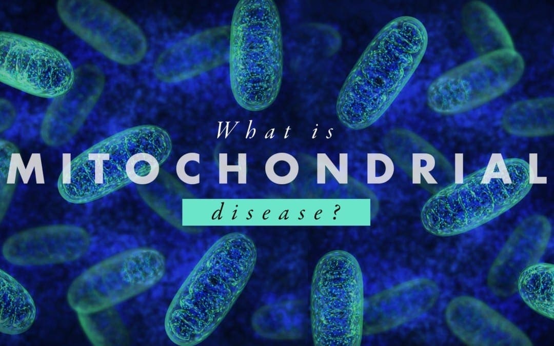
Mitochondria are the “energy factory” of the human body. Several thousand mitochondria can be found in nearly every cell. Mitochondria also play several fundamental roles in the body, such as converting chemicals from the foods we eat into energy as well as to process oxygen. Mitochondria produce 90 percent of the energy the human body requires to function accordingly. The purpose of the following article is to describe an overview of mitochondrial disease and well-being. �
What are Mitochondrial Diseases?
Mitochondrial diseases are characterized as chronic, genetic, and often inherited health issues which ultimately occur when mitochondria fail to produce enough energy for the human body to function properly. Mitochondrial diseases may develop from birth however they can frequently develop at any age. Mitochondrial disease can affect any region of the human body, including the cells of the brain, muscles, heart, liver, kidneys, pancreas, eyes, ears, and nerves, among other structures. �
When the mitochondria don’t function as well as they should because of another health issue, mitochondrial dysfunction occurs. Furthermore, many health issues can cause secondary dysfunction and result in other neurological diseases, such as Alzheimer’s disease, Lou Gehrig’s disease, and muscular dystrophy. People with secondary dysfunction don’t have genetic mitochondrial disease and do not need to be concerned about the ongoing development or worsening of symptoms. �
What are the Symptoms of Mitochondrial Disease?
Symptoms of mitochondrial disease depend on which cells of the human body are affected. Symptoms can develop at any age, involve one or more organs, and may range from mild to severe. Even patients within the same household, having the exact same mitochondrial disease can have gaps in symptoms, severity, and age of onset or beginning of symptoms. �
Symptoms of mitochondrial diseases can include: �
Poor growth
Muscle pain, muscle weakness, exercise intolerance, low muscle tone
Vision and/or hearing problems
Learning disabilities, delays in development, mental retardation
In many people, primary mitochondrial disease is a genetic health issue which can be inherited in several ways. To understand inheritance types, it is helpful to find out more about genes and DNA. Genes are substances which provide us our traits, like brown eyes or blue eyes. Genes contain DNA, which is the “blueprint” which gives each person their distinctive make-up. �
In normal circumstances, a child inherits one gene from the father and one gene from the mother. A child with a mitochondrial disease doesn’t receive the pair of genes from the parents. The gene has mutated or has become defective. Learning how the mitochondrial disease is inherited helps predict the prospect of passing the disease(s) to children. �
Inheritance types of mitochondrial disease are: �
Autosomal recessive inheritance: The child receives one mutated copy of a gene from each parent. There is a 25 percent chance that each child in the family will inherit a mitochondrial disease.
Autosomal dominant inheritance: The child receives one mutated copy of a gene from either parent. There is a 50 percent chance that each child in the family will inherit a mitochondrial disease.
Mitochondrial inheritance: In this unique type of inheritance, the mitochondria contain their own DNA. Only mitochondrial disorders caused by mutations in the mitochondrial DNA are exclusively inherited from mothers. There is a 100 percent chance that each child in the family will inherit a mitochondrial disease.
Random mutations: Occasionally, genes develop a mutation of their own which is not inherited from a parent.
How are Mitochondrial Diseases Diagnosed?
Mitochondrial diseases can be difficult to diagnose by a healthcare professional because mitochondrial diseases can ultimately affect a variety of organs and tissues in the human body and patients can also have a variety of symptoms. There is currently no single lab test or diagnostic test which can confirm the identification of mitochondrial disease. That is why a referral to a medical facility with healthcare professionals who focus on these diseases is essential to making the diagnosis. �
Diagnosis begins with a series of evaluations and tests which may include: �
A review of a patient�s family history
A complete physical evaluation
A neurological evaluation
A metabolic evaluation which includes blood and urine tests, and, if needed, a cerebral spinal fluid test
Other evaluations, determined by the regions of the human body and the patient’s symptoms which may include: �
Magnetic resonance imaging (MRI) or spectroscopy (MRS) for neurological symptoms
Retinal exam or electroretinogram (ERG) for vision symptoms
Electrocardiogram (EKG) or echocardiogram for symptoms of heart disease
Audiogram or auditory-brainstem evoked responses (ABER) for hearing symptoms
Blood test to detect thyroid dysfunction if the patient has thyroid problems
Blood test to perform genetic DNA testing
Testing may include biochemical testing. Biopsies of skin and muscle tissue may also be utilized for diagnosis. �
How are Mitochondrial Diseases Treated?
Unfortunately, there is no cure for mitochondrial disease, however, treatment can help reduce symptoms or slow the decline of overall well-being. Treatment varies from patient to patient and depends on the severity and the mitochondrial disease characterized. There is absolutely no way to predict a patient’s reaction or forecast how that person will be affected in the long-term. No two people respond the same way to the same treatment even if they have the same mitochondrial disease. �
Treatments for mitochondrial disease may include: �
Vitamins and supplements, including Coenzyme Q10; B complex vitamins, such as thiamine (B1) and riboflavin (B2), Alpha lipoic acid, L-carnitine (Carnitor), Creatine, and L-Arginine.
Exercise and physical activity, including endurance exercises and resistance/strength training to increase muscle strength. Endurance exercises include walking, running, swimming, dancing, cycling and others. Resistance/strength training includes exercises such as sit-ups, arm curls, knee extensions, weight lifting and others.
Conserving energy. Don�t try to do too much in a short period of time. Pace yourself.
Other treatments including speech therapy, respiratory therapy, physical therapy, and chiropractic care, among others.
Avoid situations which can make the health issue worse. This includes exposure to cold and/or warmth, starvation, lack of sleep, stressful situations, and usage of alcohol, smokes and monosodium glutamate or MSG, a flavor enhancer commonly added to Chinese foods, canned vegetables, soups, as well as processed meats, among other processed foods. �
Mitochondrial diseases are long-term, genetic, and frequently inherited health issues which occur when the mitochondria fail to produce enough energy for the human body to function accordingly. According to research studies, approximately one in 5,000 people has a genetic mitochondrial disease. Chiropractic care is an alternative treatment option which can help relieve symptoms associated with a variety of health issues, including mitochondrial diseases. Many chiropractors are qualified and experienced in the treatment of neurological diseases. – Dr. Alex Jimenez D.C., C.C.S.T. Insight
The purpose of the article above is to describe mitochondrial disease and its effect on overall health and wellness. Neurological diseases are associated with the brain, the spine, and the nerves. The scope of our information is limited to chiropractic, musculoskeletal and nervous health issues as well as functional medicine articles, topics, and discussions. To further discuss the subject matter above, please feel free to ask Dr. Alex Jimenez or contact us at 915-850-0900 . �
Curated by Dr. Alex Jimenez �
Additional Topic Discussion: Chronic Pain
Sudden pain is a natural response of the nervous system which helps to demonstrate possible injury. By way of instance, pain signals travel from an injured region through the nerves and spinal cord to the brain. Pain is generally less severe as the injury heals, however, chronic pain is different than the average type of pain. With chronic pain, the human body will continue sending pain signals to the brain, regardless if the injury has healed. Chronic pain can last for several weeks to even several years. Chronic pain can tremendously affect a patient’s mobility and it can reduce flexibility, strength, and endurance.
Formulas for Methylation Support
XYMOGEN�s Exclusive Professional Formulas are available through select licensed health care professionals. The internet sale and discounting of XYMOGEN formulas are strictly prohibited.
Proudly,�Dr. Alexander Jimenez makes XYMOGEN formulas available only to patients under our care.
Please call our office in order for us to assign a doctor consultation for immediate access.
If you are a patient of Injury Medical & Chiropractic�Clinic, you may inquire about XYMOGEN by calling 915-850-0900.
�
For your convenience and review of the XYMOGEN products please review the following link.*XYMOGEN-Catalog-Download �
* All of the above XYMOGEN policies remain strictly in force. �
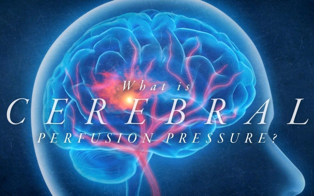
Cerebral perfusion pressure, or CPP, is the net pressure gradient which carries oxygen to brain tissue. It is measured by the difference between the mean arterial pressure, or MAP, and the Intracranial Pressure, or ICP,� which is measured in millimeters of mercury (mm Hg). Regulating CPP is fundamental in the treatment of patients with intracranial pathology, including shock, hemodynamic distress, and traumatic brain injury. �
Although the average CPP is generally between 60 and 80 mm Hg, these values may change to the left or to the right depending on individual physiology. MAP and ICP has to be measured together because CPP is a calculated measure. Regulating CPP at hemodynamically unstable conditions with abnormal ICP or in cases of intracranial pathology will reduce the chance of ischemic brain injury. �
CPP = MAP – ICP
Cerebral Perfusion Pressure Physiology
CPP and ICP
At its own average range of 60 to 80 mm Hg, the CPP is determined by the ICP and the mean arterial pressure. Under regular standards, the ICP is between 5 and 10 mm Hg which has a reduced effect on the CPP than the MAP in clinical circumstances not associated with intracranial pathology. ICP is generally measured through intracranial pressure transduction.
Physiologically, the ICP is a function of intracranial compliance. Intracranial compliance is the relationship between the ICP and the volume of the intracranial cavity including cerebrospinal fluid, or CSF, brain tissue as well as arterial and venous blood volume. Because the skull is a fixed and rigid anatomic space, the ICP can increase if the intracranial volume increases while intracranial compliance decreases. As the ICP increases or intracranial compliance decreases, CPP also decreases. �
Several processes determine that ICP continues to stay within the average range for the longest extended period of time possible, especially throughout periods of affected intracranial volume and compliance. As volume adds to the intracranial space, CSF can shift into the spinal subarachnoid space, causing the ICP to continue significantly unchanged. As volume increases due to a growing space-occupying lesion, brain tissue edema or blood, this process ultimately becomes overwhelming, and ICP begins to increase substantially. �
Cerebral blood flow, or CBF, is also a fundamental factor in ICP homeostasis. Cerebral auto-regulation makes sure that steady blood flow is maintained in the brain over a wide range of physiologic alterations. When blood pressure decreases, auto-regulation causes cerebral vasodilation and an increase in CBF and cerebral blood volume, maintaining ICP and CPP. However, when blood pressure increases, auto-regulation causes cerebral vasoconstriction and a decrease in CBF with a decrease in cerebral blood volume, also regulating ICP and CPP. Too many changes outside of average CBF ranges can cause brain ischemia and injury. �
CPP and MAP
Because ICP in its average ranges is a considerably small number, the CPP generally depends on the mean arterial pressure. MAP is the normal blood pressure during one cardiac cycle which can be measured through invasive hemodynamic monitoring or calculated by the systolic blood pressure, plus two times the diastolic blood pressure, divided by three. The average range of MAP is 70 to 100 mm Hg. �
The average arterial pressure can be affected due to everyday activities, such as rest, stress, and exercise or physical activities. However, if the ICP continues to stay the same, the average arterial pressure can change across its significantly wide range without tremendously decreasing or increasing the CPP. As a matter of fact, CPP and CBF will continue to stay considerably unchanged across a wider range of MAP (50 � 150 mm Hg) than normal due to cerebral auto-regulation and vasoconstriction or vasodilation of cerebral vasculature. �
For patients with hypertension, the auto-regulation setpoint changes, decreasing the average arterial pressure associated with the patient�s normal arterial pressure, which causes vasodilation to increase CBF. Patients with lower than normal average arterial pressure at baseline will have auto-regulatory vasoconstriction as a reaction to an increase in their significant average MAP, to return CBF to baseline. When looking at CBF and CPP in the context of the patient�s average MAP, it is clinically significant based on the regulation of intracranial pathology and hemodynamic derangements. �
Cerebral Perfusion Pressure Complications
Diagnosing and treating cerebral perfusion pressure complications necessitates measuring both the ICP and the MAP. The MAP may be quantified through the utilization of invasive hemodynamic processes, most frequently cannulation of a peripheral artery such as the radial or femoral artery. The MAP may also be measured with a non-invasive blood pressure cuff by applying the formula mentioned above utilizing the systolic and diastolic blood pressures. � Intracranial pressure is generally measured through an intracranial pressure transduction device. The most common and most accurate method or technique is utilizing an intraventricular monitor. The intraventricular dimension of ICP is the normal standard. An intraventricular catheter is inserted into a hole drilled in the skull and into the lateral ventricle to gauge the pressure of the CSF. The benefit of an intraventricular catheter is that CSF could be eliminated, if needed, to decrease ICP. Considerable complications for the ICP include a possibility of bleeding, infection, and difficulty with proper placement. Options include sub-dural and intra-parenchymal monitors. �
The ICP can be measured non-invasively through several methods and techniques, including transcranial Doppler ultrasonography or TCD. TCD utilizes a temporal window to evaluate the speed of blood flow through the middle cerebral artery. Systolic and diastolic average flow velocity is utilized to determine a pulsatility index. The pulsatility index was determined to be closely associated with ICP in several research studies as well as be associated with ICP in other research studies. Therefore, it is not suggested to use TCD as a substitute for direct ICP dimension. Invasive diagnosis and treatment of the MAP through an arterial cannula and the ICP through an intraventricular catheter will give a continuous and accurate calculation of CPP. �
Cerebral Perfusion Pressure Clinical Significance
Two general types of pathologic health issues can ultimately occur where the regulation of the CPP is fundamental, such as intracranial pathology, where ICP regulation is essential and hemodynamic instability/shock where MAP regulation is the most essential. Intracranial pathology involves space-occupying lesions, such as tumors, epidural and subdural hematoma or severe intraparenchymal hemorrhage and cerebral edema as seen after ischemic injury, traumatic brain injury or acute hepatic encephalopathy. In these circumstances, average CPP depends on decreasing the ICP into a normal range as soon as possible while regulating the MAP. When CPP is normal, it’s fundamental to keep in mind that every individual’s brain tissue has a CPP that is “normal” in the context of that individual patient’s physiology, which may be affected by other health issues, such as hypertension or cardiovascular disease. Moving towards a more dynamic direction of the average CPP utilizing the patient’s personal auto-regulatory capacity. These diagnosis and treatment approaches involve more frequent and sophisticated monitoring and might not be readily available for widespread utilization. �
In the instance of considerable traumatic brain injury, significant cerebral edema can decrease intracranial compliance and CSF, developing an increased ICP or intracranial hypertension. Auto-regulatory mechanisms and techniques may or may not function normally and when ICP continues to be elevated, CPP will decrease causing further injury through an ischemic process. In circumstances such as these, together with starting the measures for decreasing the ICP, it is essential to prevent hypotension (MAP – ICP = CPP) and in some instances, allowing hypertension to reasonably occur. �
In circumstances of instability, the ICP is considerably stable as cerebral auto-regulation is undamaged. In the instance of hypotension, the MAP decreases due to blood loss, or hemorrhagic shock, intravascular leak, or distributive shock, and decreased cardiac output, or cardiogenic shock, and the CPP also decreases. It’s the association between MAP and CPP which carries resuscitation guidelines to recommend regulating a MAP greater than or equal to 65 mm Hg. With a normal ICP, this threshold must make sure that a CPP of 55 to 60, the minimum necessary to stop cerebral ischemic injury, is ultimately maintained. As in the circumstance of ICP and cerebral auto-regulation, the goal of MAP is to be within the context of an individual patient’s evaluation hemodynamic function. Patients with untreated hypertension must have increased MAP goals to maintain proper CBF and CPP. �
As previously mentioned in the following article, cerebral perfusion pressure, or CPP, is the net pressure gradient which affects cerebral blood flow to the brain, also known as brain perfusion. According to healthcare professionals, the CPP, or cerebral perfusion pressure, must be constantly regulated within a specific limit because too little pressure or too much pressure could potentially cause a variety of brain health issues. Cerebral perfusion pressure may be associated with a variety of neurological diseases. – Dr. Alex Jimenez D.C., C.C.S.T. Insight
The purpose of the article is to discuss cerebral perfusion pressure and its association with neurodegenerative diseases. Neurological diseases are associated with the brain, the spine, and the nerves. The scope of our information is limited to chiropractic, musculoskeletal and nervous health issues as well as functional medicine articles, topics, and discussions. To further discuss the subject matter above, please feel free to ask Dr. Alex Jimenez or contact us at 915-850-0900 . �
Curated by Dr. Alex Jimenez �
Additional Topic Discussion: Chronic Pain
Sudden pain is a natural response of the nervous system which helps to demonstrate possible injury. By way of instance, pain signals travel from an injured region through the nerves and spinal cord to the brain. Pain is generally less severe as the injury heals, however, chronic pain is different than the average type of pain. With chronic pain, the human body will continue sending pain signals to the brain, regardless if the injury has healed. Chronic pain can last for several weeks to even several years. Chronic pain can tremendously affect a patient’s mobility and it can reduce flexibility, strength, and endurance.
Formulas for Methylation Support
XYMOGEN�s Exclusive Professional Formulas are available through select licensed health care professionals. The internet sale and discounting of XYMOGEN formulas are strictly prohibited.
Proudly,�Dr. Alexander Jimenez makes XYMOGEN formulas available only to patients under our care.
Please call our office in order for us to assign a doctor consultation for immediate access.
If you are a patient of Injury Medical & Chiropractic�Clinic, you may inquire about XYMOGEN by calling 915-850-0900.
�
For your convenience and review of the XYMOGEN products please review the following link.*XYMOGEN-Catalog-Download �
* All of the above XYMOGEN policies remain strictly in force. �
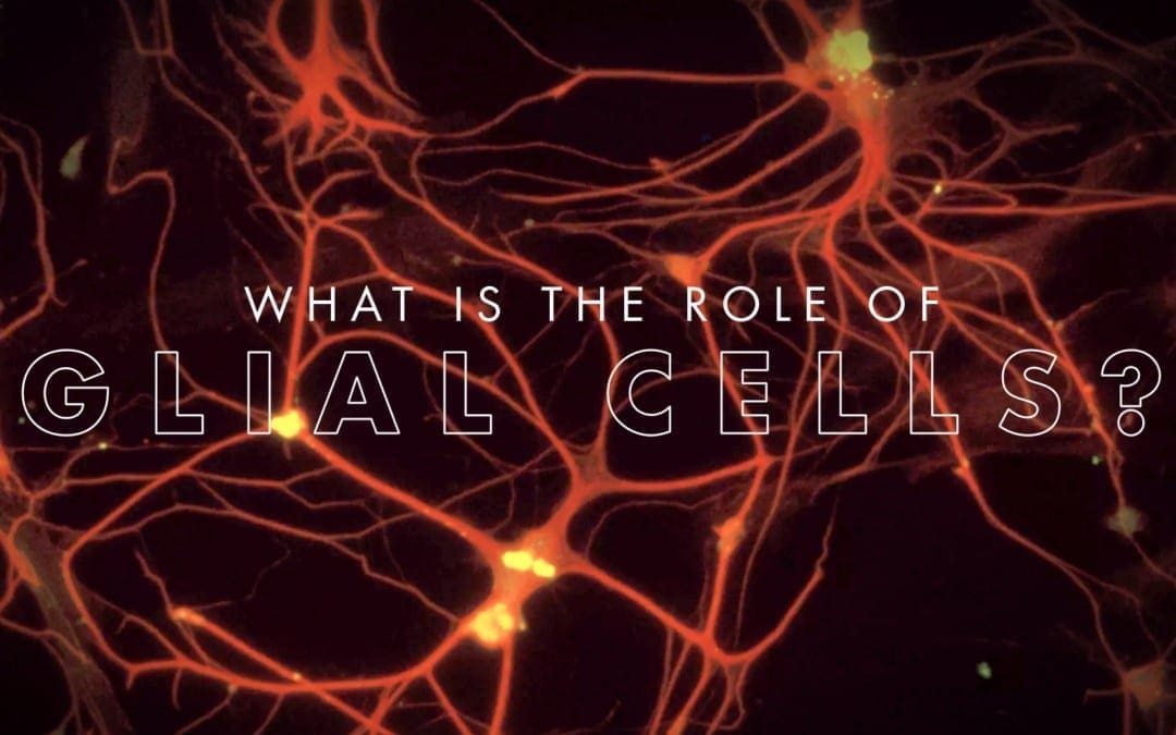
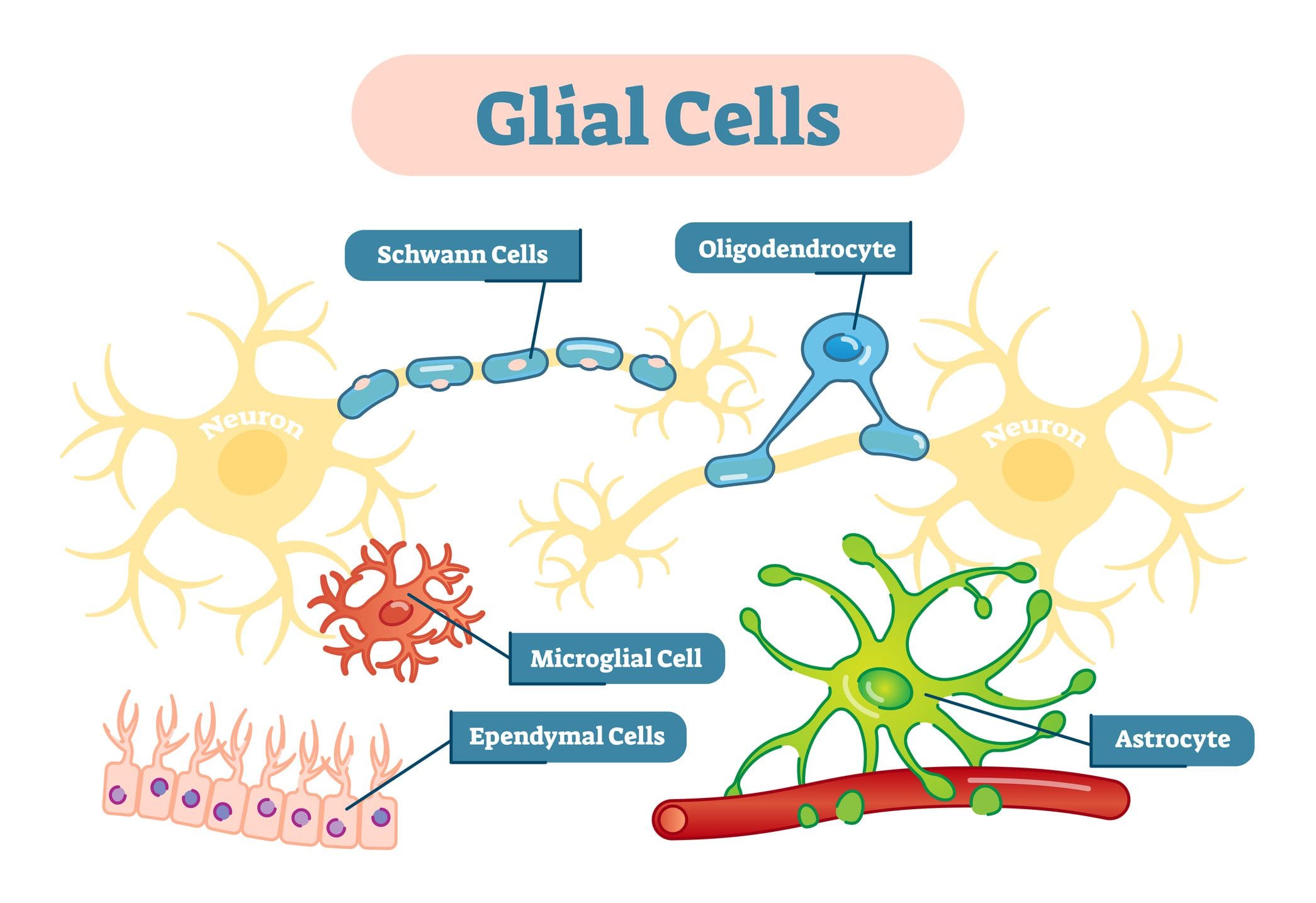
You have probably heard about the “gray matter” of the brain which is made up of cells known as neurons, however, a lesser-known type of brain cell is ultimately what makes up the “white matter” of the brain.� These are known as glial cells. �
Glial cells, also known as glia or neuroglia, were only considered to simply offer structural support. The term “glia” literally translates to “neural adhesive.” However, relatively recent research studies have demonstrated that they play a variety of roles in the brain and the nerves which run throughout the entire human body. However, there is more left to find out. �
Types of Glial Cells
Glial cells commonly offer support to the neurons. Without them, several of the most fundamental roles would never be achieved although they may not perform these roles themselves. Glial cells come in numerous forms, each of which performs certain functions to keep the brain functioning properly or not, in case of a neurological disease which affects the glial cells. �
The central nervous system, or CNS, is made up of the brain, the spinal cord, and the nerves. Five types of glial cells include: �
Astrocytes
Oligodendrocytes
Microglia
Ependymal cells
Radial glia
Moreover, there are also glial cells on the peripheral nervous system, or PNS, which is made up of the nerves in the upper and lower extremities, away from the spine. The two types of glial cells found in the peripheral nervous system include: �
Schwann cells
Satellite cells
� �
Astrocytes
The most common type of glial cell in the central nervous system is the astrocyte, also known as astroglia. The “astro” part of the name refers to how they look like stars with projections coming out all over the glial cell. Protoplasmic astrocytes have thick projections with lots of branches. Fibrous astrocytes have long, slender arms. The fibrous ones are found in the white matter while others are found among neurons in the gray matter.� Astrocytes play several major roles, including: �
Developing the blood-brain barrier or BBB. The BBB is similar to a strict security system which only allows substances which are supposed to be in the brain. This filtering system is essential for maintaining brain health.
Regulating the substances around neurons. Neurons communicate utilizing chemical messengers known as neurotransmitters. Once a chemical has transmitted a message to a cell, it essentially stays there cluttering things up until an astrocyte recycles it through a process known as reuptake. The reuptake process is generally the main target of numerous medications, including anti-depressants. Astrocytes also clean up what’s left behind when a neuron dies, as well as excess potassium ions, which are chemicals that play a fundamental role in nerve function.
Regulating blood flow to the brain. For the brain to process information accordingly, it needs a certain amount of blood to flow throughout all of its different regions. An active region receives more blood flow than an inactive one.
Synchronizing the activity of axons. Axons are characterized as long, thread-like elements of the neurons and the nerve cells which ultimately conduct electricity to help transmit messages from one cell to another.
Astrocyte dysfunction has been potentially connected to a wide variety of neurological diseases, including: �
Amyotrophic lateral sclerosis (ALS or Lou Gehrig’s disease)
Huntington’s chorea
Parkinson’s disease
Animal models of astrocyte-related disorders are helping researchers learn more about these neurological diseases. �
Oligodendrocytes
Oligodendrocytes develop from stem cells. The term is made up of Greek words which, all together, mean “cells with several branches.” Their main role is to help information move faster. Oligodendrocytes appear like white spikey balls. Their purpose is to make a protective layer, similar to the plastic insulation on electric wires. This layer is known as the myelin sheath. �
The myelin sheath is not constant. There is a gap between each membrane which is known as the”node of Ranvier,” and it is the node which helps electrical signals move effectively along neural cells. The signal is transmitted from one node to the next, which increases the velocity of the nerve conduction whilst also reducing how much energy it takes to transmit it. �
Messages along myelinated nerves may travel as fast as 200 miles per second. At birth, you only have a few myelinated axons, and the quantity of these keeps growing until you’re about 25 to 30 years old. Myelination is thought to play an important role in intelligence. Oligodendrocytes also supply stability and transmit energy from blood cells into the axons. �
The expression “myelin sheath” may be familiar to you because of its association with multiple sclerosis. In multiple sclerosis, it is believed that the human body’s immune system attacks the myelin sheaths, which leads to the breakdown of these neurons and ultimately causes impaired brain functioning. Spinal cord injuries may also cause damage to these structures. � Other neurological diseases believed to be associated with oligodendrocyte dysfunction include: �
Leukodystrophies
Tumors known as oligodendrogliomas
Schizophrenia
Bipolar disorder
Several research studies suggest that oligodendrocytes may become affected by the neurotransmitter glutamate, which, among other functions, stimulates regions of the brain so that you’re able to focus and learn new information. Nonetheless, in high levels, glutamate can be considered an “excitotoxin,” which means that it may overstimulate cells until they die. �
Microglia
Microglia are tiny glial cells. They act as the brain’s dedicated immune system, which is necessary since the BBB isolates the brain from the rest of the human body. Microglia are attentive to indications of disease and injury. If they find a problem, they are in charge of taking care of it, even if it ultimately means clearing away dead cells or getting rid of a toxin or pathogen. �
If they respond to an injury, microglia cause inflammation as part of the recovery process. In some cases, such as in Alzheimer’s disease, they might become hyper-activated and cause too much inflammation. That is thought to cause amyloid plaques and other health issues connected with the neurological disease, among a variety of other brain health issues. � Along with Alzheimer’s disease, other neurological diseases which may be associated with microglial malfunction include: �
Fibromyalgia
Chronic neuropathic pain
Autism spectrum disorders
Schizophrenia
Microglia have been thought to play many fundamental roles beyond that, including learning-associated plasticity and guiding the development of the brain. The brain produces many connections between neurons which allow them to pass information back and forth. The brain produces a lot more of these than we need, which is not always efficient. �
Microglia detect unnecessary synapses and they clean them out. Microglial research has really taken off in recent decades, leading to an ever-increasing comprehension of their roles in both health and disease in the central nervous system. �
Ependymal Cells
Ependymal cells are primarily known for creating a membrane known as the ependyma, and it can be described as a thin membrane lining the central canal of the spinal cord and the ventricles or passageways of the brain. They also create cerebrospinal fluid. Ependymal cells are extremely small and they lineup closely together to make the membrane. �
Inside the ventricles, are the cilia, which look like small hairs which move back and forth to help circulate the cerebrospinal fluid. Cerebrospinal fluid provides nutrients and removes waste products in the brain. Additionally, it serves as a cushion and shock absorber between the skull and the brain. It’s also essential for homeostasis in the brain, regulating its temperature along with other attributes which keep its potential and functioning. Ependymal cells are also included in the BBB. �
Radial Glia
Radial glia are believed to be a type of stem cell, which means that they create other types of cells. In the developing brain, they’re the”parents” of neurons, astrocytes, and oligodendrocytes. They also supply scaffolding for developing neurons, thanks to long fibers which direct young brain cells into position as the brain forms in a human embryo. Their role as stem cells, especially as founders of neurons, is ultimately what makes them the focus of research studies regarding how to repair brain damage from injury or illness. Later in life, the radial glia perform important roles in neuroplasticity as well. �
Schwann Cells
Schwann cells are known after the physiologist Theodor Schwann, who discovered them. They function a lot like oligodendrocytes in which they supply myelin sheaths for axons, but they develop in the peripheral nervous system, or PNS, rather than in the central nervous system or CNS. However, Schwann cells form spirals directly across the axon. �
Ranvier’s nodes are found between the membranes of oligodendrocytes and these help in neural transmission in precisely the same exact way. Schwann cells can also be part of the PNS’s immune system. They ultimately have the ability to consume the axons of the nerve and give a protected path for a brand new axon to develop when another nerve cell is damaged. Neurological diseases involving abnormal Schwann cells include: �
Guillain-Barre’ syndrome
Charcot-Marie-Tooth disorder
Schwannomatosis
Chronic inflammatory demyelinating polyneuropathy
Leprosy
Several research studies on bronchial Schwann cells for spinal cord injury and other types of peripheral nerve damage have been promising. Schwann cells are implicated in certain types of chronic pain. Their activation following nerve damage may contribute to dysfunction in a type of nerve fiber known as nociceptors, which feel external factors like heat and cold. �
Satellite Cells
Satellite cells get their name due to the way they surround certain neurons, with several satellites forming a sheath around the cellular surface. Researchers have only just started to learn about these cells but they’re believed to be similar to astrocytes. The main role of satellite cells is believed to be the regulation of the surroundings around the nerves. �
The nerves which have satellite cells make up something known as ganglia, which are clusters of nerve cells in the autonomic nervous system and sensory apparatus. The autonomic nervous system regulates internal organs, even while the sensory system is what enables people to see, hear, taste, touch, and smell. Satellite cells provide nourishment to the neuron and absorb heavy metal toxins, such as lead and mercury, to stop them from damaging the nerves and other structures. �
They are also believed to assist transport several neurotransmitters and other substances, including: �
Glutamate
GABA
Norepinephrine
Adenosine triphosphate
Substance P
Capsaicin
Acetylcholine
Much like microglia, satellite cells detect and respond to injury and inflammation. However, their role in repairing cell damage isn’t yet fully well understood. Satellite cells have been connected to chronic pain between peripheral tissue injury, nerve damage, and a systemic heightening of pain, or hyperalgesia, which can ultimately result from chemotherapy. �
Glial cells, also known as glia or neuroglia, are characterized as non-neuronal cells which are ultimately found in the central nervous system, or CNS, and the peripheral nervous system, or PNS. There are various types of glial cells, including astrocytes, oligodendrocytes, microglia, ependymal cells, and radial glia in the CNS and Schwann cells and satellite cells in the PNS. The glial cells play many fundamental roles in the human nervous system. – Dr. Alex Jimenez D.C., C.C.S.T. Insight
The purpose of the article is to discuss the types of glial cells associated with the brain and neurodegenerative diseases. Neurological diseases are associated with the brain, the spine, and the nerves. The scope of our information is limited to chiropractic, musculoskeletal and nervous health issues as well as functional medicine articles, topics, and discussions. To further discuss the subject matter above, please feel free to ask Dr. Alex Jimenez or contact us at 915-850-0900 . �
Curated by Dr. Alex Jimenez �
Additional Topic Discussion: Chronic Pain
Sudden pain is a natural response of the nervous system which helps to demonstrate possible injury. By way of instance, pain signals travel from an injured region through the nerves and spinal cord to the brain. Pain is generally less severe as the injury heals, however, chronic pain is different than the average type of pain. With chronic pain, the human body will continue sending pain signals to the brain, regardless if the injury has healed. Chronic pain can last for several weeks to even several years. Chronic pain can tremendously affect a patient’s mobility and it can reduce flexibility, strength, and endurance.
Formulas for Methylation Support
XYMOGEN�s Exclusive Professional Formulas are available through select licensed health care professionals. The internet sale and discounting of XYMOGEN formulas are strictly prohibited.
Proudly,�Dr. Alexander Jimenez makes XYMOGEN formulas available only to patients under our care.
Please call our office in order for us to assign a doctor consultation for immediate access.
If you are a patient of Injury Medical & Chiropractic�Clinic, you may inquire about XYMOGEN by calling 915-850-0900.
�
� For your convenience and review of the XYMOGEN products please review the following link.*XYMOGEN-Catalog-Download �
* All of the above XYMOGEN policies remain strictly in force. �
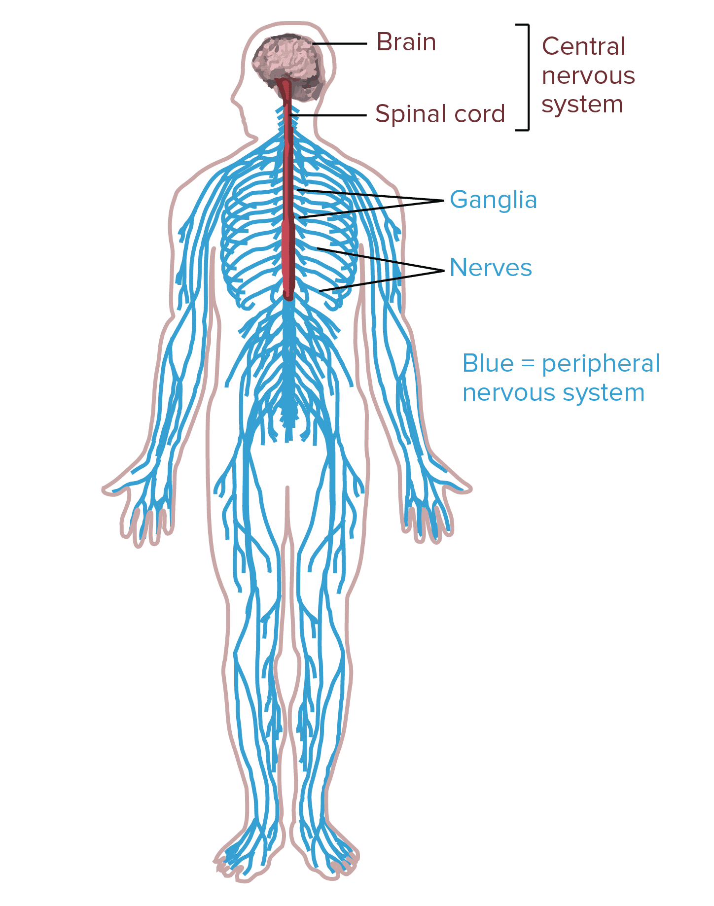
In humans, the nervous system consists of the central nervous system and the peripheral nervous system. The central nervous system, or CNS, consists of the brain and the spinal cord. It is in the CNS where the review of information occurs. The peripheral nervous system, or PNS, consists of the neurons and parts of neurons outside the CNS, including sensory neurons and motor neurons. Sensory neurons bring signals into the CNS, and motor neurons carry signals out of the CNS. �
The cell bodies of PNS neurons, such as the motor neurons which control skeletal muscles, are found in the CNS. These motor neurons have long extensions, known as axons, which run from the CNS all the way to the muscles with which they connect with or innervate. The cell bodies of additional PNS neurons, such as the sensory neurons which provide information on touch, pain, position, and temperature, are found outside the CNS, in which they are found in clusters known as ganglia. The axons of peripheral nerves which run through a common pathway are bundled together to form nerves. �
Types of Neurons
According to their roles, the neurons within the human nervous system can be separated into three different categories, including the sensory neurons, the motor neurons, and the interneurons. Below, we will describe the types of neurons. �
Sensory Neurons
The sensory neurons get information about what’s going on inside and outside the human body and they bring that information into the CNS where it could become processed. By way of instance, if you pick up a hot coal, the sensory neurons with nerve endings in your fingertips would communicate the information to your CNS that the hot coal is really hot. �
Motor Neurons
The motor neurons get information from other neurons and they communicate commands to your muscles, organs, and glands. In the previous circumstance where you picked up a hot coal, the motor neurons innervating the structures on your fingers would cause your hand to let go of the hot coal. This is only one example of the role of motor neurons. �
Interneurons
The interneurons, which can only be found in the CNS, connect one neuron to another. They get information from other neurons and communicate information to other neurons. When picking up a hot coal, the signals from the sensory neurons in your palms communicate to the interneurons on the spinal cord. Several of these interneurons communicate to the motor neurons controlling your finger muscles and cause your hand to let go of the hot coal. The motor neurons may communicate the signals to the interneurons in the spinal cord where it would ultimately create the perception of pain in the brain. �
Interneurons are the most numerous types of neurons and they are involved in processing information, both through basic neural circuits, such as those triggered by picking up a hot coal, as well as in much more complicated circuits in the brain. Different combinations of interneurons in the brain and spinal cord allow you to draw the conclusion that objects which look similar to a lump of hot coal shouldn’t be picked up and they will also help keep that information for future reference. �
Anatomy of a Neuron
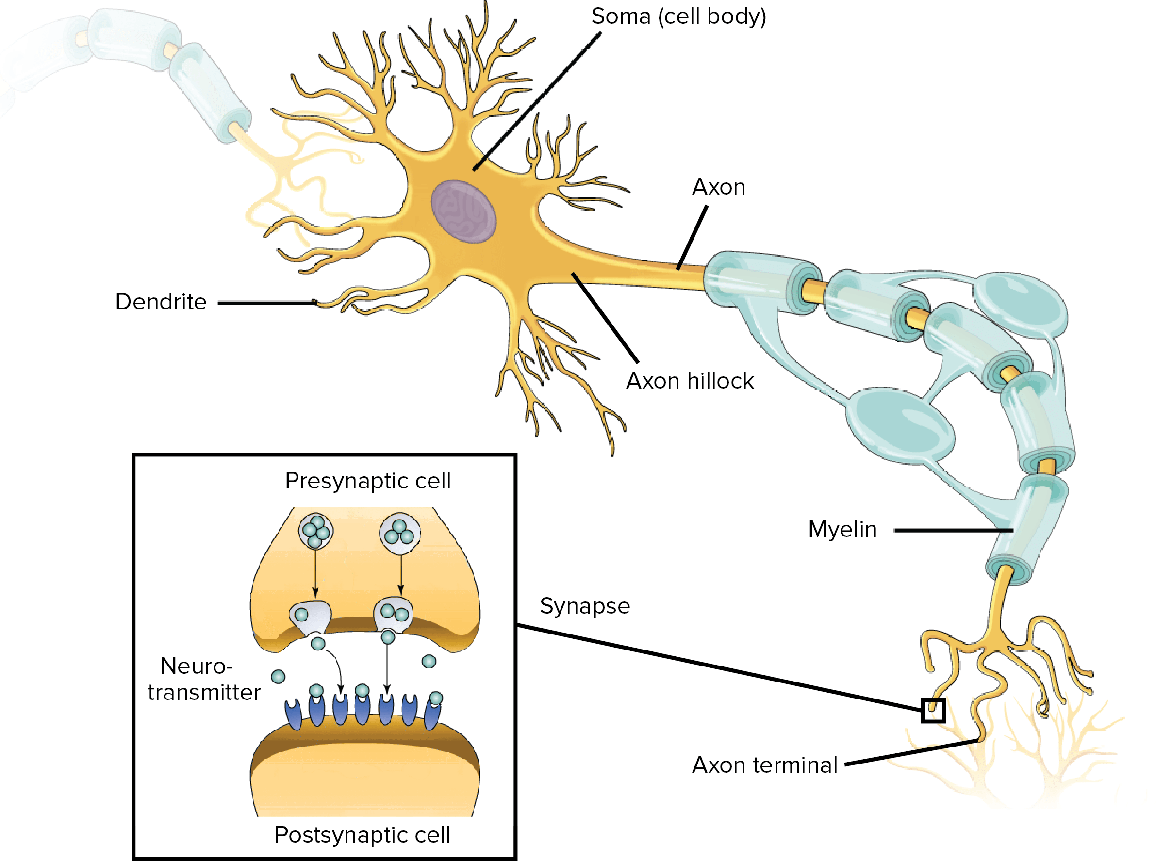
Neurons, similar to other cells, consist of a cell body known as the soma. The nucleus of the neuron is found in the soma. Neurons need to create proteins and most neuronal proteins are synthesized in the soma. Various processes, known as appendages or protrusions, run from the cell body. These include many small, branching processes, known as dendrites, and another process which is generally longer than the dendrites, known as the axon. It is possible to generalize that most neurons have three standard functions. These neuronal functions are mirrored in the anatomy of the neuron, including: �
Communicating information or signals.
Combining incoming signals to determine whether or not the information should be passed along.
Communicate information or signals to target cells, including muscles, glands, or other neurons.
Dendrites
The first two functions of the neuron, receive and process incoming signals or information, generally occur in the dendrites and cell body. Incoming signals can be either excitatory, which means that they tend to make the neuron generate an electrical impulse, or even inhibitory, which means that they tend to keep the neuron from generating an electrical impulse. �
Most neurons receive many incoming signals or information throughout the dendrites. A single neuron can have more than one pair of dendrites and they may receive thousands of incoming information or signals. Whether or not a neuron is excited into firing an electrical impulse is dependent on the amount of each of the excitatory and inhibitory signals, or information, it receives. If the neuron does end up firing an electrical impulse, the action potential or nerve impulse runs down the axon. �
Axons
The axon separates into many branches and develops bulbous swellings known as axon terminals or neural terminals. These axon terminals communicate with target cells. Axons are different from dendrites in several ways, as demonstrated below. �
The dendrites generally taper and are frequently covered with little bumps known as spines. The axon generally stays the same diameter for most of its length and doesn’t have spines.
The axon exits from the cell body through a special region known as the axon hillock.
Last but not least, many axons are covered with a special insulating compound known as the myelin, which helps them communicate the nerve impulse quickly. The myelin is never found on dendrites.
Synapses
Neuron-to-neuron communications are created on the dendrites and cell bodies of other neurons. These connections, known as synapses, are regions where information is taken from the first neuron, or the presynaptic neuron, to the target neuron, or the postsynaptic neuron. The synaptic connections between neurons and skeletal muscles are known as neuromuscular junctions and the connections between neurons and smooth muscle cells or glands are known as neuroeffector junctions. �
Signals communicate through chemical messengers known as neurotransmitters. When an action potential runs down an axon and reaches the axon terminal, it triggers the release of neurotransmitters from the presynaptic cell. Neurotransmitters run through the synapse and connect to membrane receptors on the postsynaptic cell, communicating excitatory or inhibitory information. The first two basic functions of the neuron are important for the third basic function of the neuron. �
The third function of the neuron, communicating signals to target cells, is also completed through the function of the axon and the axon terminals. Just as one neuron may communicate through many presynaptic neurons, it may also ultimately communicate through synaptic connections on numerous postsynaptic neurons throughout different axon terminals. �
Glial Cells
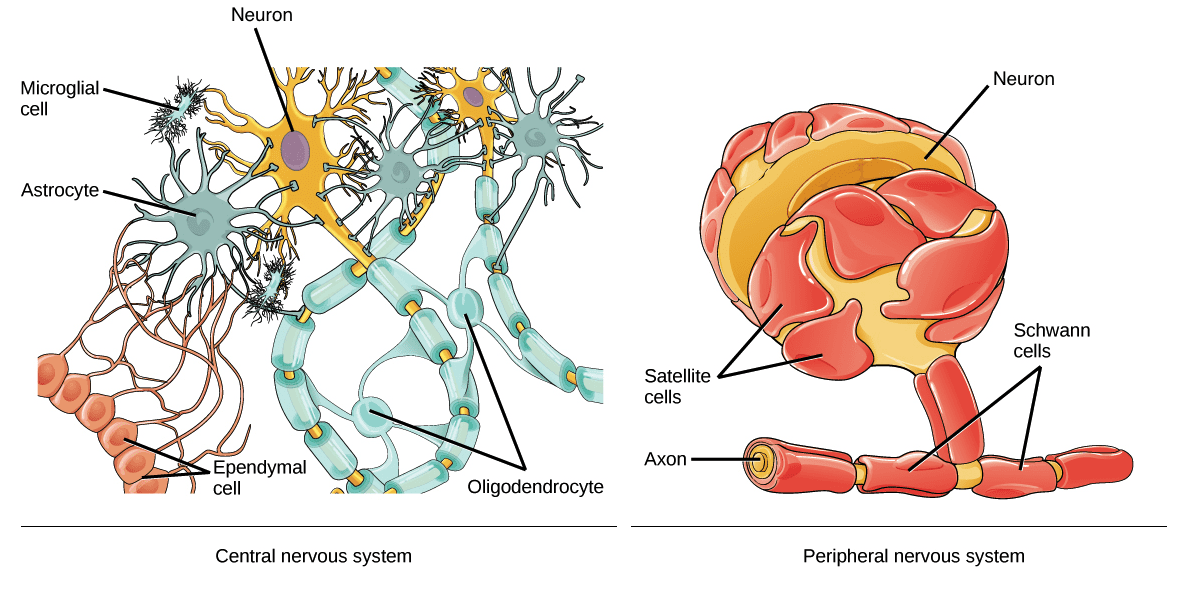
The glia, or glial cells, are fundamental to the nervous system. There are more glial cells in the brain than there are neurons. There are four types of glial cells in the adult human nervous system. Three of these, the astrocytes, the oligodendrocytes, and the microglia, are only found in the central nervous system or the CNS. The fourth, the Schwann cells, are only found in the peripheral nervous system or the PNS. Below, we will discuss the four types of glial cells, or glia, and their functions. �
Astrocytes are the most numerous types of glial cell. There are also many different types of astrocytes and they each have a variety of different functions, such as regulating blood flow in the brain, maintaining the composition of the fluid which surrounds the neurons, and maintaining communications between nerves in the synapse. During development, astrocytes help neurons find their way and add to the development of the blood-brain barrier, which also helps protect the brain. � Microglia are associated to the macrophages of the immune system and act as scavengers to remove dead cells and debris. �
The oligodendrocytes of the CNS and the Schwann cells of the PNS share a similar function. Both types of glia, or glial cells, create myelin, or the insulating compound which develops a sheath around the axons of many neurons. Myelin increases the speed with which an action potential runs down the axon and it plays a fundamental role in nervous system function. �
Additional types of glial cells, along with the four main types of glia, include satellite glial cells and ependymal cells. �
Satellite glial cells cover the cell bodies of neurons in PNS ganglia. Satellite glial cells are believed to support the role of the nerves and function as a protective barrier, however, their role is still misunderstood. Ependymal cells, which line the ventricles of the brain and the central canal of the spinal cord, have hairlike cilia which help improve the flow of the cerebrospinal fluid found within the ventricles and spinal tract. The human nervous system is necessary for our function. �
Neurons are special cells found within the nervous system which communicate with other neurons in unique ways. The neuron is the basic working unit of the brain and it is designed to communicate information, or signals, to muscles, organs, gland, and other nerve cells. Most neurons consist of a cell body, an axon, and dendrites. The cell body contains the nucleus and the cytoplasm. Understanding the structure and function of the neuron is fundamental for overall health and wellness. – Dr. Alex Jimenez D.C., C.C.S.T. Insight
The purpose of the article above is to discuss the purpose of functional neurology in the treatment of neurological disease. Neurological diseases are associated with the brain, the spine, and the nerves. The scope of our information is limited to chiropractic, musculoskeletal and nervous health issues as well as functional medicine articles, topics, and discussions. To further discuss the subject matter above, please feel free to ask Dr. Alex Jimenez or contact us at 915-850-0900 . �
Curated by Dr. Alex Jimenez �
Additional Topic Discussion: Chronic Pain
Sudden pain is a natural response of the nervous system which helps to demonstrate possible injury. By way of instance, pain signals travel from an injured region through the nerves and spinal cord to the brain. Pain is generally less severe as the injury heals, however, chronic pain is different than the average type of pain. With chronic pain, the human body will continue sending pain signals to the brain, regardless if the injury has healed. Chronic pain can last for several weeks to even several years. Chronic pain can tremendously affect a patient’s mobility and it can reduce flexibility, strength, and endurance.
Formulas for Methylation Support
XYMOGEN�s Exclusive Professional Formulas are available through select licensed health care professionals. The internet sale and discounting of XYMOGEN formulas are strictly prohibited.
Proudly,�Dr. Alexander Jimenez makes XYMOGEN formulas available only to patients under our care.
Please call our office in order for us to assign a doctor consultation for immediate access.
If you are a patient of Injury Medical & Chiropractic�Clinic, you may inquire about XYMOGEN by calling 915-850-0900.
�
For your convenience and review of the XYMOGEN products please review the following link.*XYMOGEN-Catalog-Download �
* All of the above XYMOGEN policies remain strictly in force. �
Exercises that focus mainly on the legs moving away from the body can increase the chance for individuals to experience sciatica, especially in athletes who have weaker interior thigh muscles.�Lifting is another physical activity that frequently causes sciatica. Athletes who have weaker core muscles can also increase their chance of experiencing a herniated disc while lifting. Improper lifting�
IFM's Find A Practitioner tool is the largest referral network in Functional Medicine, created to help patients locate Functional Medicine practitioners anywhere in the world. IFM Certified Practitioners are listed first in the search results, given their extensive education in Functional Medicine




 �
�