Individuals that experience nerve pain in the foot could be caused by a number of different conditions, can recognizing the most common causes help in developing an effective treatment plan?
Nerve Pain In The Foot
These sensations can feel like a burning, shooting, electrical, or stabbing pain and can happen while in motion or at rest. It can occur on the top of the foot or through the arch. The area closest to the nerve may be sensitive to the touch. A number of different conditions can cause nerve pain in the foot, including:
Morton’s neuroma
Pinched nerve
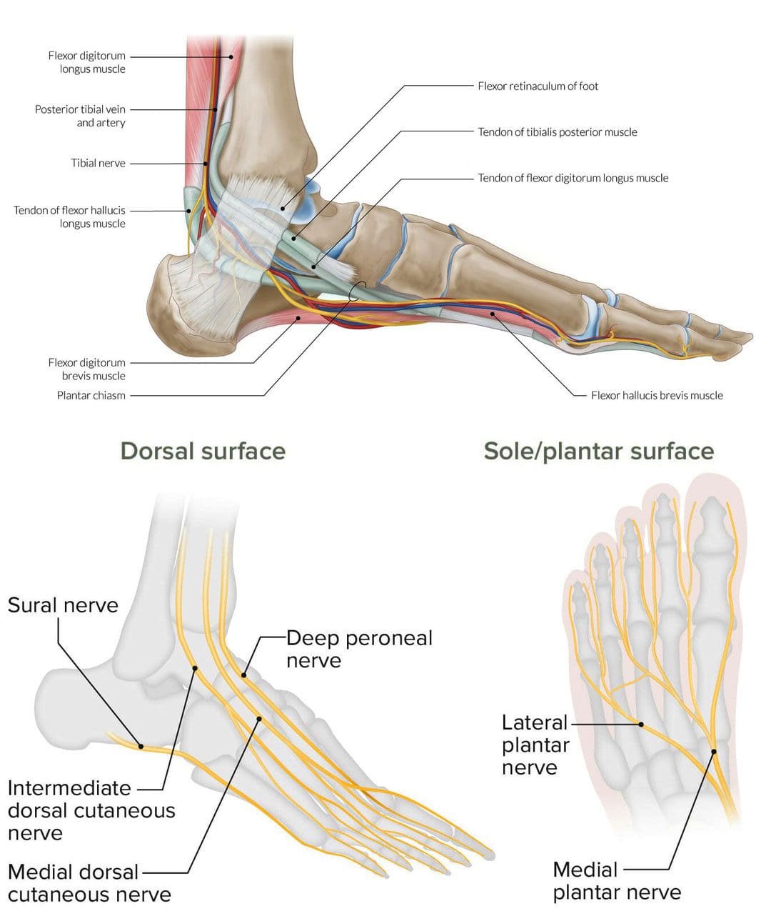
Tarsal tunnel syndrome
Diabetic peripheral neuropathy
Herniated disc
Morton’s Neuroma
Morton’s neuroma involves the nerve that runs between the third and fourth toes, but can sometimes occur between the second and third toes becoming thicker. Typical symptoms include a burning or shooting pain in the area, usually while walking. (Nikolaos Gougoulias, et al., 2019) Another common symptom is the sensation of pressure beneath the toes like the sock is bunched up underneath. Treatments can include:
Arch supports
Cortisone injections to decrease swelling
Footwear modifications – can include lifts, orthotics combined with metatarsal pads, and rocker soles, to provide cushion where needed.
Things that increase the risk of developing the condition include:
Regularly wearing high-heels – the condition occurs more frequently in women.
Shoes that are too tight.
Participating in high-impact sports like running.
Having flat feet, high arches, bunions, or hammertoes.
Pinched Nerve
A pinched nerve can feel like shooting or burning pain. Nerve entrapment can occur in various regions of the foot or the area on top of the foot may feel sensitive. Causes can be caused by: (Basavaraj Chari, Eugene McNally. 2018)
Trauma that causes swelling.
Blunt impact.
Tight shoes.
Treatment can include:
Massage
Physical therapy
Rest
Footwear modifications
Anti-inflammatories.
Things that increase the risk of developing a pinched nerve in the foot include:
Poor-fitting footwear.
Repetitive stress injury.
Trauma to the foot.
Obesity.
Rheumatoid arthritis.
Tarsal Tunnel Syndrome
Another type of nerve entrapment is tarsal tunnel syndrome. Tarsal tunnel syndrome is “anything that produces compression on the posterior tibial nerve.” (American College of Foot and Ankle Surgeons. 2019) The tibial nerve is located near the heel. Symptoms include numbness and foot cramps, burning, tingling, or shooting sensations that often radiate from the instep/arch. Both can worsen while the foot is at rest, like when sitting or sleeping. Treatment can consist of:
Placing padding in the shoe where the foot is being compressed to relieve the pain.
Custom foot orthotics.
Cortisone shots or other anti-inflammatory treatments.
Surgery may be necessary to release the nerve.
Conditions that compress the tibial nerve and can lead to tarsal tunnel syndrome include:
Flat feet
Fallen arches
Ankle sprain
Diabetes
Arthritis
Varicose veins
Bone spurs
Diabetic Peripheral Neuropathy
Long-term high blood sugar/glucose associated with diabetes can lead to a form of nerve damage known as peripheral neuropathy. (Centers for Disease Control and Prevention. 2022) Neuropathy pain feels like burning or shooting pain, or the sensation of walking on bubble wrap that usually shows up overnight. The pain can come and go as well as a gradual loss of feeling in the feet that begins in the toes and moves up the foot. It’s estimated that around half of individuals with diabetes will eventually develop neuropathy. (Eva L. Feldman, et al., 2019) Treatments can include:
Physical therapy massage to increase circulation.
Topical treatments with capsaicin.
Vitamin B.
Blood sugar management.
Alpha lipoic acid.
Medication.
Individuals with diabetes have an increased risk of developing peripheral neuropathy if:
Blood sugar is not well-controlled.
Diabetes has been present for many years.
Kidney disease.
Smoke.
Overweight or obese.
Herniated Disc
Nerve pain in the foot can be caused by spinal issues. A herniated disc in the lower back can irritate and compress the nerves, causing pain that radiates down the leg and foot. Additional symptoms usually include muscle weakness in the legs and/or numbness and tingling. Most herniated discs don’t require surgery and get better with conservative treatment. (Wai Weng Yoon, Jonathan Koch. 2021) If symptoms don’t improve or worsen, a healthcare provider may recommend surgery. Herniated discs are most common in young and middle-aged adults. Increased chances of developing a herniated disc can come from:
Degenerative changes in the spine from normal age wear and tear.
Physically demanding job.
Lifting incorrectly.
Overweight or obese.
Genetic predisposition – family history of herniated discs.
Spinal Stenosis
Spinal stenosis occurs when the spaces in the spine begin to narrow, creating pressure on the spinal cord and nerve roots. It is usually caused by wear and tear on the spine as the body ages. Stenosis in the lower back can cause burning pain in the buttocks and leg. As it progresses pain can radiate into the feet along with numbness and tingling. Conservative treatment consists of physical therapy exercises and non-steroidal anti-inflammatory medications/NSAIDs. (Jon Lurie, Christy Tomkins-Lane. 2016) Cortisone injections can be beneficial and if the condition worsens, surgery may be an option. Risk factors include:
Physical trauma – after surgery or an automobile or sports accident.
Certain cancer, antiviral medications, or antibiotics.
Complex regional pain syndrome.
Tumors that irritate and/or compress a nerve.
Liver or kidney disease.
Infectious diseases – Lyme disease complications or viral infections.
Nerve pain in the foot is definitely a reason to see a healthcare provider. Early diagnosis can help prevent symptom progression and future problems. Once the cause of the pain has been identified, the healthcare team can work together to develop a personalized treatment plan to release compressed nerves and restore mobility and function. See a healthcare provider right away if the pain and symptoms worsen, or if there are difficulties standing or walking.
Chiropractic After Accidents and Injuries
References
Gougoulias, N., Lampridis, V., & Sakellariou, A. (2019). Morton’s interdigital neuroma: instructional review. EFORT open reviews, 4(1), 14–24. https://doi.org/10.1302/2058-5241.4.180025
Chari, B., & McNally, E. (2018). Nerve Entrapment in Ankle and Foot: Ultrasound Imaging. Seminars in musculoskeletal radiology, 22(3), 354–363. https://doi.org/10.1055/s-0038-1648252
Feldman, E. L., Callaghan, B. C., Pop-Busui, R., Zochodne, D. W., Wright, D. E., Bennett, D. L., Bril, V., Russell, J. W., & Viswanathan, V. (2019). Diabetic neuropathy. Nature reviews. Disease primers, 5(1), 42. https://doi.org/10.1038/s41572-019-0097-9
Yoon, W. W., & Koch, J. (2021). Herniated discs: when is surgery necessary?. EFORT open reviews, 6(6), 526–530. https://doi.org/10.1302/2058-5241.6.210020
Lurie, J., & Tomkins-Lane, C. (2016). Management of lumbar spinal stenosis. BMJ (Clinical research ed.), 352, h6234. https://doi.org/10.1136/bmj.h6234
Staff, N. P., & Windebank, A. J. (2014). Peripheral neuropathy due to vitamin deficiency, toxins, and medications. Continuum (Minneapolis, Minn.), 20(5 Peripheral Nervous System Disorders), 1293–1306. https://doi.org/10.1212/01.CON.0000455880.06675.5a
Can combining chiropractic treatment with the common therapies of medication, exercise, and/or physical therapy help relieve sciatic endometriosis pain symptoms?
Sciatic Endometriosis
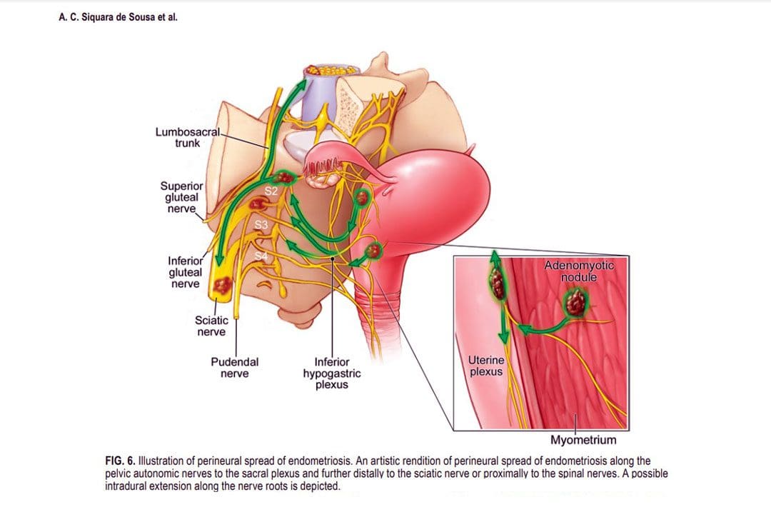
Sciatic endometriosis is a condition in which endometrial cells (tissue that resembles the lining of the uterus) grow outside of the uterine lining and compress the sciatic nerve. This places stress and pressure on the nerve causing back, pelvic, hip, and leg pain, especially before and during the menstrual cycle. It can also cause pain, irregular periods, and infertility. (The American College of Obstetricians and Gynecologists. 2021)
These areas of endometrial tissue growth are also known as lesions or implants.
Women with sciatic endometriosis often experience leg pain and weakness around the time of their menstrual cycle. (Lena Marie Seegers, et al., 2023)
Sciatic endometriosis can also cause pain when urinating, during a bowel movement, during sex, and fatigue, and irregular vaginal bleeding.
The abnormal growth may be caused by higher-than-normal levels of estrogen.
Researchers believe that endometriosis is related to retrograde menstruation, which causes menstrual blood to flow back into the pelvis instead of out through the vagina. (World Health Organization. 2023)
Sometimes, the cells grow in the area of the pelvis right above the sciatic nerve. (Adaiah Yahaya, et al., 2021)
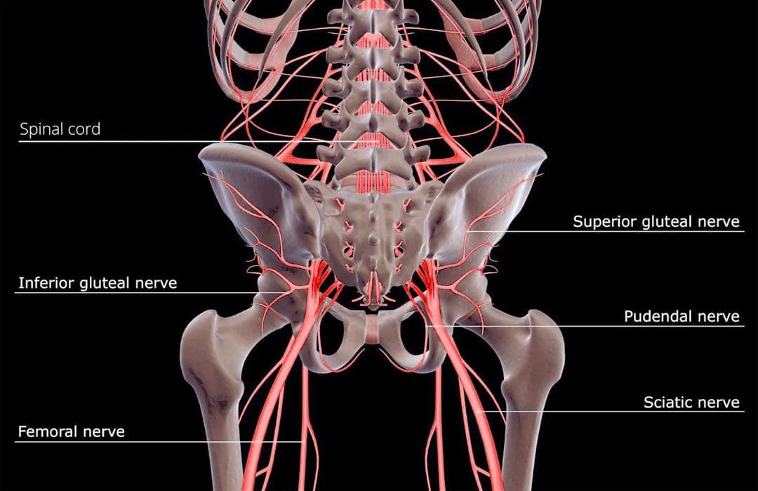
The sciatic nerve is the longest nerve in the body and travels down the back of each leg. (Johns Hopkins Medicine. 2023)
When endometrial lesions place pressure on the sciatic nerve, they can cause irritation and inflammation leading to severe pelvic pain, which makes it harder to conceive. (Liang Yanchun, et al., 2019)
Symptoms
Some women with endometriosis experience no symptoms or misinterpret the symptoms as typical premenstrual syndrome/PMS signs. The most common signs and symptoms of sciatic endometriosis include:
Difficulty walking or standing.
Loss of sensation, muscle weakness, and reflex alteration.
Limping.
Balance problems.
Bloating and nausea.
Constipation or diarrhea before or after a period.
Painful, heavy, and/or irregular periods.
Bleeding between periods.
Pain during sex, urination, and bowel movements.
Pain in the stomach, pelvis, lower back, hips, and buttocks. (MedlinePlus. 2022)
Weakness, numbness, tingling, burning, or dull aching sensations in the back of one or both legs.
Endometriosis, including sciatic endometriosis, typically cannot be diagnosed with a pelvic examination or ultrasound by themselves. A healthcare provider may need to perform a biopsy using laparoscopy and discuss menstrual cycles, symptoms, and medical history.
The laparoscopy procedure involves making tiny incisions and taking a tissue sample with tools attached to a thin tube with a camera. (MedlinePlus. 2022)
Imaging tests, like magnetic resonance imaging/MRI, and computed tomography/CT scans, can help provide essential information about the location and size of any endometrial lesions. (The American College of Obstetricians and Gynecologists. 2021)
Treatment
Symptoms can sometimes be temporarily relieved with over-the-counter/OTC pain relievers. Depending on the condition and severity a healthcare provider may prescribe hormonal treatment to prevent new endometrial implants from growing. These can include:
Hormonal birth control.
Progestin – a synthetic form of progesterone.
Gonadotropin-releasing hormone – GnRH agonists.
If pain persists or worsens, individuals may need to undergo surgery to remove the tissue.
The American College of Obstetricians and Gynecologists. Endometriosis.
Seegers, L. M., DeFaria Yeh, D., Yonetsu, T., Sugiyama, T., Minami, Y., Soeda, T., Araki, M., Nakajima, A., Yuki, H., Kinoshita, D., Suzuki, K., Niida, T., Lee, H., McNulty, I., Nakamura, S., Kakuta, T., Fuster, V., & Jang, I. K. (2023). Sex Differences in Coronary Atherosclerotic Phenotype and Healing Pattern on Optical Coherence Tomography Imaging. Circulation. Cardiovascular imaging, 16(8), e015227. https://doi.org/10.1161/CIRCIMAGING.123.015227
World Health Organization. Endometriosis.
Yahaya, A., Chauhan, G., Idowu, A., Sumathi, V., Botchu, R., & Evans, S. (2021). Carcinoma arising within sciatic nerve endometriosis: a case report. Journal of surgical case reports, 2021(12), rjab512. https://doi.org/10.1093/jscr/rjab512
Johns Hopkins Medicine. Sciatica.
Yanchun, L., Yunhe, Z., Meng, X., Shuqin, C., Qingtang, Z., & Shuzhong, Y. (2019). Removal of an endometrioma passing through the left greater sciatic foramen using a concomitant laparoscopic and transgluteal approach: case report. BMC women’s health, 19(1), 95. https://doi.org/10.1186/s12905-019-0796-0
MedlinePlus. Endometriosis.
Center for Endometriosis Care. Sciatic endometriosis.
Chen, S., Xie, W., Strong, J. A., Jiang, J., & Zhang, J. M. (2016). Sciatic endometriosis induces mechanical hypersensitivity, segmental nerve damage, and robust local inflammation in rats. European journal of pain (London, England), 20(7), 1044–1057. https://doi.org/10.1002/ejp.827
Siquara de Sousa, A. C., Capek, S., Howe, B. M., Jentoft, M. E., Amrami, K. K., & Spinner, R. J. (2015). Magnetic resonance imaging evidence for perineural spread of endometriosis to the lumbosacral plexus: report of 2 cases. Neurosurgical focus, 39(3), E15. https://doi.org/10.3171/2015.6.FOCUS15208
Are treatments more successful when patients know key terms that describe their back pain and associated conditions?
Nerve Pain Types
When individuals need to better understand their spine diagnosis, being able to distinguish between key terms can make a significant difference in understanding the development of a personalized treatment plan. Terms that describe back pain and various associated conditions can include:
Sciatica
Radiating and Referred pain
Radiculopathy
Radiculitis
Neuropathy
Neuritis
Causes of Back Pain
Back pain symptoms are most commonly caused by the continued practice of unhealthy/poor posture and overcompensated and weakened muscles. Even for individuals that exercise regularly, the movement choices that are made throughout the day can disrupt the way the muscles, tendons, ligaments, and fascia function to maintain proper body alignment.
Injuries to, and conditions of, the structures of the spinal column like the bones, discs, and nerves, are generally more serious than posture problems and soft tissue-related pain.
Depending on the diagnosis, structural problems can cause symptoms related to nerve compression, irritation, and/or inflammation. (Michigan Medicine, 2022)
Spine and Nervous System
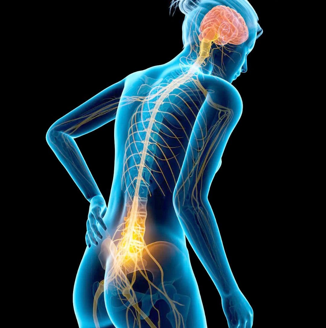
The peripheral nerves extend out to the extremities with sensation and movement capabilities.
Nerve roots exit the spinal canal which is part of the peripheral nervous system.
The branching of nerves from the spinal cord and exit out of the foramina occurs at every level of the spine.
Terms
There are different medical terms when getting a spine diagnosis or going through the treatment process.
Radiculopathy
Radiculopathy is an umbrella term, describing any disease process that affects a spinal nerve root and is something that’s happening to the body.
When a healthcare provider informs you that your pain is due to radiculopathy, a number of more specific diagnoses, clinical signs, and symptoms may be included as part of the description.
Common causes of radiculopathy include herniated disc/s and spinal stenosis.
Less common causes can include a synovial cyst or tumor that presses on the nerve root. (Johns Hopkins Medicine, 2023)
Radiculopathy can occur in the neck, low back, or in the thoracic area.
Often, radiculopathy is brought on by some form of compression of the nerve root.
For example, extruded material from a herniated disc can land on a nerve root, causing pressure to build.
This can cause symptoms associated with radiculopathy, including numbness, weakness, pain, or electrical sensations. (Johns Hopkins Medicine, 2023)
Even though there’s a spinal nerve root on either side of the spinal column, injury, trauma, or issues stemming from degeneration affect the nerves in an asymmetric fashion. Degenerative changes, known as normal wear and tear, typically occur in this fashion. Using the previous herniated disc example, the material that leaks from the disc structure tends to travel in one direction. When this is the case, the symptoms tend to be experienced on the side where the nerve root makes contact with the disc material, but not the other side. (American Association of Neurological Surgeons, 2023)
Radiculitis
Radiculitis is a form of radiculopathy but it is about inflammation and not compression. (Johns Hopkins Medicine, 2023)
Radicu– refers to the spinal nerve root.
The suffix – itis refers to inflammation.
The word refers to a spinal nerve root that is inflamed and/or irritated rather than compressed.
In disc herniations, it is the gel substance that contains various chemicals that is inflammatory.
When the gel substance makes contact with nerve roots, an inflammatory response is triggered. (Rothman SM, Winkelstein BA 2007)
Radiating or Referred Pain
Radiating pain follows the path of one of the peripheral nerves that transmit sensory information like heat, cold, pins and needles, and pain.
Referred pain is experienced in a different area of the body that is away from the pain source which tends to be an organ. (Murray GM., 2009)
It can be brought on by myofascial trigger points or visceral activity.
An example of referred pain is symptoms in the jaw or arm when an individual is having a heart attack. (Murray GM., 2009)
Radicular
The terms radicular pain and radiculopathy tend to get confused.
Radicular pain is a symptom of radiculopathy.
Radicular pain radiates from the spinal nerve root to either part or all the way down the limb/extremity.
However, radicular pain does not represent the complete symptoms of radiculopathy.
Radiculopathy symptoms also include numbness, weakness, or electrical sensations like pins and needles, burning, or shock that travels down the extremity. (Johns Hopkins Medicine, 2023)
Neuropathy
Neuropathy is another umbrella term that refers to any dysfunction or disease that affects the nerves.
It’s usually classified according to the cause, like diabetic neuropathy, or the location.
Neuropathy can occur anywhere in the body – including the peripheral nerves, the autonomic nerves/organ nerves, or nerves that are located inside the skull and innervate the eyes, ears, nose, etc.
Peripheral nerves are the long, thin strands that supply sensation, feeling, and movement impulses to all areas of the body located outside the central nervous system.
Piriformis syndrome is where a tight buttock/piriformis muscle constricts the sciatic nerve, which runs underneath. (Cass SP. 2015)
Chiropractic
Chiropractic adjustments, non-surgical decompression, MET, and various massage therapies can relieve symptoms, release stuck or trapped nerves and restore function. Through the treatments, the chiropractor and therapists will explain what is happening and why they are using a specific technique. Knowing a little about how the neuromusculoskeletal system operates can help the healthcare provider and the patient in developing and adjusting effective treatment strategies.
Sciatica During Pregnancy
References
Michigan Medicine. Upper and Middle Back Pain.
American Academy of Neurological Surgeons. Anatomy of the Spine and Peripheral Nervous System.
Johns Hopkins Medicine. Health Conditions. Radiculopathy.
American Association of Neurological Surgeons. Herniated Disc.
American Academy of Orthopaedic Surgeons. OrthoInfo. Cervical Radiculopathy (Pinched Nerve).
Rothman, S. M., & Winkelstein, B. A. (2007). Chemical and mechanical nerve root insults induce differential behavioral sensitivity and glial activation that are enhanced in combination. Brain Research, 1181, 30–43. https://doi.org/10.1016/j.brainres.2007.08.064
Murray G. M. (2009). Guest Editorial: referred pain. Journal of applied oral science: Revista FOB, 17(6), i. https://doi.org/10.1590/s1678-77572009000600001
American Academy of Orthopaedic Surgeons. OrthoInfo. Carpal Tunnel Syndrome.
Bostelmann, R., Zella, S., Steiger, H. J., & Petridis, A. K. (2016). Could Spinal Canal Compression be a Cause of Polyneuropathy? Clinics and practice, 6(1), 816. https://doi.org/10.4081/cp.2016.816
Cleveland Clinic. Mononeuropathy.
American Association of Neurological Surgeons. Glossary of Neurosurgical Terminology.
National Institutes of Health. U.S. National Library of Medicine. Medline Plus. Peripheral Nerve Disorders.
Cleveland Clinic. Spinal Stenosis.
Cass S. P. (2015). Piriformis syndrome: a cause of non-discogenic sciatica. Current sports medicine reports 14(1), 41–44. https://doi.org/10.1249/JSR.0000000000000110
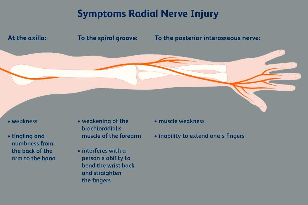
The brachial plexus is a network of nerves that begin in the cervical/neck spinal cord and travel down the cervicoaxillary canal into the armpit. Forming in the area of the shoulder joint at the branch junction of the brachial plexus, the radial nerve extends down the arm, through the elbow joint, into the forearm, across the wrist, and tips of the fingers. The nerves are susceptible to injury that can cause abnormal function leading to unusual sensations and impaired muscle function.
Radial Nerve
One of the major nerves of the upper extremity.
There is one brachial plexus on each side of the body that carries the nerves to each arm.
The radial nerve has two major functions.
One is to provide sensations in the hands, forearms, arms, and fingers.
The other is to deliver messages to muscles about when to contract.
Motor Function
The radial nerve transmits signals to the muscles of the back of the arm and forearm on when to contract.
Individuals who have abnormal radial nerve function can experience weakness of the muscles and symptoms like wrist drop.
A wrist drop occurs when the back forearm muscles cannot support the wrist, causing the individual to hold the wrist in a flexed posture.
Abnormal radial nerve function can cause symptoms of numbness or tingling in the back of the hand.
Conditions
Associated conditions to the radial nerve include lacerations, contusions, fractures, and palsies.
Nerve Contusion
A contusion typically occurs through blunt force trauma that can crush and smash the nerve area.
This causes abnormal or no function.
A nerve contusion can occur from a personal, work, or sports injury or other conditions that generate intense pressure on the nerve/s.
Nerve Lacerations
A laceration occurs when there is a penetrating injury that cuts and/or severs the nerve.
This injury can occur from stab wounds or sliced by broken glass, metal, etc.
Fractures
Broken bones of the upper extremity can lead to extended damage to the nerves near the damaged bone.
The most common type of fracture associated with radial nerve malfunction is fractures to the humerus bone.
The nerve wraps tightly around the humerus and can be injured with a fracture.
Most fracture-related radial nerve injuries heal on their own and do not require surgery.
However, the way the injury heals can be the difference between normal function and chronic pain.
Crutch Palsy
Crutch palsy is pressure on the radial nerve in the armpit resulting from using crutches incorrectly.
To use crutches properly, the individual needs to support their body weight through the hands.
However, many tend to place pressure around the armpit at the top of the crutch, causing irritation to the nerve in that area.
Padding the top of crutches and using the proper form can prevent the condition.
Saturday Night Palsy
Saturday night palsy is the abnormal function of the radial nerve after sleeping in a position that causes direct pressure against the nerve.
This often occurs when an individual falls asleep with their arm draped over an armrest on a chair.
The name comes from when individuals are intoxicated and fall asleep in a location other than the bed and in awkward positions.
Treatment
Nerve injuries often cause symptoms at different locations other than where the nerve damage is, complicating diagnosis. Determining the specific location of nerve damage is the first step in developing an appropriate treatment plan. Once the location has been identified, steps can be taken to prevent worsening damage to the nerve.
The objective is to relieve the pressure from the irritation or compression.
Massage to relax the area and increase blood circulation.
Decompression to physically restore alignment.
Adjustments to restore body balance.
Exercises and stretches to maintain treatment, strengthen the muscles, and prevent injuries.
In cases where there is structural damage, surgery may be necessary to remove pressure or repair damage.
Avoid Surgery
References
Ansari FH, Juergens AL. Saturday Night Palsy. [Updated 2023 Apr 24]. In: StatPearls [Internet]. Treasure Island (FL): StatPearls Publishing; 2023 Jan-. Available from: https://www.ncbi.nlm.nih.gov/books/NBK557520/
Barton, N J. “Radial nerve lesions.” The Hand vol. 5,3 (1973): 200-8. doi:10.1016/0072-968x(73)90029-6
Daly, Michael, and Chris Langhammer. “Radial Nerve Injury in Humeral Shaft Fracture.” The Orthopedic Clinics of North America vol. 53,2 (2022): 145-154. doi:10.1016/j.ocl.2022.01.001
DeCastro A, Keefe P. Wrist Drop. [Updated 2022 Jul 18]. In: StatPearls [Internet]. Treasure Island (FL): StatPearls Publishing; 2023 Jan-. Available from: https://www.ncbi.nlm.nih.gov/books/NBK532993/
Eaton, C J, and G D Lister. “Radial nerve compression.” Hand Clinics vol. 8,2 (1992): 345-57.
Glover NM, Murphy PB. Anatomy, Shoulder and Upper Limb, Radial Nerve. [Updated 2022 Aug 29]. In: StatPearls [Internet]. Treasure Island (FL): StatPearls Publishing; 2023 Jan-. Available from: https://www.ncbi.nlm.nih.gov/books/NBK534840/
Ljungquist, Karin L et al. “Radial nerve injuries.” The Journal of hand surgery vol. 40,1 (2015): 166-72. doi:10.1016/j.jhsa.2014.05.010
A nerve becomes pinched/compressed when added pressure is placed on it by surrounding structures that can include muscles, bones, ligaments, tendons, or a combination. This injures and damages the nerve causing function problems and symptoms and sensations in that area or other parts of the body that are supplied by that nerve. Medical practitioners refer to this as nerve compression or entrapment. Although compressed nerves are more commonly associated with the neck, arms, hands, elbows, and lower back, any nerve in the body can experience irritation, spasms, inflammation, and compression. The causes and treatment of a compressed nerve in the knee.
Compressed Nerve In The Knee
There’s only one nerve that goes through the knee that has an increased risk of getting compressed. It’s a branch of the sciatic nerve called the peroneal nerve. The nerve goes around the outside of the knee before traveling down the outside of the lower leg. At the bottom of the knee, it lies between the bone and skin, making it vulnerable to irritation or compression by anything that can put pressure on the outside of the knee.
Causes
Traumatic injuries over time can lead to pressure on the nerve from inside the knee. Common causes of a compressed nerve in the knee include:
Frequently Crossing Legs
Compression by the opposite knee, while the legs are crossed is the most common cause.
Knee Brace
A too-tight or strong brace can compress the leg and nerve.
Thigh-High Compression Stockings
Designed to maintain pressure on the legs, if too tight these stockings can compress the nerve.
Squatting Posture For Long Periods
This position places pressure on the side of the knee.
Fractures
A fracture of the large lower leg bone/tibia or sometimes the small bone/fibula near the knee can entrap the nerve.
Lower Leg Cast
The portion of the cast around the knee can be tight and compress the nerve.
Tell the doctor if a cast or brace feels tight or is causing numbness or pain in the leg.
Knee-High Boots
The top of a boot can land right below the knee and be too tight pinching the nerve.
Knee Ligament Injury
The nerve can become compressed due to bleeding or inflammation from an injured ligament.
Knee Surgery Complications
This is rare, but the nerve can inadvertently get pinched during knee replacement surgery or an arthroscopic procedure.
Prolonged Bed Rest
When lying down the legs tend to rotate outward and the knees flex.
In this position, the mattress can place pressure on the nerve.
Tumors or Cysts
Tumors or cysts can develop right on top or next to a nerve irritating and compressing the area.
Abdominal or Gynecologic Surgery
The equipment used to keep the legs rotated outward and the knees flexed for gynecologic and abdominal surgeries can compress the nerve.
Symptoms
The peroneal nerve supplies sensation and movement to the outside of the lower leg and the top of the foot. When compressed, it becomes inflamed, which causes the symptoms of a compressed nerve. Usually, only the lining/myelin sheath around the nerve is what gets injured. However, when the nerve gets damaged, the symptoms are similar but more severe. Common symptoms include:
Weakness that limits the ability to lift the foot toward the leg aka dorsiflexion.
This causes dragging the foot when walking.
The ability to turn the foot outward and extend the big toe is also affected.
Symptoms can be felt on the outside of the lower leg and on the top of the foot and include:
Tingling or pins and needles sensations.
Numbness.
Loss of sensation.
Pain.
Burning.
For individuals that have had a pinched nerve for two or more weeks, the muscles supplied by the nerve can begin to waste away or atrophy.
Symptoms can be intermittent or continuous depending on the cause.
The other common cause is a pinched nerve in the lumbar/lower spine.
When this is the cause, sensations, and pain will present in the lower back or the back and outside of the thigh.
Diagnosis
A doctor will look at medical history and perform an examination to make a diagnosis, determine the cause, and lay out a personalized treatment plan. The nerve in the knee can be felt as it travels around the top of the tibia, so a doctor may tap on it. If there is shooting pain down the leg, a pinched nerve may be present. Tests a doctor may order can include:
Knee X-ray
Shows any bone fractures or abnormal masses.
Knee MRI
Can confirm the diagnosis
Shows masses within the nerve.
Shows details of fractures or other problems in the bones.
Electromyogram – EMG
Tests electrical activity in the muscles.
Nerve Conduction Test
Tests the signal speed of the nerve.
Treatment
Treatment is aimed at reducing pain and improving mobility.
Over-the-Counter Pain Medication
OTC medication can reduce inflammation and improve symptoms short term.
Ice and Heat
Applying either heat or ice for 15 to 20 minutes at a time can provide relief from the symptoms.
An ice pack can make symptoms worse if it adds more pressure on the nerve.
Chiropractic and Physical Therapy
Chiropractic and physical therapy can release the compressed nerve, realign the structures, strengthen the muscles, and provide gait training.
Orthotic Boot
If walking gait is affected because the foot cannot bend, an orthotic boot can help.
This is a support that maintains the foot in a neutral position to walk normally.
Corticosteroid Injection
A corticosteroid injection can reduce inflammation and relieve pressure on the nerve.
Surgery
The nerve can suffer permanent damage if it has been pinched for a long time.
If that happens, surgery cannot repair the damage.
A doctor can perform surgery to correct a fracture, tumor, or other invasive problem causing a compressed nerve.
If conservative treatment doesn’t work, a peroneal nerve decompression procedure can be done to remove the pressure.
If surgery is needed, symptoms can disappear immediately, but it takes around four months to recover and rehabilitate.
Injury Rehabilitation
References
Krych, Aaron J et al. “Is peroneal nerve injury associated with worse function after knee dislocation?.” Clinical orthopedics and related research vol. 472,9 (2014): 2630-6. doi:10.1007/s11999-014-3542-9
Lezak B, Massel DH, Varacallo M. Peroneal Nerve Injury. [Updated 2022 Nov 14]. In: StatPearls [Internet]. Treasure Island (FL): StatPearls Publishing; 2023 Jan-. Available from: https://www.ncbi.nlm.nih.gov/books/NBK549859/
Soltani Mohammadi, Sussan, et al. “Comparing the squatting position and traditional sitting position for ease of spinal needle placement: a randomized clinical trial.” Anesthesiology and pain medicine vol. 4,2 e13969. 5 Apr. 2014, doi:10.5812/aapm.13969
Stanitski, C L. “Rehabilitation following knee injury.” Clinics in sports medicine vol. 4,3 (1985): 495-511.
Xu, Lin, et al. Zhongguo gu Shang = China Journal of Orthopedics and Traumatology vol. 33,11 (2020): 1071-5. doi:10.12200/j.issn.1003-0034.2020.11.017
Yacub, Jennifer N et al. “Nerve injury in patients after hip and knee arthroplasties and knee arthroscopy.” American Journal of physical medicine & Rehabilitation vol. 88,8 (2009): 635-41; quiz 642-4, 691. doi:10.1097/PHM.0b013e3181ae0c9d
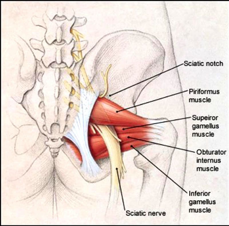
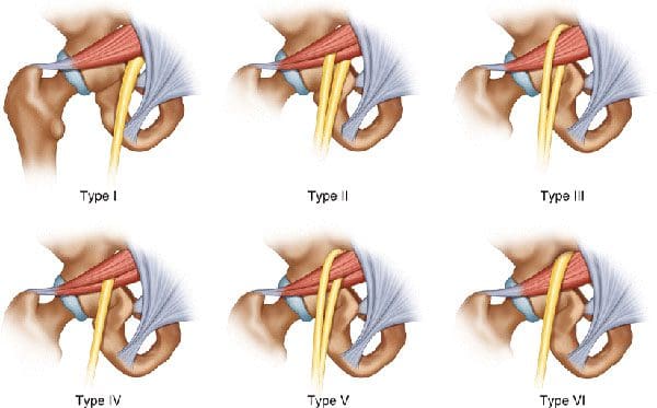
The piriformis is a large and powerful muscle beneath the gluteal/buttocks muscles. It runs from the bottom of the sacrum, where the base of the spine and pelvis converge to the top of the femur. This muscle plays a critical role in running motion; it helps externally rotate the hips and upper leg outward, provides hip flexibility and stability, and stabilizes the pelvis. The sciatic nerve passes next to, over, under, or through the piriformis muscle. When the piriformis contracts or spasms, it can irritate, become entangled and compress the nerve, resulting in painful symptoms. This can lead to various problems and is how piriformis syndrome occurs.
Running Piriformis Syndrome
The proper function of the piriformis muscle is essential for athletes who participate in running sports. Repetitive activities, like running, can fatigue the muscle and irritate and inflame the nerve.
Symptoms
Piriformis syndrome can be challenging to diagnose because it can be confused for a herniated disc, sciatica, a proximal hamstring strain/high hamstring tendinitis, or lower back problems. A few symptoms that can help determine whether the piriformis is the cause include:
Sitting, Stairs, Squatting Discomfort or Pain
Individuals don’t always experience discomfort while running.
Instead, it’s sitting, climbing stairs, and squatting where pain symptoms present.
Pain while running, specifically an overstretched sensation when going up a hill or increasing speed, is more associated with a proximal hamstring strain.
Tenderness
The area around the piriformis is tender.
Applying pressure can cause discomfort or pain around the area and radiate down the leg.
Centered Pain
Piriformis syndrome is usually felt in the middle of the glutes.
A proximal hamstring strain typically causes non-radiating pain at the bottom of the glutes, where the hamstrings connect to the pelvis.
Causes
Pelvic misalignment.
Pelvic misalignments created by other conditions, like a tilted pelvis, functional leg-length discrepancy, or practicing unhealthy posture, make the piriformis work harder to compensate, which leads to tightness and/or spasms.
Sudden increases in distance or workout intensity can worsen any weakness in the piriformis and other gluteal muscles.
Continuing to run, which is possible, can worsen and prolong the condition.
When running, the muscle’s signal transmissions are interrupted by inflammation and/or compression and cannot synchronize with each other.
The result is the inability to withstand the repetitive strain of running.
Resting may not be enough to alleviate piriformis syndrome. This is especially true if the problem involves spine and pelvic misalignment. Chiropractic can provide significant relief from running piriformis syndrome. A combination of spinal, pelvic, and extremity adjustments, therapeutic massage, MET, decompression, stretches, and anti-inflammatory nutrition will take the pressure off overly tight areas, realign the body, and maintain nervous system function.
Running form could be evaluated and checked for leg-length discrepancies and muscle-strength imbalances.
Running can continue if the individual can do so without pain or symptoms.
But it is recommended to avoid slanted surfaces, which increase the risk of pelvic misalignment.
Avoid long runs, which increase the chance of overload and fatigue.
The goal is to relax and release the piriformis.
If it’s impinging on the sciatic nerve, loosening and releasing the muscle will significantly lessen radiating pain.
Orthotics may be recommended for excessive overpronation or inward movement of the foot when landing.
Other treatments to stop piriformis spasms.
Ice and take over-the-counter anti-inflammatory medications can be used during acute phases when the area is tender.
Work out tight spots using a foam roller or percussive massager.
Stretching and loosening the muscle before and after runs can help it relax and increase blood flow.
Ahmad Siraj, Sidra, and Ragini Dadgal. “Physiotherapy for Piriformis Syndrome Using Sciatic Nerve Mobilization and Piriformis Release.” Cureus vol. 14,12 e32952. 26 Dec. 2022, doi:10.7759/cureus.32952
Chang A, Ly N, Varacallo M. Piriformis Injection. [Updated 2022 Sep 4]. In: StatPearls [Internet]. Treasure Island (FL): StatPearls Publishing; 2023 Jan-.
Heiderscheit, Bryan, and Shane McClinton. “Evaluation and Management of Hip and Pelvis Injuries.” Physical Medicine and rehabilitation clinics of North America vol. 27,1 (2016): 1-29. doi:10.1016/j.pmr.2015.08.003
Julsrud, M E. “Piriformis syndrome.” Journal of the American Podiatric Medical Association vol. 79,3 (1989): 128-31. doi:10.7547/87507315-79-3-128
Kraus, Emily, et al. “Piriformis Syndrome With Variant Sciatic Nerve Anatomy: A Case Report.” PM & R: the Journal of Injury, Function, and Rehabilitation vol. 8,2 (2016): 176-9. doi:10.1016/j.pmrj.2015.09.005
Lenhart, Rachel, et al. “Hip muscle loads during running at various step rates.” The Journal of Orthopedic and sports physical therapy vol. 44,10 (2014): 766-74, A1-4. doi:10.2519/jospt.2014.5575
Sulowska-Daszyk, Iwona, and Agnieszka Skiba. “The Influence of Self-Myofascial Release on Muscle Flexibility in Long-Distance Runners.” International Journal of environmental research and public health vol. 19,1 457. Jan 1, 2022, doi:10.3390/ijerph19010457
The nervous system communicates with the entire body and reacts to internal and external changes using electrical and chemical impulses to send and receive messages. Messages travel/synapse from one neuron to another using specialized chemicals known as neurotransmitters. Paresthesia refers to sensations of numbness, tingling, prickling, skin crawling, itching, or burning, usually in the arms, hands, legs, and/or feet, but can affect other areas of the body. Chiropractic care, massage therapy, decompression therapy, and functional medicine can relieve tissue and nerve compression, improve flexibility, range of motion, and mobility, and strengthen the muscles surrounding the affected nerve to maintain optimal health and prevent worsening or further injury.
Paresthesia
The sensation comes on without warning and is usually painless and described as tingling or numbness. There are different causes of paresthesia, including:
Some individuals have chronic or long-term paresthesia, which can be a sign of a more serious nerve injury or condition. Added physical stress can cause surrounding tissues to irritate or entangle the nerve leading to building pressure. This pressure causes paresthesia in the area interrupting circulation and function. A pinched nerve can happen anywhere in the body, like the neck, shoulder, wrist, back, and face.
A herniated disc in the lower spine can cause back pain and paresthesia in the leg or foot on the affected side.
Carpal tunnel syndrome is a pinched nerve in the wrist that causes numbness and tingling in the hand and fingers.
Pinched nerve symptoms can be intermittent or constant.
Usually, a temporary sensation is caused when pressure is placed on the affected nerve.
Once that pressure is relieved, the discomfort goes away.
Individuals with an Increased Risk
Overuse Injury
Individuals with jobs or hobbies requiring repetitive motions are at a higher risk for nerve compression, paresthesia, or injury.
Anyone can get a pinched nerve, and most individuals will experience paresthesia at some point.
Weight and water gain can cause swelling and increase pressure on nerves.
Thyroid Disease
This puts individuals at risk for carpal tunnel syndrome.
Rheumatoid arthritis
This causes inflammation, which can also compress nerves in the joints.
Diagnosis
To diagnose paresthesis, a doctor will look at the individual’s medical history and ask questions about the symptoms. They will perform a physical examination and, depending on the findings, may recommend tests that can include:
Nerve Conduction Study
This measures how fast nerve impulses travel in the muscles.
Electromyography – EMG
To look at the electrical activity of how nerves and muscles interact.
Magnetic Resonance Imaging – MRI
This looks at the different areas of the body in high definition.
Ultrasound
Used to produce images, this can be applied to the smaller areas to look for nerve compression or damage.
Chiropractic
Treatment options depend on the cause of the paresthesia. Body misalignments can cause nerve interference that can lead to health problems such as migraines, or can disrupt nerve communication and block proper circulation. Chiropractic care focuses on treating the nervous system and is a safe and effective method for treating nerve problems that cause discomfort and sensations. After a thorough examination of problem areas, massage, decompression, and chiropractic adjustments will:
Bova, Joseph, and Adam Sergent. “Chiropractic management of a 24-year-old woman with idiopathic, intermittent right-sided hemiparesthesia.” Journal of chiropractic medicine vol. 13,4 (2014): 282-6. doi:10.1016/j.jcm.2014.08.002
Christensen, Kim D, and Kirsten Buswell. “Chiropractic outcomes for managing radiculopathy in a hospital setting: a retrospective review of 162 patients.” Journal of chiropractic medicine vol. 7,3 (2008): 115-25. doi:10.1016/j.jcm.2008.05.001
Freihofer, H P Jr. “Parästhesien” [Paresthesia]. Schweizerische Monatsschrift fur Zahnheilkunde = Revue mensuelle suisse d’odonto-stomatologie vol. 89,2 (1979): 124-5.
Karne, Sampada Swapneel, and Nilima Sudhakar Bhalerao. “Carpal Tunnel Syndrome in Hypothyroidism.” Journal of Clinical and diagnostic research: JCDR vol. 10,2 (2016): OC36-8. doi:10.7860/JCDR/2016/16464.7316
IFM's Find A Practitioner tool is the largest referral network in Functional Medicine, created to help patients locate Functional Medicine practitioners anywhere in the world. IFM Certified Practitioners are listed first in the search results, given their extensive education in Functional Medicine