Can physical therapies help relieve muscle contractures in individuals who have endured prolonged bed rest, inactivity, or lack of use of certain muscle groups?
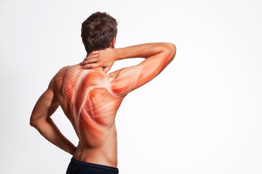
Muscle Contracture
A muscle contracture, or contracture deformity, is caused when a muscle loses elasticity. This causes permanent shortening and tightening of muscle fibers, which reduces flexibility and makes movement difficult. Muscles that cannot move and stretch cause the surrounding joints to lose mobility and develop pain symptoms. When trying to stretch the contracted muscle, the individual will feel the muscle become very rigid, which can increase pain. (Lieber, R. L., and Fridén, J. 2019) Delaying treatment can potentially cause irreversible and chronic symptoms.
Commonly Affected Muscles
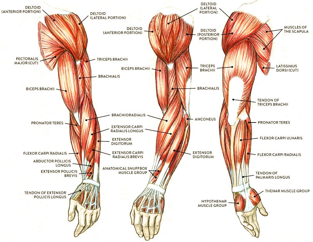
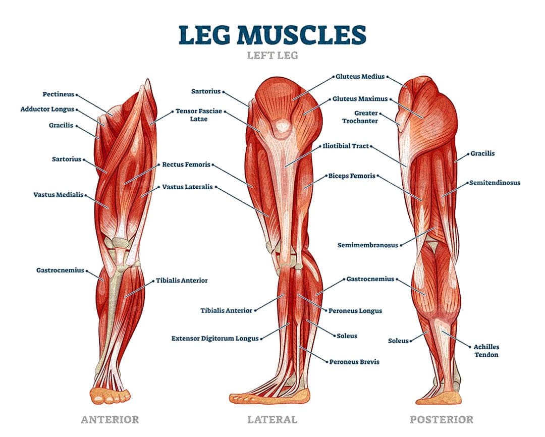
Flexor muscles bend the joints and are those most affected by contractures. The stiffening and tightening prevent the body parts from moving out and away. The most common include:
Wrist and Finger Flexors
Muscle groups that bend the wrist and fingers.
Biceps
The primary elbow flexor that bends the arm.
Gastrocnemius and Soleus
The calf muscles which allow the ankle to point the foot/plantarflexion.
Hamstrings
A group of three muscles behind the thigh that bend the knee.
Causes
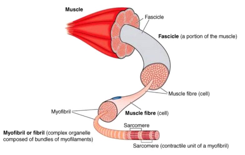
The permanent shortening of muscle fibers and changes in muscle structure cause muscle contractures or stiffer-than-normal tissue that is difficult to stretch. Sarcomeres are structural units of muscles that cause fibers to contract.
With contractures, the sarcomeres overly lengthen when the muscle fibers tighten. This increase in sarcomere length prevents the muscle from contracting normally, resulting in weakness. Muscle fibers are encased in an extracellular matrix, a mesh composed of collagen and other proteins that help transmit force and provide muscle contraction. Muscle contractures cause the amount of collagen within the extracellular matrix to increase, causing a stiffening of fibers that restricts movement. (Lieber, R. L., and Fridén, J. 2019)
Muscle contractures also form from decreased satellite cells. Satellite cells are specialized stem cells that can rebuild muscle and are necessary for muscle regeneration and repair. Without the proper amount of satellite cells, other cells like fibroblasts significantly increase in the muscle tissue, causing the fibers to become stiff and fibrotic or more fibrous. These changes to the sarcomeres, collagen within the extracellular matrix, and decreased satellite cells all result from conditions in which neurological input to the brain and spinal cord muscles becomes reduced. This is caused by lack of use, injury, or neurological and neuromuscular conditions. (Lieber, R. L., and Fridén, J. 2019)
Cerebral Palsy
Contractures often occur from upper motor neuron lesions, which prevent signals from the brain and spinal cord from reaching the motor neurons that control muscle contraction. When these signals are weakened or blocked, muscles become stiff and weak from lack of stimulation. (Lieber, R. L., and Fridén, J. 2019)
Cerebral palsy is a group of disorders affecting mobility caused by an upper motor neuron lesion that is present at birth and is the most common motor disability in children. It causes:
Cognitive impairment
Decreased muscle strength
Problems with movement, coordination, and functional motions.
Because cerebral palsy prevents the muscles of the legs from being sufficiently stimulated, contractures commonly develop in the hips, knees, and ankles. Individuals can have a 75% decrease in satellite cells to repair muscle tissue and prevent muscle fibrosis or stiffening. Specific genes linked to collagen production are also altered, causing irregular changes to the extracellular matrix of muscles. (Lieber, R. L., and Fridén, J. 2019)
Muscular Dystrophy
Muscular dystrophy is a group of inherited neuromuscular disorders characterized by muscle weakness and wasting. Deficient nerve supply to muscles causes them to become stiff and tight, inhibiting the functional range of motion needed to move joints and activate muscles to move. Clinical research suggests that individuals with muscular dystrophy have decreased levels of satellite cells to repair, increasing the risk of developing muscle contracture. (Lieber, R. L., and Fridén, J. 2019)
Disuse-induced Muscle Wasting or Disuse Atrophy
When muscles are not used for some time because of hospitalization, prolonged bed rest, or immobilization from wearing braces, splints, or casts after injuries, the blood circulation and electrical signaling from nerves to muscles decreases. This results in weakness, increased muscle tightness and stiffness, and muscle wasting/atrophy. Over time, stiff and tight muscles can progress to contractures that become extremely difficult to stretch.
Trauma or Injury
Muscle or tendon injuries can cause contractures as scar tissue develops, joining muscle fibers and joints together. This can significantly restrict movement. Large burns can also cause skin, muscles, and joint contractures. The range of motion can become significantly limited, and the changes can become irreversible if not aggressively treated.
Other Causes
Other forms of upper motor neuron lesions that can cause contractures because of weak or blocked electrical input to muscles as a result of brain or spinal cord damage include:
Neuromuscular disorders like spinal muscular atrophy – SMA.
Conditions that cause inflammation and joint stiffening, like juvenile rheumatoid arthritis.
A history of diabetes also increases the risk of developing contractures affecting finger flexors, like Dupuytren’s contractures and stenosing tenosynovitis
or trigger finger. (Lieber, R. L., and Fridén, J. 2019)
Symptoms
Symptoms include:
Extremely stiff and tight muscles resistant to stretching.
Pain from the inability to stretch.
Loss of range of motion.
Impaired joint mobility.
Severe contractures can interfere with the functional range of motion needed to move joints to complete normal tasks and movements, such as standing up from a chair and walking.
Treatment
Physical Therapy
Physical therapies can help reduce the severity through stretching and soft tissue mobilization to decrease tightness. (Lieber, R. L., and Fridén, J. 2019)
Specialized braces or splints can be custom-made to fit different body parts.
These provide a prolonged low-intensity stretch over a period of time to increase muscle length.
Once the muscle has stretched, a new brace or splint may be needed to adjust to the increased range of motion. (Lieber, R. L., and Fridén, J. 2019)
Surgery
In severe cases where muscle contractures limit the functional range of motion needed for activities of daily living or ADLs, surgical release of the contracted tissue may be recommended. This surgery can improve functional movements like walking, getting in and out of bed, and standing up from chairs. The tight muscles can be surgically cut, and the tendons can be lengthened to allow more mobility. (Lieber, R. L., and Fridén, J. 2019)
The causes of muscle contracture are not always avoidable, but various treatment options are available to help loosen up tight muscles and preserve or restore the range of motion. It’s important to move daily and stretch common areas like the fingers, arms, and legs to reduce the risk of muscle tightness and prevent contractures from developing. It is imperative to seek medical treatment for severe contractures resulting from neuromuscular disorders, including physical and occupational therapy, to prevent contractures from worsening and regaining as much functional range as possible.
Injury Medical Chiropractic and Functional Medicine Clinic uses an integrated approach personalized to the individual that focuses on what works for them and treats injuries and chronic pain syndromes through personalized care plans that improve ability through flexibility, mobility, and agility programs to relieve pain. Our providers use an integrated approach to create personalized care plans for each patient, including Functional Medicine, Acupuncture, Electro-Acupuncture, and Sports Medicine principles. Our goal is to relieve pain naturally by restoring health and function to the body. If other treatment is needed, Dr. Jimenez has teamed up with top surgeons, clinical specialists, medical researchers, and rehabilitation providers to provide the most effective treatments.
Chiropractic Treatment for Cerebral Palsy
References
Lieber, R. L., & Fridén, J. (2019). Muscle contracture and passive mechanics in cerebral palsy. Journal of applied physiology (Bethesda, Md. : 1985), 126(5), 1492–1501. https://doi.org/10.1152/japplphysiol.00278.2018
Can incorporating electrical muscle stimulation help control pain, strengthen muscles, increase physical function, retrain lost movements, and/or manage inflammation for individuals experiencing neck and back pain?
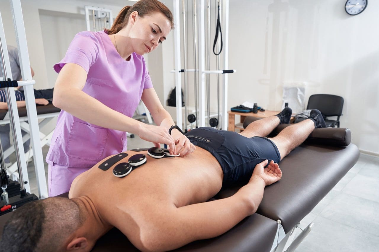
Female doctor placing myostimulation physical therapy equipment on patient’s back
Electric Muscle Stimulation
Electrical muscle stimulation or E-stim is a physical therapy used to reactivate the muscles’ ability to contract. E-stim uses devices that transmit electrical impulses through the skin to target nerves and/or muscles. The most common forms include
Transcutaneous electric nerve stimulation, or TENS, is the most well-known type of electrical stimulation that offers devices that can be used at home or on the go.
Electrical muscle stimulation or EMS.
In physical therapy, E-stim stimulates muscles to contract, strengthening them and encouraging blood circulation.
Blood circulation can directly affect the condition of muscle tissue.
Electrical muscle stimulation is also used in spinal cord injury and other neuromuscular conditions. (Ho, C. H. et al., 2014)
E-stim
During treatment, electrodes are hooked to an electric stimulation machine and placed around the affected neck or back area.
The electrodes will be placed on the skin for most neck or back injuries.
The placement of the electrodes depends on the reason for treatment and the depth or superficiality of the electrical stimulation.
The electrodes are often placed near a motor point of a muscle to ensure the correct contraction.
The therapist will adjust the controls of the stimulation machine to achieve thorough muscle contraction with minimal discomfort.
Stimulation can last 5 – 15 minutes, depending on the treatment plan and injury severity.
Spinal Joint Stabilization
Activation of the muscles may help increase spinal joint stability, improving problems with spinal instability. (Ho, C. H. et al., 2014) Electric muscle stimulation is thought to enhance the exercise program a therapist prescribes to help maintain joint stability. Electrical stimulation may also help build muscle strength and endurance. (Veldman, M. P. et al., 2016) Muscle endurance is the repetitions a muscle can contract before it fatigues.
Healing and Pain Management
Electric muscle stimulation therapy can enhance tissue healing and help manage inflammation by reducing swelling and increasing circulation. It can reduce pain sensations by blocking nerve transmission at the spinal cord. (Johnson, M. I. et al., 2019) A healthcare professional may suggest a TENS or take-home electric stimulation unit to manage symptoms. (Johnson, M. I. et al., 2019)
Treatment
Interdisciplinary therapies tailored to an individual’s specific back or neck pain have been found to provide positive results. Exercise, yoga, short-term cognitive behavioral therapy, biofeedback, progressive relaxation, massage, manual therapy, and acupuncture are recommended for neck or back pain. (Chou, R. et al., 2018) Taking non-steroidal anti-inflammatory medications may also help. Electrical muscle stimulation could be an effective neck or back treatment.
Individuals unsure whether they need or would benefit from electrical should discuss symptoms and conditions with a primary physician, healthcare provider, or specialist to guide them in the right direction and determine the best treatment. Injury Medical Chiropractic and Functional Medicine Clinic focuses on what works for the patient and strives to better the body through researched methods and total wellness programs. Using an integrated approach, we treat injuries and chronic pain syndromes through personalized care plans that improve ability through flexibility, mobility, and agility programs personalized to the individual to relieve pain. If other treatment is needed, Dr. Jimenez has teamed up with the top surgeons, clinical specialists, medical researchers, and premier rehabilitation providers to provide the most effective treatments.
Thoracic Spine Pain
References
Ho, C. H., Triolo, R. J., Elias, A. L., Kilgore, K. L., DiMarco, A. F., Bogie, K., Vette, A. H., Audu, M. L., Kobetic, R., Chang, S. R., Chan, K. M., Dukelow, S., Bourbeau, D. J., Brose, S. W., Gustafson, K. J., Kiss, Z. H., & Mushahwar, V. K. (2014). Functional electrical stimulation and spinal cord injury. Physical medicine and rehabilitation clinics of North America, 25(3), 631–ix. https://doi.org/10.1016/j.pmr.2014.05.001
Veldman, M. P., Gondin, J., Place, N., & Maffiuletti, N. A. (2016). Effects of Neuromuscular Electrical Stimulation Training on Endurance Performance. Frontiers in physiology, 7, 544. https://doi.org/10.3389/fphys.2016.00544
Johnson, M. I., Jones, G., Paley, C. A., & Wittkopf, P. G. (2019). The clinical efficacy of transcutaneous electrical nerve stimulation (TENS) for acute and chronic pain: a protocol for a meta-analysis of randomised controlled trials (RCTs). BMJ open, 9(10), e029999. https://doi.org/10.1136/bmjopen-2019-029999
Chou, R., Côté, P., Randhawa, K., Torres, P., Yu, H., Nordin, M., Hurwitz, E. L., Haldeman, S., & Cedraschi, C. (2018). The Global Spine Care Initiative: applying evidence-based guidelines on the non-invasive management of back and neck pain to low- and middle-income communities. European spine journal : official publication of the European Spine Society, the European Spinal Deformity Society, and the European Section of the Cervical Spine Research Society, 27(Suppl 6), 851–860. https://doi.org/10.1007/s00586-017-5433-8
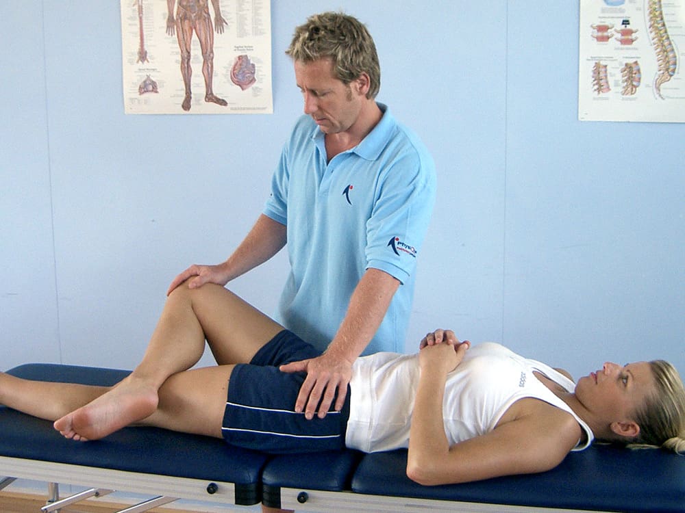
Can athletic individuals incorporate MET (muscle energy techniques) therapy to reduce the pain-like effects of adductor strain?
Introduction
The body’s lower extremities have an important role as they provide stability and mobility to the individual. Many athletes utilize their lower extremities by adding much power to exert the energy to win matches or competitions. The various muscles, soft tissues, ligaments, and joints help support the body’s skeletal structure and can succumb to injuries from repetitive motions or environmental factors. One of the muscles that can be affected by constant repetitive motions and environmental factors is the adductor muscles, which can cause many athletes to be in continuous pain and affect their performance during competitions. Luckily, there is a technique that many treatments offer to reduce muscle strain in the adductors and provide relief to the lower extremities. Today’s article looks at how adductor strain can affect many individuals, how MET therapy can help with an adductor strain, and its positive effect on athletic individuals. We discuss with certified medical providers who consolidate our patients’ information to assess the pain-like effects of an adductor strain in the lower extremities. We also inform and guide patients on how MET therapy can help stretch and strengthen tight adductor muscles to reduce strain and provide relief. We also encourage our patients to ask their associated medical providers many intricate and important questions about incorporating MET and other non-surgical therapies into their personalized treatment plan for a healthier lifestyle. Dr. Jimenez, D.C., includes this information as an academic service. Disclaimer.
How Does Adductor Strain Affect Individuals?
Do you feel tightness along your thighs and legs after a long day at work? Do you experience instability when walking from one location to another? Or do you feel pain when stretching your thighs that causes temporary relief? Many individuals experiencing pain in their lower extremities will often think it is hip pain, but their adductor muscles are in pain. The adductor muscles consist of three muscles that provide torque to the lower extremities by allowing them to move inward when a person is walking and help keep the trunk muscles steady. So, when many athletes begin to make constant repetitive motions while performing, it can cause issues for the adductors. As a common injury to many athletes, adductor strain can put exaggerated stress on the actual tendon, leading to biomechanical abnormalities affecting the musculoskeletal system. (Kiel & Kaiser, 2024a) Also, when athletes start to use constant repetitive motions during an increased volume or intensity of the training workload, it can cause stress factors in the lower extremities. (Kiel & Kaiser, 2024b) This, in turn, can have many individuals feel like they are experiencing hip and groin pain when it is, in fact, stress fractures in the adductor muscles causing myofascial pain.
So, for athletic individuals dealing with adductor strain, primary doctors need to differentiate between adductor strain and regular muscle strain in the lower extremities, as the pain symptoms sometimes have overlapping risk profiles with acute onset pain symptoms associated with distinct injury mechanisms. (McHugh et al., 2023) This is because when athletes overuse their adductor muscles, it causes pain, as many injuries within the adductors are associated with the hips and groin region. (Koscso et al., 2022) However, there are ways for athletes to find the relief they seek to reduce adductor strain and return to their routine.
Movement Medicine- Video
How MET Therapy Helps With Adductor Strain
For athletes and individuals engaged in physical activity, MET therapy can be a valuable part of the recovery process for adductor strain. MET (muscle energy technique) therapy, a form of osteopathic manipulative medicine, is used by pain specialists such as chiropractors, massage therapists, and sports physicians to alleviate pain symptoms in the musculoskeletal system. By using gentle, controlled muscle contractions, these specialists can improve musculoskeletal function by mobilizing joints, stretching tight muscles and fascia, and improving circulation and lymphatic flow. (Waxenbaum et al., 2024) Many pain specialists, including chiropractors and massage therapists, incorporate MET therapy into their practices due to its effectiveness in addressing muscular imbalances and alignment issues that contribute to pain and limited mobility in the lower extremities.
The Positive Effect Of MET Therapy
One of the positive effects of MET therapy for adductor strain is that when athletes and individuals start to utilize it as part of their recovery, their pain is reduced, and muscle mobility is increased since there are changes in the viscoelastic properties in the soft tissue. (Thomas et al., 2019) For the adductor muscles, MET therapy helps with:
Increasing muscle length & flexibility
Reduce muscle tension
Improving blood flow and promoting healing
Enhance joint function
MET therapy, when incorporated for pain relief for adductor strain, can put many individuals at ease as it actively focuses on muscle relaxation, lengthening, and strengthening the affected muscles. MET therapy can be combined with other therapies in a person’s personalized treatment plan to enhance mobility, be mindful of what is causing pain and discomfort to their bodies, and live a healthier lifestyle.
Koscso, J. M., McElheny, K., Carr, J. B., 2nd, & Hippensteel, K. J. (2022). Lower Extremity Muscle Injuries in the Overhead Athlete. Curr Rev Musculoskelet Med, 15(6), 500-512. https://doi.org/10.1007/s12178-022-09786-z
McHugh, M. P., Nicholas, S. J., & Tyler, T. F. (2023). Adductor Strains in Athletes. Int J Sports Phys Ther, 18(2), 288-292. https://doi.org/10.26603/001c.72626
Thomas, E., Cavallaro, A. R., Mani, D., Bianco, A., & Palma, A. (2019). The efficacy of muscle energy techniques in symptomatic and asymptomatic subjects: a systematic review. Chiropr Man Therap, 27, 35. https://doi.org/10.1186/s12998-019-0258-7
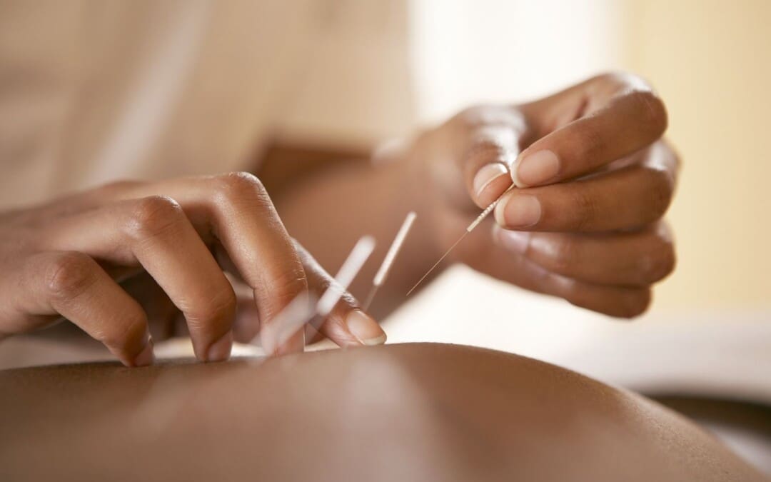
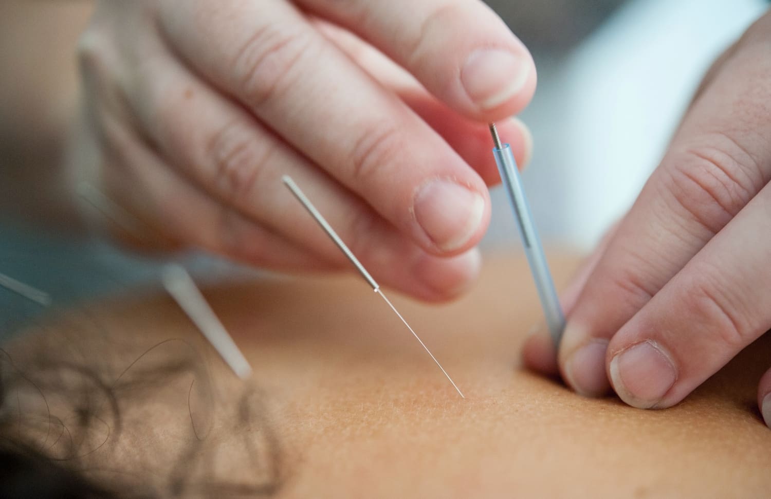
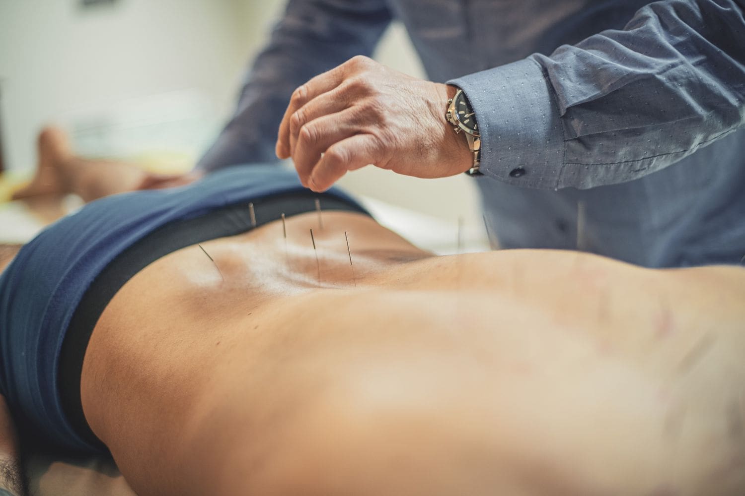
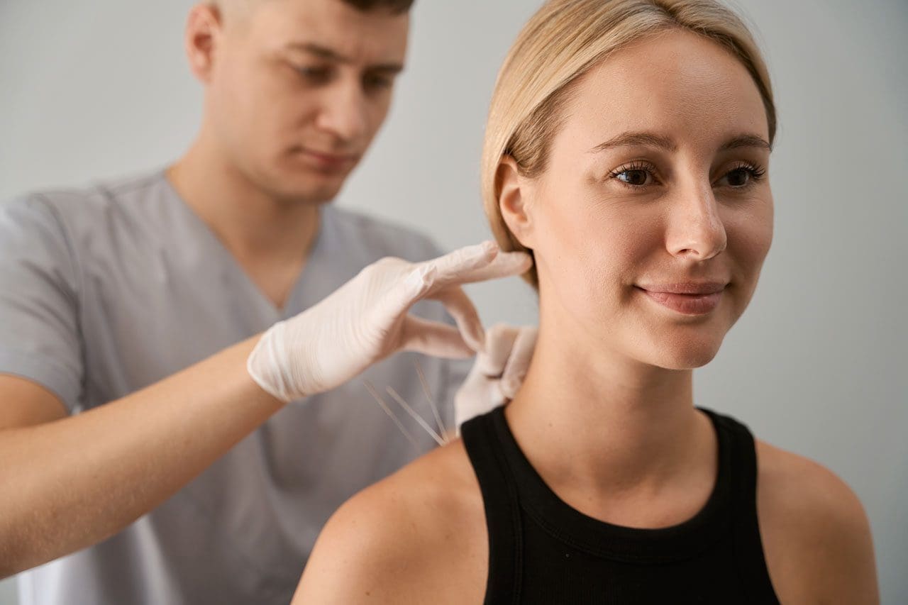
Can individuals dealing with muscle pain find relief from acupuncture therapy to get back to their daily activities and well-being?
Introduction
Many people worldwide have dealt with pain in their musculoskeletal system that has affected their daily routine. Some of the most common factors that people have experienced muscle pain include sedentary lifestyles from working at a desk job or physical demands from an active lifestyle. The muscles, tendons, ligaments, and soft tissues can become overstretched and overworked, causing the muscles to become weak. At the same time, visceral somatic issues in the neck, shoulders, and back can affect the upper and lower extremities, leading to a life of disability. Many factors that can contribute to the development of muscle pain can impact a person’s routine and cause them to find various techniques to reduce the muscle pain in their bodies. Since muscle pain can be in acute or chronic form, many individuals who are seeking treatment for their ailments can look into non-surgical therapies like acupuncture to not only reduce muscle pain but also find the relief they are looking for. Today’s article focuses on how muscle pain can affect a person’s well-being, how the essence of acupuncture can be beneficial for muscle pain, and how people can integrate acupuncture therapy as part of a wellness routine. We talk with certified medical providers who consolidate our patients’ information to assess how muscle pain can impact a person’s well-being. We also inform and guide patients on how acupuncture therapy can benefit the body by reducing the effects of muscle pain. We encourage our patients to ask their associated medical providers intricate and important questions about incorporating acupuncture therapy into a wellness routine to reduce muscle pain and its referred symptoms. Dr. Jimenez, D.C., includes this information as an academic service. Disclaimer.
How Muscle Pain Can Affect A Person’s Well-Being
Do you feel the effects of tiredness and weakness in your upper and lower extremity muscles? Have you experienced general soreness or aches in your neck, shoulders, or back? Or does twisting and turning your body cause temporary relief to your body, only for it to be worse throughout the day? When it comes to muscle pain can be a multi-factorial condition where that can involve complex interactions on a person’s structure, physical, social, lifestyle, and comorbid health factors that can come into play as contributing factors for people to experience long-term pain and disability. (Caneiro et al., 2021) As many individuals start to do repetitive motions or stay in sedentary positions, muscle pain can develop when they stretch or try to move their muscles while doing their routine. The burden of muscle pain often correlates with socioeconomic factors that can cause many people, both young and old, to substantially limit their mobility and engagement in their routine, which predisposes increased risk factors to other chronic conditions they may have. (Dzakpasu et al., 2021)
When many individuals are dealing with muscle pain in its acute or chronic form, many often don’t realize that when the affected muscles in the upper and lower body quadrants are coping with pain, there is associated pain and stiffness from how active or inactive the muscles are can affect the soft tissue causing high mechanical stress to the affect the skeletal joints. (Wilke & Behringer, 2021) When this happens, many people will start to experience referred muscle pain in their bodies, causing issues with their mobility, flexibility, and stability. Coincidentally, muscle pain can also be a symptom of many people who have various pains in their bodies that have impacted their lives prior; seeking treatment can reduce the effects of muscle pain and help them take back their routine to lead a healthier lifestyle.
Movement Medicine- Video
The Essence Of Acupuncture For Muscle Pain
When many people are dealing with muscle pain, they are seeking treatments that are not only affordable but also can be effective in reducing the overlapping risk profiles that are affecting the body, causing muscle pain. Many treatments like chiropractic care, decompression, and massage therapy are non-surgical and are effective through consecutive sessions. One of the oldest and most effective treatments that can help reduce muscle pain in the body is acupuncture therapy. Acupuncture is a holistic treatment derived from Traditional Chinese Medicine that utilizes small, solid, thin needles inserted by professional acupuncturists to various acupoints. The main philosophy is that acupuncture provides relief to the body as it helps improve the body’s energy flow while maintaining a person’s overall health and vitality. (Zhang et al., 2022) When a person is dealing with muscle pain, the muscle fibers can develop tiny nodules known as trigger points that can induce pain in the affected muscle quadrants. With acupuncture needles placed in the affected area, local and referred pain is reduced, muscle blood flow and oxygen are returned to the body, and the muscle’s range of motion is improved. (Pourahmadi et al., 2019) Some of the benefits that acupuncture therapy provides include:
Increased circulation
Inflammation reduction
Endorphin release
Relaxing muscle tension
Integrating Acupuncture As Part Of A Wellness Routine
Many individuals who are seeking acupuncture therapy as part of their wellness journey can see the positive benefits of acupuncture and can combine it with other therapies to reduce the chances of muscle pain from returning. While acupuncture can help stimulate the nerves and restore motor function, treatments like joint mobilization can help stretch the affected muscles and joints to improve the body’s range of motion. (Lee et al., 2023) With many individuals seeking acupuncture treatment to reduce muscle pain, many can make small changes in their routine to prevent the pain from causing overlapping risk profiles to their bodies. When addressing the root causes of pain and promoting the body’s innate healing abilities, acupuncture can help restore balance, alleviate discomfort, and enhance overall well-being.
References
Caneiro, J. P., Bunzli, S., & O’Sullivan, P. (2021). Beliefs about the body and pain: the critical role in musculoskeletal pain management. Braz J Phys Ther, 25(1), 17-29. https://doi.org/10.1016/j.bjpt.2020.06.003
Dzakpasu, F. Q. S., Carver, A., Brakenridge, C. J., Cicuttini, F., Urquhart, D. M., Owen, N., & Dunstan, D. W. (2021). Musculoskeletal pain and sedentary behaviour in occupational and non-occupational settings: a systematic review with meta-analysis. Int J Behav Nutr Phys Act, 18(1), 159. https://doi.org/10.1186/s12966-021-01191-y
Lee, J. E., Akimoto, T., Chang, J., & Lee, H. S. (2023). Effects of joint mobilization combined with acupuncture on pain, physical function, and depression in stroke patients with chronic neuropathic pain: A randomized controlled trial. PLOS ONE, 18(8), e0281968. https://doi.org/10.1371/journal.pone.0281968
Pourahmadi, M., Mohseni-Bandpei, M. A., Keshtkar, A., Koes, B. W., Fernandez-de-Las-Penas, C., Dommerholt, J., & Bahramian, M. (2019). Effectiveness of dry needling for improving pain and disability in adults with tension-type, cervicogenic, or migraine headaches: protocol for a systematic review. Chiropr Man Therap, 27, 43. https://doi.org/10.1186/s12998-019-0266-7
Wilke, J., & Behringer, M. (2021). Is “Delayed Onset Muscle Soreness” a False Friend? The Potential Implication of the Fascial Connective Tissue in Post-Exercise Discomfort. Int J Mol Sci, 22(17). https://doi.org/10.3390/ijms22179482
Zhang, B., Shi, H., Cao, S., Xie, L., Ren, P., Wang, J., & Shi, B. (2022). Revealing the magic of acupuncture based on biological mechanisms: A literature review. Biosci Trends, 16(1), 73-90. https://doi.org/10.5582/bst.2022.01039
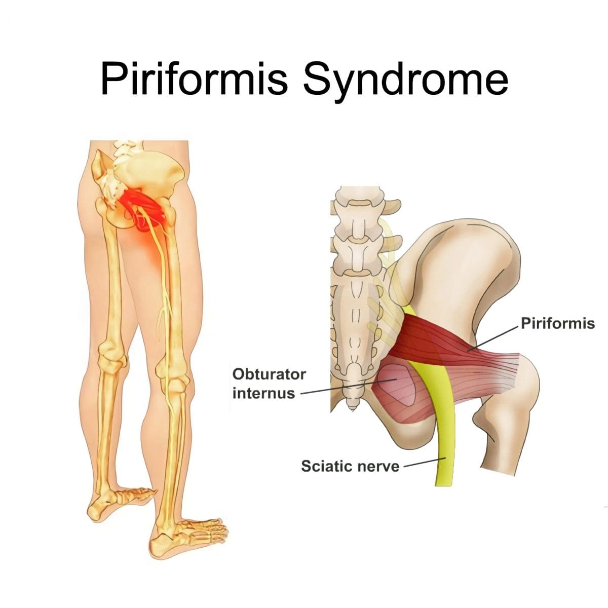
Can physical therapy treatment protocols aimed at improving range of motion and flexibility around the hip and relieving inflammation around the sciatic nerve help individuals experiencing deep buttock pain or piriformis syndrome?
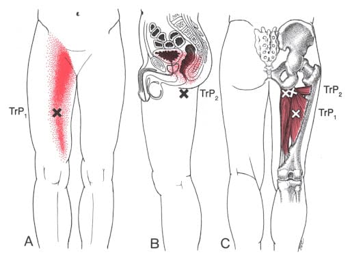
Deep Buttock Pain
Piriformis syndrome, a.k .a. deep buttock pain, is described as sciatic nerve irritation from the piriformis muscle.
The piriformis is a small muscle behind the hip joint in the buttocks.
It is about one centimeter in diameter and functions in the hip joint’s external rotation or turning outward.
The piriformis muscle and tendon are close to the sciatic nerve, which supplies the lower extremities with motor and sensory functions.
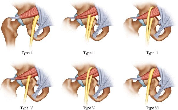
Depending on an individual’s anatomic variation of the muscle and tendon:
The two cross over, under, or through each other behind the hip joint in the deep buttock.
This relationship is thought to irritate the nerve, leading to sciatica symptoms.
Piriformis Syndrome
When diagnosed with piriformis syndrome, it is thought that the muscle and tendon bind to and/or spasm around the nerve, causing irritation and pain symptoms.
The theory supported is that when the piriformis muscle and its tendon tighten, the sciatic nerve becomes compressed or pinched. This decreases blood circulation and irritates the nerve from the pressure. (Shane P. Cass 2015)
Tenderness with pressure on the piriformis muscle.
Discomfort in the back of the thigh.
Deep buttock pain behind the hip.
Electric sensations, shocks, and pains travel down the back of the lower extremity.
Numbness in the lower extremity.
Some individuals develop symptoms abruptly, while others go through a gradual increase.
Diagnosis
Doctors will order X-rays, MRIs, and nerve conduction studies, which is normal.
Because piriformis syndrome can be challenging to diagnose, some individuals with minor hip pain may receive a piriformis syndrome diagnosis even if they don’t have the condition. (Shane P. Cass 2015)
It is sometimes referred to as deep buttock pain. Other causes of this type of pain include back and spinal problems like:
Herniated discs
Spinal stenosis
Radiculopathy – sciatica
Hip bursitis
A piriformis syndrome diagnosis is usually given when these other causes are eliminated.
When the diagnosis is uncertain, an injection is administered in the area of the piriformis muscle. (Danilo Jankovic et al., 2013)
Different medications can be used, but the injection itself is used to help determine the specific location of the discomfort.
When an injection is given into the piriformis muscle or tendon, it is often administered by ultrasound guidance to ensure the needle delivers the medication to the correct location. (Elizabeth A. Bardowski, J. W. Thomas Byrd 2019)
Avoiding activities that cause symptoms for at least a few weeks.
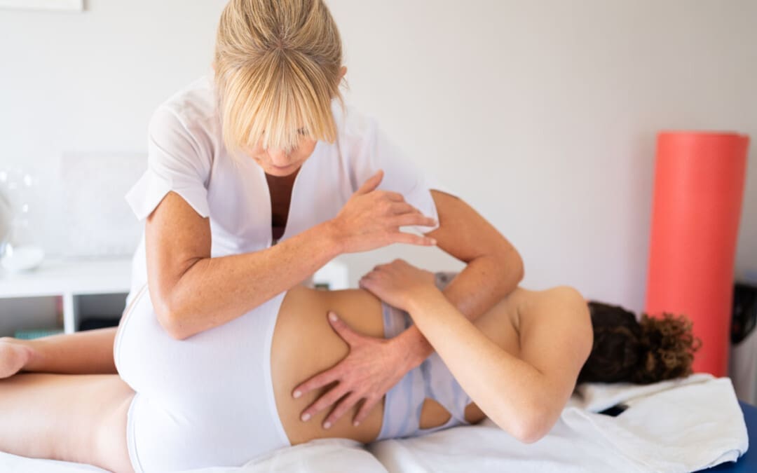
Physical Therapy
Emphasize stretching and strengthening the hip rotator muscles.
Non-Surgical Decompression
Gently pulls the spine to release any compression, allowing optimal rehydration and circulation and taking the pressure off the sciatic nerve.
Therapeutic Massage Techniques
To relax and release muscle tension and increase circulation.
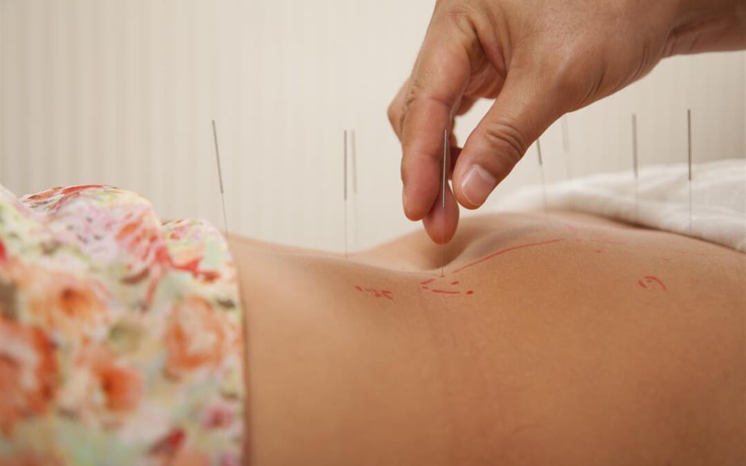
Acupuncture
To help relax the piriformis muscle, sciatic nerve, and surrounding area.
Relieve pain.
Chiropractic Adjustments
Realignment rebalances the spine and musculoskeletal system to alleviate pain.
Anti-Inflammatory Medication
To decrease inflammation around the tendon.
Cortisone Injections
Injections are used to decrease inflammation and swelling.
Botulinum Toxin Injection
Injections of botulinum toxin paralyze the muscle to relieve pain.
Surgery
Surgery can be performed in rare cases to loosen the piriformis tendon, known as a piriformis release. (Shane P. Cass 2015)
Surgery is a last resort when conservative treatments have been tried for at least 6 months with little to no relief.
Recovery can take several months.
Sciatica Causes and Treatment
References
Cass S. P. (2015). Piriformis syndrome: a cause of nondiscogenic sciatica. Current sports medicine reports, 14(1), 41–44. https://doi.org/10.1249/JSR.0000000000000110
Jankovic, D., Peng, P., & van Zundert, A. (2013). Brief review: piriformis syndrome: etiology, diagnosis, and management. Canadian journal of anaesthesia = Journal canadien d’anesthesie, 60(10), 1003–1012. https://doi.org/10.1007/s12630-013-0009-5
Bardowski, E. A., & Byrd, J. W. T. (2019). Piriformis Injection: An Ultrasound-Guided Technique. Arthroscopy techniques, 8(12), e1457–e1461. https://doi.org/10.1016/j.eats.2019.07.033
Can individuals dealing with joint pain incorporate acupuncture therapy to manage lupus symptoms and restore body mobility?
Introduction
The immune system is highly important to the body as its main job is to protect vital structures from foreign invaders that can cause pain-like issues and discomfort. The immune system has a healthy relationship with the different body systems, including the musculoskeletal system, as the inflammatory cytokines help heal muscle and tissue damage when the body is injured. Over time, however, when normal environmental and genetic factors start to develop in the body, the immune system will begin to send out these cytokines to healthy, normal cells. To that point, the body starts at risk of developing autoimmune diseases. Now, autoimmune diseases in the body can cause havoc over time when they are not managed, leading to chronic disorders that can cause overlapping symptoms in the musculoskeletal system. One of the most common autoimmune diseases is systemic lupus erythematosus or lupus, and it can cause a person to be in consistent pain and discomfort while correlating with muscle and joint pain. Today’s article looks at the factors and effects of lupus, the burden of joint pain in lupus, and how holistic approaches like acupuncture can help manage lupus while restoring body mobility. We talk with certified medical providers who consolidate our patients’ information to assess how to minimize the pain effects caused by lupus on the joints. We also inform and guide patients on how acupuncture can help manage lupus and combine other therapies to reduce its pain-like symptoms affecting the musculoskeletal system. We encourage our patients to ask their associated medical providers intricate and important questions about incorporating acupuncture therapy to relieve the inflammatory effects of lupus while finding natural ways to restore mobility. Dr. Jimenez, D.C., includes this information as an academic service. Disclaimer.
The Factors & Effects Of Lupus
Have you been experiencing joint pain in your upper or lower extremities, making it difficult to function throughout the day? Have you been feeling the constant effects of fatigue? Many individuals experiencing these pain-like issues could risk developing systemic lupus erythematosus. In this autoimmune disease, the body’s own immune system mistakenly starts to attack its tissues, thus leading to inflammation and a range of pain-like symptoms. Lupis is tricky to diagnose because of its complex immune dysregulation that can lead to an overproduction of cytokines that can affect the body. (Lazar & Kahlenberg, 2023) At the same time, lupus can affect a diverse population, with symptoms and severity varying depending on how mild or severe the factors affect the body. Lupus can impact various body parts, including the joints, skin, kidneys, blood cells, and other vital body parts and organs, as environmental and hormonal factors can influence its development. (Tsang & Bultink, 2021) Additionally, lupus can be closely associated with other comorbidities that are causing overlapping risk profiles with inflammation that can affect the joints in the musculoskeletal system.
The Burden of Joint Pain In Lupus
Lupus is tricky to diagnose since it often mimics other ailments; the most common pain symptom that lupus affects is the joints. Individuals with lupus experience joint pain, which can cause inflammatory effects and structural damage to the joints, tendons, muscles, and bones, causing pathological abnormalities. (Di Matteo et al., 2021) Since lupus causes inflammatory effects in the joints, many individuals will think that they are experiencing inflammatory arthritis, and it can cause overlapping risk profiles as it is accompanied by lupus, thus causing localized pain in the joints regardless of its origin. (Senthelal et al., 2024) Joint pain in lupus individuals can significantly hinder daily activities, reducing mobility and overall quality of life as they are trying to find relief.
Unlocking The Secrets of Inflammation-Video
A Holistic Approach to Managing Lupus
While standard treatments for lupus involve medication and immunosuppressants to reduce the inflammation caused by lupus, many people want to seek out holistic approaches to manage lupus and reduce the inflammatory effects from affecting their joints by making small changes in their lives. Many people incorporate anti-inflammatory foods rich in antioxidants to dampen the inflammatory effects. Various supplements, like vitamin D, calcium, zinc, etc., can help reduce inflammation caused by lupus and strengthen bone health. Additionally, non-surgical treatments can even improve cardiorespiratory capacity and decrease fatigue while improving psychological function, which can help improve a person’s quality of life by managing the symptoms caused by lupus. (Fangtham et al., 2019)
How Acupuncture Could Help Lupus & Restore Mobility
One of the oldest forms of non-surgical and holistic approaches to reducing inflammation and managing lupus is acupuncture. Acupuncture involves solid, thin needles used by highly trained professionals to be inserted into specific body points to balance the body’s qi (energy) by stimulating the nervous system and releasing beneficial chemicals into the affected muscles, spinal cord, and brain. Additionally, acupuncture, with its minimal side effects and holistic approach, can help manage lupus. This is because when acupuncture needles are placed at the acupoints of the body, it can disrupt the pain signals that are causing pain in the affected area and regulate the inflammatory cytokines from lupus to provide relief. (Wang et al., 2023) This is due to its philosophy of addressing not only the physical pain but also the emotional and psychological symptoms of living with a chronic condition like lupus.
Additionally, acupuncture can help restore joint mobility while managing lupus through consecutive treatments, as many people notice that their joint mobility is improved and their pain is diminished. This is because the insertion and manipulation of the needles in the body’s acupoints cause alterations in afferent sensory input to the central nervous system, which increases alpha motoneuron excitability and reduces inflammation. (Kim et al., 2020) When individuals are dealing with lupus and are trying to find alternative holistic methods to relieve inflammation and joint pain caused by lupus, acupuncture, and non-surgical treatments can offer a ray of hope in managing the daily challenges of lupus.
References
Di Matteo, A., Smerilli, G., Cipolletta, E., Salaffi, F., De Angelis, R., Di Carlo, M., Filippucci, E., & Grassi, W. (2021). Imaging of Joint and Soft Tissue Involvement in Systemic Lupus Erythematosus. Curr Rheumatol Rep, 23(9), 73. https://doi.org/10.1007/s11926-021-01040-8
Fangtham, M., Kasturi, S., Bannuru, R. R., Nash, J. L., & Wang, C. (2019). Non-pharmacologic therapies for systemic lupus erythematosus. Lupus, 28(6), 703-712. https://doi.org/10.1177/0961203319841435
Kim, D., Jang, S., & Park, J. (2020). Electroacupuncture and Manual Acupuncture Increase Joint Flexibility but Reduce Muscle Strength. Healthcare (Basel), 8(4). https://doi.org/10.3390/healthcare8040414
Tsang, A. S. M. W. P., & Bultink, I. E. M. (2021). New developments in systemic lupus erythematosus. Rheumatology (Oxford), 60(Suppl 6), vi21-vi28. https://doi.org/10.1093/rheumatology/keab498
Wang, H., Wang, B., Huang, J., Yang, Z., Song, Z., Zhu, Q., Xie, Z., Sun, Q., & Zhao, T. (2023). Efficacy and safety of acupuncture therapy combined with conventional pharmacotherapy in the treatment of systemic lupus erythematosus: A systematic review and meta-analysis. Medicine (Baltimore), 102(40), e35418. https://doi.org/10.1097/MD.0000000000035418
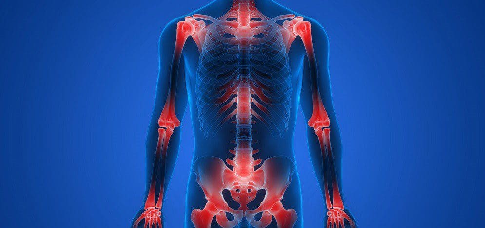
For individuals dealing with musculoskeletal pain, can incorporating acupuncture and electroacupuncture therapy provide beneficial results?
Introduction
The upper and lower body quadrants are surrounded by muscles, soft tissues, and ligaments that allow the body to be mobile with feelings of pain or discomfort. Each muscle group has an important job providing sensory-motor functions like grasping objects, moving extremities, supporting the body in a correct posture, and stabilizing vertical axial weight. However, many people have adopted various habits from environmental factors or have been through traumatic injuries that can cause referred muscle pain in the upper and lower body quadrants. When this happens, it can lead to a life of disability, pain, and discomfort over time if it is not treated right away. To that point, musculoskeletal pain can also cause overlapping risk profiles with other comorbidities that can be pre-existing in the body. Fortunately, numerous treatments can help reduce musculoskeletal pain and benefit the body. Today’s article looks at two different non-surgical therapies, how each is beneficial to reducing musculoskeletal pain, and how effective they can help many people with musculoskeletal pain. We talk with certified medical providers who consolidate our patients’ information to assess how to reduce the pain-like effects of musculoskeletal pain with non-surgical treatments. We also guide patients on how these non-surgical treatments can help lessen the referred pain caused by various environmental factors affecting their musculoskeletal system. We encourage our patients to ask their associated medical providers intricate and important questions about incorporating non-surgical treatments into their health and wellness treatments. Dr. Jimenez, D.C., includes this information as an academic service. Disclaimer.
The Traditional Touch Of Acupuncture
After a long workday, do you feel soreness in your arms, legs, or feet? Have you experienced any symptoms of numbness or stiffness in the upper or lower portions of your body? Or do you feel muscle aches and pains after waking up in the morning? Around the world, many individuals have dealt with musculoskeletal pain at some point, which causes many people to miss out on numerous activities. Musculoskeletal pain is a multifactorial condition that any individual can develop over time. Some biological mechanisms contributing to the development of musculoskeletal pain can be heterogeneous, cardiometabolic, and systemic inflammation that can affect the body. (Dzakpasu et al., 2021) When many people are doing repetitive motions or have dealt with injuries, it can cause the various muscles to be overstretched, tightened, or weak, which can cause individuals to feel miserable and seek treatment. When people go to get treatment for their musculoskeletal pain, many people will tell their doctors about their pain experience and how it impacts their daily social well-being. By gaining information about how musculoskeletal pain negatively affects their lives, a multidisciplinary approach to pain management that emphasizes rehabilitation and non-surgical treatments can be the first step in effectively managing musculoskeletal pain. (Welsh et al., 2020)
Now, non-surgical treatments vary depending on the severity of musculoskeletal pain the person is experiencing. Since musculoskeletal pain is a multifactorial condition, many people could experience comorbidities that cause overlapping risk profiles that correlate with musculoskeletal pain, hence why many people incorporate non-surgical treatments since it is affordable and can be combined with other treatments. One of the oldest therapies that is still practiced today is acupuncture. Now, acupuncture involves the insertion of thin, solid needles into the body’s acupoints to restore the normal flow of energy through the body’s pathways. Highly trained professionals do acupuncture, and it is safe and effective for the person dealing with musculoskeletal pain. Additionally, acupuncture can positively affect the body as it can help change the pain perception of the affected muscle. (Kelly & Willis, 2019)
How Acupuncture Benefits Muscle Pain
Acupuncture can also provide beneficial results to individuals by emphasizing the mobilization of self-healing mechanisms to restore the body’s homeostasis to normal. (Wang et al., 2023) Some of the beneficial properties that people can experience with acupuncture include:
Provides natural pain relief by stimulating the release of endorphins in the affected muscle.
Reducing muscle inflammation in the affected muscle group area.
Improving blood flow circulation to decrease muscle stiffness and soreness.
Reducing stress and muscle tension in the affected area.
At the same time, acupuncture therapy for muscle pain can help reduce the inhibitory effects and modulate the feeling of pain, which then modifies central sensitization. (Zhu et al., 2021)
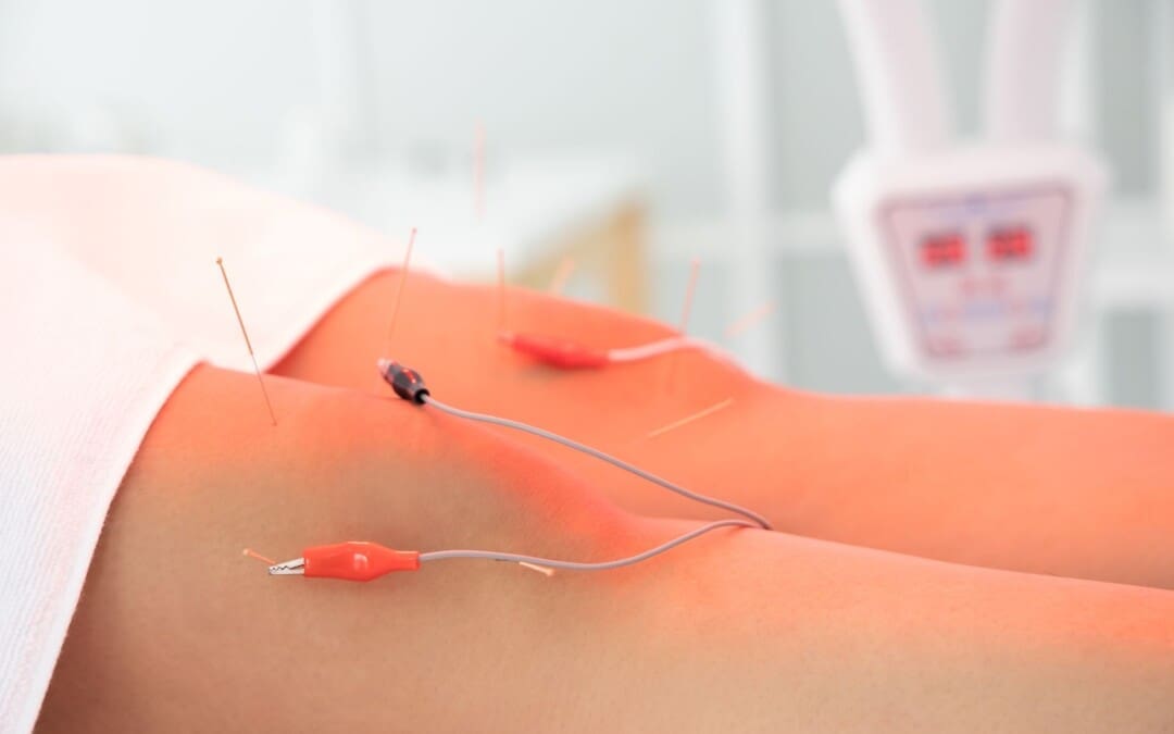
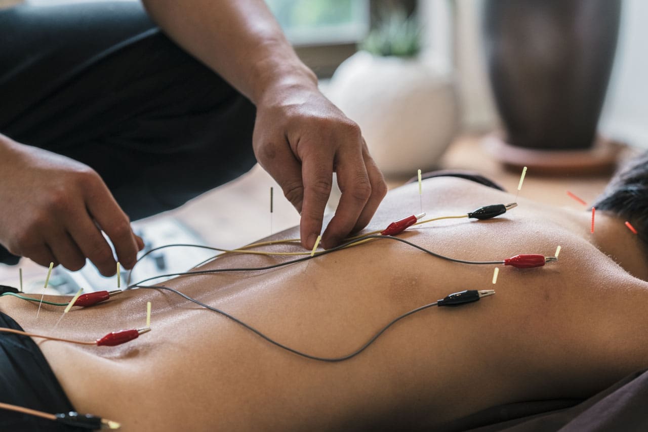
The Modern Twist Of Electroacupuncture
Now, electroacupuncture is a different form of acupuncture that uses the application of acupuncture needles and electric stimulation on the affected muscle. At the same time, when people are getting treated with electroacupuncture, their somatosensory afferent nerves provide pain relief. They are blocked to stop the pain signals from reaching the central nervous system. (Chen et al., 2021) This is because adding electric stimulation can enhance the therapeutic effects of the acupuncture points in the body.
How Electroacupuncture Benefits Muscle Pain
Regarding reducing muscle pain, electroacupuncture is more effective as acupuncturists can help adjust the intensity of the electric currents on the affected muscle to ensure comfort. Some of the benefits that electroacupuncture provides include:
Enhanced pain relief as the electric current can stimulate endorphin release.
Muscle relaxation from spasms in the affected muscle group.
Increased the healing rate by stimulating deeper muscles.
Help enhance muscle strength and flexibility to improve functionality.
Electroacupuncture can relieve pain and even adjust the biomechanical properties of the extensor-flexor muscles to improve abnormal joint loading caused by musculoskeletal pain. (Shi et al., 2020)
How These Two Treatments Help With Musculoskeletal Pain?
When it comes to acupuncture and electroacupuncture, it all depends on the severity of musculoskeletal pain affecting the body. Many people prefer traditional acupuncture for acute musculoskeletal pain in a more holistic approach. In comparison, others might prefer electroacupuncture to reduce the chronic pain effects of musculoskeletal pain. However, both of these treatments are non-surgical. They can be combined with other therapies like physical therapy or chiropractic care to help stimulate the body’s natural healing factor and relieve musculoskeletal pain. When these two treatments are combined with other therapies, the affected muscles are strengthened and provide mobility function back into the extremities. When people start thinking about their well-being, they can utilize these treatments to reduce the comorbidities associated with musculoskeletal pain that is affecting them. Thus allowing them to make small, healthy changes to their routine and live pain-free lives.
Beyond Adjustments: Chiropractic and Integrative Healthcare- Video
References
Chen, L., Wang, X., Zhang, X., Wan, H., Su, Y., He, W., Xie, Y., & Jing, X. (2021). Electroacupuncture and Moxibustion-Like Stimulation Relieves Inflammatory Muscle Pain by Activating Local Distinct Layer Somatosensory Afferent Fibers. Front Neurosci, 15, 695152. https://doi.org/10.3389/fnins.2021.695152
Dzakpasu, F. Q. S., Carver, A., Brakenridge, C. J., Cicuttini, F., Urquhart, D. M., Owen, N., & Dunstan, D. W. (2021). Musculoskeletal pain and sedentary behaviour in occupational and non-occupational settings: a systematic review with meta-analysis. Int J Behav Nutr Phys Act, 18(1), 159. https://doi.org/10.1186/s12966-021-01191-y
Shi, X., Yu, W., Wang, T., Battulga, O., Wang, C., Shu, Q., Yang, X., Liu, C., & Guo, C. (2020). Electroacupuncture alleviates cartilage degradation: Improvement in cartilage biomechanics via pain relief and potentiation of muscle function in a rabbit model of knee osteoarthritis. Biomed Pharmacother, 123, 109724. https://doi.org/10.1016/j.biopha.2019.109724
Wang, M., Liu, W., Ge, J., & Liu, S. (2023). The immunomodulatory mechanisms for acupuncture practice. Front Immunol, 14, 1147718. https://doi.org/10.3389/fimmu.2023.1147718
Welsh, T. P., Yang, A. E., & Makris, U. E. (2020). Musculoskeletal Pain in Older Adults: A Clinical Review. Med Clin North Am, 104(5), 855-872. https://doi.org/10.1016/j.mcna.2020.05.002
Zhu, J., Li, J., Yang, L., & Liu, S. (2021). Acupuncture, from the ancient to the current. Anat Rec (Hoboken), 304(11), 2365-2371. https://doi.org/10.1002/ar.24625
IFM's Find A Practitioner tool is the largest referral network in Functional Medicine, created to help patients locate Functional Medicine practitioners anywhere in the world. IFM Certified Practitioners are listed first in the search results, given their extensive education in Functional Medicine
 With contractures, the sarcomeres overly lengthen when the muscle fibers tighten. This increase in sarcomere length prevents the muscle from contracting normally, resulting in weakness. Muscle fibers are encased in an extracellular matrix, a mesh composed of collagen and other proteins that help transmit force and provide muscle contraction. Muscle contractures cause the amount of collagen within the extracellular matrix to increase, causing a stiffening of fibers that restricts movement. (Lieber, R. L., and Fridén, J. 2019)