Understanding Compression Injuries, Nerve Damage, and Whiplash from Car Accidents: A Comprehensive Guide
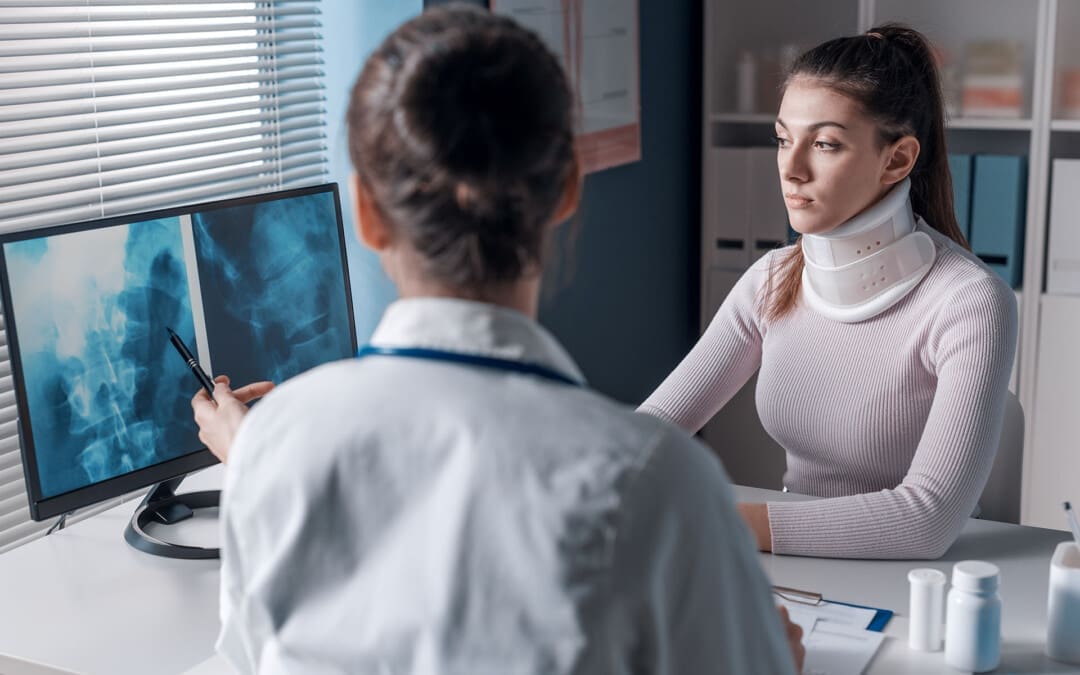
Car accidents, also known as motor vehicle accidents (MVAs), are a leading cause of injuries worldwide, often resulting in conditions that can significantly impact a person’s quality of life. Among the most common injuries are compression injuries, nerve damage, and indirect trauma like whiplash. These injuries can lead to chronic pain, mobility issues, and even long-term disabilities if not properly diagnosed and treated. In El Paso, Texas, where thousands of accidents occur annually, clinicians like Dr. Alexander Jimenez, a chiropractor and nurse practitioner, specialize in helping victims recover using advanced diagnostic tools and integrative care.
This blog explores the medical causes of compression injuries, nerve damage, and whiplash from MVAs; their connection to peripheral neuropathies; and the innovative approaches used by Dr. Jimenez to diagnose and treat these conditions. We’ll also discuss the impact of these injuries in El Paso’s personal injury cases and how accurate diagnostics bridge the gap between medical care and legal documentation. This guide aims to clarify and make complex medical concepts more accessible.
What Are Compression Injuries, Nerve Damage, and Whiplash?
Compression Injuries
The force of an MVA can squeeze or press parts of the body, such as bones, muscles, or nerves, resulting in compression injuries. For example, a sudden impact can compress spinal discs or vertebrae, leading to pain and restricted movement. These injuries often affect the spine, causing issues like herniated discs or fractures that may press on nerves.
Nerve Damage
Nerve damage, also called neuropathy, occurs when nerves are stretched, compressed, or torn. Nerves act like the body’s wiring, carrying signals between the brain and other parts. When damaged, they can cause symptoms like sharp pain, numbness, tingling, or weakness. In MVAs, nerve damage often results from trauma to the spine or limbs, disrupting normal function.
Whiplash
Whiplash is a common MVA injury, especially in rear-end collisions. The sudden jerking of the head forward and then backward strains the muscles, ligaments, and nerves of the neck. This rapid motion can cause inflammation or compression of nerves, leading to neck pain, headaches, and sometimes long-term issues. Research suggests that about 50% of whiplash patients experience neck pain for at least a year after the accident (Carroll et al., 2008).
Medical Causes of Compression Injuries and Nerve Damage in MVAs
MVAs can cause various injuries due to the sudden and forceful movements involved. Below, we examine the primary causes of compression injuries and nerve damage, drawing on clinical insights.
Whiplash and Nerve Compression
Whiplash occurs when the neck undergoes rapid acceleration and deceleration, often in rear-end collisions. This motion can inflame tissues around the cervical spine (neck) or compress nerves, leading to pain, stiffness, and numbness in the arms or hands. According to Houston Pain Specialists, whiplash is a primary cause of nerve pain in MVAs due to its impact on soft tissues and nerves.
Herniated Discs
The spine’s intervertebral discs act as cushions between vertebrae. In an MVA, the force of impact can cause these discs to shift or rupture, a condition known as a herniated disc. When the disc’s inner material protrudes, it can press on nearby nerves, causing pain, numbness, or weakness. The Russo Firm notes that herniated discs are a common cause of peripheral neuropathy, disrupting nerve signals to the limbs.
Spinal Cord Injuries
Severe MVAs can directly injure the spinal cord, the bundle of nerves running through the spine. These injuries may cause partial or complete loss of sensation and movement below the injury site. Spinal cord damage is less common but can lead to permanent nerve damage, affecting functions like walking or breathing.
Pinched Nerves
A pinched nerve occurs when surrounding tissues, such as bones or swollen muscles, compress a nerve. In MVAs, displaced vertebrae or inflamed tissues can pinch nerves, causing sharp pain or tingling. This is often observed in the neck or lower back, contributing to conditions such as radiculopathy.
Inflammation and Swelling
After an MVA, the body responds to trauma with inflammation, which can cause swelling around injured areas. This swelling may press on nerves, leading to pain and potential chronic nerve damage if untreated. Inflammation is a key factor in prolonged symptoms, as noted by Houston Pain Specialists.
Scar Tissue Formation
As the body heals, scar tissue can form around injured areas. This tissue may entrap or compress nerves, worsening pain over time. Scar tissue is a significant concern in cases of chronic nerve pain, as it can create lasting pressure on nerves.
Severe Stretching or Compression
The intense forces in an MVA can stretch or compress nerves beyond their normal range, causing immediate damage. This may lead to ongoing pain or neurological symptoms if the nerves don’t heal properly, as explained by Houston Pain Specialists.
Cause
Description
Common Symptoms
Whiplash
Rapid neck movement can inflame or compress nerves.
Neck pain, numbness, headaches
Herniated Discs
Disc rupture presses on nerves.
Pain, numbness, weakness in limbs
Spinal Cord Injuries
Direct trauma to the spinal cord.
Loss of sensation, paralysis
Pinched Nerves
Compression by bones or tissues.
Sharp pain, tingling
Inflammation and Swelling
Swelling presses on nerves.
Pain, reduced mobility
Scar Tissue Formation
Scar tissue entraps nerves post-healing.
Chronic pain, nerve irritation
Severe Stretching/Compression
Direct nerve damage from impact.
Persistent pain, neurological symptoms
Peripheral Neuropathies from MVA Injuries
Peripheral neuropathy refers to damage to the peripheral nerves, which connect the brain and spinal cord to the rest of the body. These nerves control movement, sensation, and autonomic functions, such as heart rate. Motor vehicle accidents (MVAs) can cause peripheral neuropathies through mechanisms such as nerve compression or trauma.
Sciatica as a Peripheral Neuropathy
Sciatica, a common peripheral neuropathy, occurs when the sciatic nerve, running from the lower back to the legs, is compressed. This type of injury often results from herniated discs or spinal misalignment caused by MVAs. Symptoms include radiating pain, numbness, and muscle weakness in the legs. Dr. Jimenez’s website highlights that ligamentous injuries, such as tears in the annulus fibrosus, can lead to disc herniation and sciatica (Jimenez, n.d.).
Symptoms and Diagnosis
Symptoms of peripheral neuropathy include:
Sharp, burning, or shooting pain
Numbness or tingling
Sensitivity to touch
Muscle weakness or coordination issues
Autonomic issues like blood pressure changes
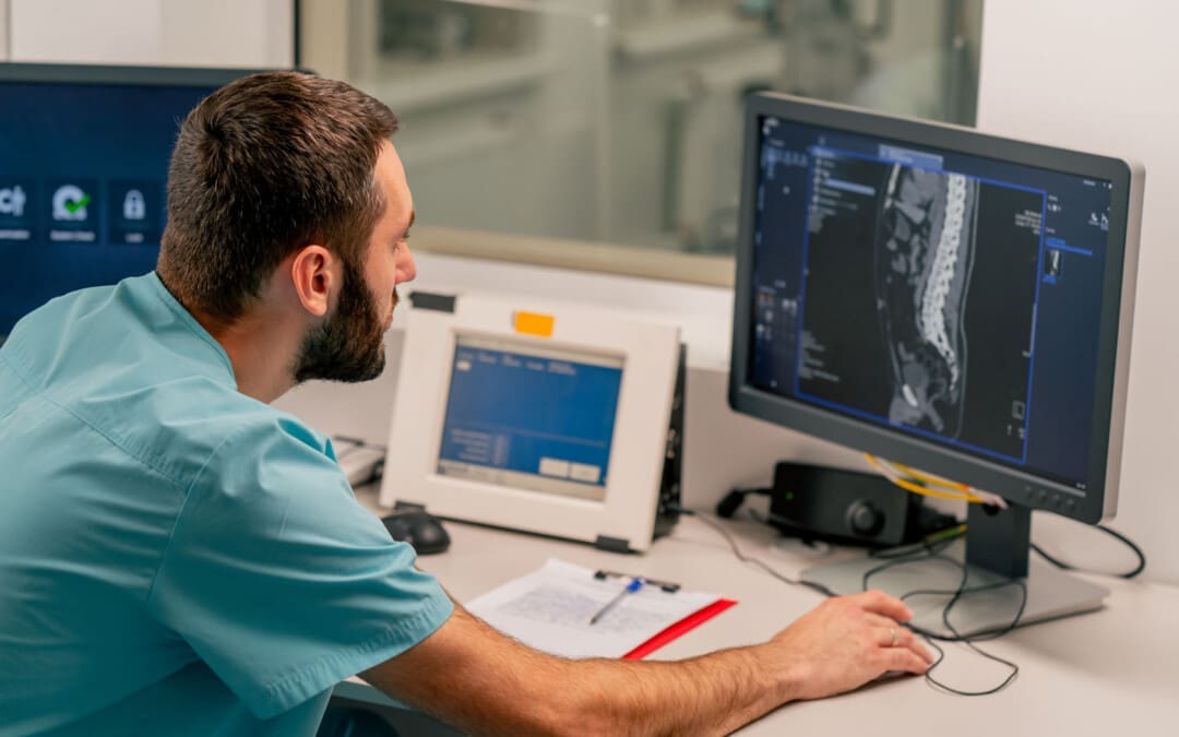
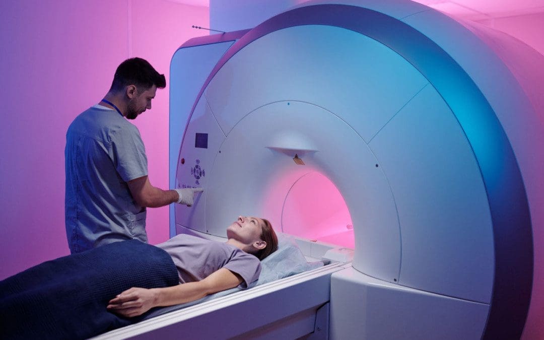
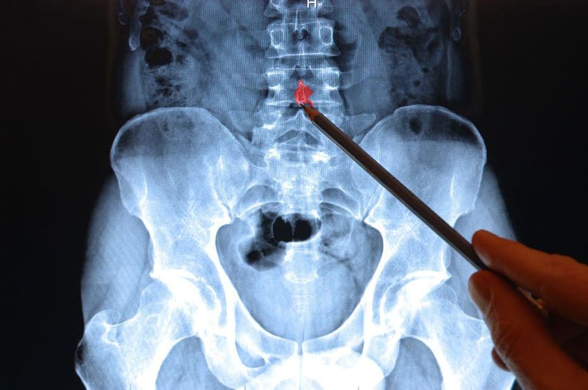
Diagnosis often involves imaging, such as MRI, to detect nerve compression, as well as clinical assessments to evaluate symptoms. Early diagnosis is crucial in preventing chronic conditions, as noted by The Russo Firm.
Dr. Alexander Jimenez’s Clinical Insights
Dr. Alexander Jimenez, DC, APRN, FNP-BC, is a leading clinician in El Paso, Texas, recognized for his integrative approach to treating motor vehicle accident (MVA) injuries. With over 30 years of experience, he holds certifications in chiropractic care, functional medicine, and nursing, allowing him to address both musculoskeletal and systemic issues.
Background and Expertise
Dr. Jimenez’s practice focuses on holistic healing, combining chiropractic techniques with functional medicine. His clinic, Injury Medical Clinic, offers treatments for chronic pain, personal injury, and complex conditions like sciatica and herniated discs. Awards such as the Top Rated El Paso Chiropractor Award from 2015 to 2024 (Three Best Rated) recognize his work.
Diagnostic Approach
Dr. Jimenez uses advanced diagnostic tools to assess MVA injuries:
MRI: Detects soft tissue injuries like ligament tears and disc herniations, which may not appear on X-rays. A case study on his website describes a 49-year-old female with a 9 mm disc bulge causing radicular pain, diagnosed via 1.5 Tesla MRI (Jimenez, 2017).
Computerized Radiographic Mensuration Analysis (CRMA) assesses how the spine moves to determine if the ligaments are loose, a condition known as Alteration of Motion Segment Integrity (A.O.M.S.I.). The diagnosis is crucial for deciding impairment ratings, which can significantly influence insurance claims (Jimenez, 2017).
Treatment Strategies
Dr. Jimenez employs chiropractic adjustments, physical therapy, and functional medicine to restore mobility and reduce pain. His integrative approach addresses both immediate injuries and underlying causes, such as inflammation or biomechanical issues, to prevent the development of chronic conditions.
Impact of El Paso’s Personal Injury Cases
El Paso experiences a high number of motor vehicle accidents (MVAs), with 19,150 reported in 2021, of which 25–27% resulted in injuries (Make Roads Safe). Common injuries include whiplash, herniated discs, and nerve damage, which can lead to long-term issues like peripheral neuropathy. Dr. Jimenez’s clinic plays a vital role in treating these victims, offering personalized care plans to restore health and support legal claims.
Case Study Example
A 49-year-old female patient involved in an MVA experienced radicular pain due to a 9 mm disc bulge, detected via MRI. Dr. Jimenez’s treatment plan, combining chiropractic care and physical therapy, helped alleviate her symptoms and provided documentation for her personal injury case (Jimenez, 2017).
Linking Diagnostic Tests and Imaging to Patient Injuries
Accurate diagnosis is crucial for effective treatment and legal documentation in motor vehicle accident (MVA) cases. Dr. Jimenez’s use of advanced imaging and diagnostic tests ensures precise identification of injuries, which is crucial for both medical and legal outcomes.
Importance of Diagnostics
Standard X-rays may miss soft tissue injuries, with 12% of spinal cord injuries showing no radiographic abnormality (Jimenez, 2017). MRI and CRMA provide detailed insights:
MRI: Visualizes ligament tears, disc herniations, and nerve compression.
CRMA: Measures spinal instability, with specific criteria for A.O.M.S.I. (e.g., >3.5 mm motion in the cervical spine). Such injuries can result in a 25–28% impairment rating, which can influence insurance settlements (Jimenez, 2017).
Legal Documentation
Insurance companies reserve significant funds (e.g., $60,000) for ligament laxity diagnoses, as they indicate serious injury. Dr. Jimenez’s detailed documentation, supported by CRMA and MRI, helps patients secure fair compensation for medical bills, lost wages, and pain and suffering.
Diagnostic Tool
Purpose
Impact on Treatment and Legal Claims
MRI
Detects soft tissue and nerve damage
Guides treatment; provides evidence for legal claims
The term “dual-scope procedures” may refer to Dr. Jimenez’s use of multiple diagnostic approaches, such as combining MRI and CRMA, to assess injuries comprehensively. This dual approach ensures a thorough understanding of both structural and functional damage, which enhances treatment plans and legal documentation.
Conclusion
Motor vehicle accidents can cause severe injuries, like compression injuries, nerve damage, and whiplash, often leading to peripheral neuropathies such as sciatica. These conditions require prompt and accurate diagnosis to prevent chronic pain and disability. In El Paso, Dr. Alexander Jimenez stands out for his expertise in treating MVA victims, using advanced tools like MRI and CRMA to link injuries to effective treatment and legal outcomes. His integrative approach ensures patients receive holistic care while supporting their pursuit of fair compensation.
If you have been involved in a motor vehicle accident (MVA), please consider seeking a medical evaluation promptly to address any potential injuries. Contact specialists like Dr. Jimenez at Injury Medical Clinic (915-850-0900) for expert care and support.
How are MRIs used to help diagnose bulging and herniated discs and help healthcare providers develop effective treatment programs for individuals experiencing back pain symptoms?
Herniated Bulging Disc MRI
A herniated bulging disc is often identified during magnetic resonance imaging (MRI); however, it’s usually an incidental finding that was done for other reasons where spinal problems and/or injuries are found. A bulging disc is relatively common, even in individuals who experience no symptoms. A herniated or bulging disc in the back can be identified with an MRI test, typically recommended when someone experiences back pain symptoms for at least six weeks. (American Academy of Neurological Surgeons, 2024) Normal wear and tear and age cause changes in the spinal disc/s cushion to bulge and become misaligned with the spine. (Brinjikji W. et al., 2015) And with a herniated disc, it can press against the spinal cord and nerves. Repeated heavy lifting, practicing unhealthy postures, a history of back injuries, or underlying health conditions are common causes.
Bulging Disc
Bulging discs are common even in healthy individuals but can be difficult to interpret independently on an MRI, so other symptoms and findings are as important in diagnosis.
Causes
A bulging disc is usually considered age-related degenerative changes that cause the disc to bulge downward with gravity. (Penn Medicine, 2018)
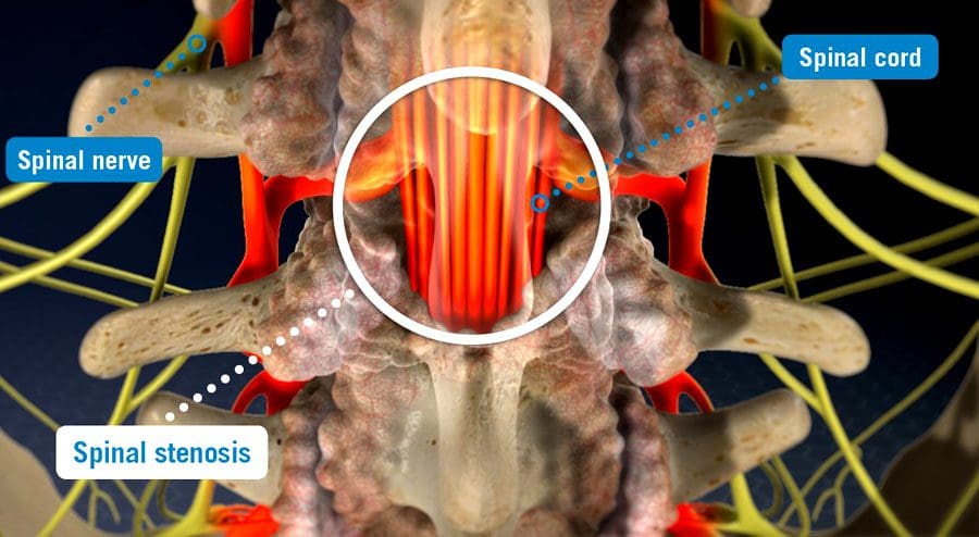
A significant bulge is expected to cause leg pain due to irritation to the nerves going down the legs. (Amin R. M., Andrade N. S., & Neuman B. J. 2017) As the condition progresses, more than one disc can be affected, leading to other spinal conditions, including spinal stenosis.
A Bulging Disc On MRI
A disc bulge will measure over 25% of the total disc circumference. Its displacement is usually 3 millimeters or less from the normal shape and position of the disc. (Radiopaedia, 2024)
Herniated Disc
A herniated disc shifts out of its correct position and compresses nearby spinal nerves, causing pain and mobility issues.
Herniated discs will measure less than 25% of the total disc circumference. However, herniation is based on the type and can include: (Wei B., & Wu H. 2023)
Disc Protrusion – the displacement is limited, and the ligaments are intact.
Disc Extrusion – part of the disc remains connected but has slipped through the annulus or outer covering of the disc.
Disc Sequestration – a free fragment has separated and broken off from the main disc.
Candidates For Spinal MRI
The MRI is generally safe for most, including those with implanted cardiac devices like newer-model pacemakers. (Bhuva A. N. et al., 2020) However, it’s important to ensure that the healthcare team is aware of cochlear implants or other devices so that necessary precautions can be taken. It is recommended for all individuals that symptoms be present for six weeks before an MRI. A specialist may want to see MRI results sooner, especially if symptoms include: (American Academy of Neurological Surgeons, 2024)
A specific injury, like a fall that caused the pain
Recent or current infection or fever with spinal symptoms
Significant weakness in arms or legs
Loss of pelvic sensation.
A history of metastatic cancer.
Loss of bladder or bowel control
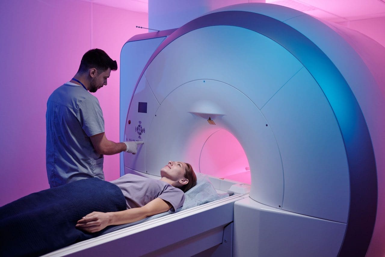
An MRI may be needed if symptoms are rapidly worsening. However, many with a disc bulge don’t have symptoms at all. In most cases, an MRI is an outpatient procedure that can be completed in an hour or less but can take longer if contrast dye is used. The healthcare provider will provide specific instructions about MRI preparation.
Treatment
Treatment for a herniated or bulging disc depends on the cause and severity of symptoms.
Over-the-counter pain relief, including nonsteroidal anti-inflammatory drugs (NSAIDs).
Physical therapy
In rare cases that have not resolved with conservative treatment, surgery may be recommended.
Remember that the MRI findings may not identify or rule out all conditions, including muscle strains or ligament injuries, which may require different treatments, such as targeted stretches and exercises. (Brinjikji W. et al., 2015) (Fujii K. et al., 2019)
Herniated Disc
Treatment depends on the cause and severity of symptoms, if any. It can include stand-alone or a combination of physical therapy, medication, and steroid injections. Cases usually resolve in six to 12 weeks (Penn Medicine, 2018). Electrical nerve stimulation may be performed through specialized devices and/or acupuncture to help with nerve compression. (National Institute of Neurological Disorders and Stroke, 2020) Surgery may be recommended if conservative treatments fail to achieve significant pain relief and healing. (Wang S. et al., 2023)
Injury Medical Chiropractic and Functional Medicine Clinic
A healthcare provider can discuss treatment options such as pain medication, physical therapy, and surgery. Injury Medical Chiropractic and Functional Medicine Clinic works with primary healthcare providers and specialists to develop an optimal health and wellness solution. We focus on what works for you to relieve pain, restore function, and prevent injury. Regarding musculoskeletal pain, specialists like chiropractors, acupuncturists, and massage therapists can help mitigate the pain through spinal adjustments that help the body realign itself. They can also work with other medical professionals to integrate a treatment plan to resolve musculoskeletal issues.
Root Causes of Spinal Stenosis
References
American Academy of Neurological Surgeons. (2024). Herniated disc. https://www.aans.org/patients/conditions-treatments/herniated-disc/
Brinjikji, W., Diehn, F. E., Jarvik, J. G., Carr, C. M., Kallmes, D. F., Murad, M. H., & Luetmer, P. H. (2015). MRI Findings of Disc Degeneration are More Prevalent in Adults with Low Back Pain than in Asymptomatic Controls: A Systematic Review and Meta-Analysis. AJNR. American journal of neuroradiology, 36(12), 2394–2399. https://doi.org/10.3174/ajnr.A4498
Penn Medicine. (2018). Bulging Disc vs. Herniated Disc: What’s The Difference? Penn Musculoskeletal and Rheumatology Blog. https://www.pennmedicine.org/updates/blogs/musculoskeletal-and-rheumatology/2018/november/bulging-disc-vs-herniated-disc
Wu, P. H., Kim, H. S., & Jang, I. T. (2020). Intervertebral Disc Diseases PART 2: A Review of the Current Diagnostic and Treatment Strategies for Intervertebral Disc Disease. International journal of molecular sciences, 21(6), 2135. https://doi.org/10.3390/ijms21062135
Amin, R. M., Andrade, N. S., & Neuman, B. J. (2017). Lumbar Disc Herniation. Current reviews in musculoskeletal medicine, 10(4), 507–516. https://doi.org/10.1007/s12178-017-9441-4
American Academy of Orthopaedic Surgeons. (2022). Herniated disk in the lower back. https://orthoinfo.aaos.org/en/diseases–conditions/herniated-disk-in-the-lower-back/
Wei, B., & Wu, H. (2023). Study of the Distribution of Lumbar Modic Changes in Patients with Low Back Pain and Correlation with Lumbar Degeneration Diseases. Journal of pain research, 16, 3725–3733. https://doi.org/10.2147/JPR.S430792
Bhuva, A. N., Moralee, R., Moon, J. C., & Manisty, C. H. (2020). Making MRI available for patients with cardiac implantable electronic devices: growing need and barriers to change. European radiology, 30(3), 1378–1384. https://doi.org/10.1007/s00330-019-06449-5
Brinjikji, W., Luetmer, P. H., Comstock, B., Bresnahan, B. W., Chen, L. E., Deyo, R. A., Halabi, S., Turner, J. A., Avins, A. L., James, K., Wald, J. T., Kallmes, D. F., & Jarvik, J. G. (2015). Systematic literature review of imaging features of spinal degeneration in asymptomatic populations. AJNR. American journal of neuroradiology, 36(4), 811–816. https://doi.org/10.3174/ajnr.A4173
Fujii, K., Yamazaki, M., Kang, J. D., Risbud, M. V., Cho, S. K., Qureshi, S. A., Hecht, A. C., & Iatridis, J. C. (2019). Discogenic Back Pain: Literature Review of Definition, Diagnosis, and Treatment. JBMR plus, 3(5), e10180. https://doi.org/10.1002/jbm4.10180
Wang, S., Zhao, T., Han, D., Zhou, X., Wang, Y., Zhao, F., Shi, J., & Shi, G. (2023). Classification of cervical disc herniation myelopathy or radiculopathy: a magnetic resonance imaging-based analysis. Quantitative imaging in medicine and surgery, 13(8), 4984–4994. https://doi.org/10.21037/qims-22-1387
National Institute of Neurological Disorders and Stroke. (2020). Low back pain fact sheet. Retrieved from https://www.ninds.nih.gov/sites/default/files/migrate-documents/low_back_pain_20-ns-5161_march_2020_508c.pdf
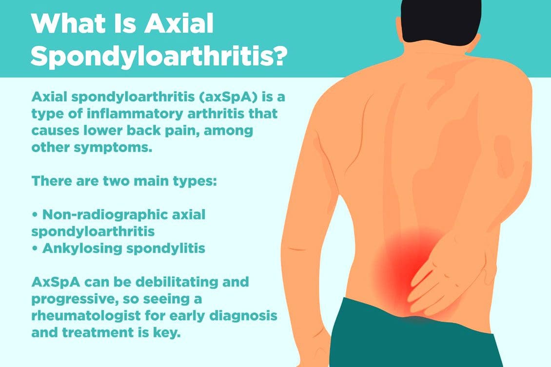
Non-radiographic axial spondyloarthritis or nr-axSpA and non-radiographic ankylosing spondylitis/AS are related. However, non-radiographic axial spondyloarthritis can present AS symptoms with active inflammation of the spine and sacroiliac/SI joints, causing back and hip pain but does not reveal joint damage on X-rays or MRIs. Injury Medical Chiropractic and Functional Medicine Clinic can explain what it means to have non-radiographic axial spondyloarthritis, how it can be managed, and what to do to prevent it from turning into ankylosing spondylitis.
Non-Radiographic Axial Spondyloarthritis
Non-radiographic axial spondyloarthritis means there are early AS symptoms but have not developed enough joint inflammation or damage to show up on an X-ray or other form of imaging. Early evidence of joint inflammation includes blurring of the joint edges and localized regions of joint erosion. It can be difficult for physicians to see these subtle changes on an x-ray.
Ankylosing Spondylitis
Ankylosing spondylitis, or AS, is a form of inflammatory arthritis that affects joints in the spine and elsewhere.
It is a chronic, inflammatory, autoimmune disease.
Medical research is still ongoing to determine the exact cause, but a genetic component is believed to be contributing factor.
Around 85% of individuals with ankylosing spondylitis have inherited the HLA-B27 gene, which is associated with multiple autoimmune conditions.
In the early stages, individuals will present lower back pain around the sacroiliac joints or the joints that connect the spine to the pelvis.
Later stages have more obvious X-ray findings, like the fusing of the sacroiliac joints and the lower spine that takes place over time.
Joint inflammation can progress, causing permanent joint damage and spine rigidity.
Most individuals with the condition can manage their symptoms with NSAIDs, chiropractic care, physical and massage therapy, and range of motion exercises.
Stage 1
There is no evidence of spinal inflammation on x-rays.
MRI provides more detailed images of bones and may reveal bone marrow edema or accumulation of fluid in the structures of the spinal bones and joints.
Individuals with non-radiographic axial spondyloarthritis, you are here.
Stage 2
There is visible inflammation of the spinal joints on the x-ray.
The sacroiliac joints between the spine and the pelvis are the most affected.
Stage 3
Chronic inflammation of the joints has caused bone loss and permanent joint damage, resulting in spine rigidity.
Symptoms of Non-Radiographic Axial Spondyloarthritis
There are differences between back pain associated with muscle strain and arthritis. Back pain symptoms include:
Starts to present before age 40.
It has a gradual onset and can go unnoticed for years.
Improves with movement or activity.
Eases up throughout the day.
Starts up in the evening when resting.
Other symptoms include:
Joint stiffness
Swollen fingers
Heel pain
Bilateral buttock discomfort and pain
Slowing Progression
Progression from non-radiographic axial spondyloarthritis to ankylosing spondylitis occurs in 10% – 20% of individuals over a two-year period. Progression factors include genetics, gender, degree of joint damage, and level of inflammatory markers at the time of diagnosis.
Early diagnosis and treatment can slow the progression before significant joint damage with anti-inflammatory therapy, rheumatological therapy, and targeted exercise.
Work with a specialist like an orthopedic spine specialist and rheumatologist that understands the disorder and is up to date on the most recent treatment modalities.
Individuals with non-radiographic axial spondyloarthritis should expect to have serial X-rays to gauge the progression of the disease.
Staying healthy and active is recommended to slow the progression of nr-AxSpA and AS.
Recent medical advances and lifestyle adjustments can slow the progression in most cases.
axSpA
References
Six tips for living well with ankylosing spondylitis. Available at https://www.mayoclinic.org/diseases-conditions/ankylosing-spondylitis/in-depth/6-tips-for-living-well-with-ankylosing-spondylitis/art-20478753. Accessed 11/07/2022.
Ankylosing spondylitis. Mayo Clinic. Available at https://www.mayoclinic.org/diseases-conditions/ankylosing-spondylitis/symptoms-causes/syc-20354808. Accessed 11/05/2022.
D. J. Pradeep, A. Keat, K. Gaffney, Predicting outcome in ankylosing spondylitis, Rheumatology, Volume 47, Issue 7, July 2008, Pages 942–945, https://doi.org/10.1093/rheumatology/ken195
Kucybała, Iwona, et al. “Radiologic approach to axial spondyloarthritis: where are we now and where are we heading?.” Rheumatology international vol. 38,10 (2018): 1753-1762. doi:10.1007/s00296-018-4130-1
Michelena, Xabier, López-Medina, Clementina, and Helena Marzo-Ortega. “Non-radiographic versus radiographic axSpA: what’s in a name?”.” National Center for Biotechnology Information. October 14, 2020. https://doi.org/10.1093/rheumatology/keaa422
Swift D. Ankylosing spondylitis: disease progression varies widely. Medpage Today. Accessed 11/05/2022.Available at https://www.medpagetoday.com/rheumatology/arthritis/49096
Spinal stenosis is when space somewhere along or within the spine begins to narrow, closing off the ability of normal/comfortable movement and nerve circulation. It can affect different areas, including the cervical/neck, lumbar/low back, and, less commonly, the thoracic/upper or mid-back regions causing tingling, numbness, cramping, pain, muscle weakness, or a combination in the back, leg/s, thighs, and buttocks. There can be various factors causing the stenosis; correct diagnosing is the first step, and where a spinal stenosis MRI comes in.
Spinal Stenosis MRI
Stenosis can be challenging to diagnose as it is more of a symptom/complication than a condition, often caused by herniated discs, bone spurs, a congenital condition, post-surgery, or after an infection. Magnetic resonance imaging/MRI is a common test used in diagnosis.
Diagnosis
A healthcare professional, like a chiropractor, physical therapist, spine specialist, or physician, will begin with understanding symptoms and medical history.
A physical exam will be conducted to learn more about the location, duration, positions, or activities that decrease or worsen the symptoms.
Additional tests include muscle strength, gain analysis, and balance testing to help better understand where the pain is coming from.
To confirm a diagnosis, imaging will be required to see what is going on.
An MRI uses computer-generated imaging to produce images that show bone and soft tissues, like muscles, nerves, and tendons, and if they are compressed or irritated.
A healthcare professional and MRI technician will go over the safety requirements before the imaging.
Because the machine uses powerful magnets, there can be no metal on or in the body, like implanted prostheses or devices that include:
A different imaging test may be used if an individual cannot have an MRI like a CT scan.
An MRI can range from several minutes to an hour or longer, depending on how many positions are necessary to isolate the injured area and get a clear image. The test is painless, but sometimes individuals are asked to maintain a specific position that could be uncomfortable. The technician/s will ask if there is discomfort and offer any help to make the experience as easy as possible.
Treatment
Not all cases of stenosis cause symptoms, but there are treatment options that a healthcare professional can recommend.
Conservative care is the first recommendation that includes chiropractic, decompression, traction, and physical therapy.
Treatment increases muscle strength, improves range of motion, improves posture and balance, decreases discomfort symptoms, and incorporates strategies to prevent and manage symptoms.
Prescription medications could be part of a larger treatment plan.
Surgery could become an option in more severe cases where conservative care is not working.
Spinal Stenosis
References
Database of Abstracts of Reviews of Effects (DARE): Quality-assessed Reviews [Internet]. York (UK): Centre for Reviews and Dissemination (UK); 1995-. Diagnosis of lumbar spinal stenosis: an updated systematic review of the accuracy of diagnostic tests. 2013. Available from: https://www.ncbi.nlm.nih.gov/books/NBK142906/
Ghadimi M, Sapra A. Magnetic Resonance Imaging Contraindications. [Updated 2022 May 8]. In: StatPearls [Internet]. Treasure Island (FL): StatPearls Publishing; 2022 Jan-. Available from: https://www.ncbi.nlm.nih.gov/books/NBK551669/
Gofur EM, Singh P. Anatomy, Back, Vertebral Canal Blood Supply. [Updated 2021 Jul 26]. In: StatPearls [Internet]. Treasure Island (FL): StatPearls Publishing; 2022 Jan-. Available from: https://www.ncbi.nlm.nih.gov/books/NBK541083/
Lurie, Jon, and Christy Tomkins-Lane. “Management of lumbar spinal stenosis.” BMJ (Clinical research ed.) vol. 352 h6234. 4 Jan. 2016, doi:10.1136/bmj.h6234
Stuber, Kent, et al. “Chiropractic treatment of lumbar spinal stenosis: a review of the literature.” Journal of chiropractic medicine vol. 8,2 (2009): 77-85. doi:10.1016/j.jcm.2009.02.001
Chiropractors and spine specialists utilize spinal imaging through X-rays, MRIs, or CT scans to figure out what is causing back problems and pain. Imaging is common. Whether chiropractic or spinal surgery, they help immensely discover back issues and allow the individual to see what is happening. Types of cases include back pain that:
X-rays for back pain can be quite helpful. An X-ray is radiation-based and is used to examine the conditions of the bone structures. X-rays are optimal for bone tissue or tissues that are ossified or calcified. They work the best with hard tissues, specifically bones. Soft tissues like muscles, ligaments, or intravertebral discs do not present as well.
Individuals undergoing a back X-ray will be scanned by a machine that generates a beam. A receiver picks registers the beam after it passes through the body and generates an image. It takes around five minutes to complete but could be longer depending on the doctor’s number of images. X-rays are helpful for insurance purposes and rule out bone conditions like compression fractures and/or bone spurs. X-rays are ordered for specific reasons and are often part of a whole-body diagnostic study. This includes MRI and/or CT scan.
CT Scan
CT stands for computed tomography. It is a series of X-rays that are digitized into images using a computer. The advantage of a CT scan to standard X-rays is that it offers different views/angles of the body and can be in 3D. CT scans are most often used in trauma cases or individuals who have had surgery. They take around five minutes. For X-rays, individuals stand up or lay under the X-ray machine as it scans the body. A CT scan has the individual lie down in a circular donut-looking machine that scans while rotating during the imaging. Individuals are recommended to wear casual loose, comfortable clothing. Sometimes a dye, or intravenous contrast, is used to get the vascular tissues to stand out, generating clearer images.
MRI
MRI is short for magnetic resonance imaging. MRIs use magnets to generate images. MRI imaging is often used in individuals that have undergone surgery. They take longer, usually around 30 to 45 minutes. No metallic objects are allowed in the MRI. Patients are asked to remove items like belts, jewelry, etc. Contrast dye can be a part of an MRI. The machine is like a tunnel. This can become challenging for individuals that have claustrophobia. Consult with a doctor and find out how to get comfortable during the process.
Other Forms of Spinal Imaging
Other forms of imaging include:
CT navigation
CT navigation shows real-time CT scans during the procedure.
Fluoroscopy
Fluoroscopy involves an X-ray beam that passes directly through the body that shows live, moving images.
Both of these types of spinal imaging are utilized during surgeries. For some cases, intraoperative imaging is used. This type of imaging uses high-tech robotics to help surgeons navigate through tight spaces during the procedure. This increases the surgeon’s accuracy and reduces the size of the incision.
Ultrasound
Ultrasound can be used for spinal conditions. This is an imaging test that uses sound waves to generate images. However, the imaging tests which are used in spinal imaging are primarily X-rays and MRIs.
Imaging Appointment
Talk with your doctor or chiropractor ahead of time to understand what to expect during the imaging process. They will let you know how to prepare and any special instructions before the appointment. Along with medical history and a physical examination, spinal imaging is an important part of the diagnostic process to find what is causing the pain and to develop the best treatment plan.
Body Composition
Short-term Effects of Coffee and Blood Pressure
The caffeine in coffee is a stimulant or substance that excites the body’s systems. When caffeine is ingested, individuals experience an increase in excitement, specifically in the cardiovascular system. This excitement causes the heart rate and blood pressure to rise and then lower back to a baseline level for healthy individuals. Coffee slightly increases short-term blood pressure. Moderate coffee consumption is safe for individuals that do not have pre-existing cardiovascular conditions.
References
United States Nuclear Regulatory Commission. (May 2021) “Doses in Our Daily Lives” https://www.nrc.gov/about-nrc/radiation/around-us/doses-daily-lives.html
X-Ray for Back Pain: Current Reviews in Musculoskeletal Medicine. (April 2009) “What is the role of imaging in acute low back pain?” https://www.ncbi.nlm.nih.gov/pmc/articles/PMC2697333/
Low back pain is one of the most common ailments for people visiting a doctor or an urgent care clinic. When the back pain becomes intense, it can get you thinking something is seriously wrong with your back. The doctor might offer an x-ray or MRI scan to put your concerns at ease.
Fortunately, most cases of low back pain, even acute pain, improve within days or a few weeks. Most cases are remedied with chiropractic, physical therapy, heat/ice therapy, and rest. And a lot of these cases do not require any form of spinal imaging. However, those are why X-ray, MRI, and CT scans are necessary to figure out what’s happening.
Strained muscle
Sprained ligament
Poor posture
These typical causes of low back pain can be painful and limit activities.
Back Pain Lasting Longer Than 2/3 Weeks
Subacute pain lasts between 4 and 12 weeks, while chronic back pain lasts three months or longer. These are not indications of a severe lower back spinal condition.
Less than 1% of people with low back pain are diagnosed with the condition that may require spine surgery:
Doctors may recommend an x-ray or MRI if the low back pain is from a traumatic injury, like a:
Slip
Fall
Automobile accident
Other potential causes of low back pain may warrant medical imaging immediately or later.
The diagnostic process starts with the evaluation of the low back symptoms and how they relate to what was found during the:
Physical exam
Neurological exam
Medical history
A doctor utilizes these results to determine whether spinal imaging is necessary, along with the type of imaging test, x-ray, or MRI and the timing to confirm a diagnosis.
A Low Back X-Ray/MRI
X-ray spinal imaging best detects bony structural problems but is not so great with soft tissue injuries. X-ray series may be performed to diagnose vertebral compression fractures.
Anterior
Posterior
Lateral views
MRI is a radiation-free test. MRIs create 3-D anatomical views of the spinal bones and soft tissues. A contrast dye like gadolinium is used to enhance and improve the quality of the images. The contrast is injected through an intravenous line in your hand or arm before or during the test. An MRI can evaluate neurological symptoms, like radiating pain or pain that develops after a cancer diagnosis.
Symptoms, Co-existing Medical Diagnoses, and Conditions that may Require Spine Imaging
Neurological symptoms
Low back pain that radiates, fans out, or downward into the buttocks, legs, and feet
Abnormal reflexes in the lower body can indicate nerve disruption
Radiation to your entire body is measured through the millisievert (mSv), also known as the effective dose. The radiation dose is the same amount every time you experience an x-ray. When undergoing an x-ray, the radiation not absorbed by the body creates the image.
The effective dose helps a doctor measure the risk for possible side effects of radiographic imaging:
CT scans use radiation as well
Specific body tissues and organs in the lower back are sensitive to radiation exposure, like the reproductive organs.
MRI Radiation-Free Why Not Just Use This Test All The Time
MRI cannot be used on all patients because of its powerful magnet technology. Pregnant women or individuals with metal inside their body, like a spinal cord stimulator, heart pacemaker, etc., cannot be scanned with an MRI.
MRI testing is also expensive; doctors do not want to prescribe unnecessary tests that increase costs. Or because of the fine detail that MRIs provide, sometimes a spinal issue can look severe but is not.
Example: An MRI of the lower back reveals a herniated disc in a patient with no back/leg pain or other symptoms.
This is why doctors bring all their findings like the symptoms, physical exam, and medical history to confirm a diagnosis and create a custom treatment plan.
Imaging Test Takeaways
If low back pain takes its toll, listen to what the doctor recommends. They might not order a lumbar x-ray or MRI immediately but remember the issues mentioned above, like neurological symptoms and co-existing medical conditions. But these tests help discover the cause or causes of the pain. Remember this is to help get patients to their optimal health and pain-free.
How to eliminate Back Pain naturally | (2020) Foot Levelers |El Paso, Tx
NCBI Resources
Imaging diagnostics is an essential element in the evaluation of spine trauma. The rapid evolution of imaging technology has tremendously changed the assessment and treatment of spine injuries. Imaging diagnostics utilizing CT and MRI, among others, are helpful in acute and chronic settings. Spinal cord and soft-tissue injuries are best evaluated by magnetic resonance imaging, or MRI, whereas computed tomography scanning or CT scans best evaluate spinal trauma or spine fracture.
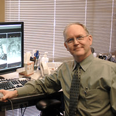
Dr. Alex Jimenez collaborates with top rated diagnosticians and imaging specialists. We are blessed to have in our association, imaging specialists that provide fast, courteous & premiere board certified specialists. In collaboration with our offices we can provide the quality of service our patients mandate and deserve.
Who We Are
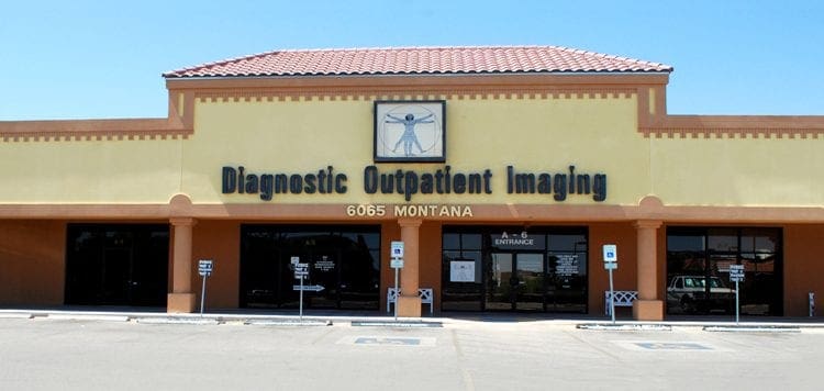
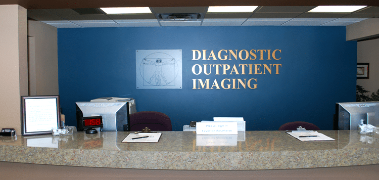
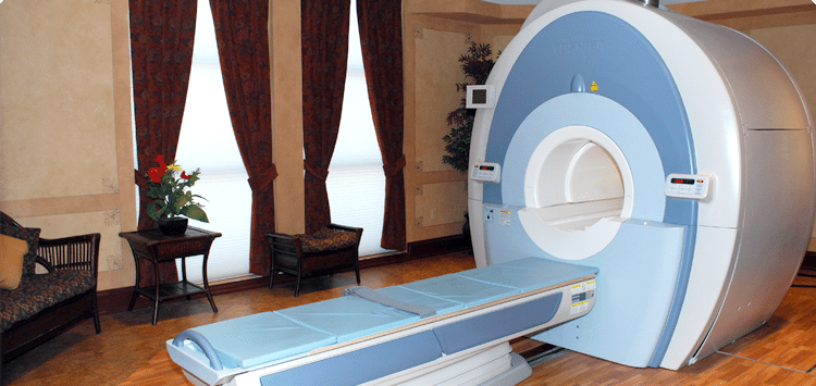
Diagnostic Outpatient Imaging (DOI) is a state-of-the-art Radiology center in El Paso, TX. It is the only center of its kind in El Paso, owned and operated by a Radiologist.
This means when you come to DOI for a radiologic exam, every detail, from the design of the rooms, the choice of the equipment, the hand-picked technologists, and the software which runs the office, is carefully chosen or designed by the Radiologist and not by an accountant.
Our market niche is one center of excellence. Our values related to patient care are: We believe in treating patients the way we would treat our family and we will do our best to ensure that you have a good experience at our clinic.
Dear Doctors,
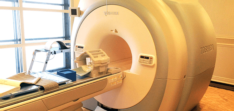
We are pleased to inform you of the arrival of our Titan 3-Tesla MRI at Diagnostic Outpatient Imaging. This is El Paso’s only radiology imaging center that offers this technology. Patients do not always realize how important image quality is: It can make the difference in the diagnosis.
3-Tesla MRI is like HD TV and once you try it, you will not want to go back. The increased magnet strength gives us many benefits at no additional expense to the patient. It gives us the ability to scan faster or to scan with higher detail. An MRI of the brain can take 20 minutes and have exceptional quality, or we can perform the scan in less time, with better quality that is achieved on most 1.5 Tesla “high field” MRIs. This is incredibly useful for children.
Our 3T MRI can perform Diffusion Tensor Imaging, MRI Spectroscopy and CSF flow studies to name just a few of its possibilities.
This scanner is not only very fast, it is very large. Our open MRI has a clearance of 35 cm. The 3T has a diameter of 71 cm! This is welcome news for nervous or claustrophobic patients, and combined with its speed, it can actually eliminate the need for sedation for some patients. 3T MRI is faster, clearer, and has more diagnostic possibilities. We are certain you and your patients will notice the difference.
Our Services
MRI’s:
DOI has three MRI’s under one roof. All are American College of Radiology (ACR) Certified.
Good
Open MRI (0.35 Tesla): This MRI perfect for claustrophobic and very large patients. There is no table weight limit on this MRI
Better
High Field 1.5 Tesla MRI- This is a eight channel MRI with high end image quality. It is in a beautiful room and has ‘pianissimo’ technology, which makes the MRI relatively quiet. This machine has been the best MRI in private practice in El Paso for years. It will soon be eclipsed by our new 3.0 Tesla MRI.
Best
High Field 3.0 Tesla MRI- This is the only 3.0 Tesla MRI in private practice in El Paso. This technology can deliver stunning image quality, which can actually make a difference in your diagnosis. The increased magnet strength gives us many benefits at no additional expense to the patient.�??It gives us the ability to scan faster, or to scan with higher detail. This is welcome news for nervous or claustrophobic patients, and as well as for children as it can actually eliminate the need for sedation in some patients. 3T is faster, clearer, more diagnostic for a better for MRI. It is like HD TV. Once you have tried it, you won’t want to go back. This MRI effectively doubles our MRI capacity. If needed most exams can be completed in under 5 minutes, instead of the normal 30-45 minutes.
Breast MRI:
DOI began Breast MRI in July 2007, being the first facility in El Paso to perform the exam. We have now performed over 2500 breast MRI’s and many MRI-guided breast biopsies. All have been interpreted and/or performed by Dr. Boushka, making him the most experienced radiologist in the city with this exam. This is the most powerful tool for the detection of Breast cancer to date.
Hours: Monday to Thursday 7 am to 9 pm Friday 7 am to 5 pm Saturday 8 am to 4 pm
Prostate MRI:
Guys, you need great medical care also. We are the only facility in El Paso performing this leading edge exam. MRI can see cancers when other imaging methods cannot. Not only can we see prostate cancers with MRI, we can perform MRI-guided prostate biopies for pathologic (definitive) diagnosis.
Monday to Thursday 7 am to 9 pm Friday 7 am to 5 pm Saturday 8 am to 4 pm
CT:
We have a 16 slice Toshiba Aquillion CT scanner, with newly updated in Dec 2013. The upgrade allows for reduced X-ray dose, higher resolution, more patient comfort, shorter breath holds and doubles the speed of the scanner. This scanner performs CT X-ray exams as helical volume acquisitions in 3D from a single patient exam. Most exams are finished in under 60 seconds, unless delayed images with contrast are indicated. Additionally we have a powerful 3D post processing workstation.
Hours Monday to Friday 7 am to 6 pm
Ultrasound:
DOI has just doubled our Ultrasound capacity with newly purchased Philips 34 XRL scanner. We have Three certified Ultrasonographers with cumulative experience of 45 years. We are confident you will find them professional and compassionate. Beverly Bruner RDMS, Sonographer, formally of Desert Imaging has joined our team.
3D OB Ultrasounds:
You better believe it. Available whenever our US department is open. No referral necessary. Images are reviewed by an actual radiologist.
Ultrasound Hours: Monday, Tuesday, Thursday 8 am to 5 pm Wednesday 8 am to 8 pm Friday 8 am to 5 pm Saturday 8 am to 12 pm
Digital Mammography
DOI was the first facility in El Paso to acquire Hologic Full Field Digital Mammography and thus we have more experience with this technology than any facility in El Paso. Our Mammographer has 20 years of experience and has her own following of patents who seek her out to perform their mammograms because of her excellent and compassionate care. Our private pay screening mammography price of $90, including the interpretation is an unbeaten price in El Paso.
Hours Mon – Fri 8am to 4pm Extended hours Wednesday until 8pm) Saturdays 8am to 12pm
Bone Denisity (DEXA)
We have a brand new, Hologic Discovery CI bone densitometer scanner. This is the latest technology.
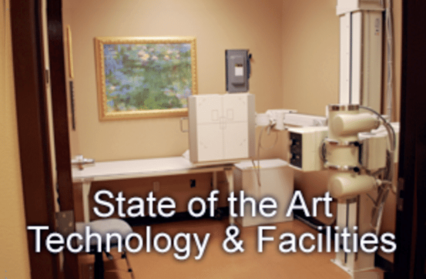
X-Ray
Our digital computed radiography was just updated February 2014. No appointments are necessary.
IFM's Find A Practitioner tool is the largest referral network in Functional Medicine, created to help patients locate Functional Medicine practitioners anywhere in the world. IFM Certified Practitioners are listed first in the search results, given their extensive education in Functional Medicine


 Please note that we can answer general questions, but anything specific to your medical case should be discussed with your physician.


