ClickCease
+1-915-850-0900 [email protected]
Select Page
Managing Finger Injuries: From Minor to Severe

Managing Finger Injuries: From Minor to Severe

For individuals dealing with finger injuries, which can occur from various causes, including overuse, jobs, sports, and more, can knowing the cause of finger pain help healthcare providers determine what steps to take for treatment?

Managing Finger Injuries: From Minor to Severe

Finger Injuries

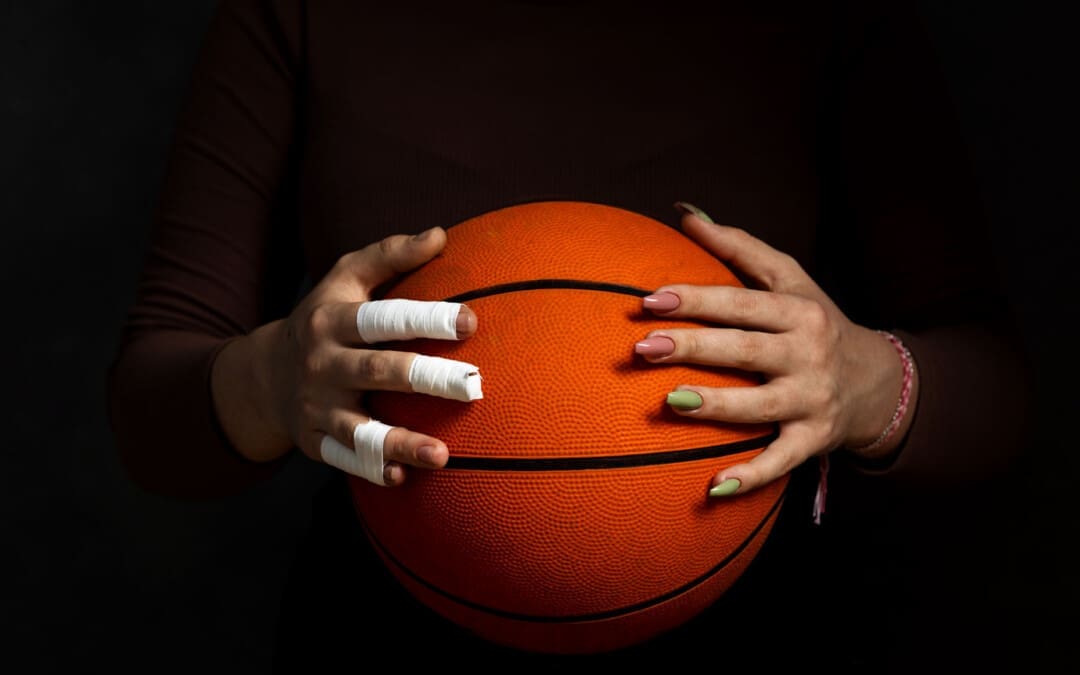
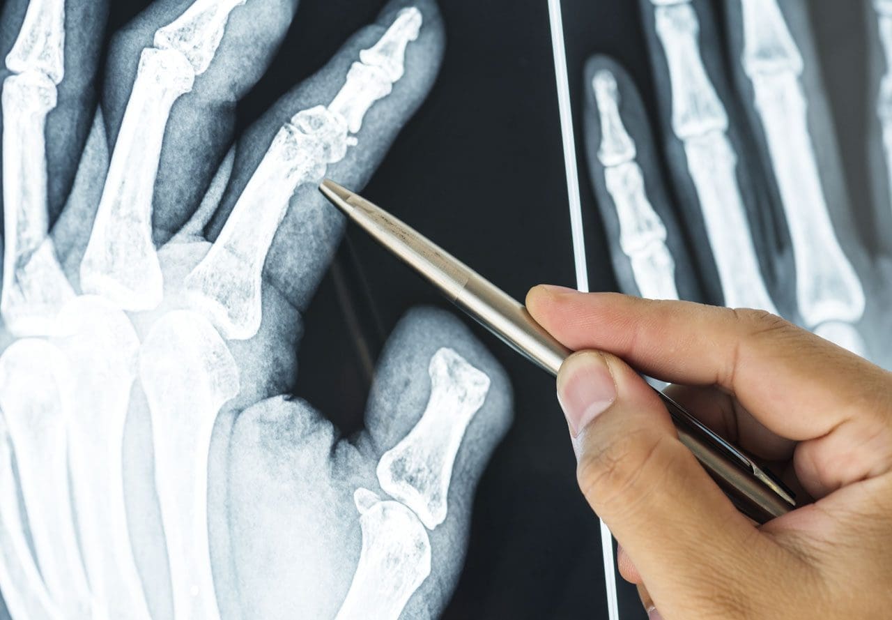
Finger injuries are common and can range from minor to serious. (van Veenendaal L. M. et al., 2014) Symptoms can result from an acute injury, including broken fingers and sprains, or chronic conditions like arthritis.

Fractures

Finger fractures can vary and can be serious and lead to permanent damage, deformity, and loss of function if not treated properly. What is important is that fractures are appropriately diagnosed so the proper treatment plan can be initiated. Most finger fractures can be addressed with simple treatments, while others may require surgery. (Oetgen M. E., and Dodds S. D. 2008)

Sprain and Dislocation

Sprains and dislocations are common finger injuries. (Prucz R. B. and Friedrich J. B. 2015) Both damage the ligaments that support the finger joints. In more severe injuries, a dislocation can occur, necessitating the finger to be put back into place or reduced. Individuals with a sprain or dislocation often notice finger swelling or stiffness for months after the injury.

Ligament Damage

Some call this injury skier’s or gamekeeper’s thumb, which results from a specific type of thumb dislocation. Here, the ulnar collateral ligament of the thumb is damaged. This ligament helps keep the thumb stable and supports grip and hand strength. However, this type of ligament injury often requires surgery. (Christensen T. et al., 2016)

Arthritis

Arthritis causes damage to normal joint surfaces where two bones come together. Fingers are one of the most common locations where arthritis occurs. (Spies C. K. et al., 2018) Two types of arthritis commonly affect the fingers: osteoarthritis and rheumatoid arthritis.

Arthritis of The Thumb

Arthritis of the thumb usually occurs at the joint where the thumb meets the wrist. This joint called the carpometacarpal/CMC joint, helps with gripping and pinching. Thumb arthritis is more common in women than men and increases in frequency over 40. (Deveza L. A. et al., 2017)

Trigger Finger

Trigger finger or stenosing tenosynovitis, is a common injury that causes pain and snapping of the fingers’ tendons, resulting in a sensation of locking or catching when bending and straightening the digits. (Makkouk A. H. et al., 2008) Other symptoms include pain and stiffness in the fingers and thumb. Treatments can vary from observation, rest, splinting, injections, and surgery.

Tendon Injuries

Mallet finger

A mallet finger is an injury to the tip of the finger. Usually, it occurs when the end of a straightened finger or thumb is hit, jamming the finger. After the injury, the individual may notice that they cannot fully straighten the tip of the finger. Treatment almost always uses a splint that has to stay on for about six weeks without removal. (Alla, S. R., Deal, N. D., and Dempsey, I. J. 2014) Very rarely is a surgical procedure necessary.

Jersey Finger

This is an injury to the finger flexor tendon. The flexor tendon pulls the finger into the palm when contracting the forearm flexor muscles. The injury occurs at the tip of the finger; typically, the tendon snaps back to the finger’s base or into the palm.

Ring Injuries

Injuries to the finger while wearing wedding bands or other finger jewelry can lead to serious complications. Even minor injuries can have devastating complications if the severity of the injury is not recognized and addressed. If an injury occurs while wearing the jewelry and there is soft tissue damage, including blood circulation being cut off, immediate medical attention is necessary.

Other Injuries

Bruises

The most common finger injury is caused by direct trauma to the skin and muscles. Symptoms include pain, swelling, tenderness, and discoloration of the skin.

Cuts and Scrapes

These can range from minor to more serious, such as injuries that cut through blood vessels, nerves, and tendons.

Injury Medical Chiropractic and Functional Medicine Clinic

After the initial inflammation and swelling have subsided, a doctor will recommend a treatment plan that usually involves physical therapy, self-performed physical rehabilitation, or supervision by a physical therapist or team. At Injury Medical Chiropractic and Functional Medicine Clinic, our areas of practice include Chronic Pain, Personal Injury, Auto Accident Care, Work Injuries, Back Injury, Low Back Pain, Neck Pain, Migraine Headaches, Sports Injuries, Severe Sciatica, Scoliosis, Complex Herniated Discs, Fibromyalgia, Chronic Pain, Complex Injuries, Stress Management, Wellness & Nutrition, Functional Medicine Treatments, and in-scope care protocols. We focus on what works for you to relieve pain and restore function. If other treatment is needed, individuals will be referred to a clinic or physician best suited to their injury, condition, and/or ailment.


Sports Injury Rehabilitation


References

van Veenendaal, L. M., de Klerk, G., & van der Velde, D. (2014). A painful finger as first sign of a malignancy. Geriatric orthopaedic surgery & rehabilitation, 5(1), 18–20. https://doi.org/10.1177/2151458514522125

Oetgen, M. E., & Dodds, S. D. (2008). Non-operative treatment of common finger injuries. Current reviews in musculoskeletal medicine, 1(2), 97–102. https://doi.org/10.1007/s12178-007-9014-z

Prucz, R. B., & Friedrich, J. B. (2015). Finger joint injuries. Clinics in sports medicine, 34(1), 99–116. https://doi.org/10.1016/j.csm.2014.09.002

Christensen, T., Sarfani, S., Shin, A. Y., & Kakar, S. (2016). Long-Term Outcomes of Primary Repair of Chronic Thumb Ulnar Collateral Ligament Injuries. Hand (New York, N.Y.), 11(3), 303–309. https://doi.org/10.1177/1558944716628482

Spies, C. K., Langer, M., Hahn, P., Müller, L. P., & Unglaub, F. (2018). The Treatment of Primary Arthritis of the Finger and Thumb Joint. Deutsches Arzteblatt international, 115(16), 269–275. https://doi.org/10.3238/arztebl.2018.0269

Deveza, L. A., Hunter, D. J., Wajon, A., Bennell, K. L., Vicenzino, B., Hodges, P., Eyles, J. P., Jongs, R., Riordan, E. A., Duong, V., Min Oo, W., O’Connell, R., & Meneses, S. R. (2017). Efficacy of combined conservative therapies on clinical outcomes in patients with thumb base osteoarthritis: protocol for a randomised, controlled trial (COMBO). BMJ open, 7(1), e014498. https://doi.org/10.1136/bmjopen-2016-014498

Makkouk, A. H., Oetgen, M. E., Swigart, C. R., & Dodds, S. D. (2008). Trigger finger: etiology, evaluation, and treatment. Current reviews in musculoskeletal medicine, 1(2), 92–96. https://doi.org/10.1007/s12178-007-9012-1

Alla, S. R., Deal, N. D., & Dempsey, I. J. (2014). Current concepts: mallet finger. Hand (New York, N.Y.), 9(2), 138–144. https://doi.org/10.1007/s11552-014-9609-y

Compressed Nerve In The Knee

Compressed Nerve In The Knee

A nerve becomes pinched/compressed when added pressure is placed on it by surrounding structures that can include muscles, bones, ligaments, tendons, or a combination. This injures and damages the nerve causing function problems and symptoms and sensations in that area or other parts of the body that are supplied by that nerve. Medical practitioners refer to this as nerve compression or entrapment. Although compressed nerves are more commonly associated with the neck, arms, hands, elbows, and lower back, any nerve in the body can experience irritation, spasms, inflammation, and compression. The causes and treatment of a compressed nerve in the knee.

Compressed Nerve In The Knee

Compressed Nerve In The Knee

There’s only one nerve that goes through the knee that has an increased risk of getting compressed. It’s a branch of the sciatic nerve called the peroneal nerve. The nerve goes around the outside of the knee before traveling down the outside of the lower leg. At the bottom of the knee, it lies between the bone and skin, making it vulnerable to irritation or compression by anything that can put pressure on the outside of the knee.

Causes

Traumatic injuries over time can lead to pressure on the nerve from inside the knee. Common causes of a compressed nerve in the knee include:

Frequently Crossing Legs

  • Compression by the opposite knee, while the legs are crossed is the most common cause.

Knee Brace

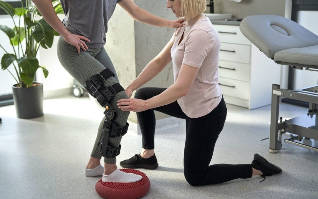
  • A too-tight or strong brace can compress the leg and nerve.

Thigh-High Compression Stockings

  • Designed to maintain pressure on the legs, if too tight these stockings can compress the nerve.

Squatting Posture For Long Periods

  • This position places pressure on the side of the knee.

Fractures

  • A fracture of the large lower leg bone/tibia or sometimes the small bone/fibula near the knee can entrap the nerve.

Lower Leg Cast

  • The portion of the cast around the knee can be tight and compress the nerve.
  • Tell the doctor if a cast or brace feels tight or is causing numbness or pain in the leg.

Knee-High Boots

  • The top of a boot can land right below the knee and be too tight pinching the nerve.

Knee Ligament Injury

  • The nerve can become compressed due to bleeding or inflammation from an injured ligament.

Knee Surgery Complications

  • This is rare, but the nerve can inadvertently get pinched during knee replacement surgery or an arthroscopic procedure.

Prolonged Bed Rest

  • When lying down the legs tend to rotate outward and the knees flex.
  • In this position, the mattress can place pressure on the nerve.

Tumors or Cysts

  • Tumors or cysts can develop right on top or next to a nerve irritating and compressing the area.

Abdominal or Gynecologic Surgery

  • The equipment used to keep the legs rotated outward and the knees flexed for gynecologic and abdominal surgeries can compress the nerve.

Symptoms

The peroneal nerve supplies sensation and movement to the outside of the lower leg and the top of the foot. When compressed, it becomes inflamed, which causes the symptoms of a compressed nerve. Usually, only the lining/myelin sheath around the nerve is what gets injured. However, when the nerve gets damaged, the symptoms are similar but more severe. Common symptoms include:

  • Weakness that limits the ability to lift the foot toward the leg aka dorsiflexion.
  • This causes dragging the foot when walking.
  • The ability to turn the foot outward and extend the big toe is also affected.
  • Symptoms can be felt on the outside of the lower leg and on the top of the foot and include:
  • Tingling or pins and needles sensations.
  • Numbness.
  • Loss of sensation.
  • Pain.
  • Burning.
  • For individuals that have had a pinched nerve for two or more weeks, the muscles supplied by the nerve can begin to waste away or atrophy.
  • Symptoms can be intermittent or continuous depending on the cause.
  • The other common cause is a pinched nerve in the lumbar/lower spine.
  • When this is the cause, sensations, and pain will present in the lower back or the back and outside of the thigh.

Diagnosis

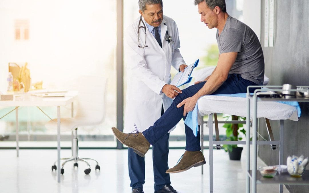
A doctor will look at medical history and perform an examination to make a diagnosis, determine the cause, and lay out a personalized treatment plan. The nerve in the knee can be felt as it travels around the top of the tibia, so a doctor may tap on it. If there is shooting pain down the leg, a pinched nerve may be present. Tests a doctor may order can include:

Knee X-ray

  • Shows any bone fractures or abnormal masses.

Knee MRI

  • Can confirm the diagnosis
  • Shows masses within the nerve.
  • Shows details of fractures or other problems in the bones.

Electromyogram – EMG

  • Tests electrical activity in the muscles.

Nerve Conduction Test

  • Tests the signal speed of the nerve.

Treatment

Treatment is aimed at reducing pain and improving mobility.

Over-the-Counter Pain Medication

  • OTC medication can reduce inflammation and improve symptoms short term.

Ice and Heat

  • Applying either heat or ice for 15 to 20 minutes at a time can provide relief from the symptoms.
  • An ice pack can make symptoms worse if it adds more pressure on the nerve.

Chiropractic and Physical Therapy

  • Chiropractic and physical therapy can release the compressed nerve, realign the structures, strengthen the muscles, and provide gait training.

Orthotic Boot

  • If walking gait is affected because the foot cannot bend, an orthotic boot can help.
  • This is a support that maintains the foot in a neutral position to walk normally.

Corticosteroid Injection

  • A corticosteroid injection can reduce inflammation and relieve pressure on the nerve.

Surgery

  • The nerve can suffer permanent damage if it has been pinched for a long time.
  • If that happens, surgery cannot repair the damage.
  • A doctor can perform surgery to correct a fracture, tumor, or other invasive problem causing a compressed nerve.
  • If conservative treatment doesn’t work, a peroneal nerve decompression procedure can be done to remove the pressure.
  • If surgery is needed, symptoms can disappear immediately, but it takes around four months to recover and rehabilitate.

Injury Rehabilitation


References

Krych, Aaron J et al. “Is peroneal nerve injury associated with worse function after knee dislocation?.” Clinical orthopedics and related research vol. 472,9 (2014): 2630-6. doi:10.1007/s11999-014-3542-9

Lezak B, Massel DH, Varacallo M. Peroneal Nerve Injury. [Updated 2022 Nov 14]. In: StatPearls [Internet]. Treasure Island (FL): StatPearls Publishing; 2023 Jan-. Available from: https://www.ncbi.nlm.nih.gov/books/NBK549859/

Soltani Mohammadi, Sussan, et al. “Comparing the squatting position and traditional sitting position for ease of spinal needle placement: a randomized clinical trial.” Anesthesiology and pain medicine vol. 4,2 e13969. 5 Apr. 2014, doi:10.5812/aapm.13969

Stanitski, C L. “Rehabilitation following knee injury.” Clinics in sports medicine vol. 4,3 (1985): 495-511.

Xu, Lin, et al. Zhongguo gu Shang = China Journal of Orthopedics and Traumatology vol. 33,11 (2020): 1071-5. doi:10.12200/j.issn.1003-0034.2020.11.017

Yacub, Jennifer N et al. “Nerve injury in patients after hip and knee arthroplasties and knee arthroscopy.” American Journal of physical medicine & Rehabilitation vol. 88,8 (2009): 635-41; quiz 642-4, 691. doi:10.1097/PHM.0b013e3181ae0c9d

Posterior Cruciate Ligament Injuries: El Paso Back Clinic

Posterior Cruciate Ligament Injuries: El Paso Back Clinic

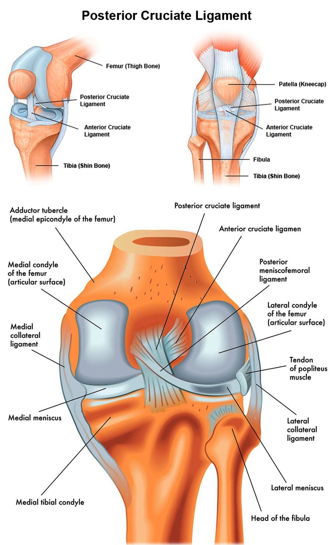
The body has around 1,000 ligaments that connect bones and joints. Ligaments are strong bands of tissue that support joint mobility and stabilize the muscles and bones. An injury to one or more ligaments can cause inflammation, swelling, discomfort, and instability. The PCL refers to the posterior cruciate ligament that runs along the back of the knee joint. This ligament connects the femur/thigh bone to the tibia/shinbone. Anyone can suffer from an injury to the posterior cruciate ligament. It can be caused by the knee hitting a dashboard in an automobile collision, a worker twisting or falling on a bent knee or a sports contact injury. The Injury Medical Chiropractic and Functional Medicine Clinic Team provide soft tissue work, trigger point therapy, and targeted non-surgical treatment through advanced therapy methods and technologies.

Posterior Cruciate Ligament Injuries: Chiropractic Wellness TeamPosterior Cruciate Ligament

The posterior cruciate ligament – PCL is located inside the knee, just behind the anterior cruciate ligament – ACL. It is one of several ligaments that connect the femur/thighbone to the tibia/shinbone. The posterior cruciate ligament keeps the tibia from moving backward.

Injury

Posterior cruciate ligament injuries are far less common than ACL – anterior cruciate tears. PCL injuries make up less than 20% of all knee ligament injuries. It is more common for PCL tears to occur with other ligament injuries. A PCL injury can cause mild, moderate, or severe damage and is rated into four different categories:

Grade I

  • A partial tear is present in the ligament.

Grade II

  • There is a partial tear.
  • The ligament can feel loose.

Grade III

  • The ligament is completely torn.
  • The knee is unstable.

Grade IV

  • The PCL is injured.
  • Other knee ligaments are damaged.

Individuals with posterior cruciate ligament injuries can have short or long-term symptoms. Typically, long-term symptoms occur when an injury slowly develops over time. In mild cases, individuals may still be able to walk, and their symptoms may be less noticeable. Common symptoms associated with PCL injuries include:

  • Difficulty placing weight on the injured knee.
  • Stiffness.
  • Walking difficulties.
  • Difficulty descending stairs.
  • A wobbly sensation inside the knee.
  • Inflammation and swelling can be mild to severe.
  • Knee pain.
  • Pain that worsens over time.
  • Over time, tears could lead to the development of osteoarthritis.

There is an increased risk of extensive damage and chronic pain conditions if left untreated.

Chiropractic Care

The continued participation in work or activity following a mild injury is the primary reason individuals undergo therapy, injections, or surgical repairs. Knee injuries need immediate attention to prevent worsening or further damage. A chiropractor will examine the knee, check the range of motion and ask about symptoms. They may request imaging tests to determine the extent of the damage. These tests may include the following:

  • X-rays.
  • Magnetic resonance imaging.
  • CT scan.

During the physical examination, they will check all the structures of the injured knee and compare them to the non-injured knee. The wounded knee may appear to sag backward when bent or could slide back too far, specifically when beyond a 90-degree angle. Treatment depends on the severity of the injury. Common treatments include:

Crutches

  • Crutches may be recommended to limit the weight placed on the knee.

Knee Brace

  • A special brace can address instability and help prevent the tibia bone from sagging backward.
  • Gravity tends to pull the bone backward when lying down.

Chiropractic and Physical Therapy

  • As the swelling goes down, a carefully personalized rehabilitation program can begin.
  • A chiropractic regimen will reset and retrain the ligament.
  • Massage therapy will minimize scar tissue and increase circulation.
  • Specific exercises will stabilize the knee, restore function, and strengthen the leg muscles that support it.
  • Strengthening the muscles in the front of the thigh/quadriceps is a key factor in a successful recovery.

Surgery

  • In severe cases, surgery may be necessary for full rehabilitation.
  • Knee arthroscopy is performed to reconstruct the ligament.
  • This procedure is less invasive compared to traditional surgical methods.

Recovery time varies from person to person. If the injury is mild, it may only take around ten days to heal. If surgery was needed, recovery could take about six to nine months. Full recovery typically requires 6 to 12 months.


Best Knee Injury Chiropractor


References

American Academy of Orthopaedic Surgeons. Posterior Cruciate Ligament Injuries. (https://orthoinfo.aaos.org/en/diseases–conditions/posterior-cruciate-ligament-injuries) Accessed 7/26/21.

Bedi A, Musahl V, Cowan JB. Management of Posterior Cruciate Ligament Injuries: An Evidence-Based Review. Journal of the American Academy of Orthopedic Surgery. 2016 May;24(5):277-89. Accessed 7/26/21.

Lu, Cheng-Chang, et al. “Twelve Weeks of a Staged Balance and Strength Training Program Improves Muscle Strength, Proprioception, and Clinical Function in Patients with Isolated Posterior Cruciate Ligament Injuries.” International journal of environmental research and public health vol. 18,23 12849. 6 Dec. 2021, doi:10.3390/ijerph182312849

Pierce, Casey M et al. “Posterior cruciate ligament tears: functional and postoperative rehabilitation.” Knee surgery, sports traumatology, arthroscopy: official journal of the ESSKA vol. 21,5 (2013): 1071-84. doi:10.1007/s00167-012-1970-1

Schüttler, K F et al. “Verletzungen des hinteren Kreuzbands” [Posterior cruciate ligament injuries]. Der Unfallchirurg vol. 120,1 (2017): 55-68. doi:10.1007/s00113-016-0292-z

Zsidai, Bálint, et al. “Different injury patterns exist among patients undergoing operative treatment of isolated PCL, combined PCL/ACL, and isolated ACL injuries: a study from the Swedish National Knee Ligament Registry.” Knee surgery, sports traumatology, arthroscopy: official journal of the ESSKA vol. 30,10 (2022): 3451-3460. doi:10.1007/s00167-022-06948-x

Delayed Injury Symptoms

Delayed Injury Symptoms

Automobile accidents and crashes can cause all kinds of damage to the body even when the accident/crash is not severe. Physical symptoms might not present at all for several days, even weeks. This is known as having delayed injury symptoms. These can include:

  • Swelling.
  • Stiffness.
  • Aching.
  • Pain that radiates all over the body.
  • Sleep problems.
  • Headaches.
  • Brain fog.
  • Disorientation.
  • Memory problems.

Chiropractic and physical therapy rehabilitation can restore the body’s alignment, stop inflammation, loosen, stretch and strengthen the musculoskeletal system restoring optimal health.

Delayed Injury Symptoms

Adrenaline

When the body is involved in a dangerous physical situation, it protects itself by releasing a surge of adrenaline. This hormone protects the body, causing the fight or flight response when in danger. Adrenaline causes several preservation responses that include:

  • Intense increase in energy.
  • Little or no pain.
  • Enlarged blood vessels and airways increase oxygen flow.
  • Increased strength from increased blood flow to the muscles.
  • Changes in vision and hearing that focus on sights and sounds all around.
  • Endorphins are released that make the body feel calm and in control.
  • Endorphins affect the way the body responds to pain and stress.

Individuals don’t start feeling aches and pains until the adrenaline and endorphins wear off. However, because everybody is different and the emergency response has turned off, the body still might not feel the injury symptoms. These are delayed injury symptoms.

Rate of Speed

When riding in a vehicle, the body moves at the same speed as the vehicle. During an impact, the vehicle stops, but the body continues moving until it stops, typically with a lot of force from the seatbelt, airbag, or other barriers. The intense momentum change can cause soft tissue damage and ligament or muscle strains from the stretching, pulling, contracting, and tearing. Also, the intervertebral discs can tear, bulge, or herniate over time, creating pressure on nerves and the surrounding tissues.

Delayed Injury Symptoms

Headaches

  • Headaches that develop days after an accident/crash are common.
  • They can signal a possible injury to the neck or head, a blood clot on the brain, or a concussion.

Numbness

  • Loss of feeling in arms and hands could indicate a whiplash-associated disorder.
  • The loss of feeling/sensation results from damage to the neck or spinal column.
  • Around 20 percent of individuals impacted by a rear-end crash develop some whiplash symptoms.

Neck or Shoulder Pain and/or Stiffness

  • Whiplash is a classic delayed symptom injury associated with accidents.
  • Most delayed whiplash injuries are caused by rear-end vehicle collisions at speeds of less than 14 miles an hour.
  • Whiplash injuries usually require x-rays, CT scans, or MRIs for proper diagnosis.

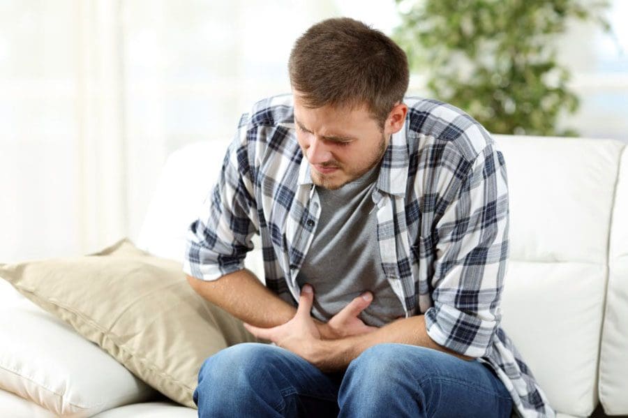
Abdominal Pain or Swelling

  • This could indicate internal bleeding.
  • Internal bleeding can remain undiscovered for hours or days.
  • This can be a life-threatening condition that needs to be diagnosed and treated by emergency medical personnel.
  • Other symptoms include:
  • Large areas of deep bruising.
  • Dizziness.
  • Fainting.

Back Aches and Pains

  • Back pain can be caused by injury to the muscles, ligaments, nerves, or damage to the vertebrae.
  • Low back pain occurs in more than half of rear-impact collisions and almost three-quarters of side-impact crashes.

Chiropractic Rehabilitation

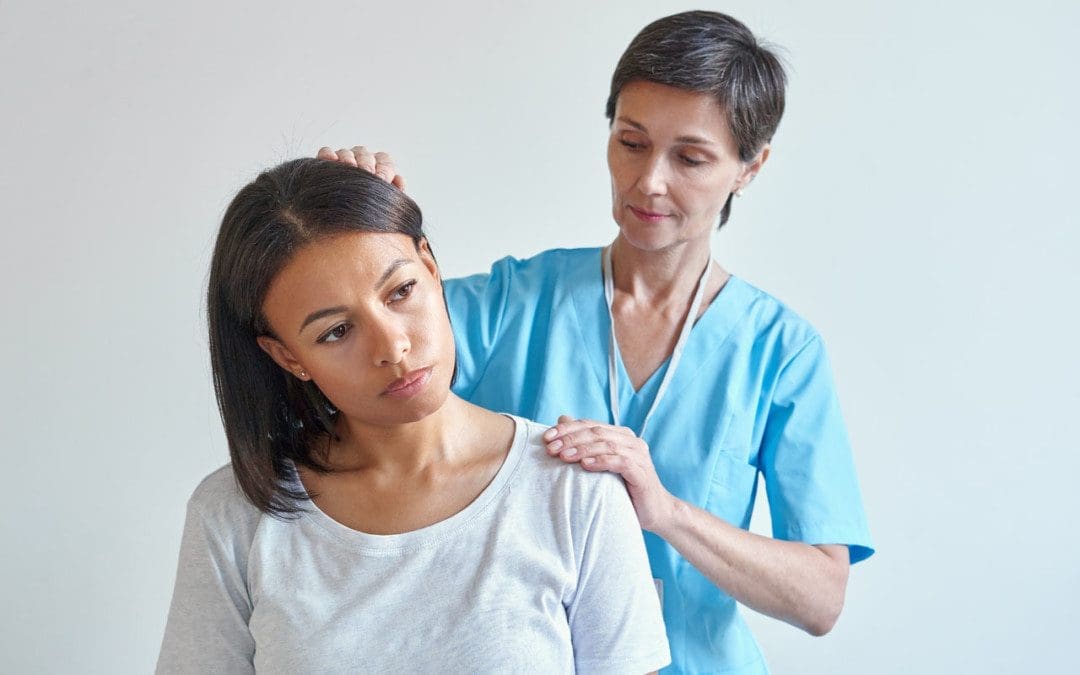
After an accident, soft tissues can sustain minimal damage; however, the minimal damage left untreated can start to worsen and turn into a painful condition. Emergency room visits are to rule out major injuries like brain/nerve injuries, bleeding, punctures, lacerated organs, fractures that require emergency stabilization. Chiropractors look for other symptoms and mechanisms that indicate damage to the body’s soft tissues and nerves to see if they have been stretched or torn and dysfunction in the nervous system.


Body Composition


Calorie Counting

Counting calories can be a stepping stone to change behavior towards food. Tracking what foods are being taken into the body promotes mindfulness of dietary habits. Studies on the subject reveal a significant association between self-monitoring and weight loss. Takeaways include:

References

Burke, Lora E et al. “Self-monitoring in weight loss: a systematic review of the literature.” Journal of the American Dietetic Association vol. 111,1 (2011): 92-102. doi:10.1016/j.jada.2010.10.008

D’Elia, Michael A et al. “Motor vehicle collision with seatbelt sign and traumatic abdominal wall hernia should raise suspicion for hollow viscus injury.” Trauma case reports vol. 22 100206. 25 May. 2019, doi:10.1016/j.tcr.2019.100206

Kacprzynski, Gregory, and Joshua Bucher. “Delayed vertebral artery dissection after mild trauma in a motor vehicle collision.” The American Journal of emergency medicine vol. 45 (2021): 678.e1-678.e2. doi:10.1016/j.ajem.2020.11.028

Olinger, Catherine, and Richard Bransford. “Upper Cervical Trauma.” The Orthopedic clinics of North America vol. 52,4 (2021): 451-479. doi:10.1016/j.ocl.2021.05.013

Sterling, Michele. “Whiplash-associated disorder: musculoskeletal pain and related clinical findings.” The Journal of manual & manipulative therapy vol. 19,4 (2011): 194-200. doi:10.1179/106698111X13129729551949

The Knee

The Knee

The Knee | MRI may be requested for:

  • Ligament injuries
  • Meniscal tears and degeneration
  • Rheumatoid arthritis
  • Osteochondral fractures
  • Tendon disruptions

Bones & Cartilage Of The Knee

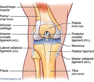
The knee joint is the largest, most complicated, and most vulnerable joint in the body, as it does not have a stable bony configuration. It consists of the tibiofemoral and patellofemoral articulations, which include the femur, tibia, and patella. The knee is a synovial joint that is enclosed by a ligament capsule. The capsule contains synovial fluid that keeps the joint lubricated (Figure 82). The knee provides flexible movement, but must also bear large weight and pressure loads. During walking, the knees support 1.5 times your body weight. When climbing stairs, they support 3-4 times your body weight. When squatting, your knees support 8 times your body weight.
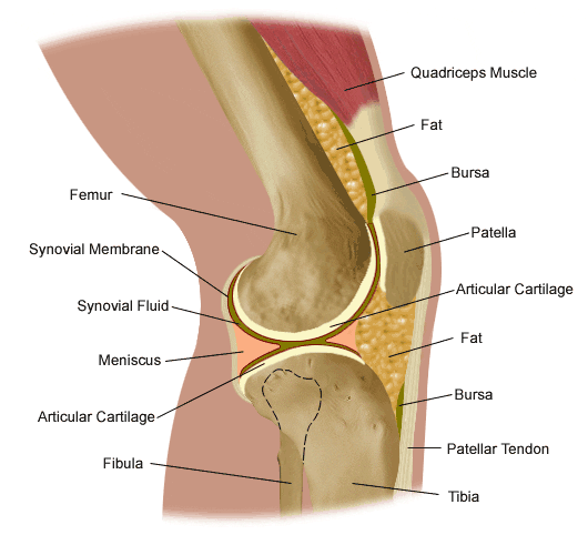
the knee

Figure 82. Anatomy of the knee.

The tibiofemoral articulation is a modified hinge joint that allows bending and straightening, but also allows for slight rotation. This articulation consists of the lateral and medial condyles of the femur resting on the lateral and medial aspects of the tibial plateau. The femoral condyles make up the distal portion of the femur, which is expanded in order to assist with weight distribution at the knee joint. The medial femoral condyle is typically larger and rounder. The condyles are united anteriorly to provide the articular surface for the patella, but they are separated posteriorly by the intercondylar notch. This notch, or fossa, is the attachment site for the cruciate ligaments, the ligaments of Humphrey and Wrisberg, and the frenulum of the patellar fat pad. A large part of the posterior distal femur is called the popliteal surface. This area is covered by fat, which separates it from the popliteal artery. The medial and lateral edges of the popliteal surface are attachment sites for muscles. Superior to the femoral condyles are the epicondyles, which are the attachment sites for muscles, tendons, and capsular ligaments. The medial epicondyle is the attachment site for the medial (or tibial) collateral ligament (Figure 83). The lateral femoral epicondyle is the attachment site for the lateral (or fibular) collateral ligament, as well as the tendon of the popliteus muscle, fibers of the iliotibial tract, and the lateral capsular ligament. Superior and posterior to the epicondyles is the most distal extent of the linea aspera, the bony ridge of the femur.

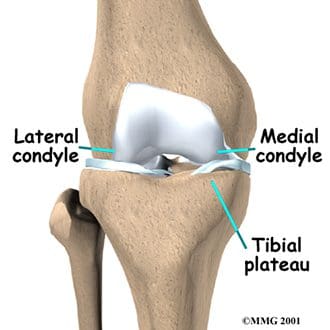
The tibia is the distal portion of the tibiofemoral articulation at the knee. The tibia is the second longest bone in the body, ranked just behind the femur. Its proximal end is flattened and expanded to provide a larger surface for the body weight that is transmitted through the femur. Like the femur, the proximal tibia has medial and lateral condyles. The medial condyle is larger, and somewhat flattened where it contacts the medial meniscus. The lateral condyle has a circular look to its femoral articular surface. The lateral tibial condyle articulates with the head of the fibula posteriorly, which is as close as the fibula comes to any involvement in the knee joint. Both the medial and lateral condyles rise in the center of the superior aspect of the tibia to form the intercondylar eminence. Posterior to this eminence are the attachments sites for the posterior horns of the medial and lateral menisci, which will be discussed with the ligaments of the knee. The medial and lateral tibial condyles, and the area of the intercondylar eminence are often grouped together and referred to as the tibial plateau (Figure 84). This is a critical weight-bearing area, and greatly affects the stability of the knee joint. The tibial tuberosity (or tubercle) is located on the anterior surface of the proximal tibial shaft. It has a smooth upper portion, and a roughened lower portion, which is the insertion site for the patellar tendon. The lateral side of the tibial tuberosity has a ridge for the attachment of fibers from the iliotibial tract. This is the strongest direct attachment site for the iliotibial tract. The IT tract, or band, helps in limiting lateral movement of the knee.
the knee

Figure 84. Tibial plateau.

the knee

Figure 83. Tibiofemoral anatomy.


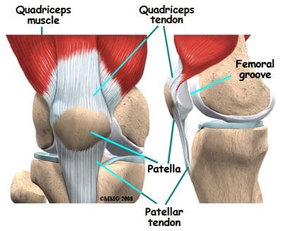
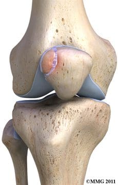
The patella is the third bone involved in the knee joint, specifically in the patellofemoral articulation. Patella means �little plate� in Latin, which describes the look and function of this sesamoid bone. The patella develops in the tendon of the quadriceps femoris muscle (Figure 85). It moves when the leg moves, and protects the knee joint by relieving friction between the bones and muscles when the knee is bent or straightened. The patellofemoral joint is a saddle-type synovial joint, allowing the patella to glide along the bottom front surface of the femur between the femoral condyles in the patellofemoral groove. Ossification of the patella is typically completed in females by age 10, and in males between the ages of 13-16. If the patella has more than one ossification center, and the additional center does not fuse, it is termed a bipartite patella (Figure 86).
the knee

Figure 86. Bipartite patella.

the knee

Figure 85. Patella location.


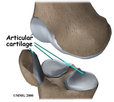
Articular, or hyaline, cartilage covers the ends of the bones involved in any joint. In the knee joint, this includes the distal end of the femur, the proximal end of the tibia, and the posterior aspect of the patella (Figure 87). In larger joints, this cartilage is approximately �� thick. Articular cartilage is white, shiny, rubbery, and slippery, enabling surfaces to slide against one another without damage. Articular cartilage is very flexible, due in part to its high water content, which also makes it highly visible on MRI. In contrast to the bones that it covers, articular cartilage has almost no blood vessels, so it is not good at repairing itself. Bones, on the other hand, have numerous blood vessels, and are good at self-repair.
the knee

Figure 87. Articular cartilage.

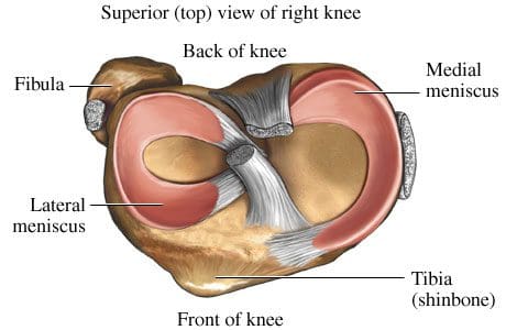
Another type of cartilage is found between the femur and tibia- the fibrous cartilage that makes up the medial and lateral menisci. The menisci, also referred to as �articular disks�, wrap around the round ends of the femur to fill the space between the femur and tibia (Figure 88). Since the menisci are more fibrous in composition, they have tensile strength and can resist pressure. They can help spread the force from our body weight over a larger area. By helping with weight distribution, the menisci protect the articular cartilage on the ends of the bones from excessive forces. The menisci are fashioned to be thicker on their outsides, creating a shallow socket on the tibial surface. They act like a wedge on the rounded distal portion of the femur, improving the overall stability of the knee joint by preventing any �rolling� of the femur. Despite how strong they sound, the menisci can crack or tear when the knee is forcefully rotated or bent. The medial meniscus is fused with the medial collateral ligament, so it is less mobile than the lateral meniscus. It is often injured when the anterior or posterior cruciate ligaments are injured. The inner 2/3 of the medial meniscus receives a limited blood supply, so the entire meniscus is usually slow to heal. The lateral meniscus suffers from fewer injuries than the medial meniscus. Meniscal tears are one of the most common causes of knee pain, with suspected meniscal tears the most common indication for an MRI of the knee joint.
the knee

Figure 88. Superior view of menisci of right knee.

Symptoms that might indicate a problem with the bones of the knee joint include locking of the joint, the knee giving way, crackling or grinding felt in the joint, and pain and swelling. Locking of the joint can be indicative of a �loose body� (bone, cartilage, or foreign object) in the joint space, which can often be removed through arthroscopy (Figure 89). A knee that gives way can indicate that the patella is out of the patellofemoral groove, which leaves the knee unstable. Crackling and grinding at the joint can result from degenerative arthritis or osteoarthritis, as well as from a dislocating patella. An increase in pain with activity can occur due to a stress fracture or bone fracture. One of the pathologic conditions that can affect the bones of the knee joint is osteochondritis dissecans, which can affect the distal femur, and was discussed previously with the femur anatomy. Various types of arthritis manifest in the bones of the knee joint, including osteoarthritis, infectious arthritis, and rheumatoid arthritis. Chondromalacia patella, also known as patellofemoral syndrome or �runner�s knee� results from an irritation of the undersurface of the patella (Figure 91). If the patella is not tracking correctly in the patellofemoral groove, the articular cartilage may rub against the knee joint (Figure 90). The cartilage degenerates, and becomes irritated and painful. This condition is most common amongst young, healthy athletes, especially females and runners that are flat-footed. Treatment is typically rest and physical therapy to stretch and strengthen the quads and hamstrings. If surgery is required, it may be to perform a �lateral release�, as the abnormal tracking of the patella can cause a tightening of the lateral tissues of the knee. The lateral release procedure cuts the tight tissues, so the patella can return to its normal position and tracking. Osgood-Schlatter disease involves the anteriorly located tibial tuberosity, and the patellar tendon that inserts on that tuberosity (Figures 92, 93). This condition affects children during their growth spurts, and is typically found more in boys. During growth spurts, contractions of the quad muscle put additional stress on the patellar tendon at its attachment site on the tibial tuberosity. This can result in multiple subacute avulsion fractures and inflammation of the tendon. Excess bone growth occurs on the tuberosity, and a lump on the tuberosity can be seen and felt. This lump can become irritated and swollen, causing knee and leg pain. This condition is typically worsened with running, jumping, and climbing stairs. Osgood-Schlatter usually resolves with rest, ice, compression and elevation, as well as maturity of the youngster�s skeleton.
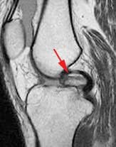
Figure 89. Intraarticular loose body.
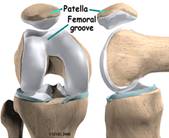
 

Figure 90. Patellofemoral groove.
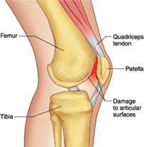
Figure 91. Patellofemoral syndrome or �runner�s knee�.
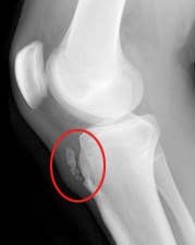


Figure 92. Xray displaying Osgood-Schlatter disease.
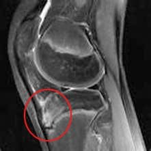
 

Figure 93. MRI displaying Osgood- Schlatter disease.


Ligaments Of The Knee

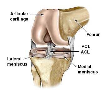
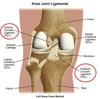
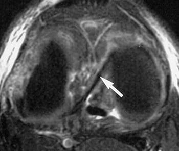
Ligaments are the tough bands of tissue that connect bones. They are considered to be �viscoelastic�, meaning they can gradually lengthen under tension, but return to their original shape when the tension is removed. However, if they are stretched for a prolonged period of time, or past a certain point, the ligaments cannot retain their original shape, and may eventually tear or snap. This is one of the reasons that a dislocated joint should be re-located as quickly as possible. If the ligaments lengthen, they leave the joint weakened and prone to future dislocations. Controlled stretching exercises to lengthen ligaments, and make the joints more supple, are part of the daily routines of athletes, gymnasts, dancers, etc. Damaged ligaments can lead to unstable joints, wearing of the cartilage, and eventually osteoarthritis. The numerous ligaments of the knee joint are the most important structures in controlling stability of the knee. Many of these ligaments were mentioned in the femur anatomy section, as they have attachments on the distal femur. The more important ligaments will be reviewed here in greater detail, in regards to their functions in the knee joint. The main intracapsular ligaments are the anterior and posterior cruciates (Figures 94, 95). Intracapsular ligaments are not very common in synovial joints. They provide stability, but permit a larger range of motion as compared to capsular or extracapsular ligaments. The anterior cruciate ligament (ACL) stretches from the lateral femoral condyle to the anterior intercondylar area of the tibia, preventing the tibia from being pushed too far anterior relative to the femur. It is the more commonly injured of the cruciate ligaments, and can be torn during twisting and bending of the knee. Women are at higher risk for ACL ruptures due to the facts that the maximum diameter of the intercondylar fossa is in its posterior aspect (the ACL attaches anteriorly), and the overall width of the intercondylar fossa is smaller in females. The posterior cruciate ligament (PCL) stretches from the medial femoral condyle to the posterior intercondylar area of the tibia, preventing posterior displacement of the tibia relative to the femur. It is the stronger of the two cruciate ligaments, and is injured less frequently; however, it can be injured from direct force or trauma. The menisci are also considered to be intracapsular structures, with connections to ligaments inside and outside the joint capsule. Two of their intracapsular ligaments are the anterior and posterior transverse meniscomeniscal ligaments. They attach the medial and lateral menisci to each other at their anterior and posterior aspects. Posterior transverse meniscal ligaments are very rare- only 1-4% of knees will have them. Two additional intermeniscal ligaments are the medial and lateral oblique meniscomeniscal ligaments (Figure 96). Their names describe their anterior horn attachment sites; they attach on the posterior horn of the opposite meniscus (i.e. medial oblique meniscomeniscal attaches to the anterior horn of the medial meniscus and posterior horn of the lateral meniscus). The oblique meniscomeniscal ligaments both traverse the intercondylar notch, and pass between the anterior and posterior cruciate ligaments (Figure 97).
the knee

Figure 94. Cruciate ligaments and menisci.
the knee

Figure 95. Posterior view of cruciate ligaments of left knee.
the knee

Figure 96. Axial fatsat T2 FSE image with arrow indicating
oblique meniscal ligament coursing from anterior horn of
medial meniscus to posterior horn of lateral meniscus.
the knee

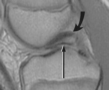
Figure 97. Sagittal dual-echo T2 through the intercondylar notch at the level of the posterior cruciate ligament (curved arrow); thin linear structure of low signal intensity inferior to PCL represents the oblique meniscomeniscal ligament (straight arrow); sometimes misinterpreted as displaced meniscal fragment.


The medial (or tibial) collateral ligament is considered a capsular ligament, as it is part of the articular capsule surrounding the synovial knee joint. It acts as mechanical reinforcement for the joint, protecting the knee from valgus force, or being bent open medially due to stress on the lateral side of the knee. The medial collateral ligament (MCL) is one of the most commonly injured of all knee ligaments, occurring in all sports, in all ages, and often times with medial meniscal tears (Figures 98-101). It has both superficial and deep components. Fibers from the superficial portion of the MCL attach to the medial epicondyle of the femur and the medial tibial condyle. Fibers from the deep medial collateral ligament attach to the medial meniscus. Proximal to the attachment point, this ligament is referred to as the meniscofemoral ligament, as it attaches the medial meniscus to the medial aspect of the femur. Distal to the meniscal attachment, the ligament is referred to as the meniscotibial (or coronary) ligament, as it attaches the medial meniscus to the medial aspect of the tibia. The meniscofemoral and meniscotibial are also referred to as the meniscocapsular or medial capsular ligaments, as they play an important role in anchoring peripheral parts of the medial meniscus in the medial side of the knee. The meniscotibial ligament is typically injured more often than the meniscofemoral ligament. The meniscotibial ligament attaches to the tibia several millimeters inferior to the articular cartilage. Its job is to stabilize and maintain the meniscus in its proper position on the tibial plateau. Disruption of the meniscotibial ligament can result in a floating meniscus or meniscal avulsion, while the meniscofemoral ligament may not be affected. The deep medial collateral ligament is short, and tightens quickly with rotation motions. It is often damaged, along with the ACL, when the mechanism of injury involves tibial rotation. Diagnosis and surgical repair of the deep medial collateral ligament can be challenging.
the knee

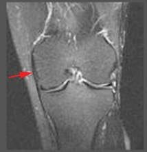
Figure 98. Normal MCL is linear,
has low signal intensity.
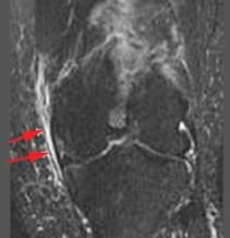
the knee

Figure 99. Grade 1 sprain shows adjacent edema, no change in signal intensity of MCL.
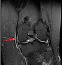
the knee

Figure 100. Grade 2 sprain or partial tear shows increased edema,
abnormal signal intensity,
thickening or thinning of ligament.
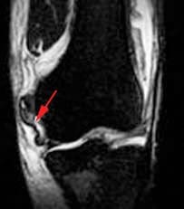
the knee

Figure 101. Grade 3 involves complete disruption of ligaments or attachments.


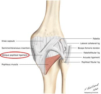
In addition to fibers of the medial collateral ligament, the deep portion of the capsular compartment of the medial knee is the location of the medial knee�s posterior support. The posterior oblique ligament is attached proximally to the medially located adductor tubercle of the femur, and distally to the tibia and the posterior aspect of the knee joint capsule. If the posterior oblique is injured, it is usually torn from its femoral origin. The posterior oblique ligament provides static resistance to valgus loads as the knee moves into full extension, as well as dynamic stabilization to valgus forces (stress from lateral side) as the knee moves into flexion. It acts as an important restraint to posterior tibial translation in cases of posterior cruciate ligament injury. The posterior oblique ligament has three �arms�. Its superior capsular �arm� becomes continuous with the posterior knee capsule, and the proximal portion of the oblique popliteal ligament. The oblique popliteal ligament is also an important posterior stabilizing structure for the knee joint Figure 102). It extends from the posteromedial aspect of the tibia, running obliquely and laterally upward to insert near the lateral epicondyle of the femur.
the knee

Figure 102. Oblique popliteal ligament in posterior view of knee.
the knee

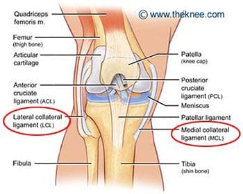
Figure 103. Medial (tibial) and lateral (fibular) collateral ligaments.


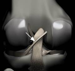
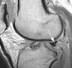
The lateral (or fibular) collateral ligament is considered an extracapsular ligament. It helps to provide joint stability and protects the lateral side of the knee from varus forces, or inside bending forces that are directed at the medial side of the knee. Injuries to the lateral collateral ligament are less common than injuries to the medial collateral, as the opposite leg can guard against medial forces that can lead to lateral collateral injuries. Injuries can occur in sports such as soccer and rugby, where the knee is extended and unprotected during running. The lateral, or fibular, collateral ligament stretches obliquely downward and backward, from the lateral epicondyle of the femur to the head of the fibula (Figure 103). It is not fused with the capsular ligament or with the lateral meniscus, so it has increased flexibility and decreased incidence of injury when compared to the medial collateral ligament. Similar to the medial meniscus, the lateral meniscus has a meniscotibial, or coronary, ligament. It connects the inferior edges of the lateral meniscus to the periphery of the tibial plateau. The lateral meniscus also has a meniscofemoral ligament that extends from the posterior horn of the lateral meniscus to the lateral aspect of the medial femoral condyle. It is given two distinct names, based on its location in relation to the posterior cruciate ligament (PCL). The ligament of Humphrey passes in front of the posterior cruciate ligament. It is less than 1/3 the diameter of the posterior cruciate ligament, but may be confused for the posterior cruciate during arthroscopy. The ligament of Wrisberg passes behind the posterior cruciate ligament, and is about � of the posterior cruciate�s diameter (Figure 104). Its femoral origin often merges with the posterior cruciate ligament. Both ligaments are present in only about 6% of knees. Approximately 70% of people have one or the other of these ligaments, with the majority possessing the more posterior ligament of Wrisberg (Figure 105). MRI is the preferred imaging modality for medial collateral or lateral collateral ligament injuries, as it can detect any associated internal knee derangements, cruciate-collateral ligament injuries, or cartilage deficiencies.
the knee

Figure 104. Rendering of posterior knee, arrow indicates Ligament of Wrisberg; courses obliquely from lateral aspect of medial femoral condyle to posterior horn of lateral meniscus,
remains posterior to PCL.
the knee

Figure 105. Arrow indicates �Wrisberg pseudo-tear�; intermediate signal
intensity line at junction of
Ligament of Wrisberg and normal posterior horn of lateral meniscus; often mistaken for a meniscal tear.

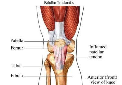
The patellar ligament is the connection between the patella and the tibia, extending from the apex (inferior aspect) of the patella to the tibial tuberosity. Technically, it is connecting two bones, so it is a ligament. However, it is most often referred to as the patellar tendon, because the superficial fibers that cover the front of the patella and extend to the tibia are continuous with the central portion of the common tendon of the quadriceps femoris muscle. The posterior surface of the patellar ligament is separated from the synovial membrane of the knee joint by a large infrapatellar pad of fat. Injuries to the patellar ligament can occur from overuse, such as sports that involve jumping and quick directional changes, as well as running-related sports. This is the ligament that is injured in jumper�s knee (or patellar tendonitis), which begins with inflammation, and can lead to degeneration or rupture of the patellar ligament and the tissue around it (Figure 106). Patients with patellar ligament injuries typically complain of pain in the area below the kneecap, which will increase with walking, running, squatting, etc. They can often be treated in the same manner as other soft tissue injuries- with rest, ice, compression and elevation. The patellar ligament attachment at the tibial tuberosity is the site of Osgood-Schlatter disease, which was discussed previously.
the knee

Figure 106. Patellar tendonitis (jumper�s knee).

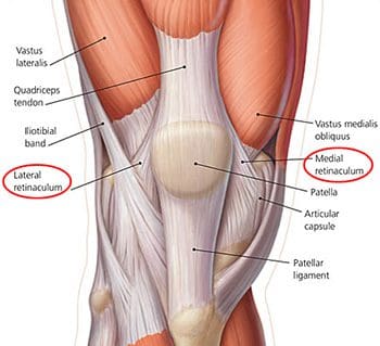
Along the sides of the patella and the patellar ligament are the medial and lateral patellar retinacula (Figure 107). They are fibrous tissue stabilizers for the patella that form from the medial and lateral portions of the quad tendons as they pass down to insert on either side of the tibial tuberosity. The lateral retinaculum is the thicker of the two, but both have superficial and deep layers. Within the deep layers are various ligaments (whose names indicate the structures they connect) that help support the patella in its position, relative to the femur below it. The deep layer of the lateral patellar retinaculum is the location where the lateral patellofemoral ligament meets the iliopatellar band, which is a tract of fibers from the iliotibial (IT) band that connects to the patella. The deep layer of the medial patellar retinaculum has three focal capsular thickenings, referred to as the medial patellofemoral, medial patellomeniscal, and medial patellotibial ligaments. The medial patellofemoral ligament is strong enough to influence patellar tracking, and acts as a major medial restraint. Imbalances in the forces that control patellar tracking during flexion and extension of the knee can lead to patellofemoral pain syndrome (runner�s knee), one of the most common causes of knee pain. This can result from overuse, trauma, muscle dysfunction, patellar hypermobility, and poor quadriceps flexibility. Typical symptoms include pain behind or around the patella that is increased with running, and activities that involve knee flexion. MRI is typically not necessary for this diagnosis. Physical therapy has been found to be effective for the treatment of patellofemoral pain syndrome.
the knee

Figure 107. Lateral and medial retinaculum.

Muscles & Tendons Of The Knee

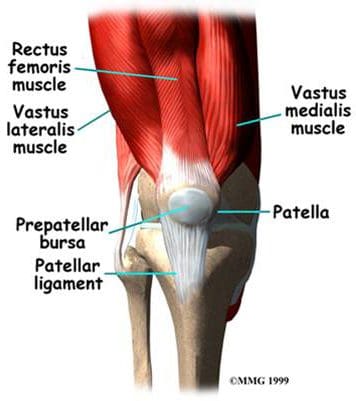
The flexor and extensor muscles of the knee have been discussed previously, as the majority of them are the anterior and posterior muscles of the thigh. We will review the thigh muscles involved in knee movement, and add two muscles of the lower leg that also affect the knee. The quadriceps femoris muscles of the anterior thigh are the main knee extensors (Figure 108). As these muscles contract, the knee joint straightens. The tendons of the vastus medialis, vastus intermedius, vastus lateralis, and rectus femoris join at the superior aspect (base) of the patella to form the patellar tendon. This tendon continues over the patella and attaches it to the tibial tuberosity (since it is connecting bone to bone, it is sometimes called the patellar ligament). The quadriceps, along with the gluteal muscles, are responsible for the thrusting forces necessary for walking, running, and jumping. The quads also help control movement of the patella, as they are attached to it by the quadriceps tendons (Figure 109). The patella increases the force exerted by the quadriceps muscles as the knee is straightened.
the knee

Figure 108. Anterior thigh muscles – knee extensors.
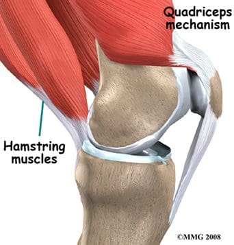
the knee

Figure 109. Quadriceps controlling the patella.


The posterior thigh muscles, also known as the hamstrings, are the main knee flexors, with assistance from the sartorius, gracilis, gastrocnemius, and popliteus muscles. The knee bends when the hamstrings contract. The hamstring muscles give the knee joint the strength needed for propulsion in running and jumping. They also help to stabilize the knee by protecting the collateral and cruciate ligaments, especially when the knee twists. The three hamstring muscles have varying attachment sites around the knee joint (Figure 110). The biceps femoris attaches to the head of the fibula and the superolateral aspect of the tibia. The semitendinosus attaches on the anterior aspect of the tibia, medial to the tibial tuberosity, crossing over the medial collateral ligament. The tendon of the semitendinosus muscle is sometimes used for cruciate ligament reconstruction. The semimembranosus attaches at the posteriomedial aspect of the medial tibial condyle. The sartorius muscle is also a knee flexor, although it is an anterior thigh muscle. It inserts on the anterior medical aspect of the tibia. The gracilis muscle of the medial thigh is one of the hip adductors, but also plays a part in knee flexion. Like the semitendinosus tendon, the tendon of the gracilis is sometimes used for cruciate ligament reconstructions. The gracilis attaches to the medial aspect of the proximal tibia.
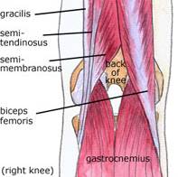
the knee

Figure 110. Posterior knee
muscles – knee flexors.

Additional flexors of the knee joint include some of the posterior muscles of the lower leg. The large superficial gastrocnemius muscle has a medial and a lateral head, which originate from the medial and lateral femoral condyles, respectively. It runs the length of the posterior lower leg, attaching to the calcaneus by the Achilles tendon. The gastrocnemius gives us the ability to flex our knee while our foot is flexed, as it connects to both joints. It is involved in standing, walking, running, and jumping. The popliteus is a deep posterior lower leg muscle that helps with knee flexion, and also rotates the tibia medially, which aids in knee stability. The popliteus originates from the outer margin of the lateral meniscus of the knee joint. It extends posteriorly and inserts on the medial aspect of the tibia, inferior to the medial tibial epicondyle.

The important tendons of the knee include the quadriceps, patellar, and hamstring tendons, and the iliotibial band (Figure 111). Tendons attach muscles to bones. These major knee tendons have all been discussed with either the bones or the muscles that they attach. The quadriceps tendon was mentioned with the quadriceps muscle as the muscle�s attachment to the patella. The quad tendon continues over the patella, then attaches the apex of the patella to the tibial tuberosity. It is then called the patellar tendon (or ligament). Hamstring tendons were discussed with the hamstring muscles, the posterior muscles that are flexors of the knee. Hamstring tendons are sometimes used for cruciate ligament reconstructions. Tendonitis, which is the inflammation of a tendon, is a common knee injury amongst athletes in a variety of sports. The iliotibial band (or IT tract) functions like a tendon, as it attaches the knee to the tensor fasciae latte muscle. The band is actually a fibrous reinforcement of the fascia lata, or deep tissue of the thigh. It runs from the ilium to the tibia. Proximally, it acts as a hip abductor, while distally it acts as lateral stabilization for the knee, and aids with medial rotation of the tibia. The IT band is in constant use during walking and running, which can lead to irritation at the point where it passes over the lateral femoral epicondyle. A �tight� IT band can cause inflammation and/or irritation at the femoral epicondyle, or at the point of insertion on the lateral tibial condyle. This condition is called IT band friction syndrome. It is common amongst runners, hikers, and cycling enthusiasts.
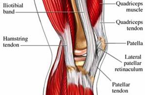
the knee

Figure 111. Tendons of the knee.

Nerves Of The Knee

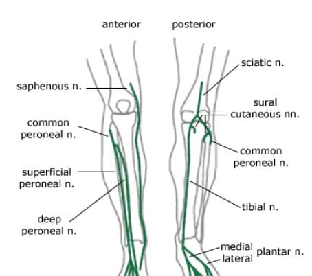
The main nerves to the knee that come from the sacral plexus of nerves are the tibial nerve and the common peroneal nerve (Figure 112). Both are branches of the sciatic nerve, and begin posteriorly, slightly above the actual knee joint. Both of these nerves, or their branches, continue through the lower leg and foot, providing sensation and muscle control. The tibial and common peroneal nerves are also both involved in cutaneous innervation, which is the supply of nerves to the skin of the knee. The tibial nerve remains posterior and more medial, branching at the medial ankle to innervate the foot. The common peroneal nerve begins posterolaterally, moving anteriorly near the neck of the fibula. It then branches into the superficial and deep peroneal nerves, which continue their anterior descent to the foot. The tibial and common peroneal nerves are the most commonly injured nerves when a knee is dislocated. Nerves can grow back, but they do so at a rate of approximately � inch per month.
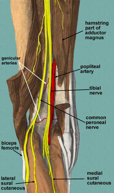
the knee

Figure 112. Sacral plexus nerves of knee.

Nerves from the lumbar plexus that affect the knee include the lateral femoral cutaneous, and the saphenous, which is a branch of the femoral nerve (Figure 113). The saphenous nerve travels more medially and gives off infrapatellar branches around the knee joint. Below the knee, the saphenous nerve sends branches to the skin of the anterior and medial lower leg. The lateral femoral cutaneous nerve sends an anterior branch to the skin of the anterior and lateral thigh, down to the area of the knee. Terminal filaments of this nerve communicate with the infrapatellar branch of the saphenous nerve, forming the peripatellar plexus.
the knee

Figure 113. Lumbar plexus nerves of knee.

Arteries & Veins Of The Knee

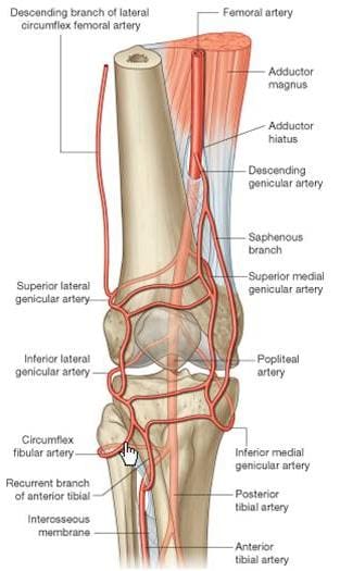
The popliteal artery, a branch of the superficial femoral artery, is the main arterial supply to the knee joint. It runs along the posterior aspect of the distal femur, behind the knee joint. At the supracondylar ridge, the popliteal artery gives off the blood supply to the knee, which consists of various genicular arteries (Figure 114). Inferior to the knee joint, the popliteal branches into the anterior and posterior tibial arteries, which supply the lower leg. The popliteal artery is a common site for both atherosclerosis and aneurysms, and is listed as the most common site for peripheral arterial aneurysms. Approximately 50% of these aneurysms are bilateral. Although they rarely rupture, popliteal aneurysms may serve as a focus for abrupt thrombotic occlusion of the involved popliteal artery, which can affect the foot on the same side. A thrombus within an aneurysm can also lead to a distal embolism. The genicular arteries are sources of continued blood flow to the knee and lower limb, in case of an obstructed popliteal artery. The descending genicular, also called the highest or supreme genicular, branches from the femoral artery, just superior to the popliteal branch. It supplies the adductor magnus and hamstring muscles, then joins with the network of genicular arteries around the knee joint. The middle genicular pierces the oblique popliteal ligament, and supplies the ligaments and synovial membrane inside the knee articulation (including the ACL and PCL). The sural artery joins the anastomoses of the genicular arteries, and also supplies muscles of the lower leg, including the large gastrocnemius muscle. The anastomotic pattern around the knee joint is supplied by the popliteal artery posteriorly, the descending genicular artery medially, and the descending branch of the lateral circumflex femoral artery laterally. The genicular arteries involved in the anastomosis are labeled as the medial and lateral superior geniculars, and the medial and lateral inferior geniculars.
the knee

Figure 114. Arteries of knee.

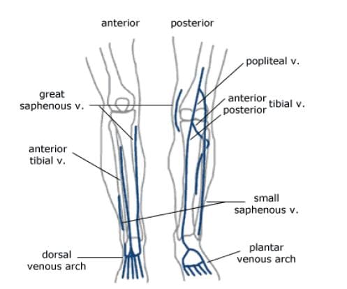
The major deep veins around the knee joint are the popliteal vein, and the anterior and posterior tibial veins (Figure 115). The popliteal vein begins at the junction of the tibial veins in the posterior aspect of the lower leg, just inferior to the knee joint. It ascends posteriorly, continuing as the femoral vein about halfway up the thigh. As deep veins typically follow the arteries, the genicular veins accompany the genicular arteries around the knee joint, then drain into the popliteal vein. The important superficial veins around the knee joint are the small and great saphenous veins. Superficial veins typically do not follow arteries, but rather travel with cutaneous nerves. The small saphenous ascends the lower leg posteriorly, angling from lateral to medial. It merges with the popliteal vein at a position slightly superior to the knee joint. The great saphenous vein, the longest vein in the body, has a medial and anterior course in the lower leg. It moves to a posterior position, but stays medial along the knee joint, moving alongside the medial epicondyle of the femur. The great saphenous then moves anteriorly again through the thigh.
the knee

Figure 115. Veins of knee.

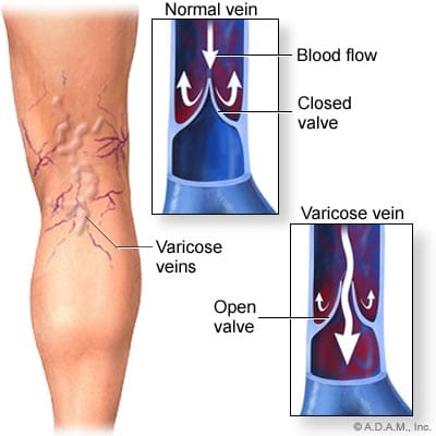
Varicose and �spider� veins are often seen in the leg in the posterior aspect of the knee joint. As mentioned previously, in the femoral vein discussion, veins have valves to ensure the �one-way� uphill flow of blood back to the heart (Figure 116). Communicating vessels, also called perforating veins, exist between the deep and superficial veins to help compensate for valves that may be incompetent, and are allowing blood reflux. If venous walls are weakened or dilated, the cusps of the valves can no longer close properly, and the valves can become incompetent. This leads to an increase in the weight of the column of blood for the veins that are �downstream� from the bad valve. Blood can pool in these veins, causing them to become varicose, where the veins swell, become tortuous, and even bulge through the skin surface. Reticular veins, which are smaller varicose veins that do not bulge through the skin, as well as very small �spider� veins are both typically less severe conditions, but both still involve the backwards flow of blood. Removal of severe varicose veins will actually help blood flow, as the blood will no longer be stagnant in the pooled areas.
the knee

Figure 116. Varicose veins around knee.

Bursae Of The Knee

The synovial knee joint is home to a large number of bursae (Figure 117). These are fluid sacs and synovial pockets that surround and sometimes communicate with the joint cavity. They facilitate friction-free movement between the bones and moving structures (tendon, muscle). Fluid or debris can collect in the bursa, or fluid can extend into the bursa from the adjacent joint in situations such as excessive friction, infection or direct trauma. This type of pathological enlargement of the bursa is referred to as bursitis, which can mimic several peripheral joint and muscle abnormalities. Radiologists must be able to accurately identify bursal pathology, especially amongst the numerous knee bursae (14 reported in some literature). We will identify a few of the more common bursa, beginning with the suprapatellar bursa. This bursa lies between a quadriceps tendon and the femur, superior to the patella (Figure 118). Fluid is commonly found here when patients have a joint effusion. Bursitis of the prepatellar bursa is also known as �housemaid�s knee�. It occurs from repetitive trauma from kneeling, as seen with housemaids, wrestlers, and carpet-layers. This bursa is found between the patella and the skin (Figure 119). Inflammation of the superficial infrapatellar bursa may be called �Clergyman�s knee�, another bursitis that can occur from excessive kneeling. This bursa is located between the distal third of the patellar tendon and the overlying skin (Figure 120).
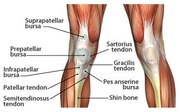
the knee

Figure 117. Bursae in the knee.

the knee

Figure 118. T2 gradient
displaying suprapatellar
bursa.
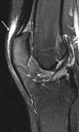
the knee

Figure 119. T2
fatsat displaying
prepatellar bursa.
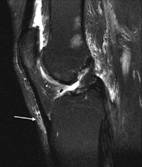
the knee

Figure 120. T2 fatsat
displaying infrapatellar
bursa.


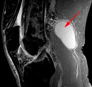
The synovial sac of the knee joint sometimes forms a posterior bulge, known as a Baker�s cyst or popliteal cyst (Figure 121). It typically forms between the tendons of the medial head of the gastrocnemius muscle and the semimembranosus muscle, posterior to the medial femoral condyle. Baker�s cysts are not true cysts, as they typically maintain open communication with the synovial sac. However, they can pinch off, and they can rupture. They are usually asymptomatic, but can be indicative of another problem of the knee, such as arthritis or a meniscal tear. Aspiration of the synovial fluid can be performed if the cyst becomes problematic. Treatment is usually necessary if a Baker�s cyst ruptures, as it can cause acute pain behind the knee, and swelling of the calf muscles. A ruptured cyst can also mimic a DVT or thrombophlebitis. Ultrasound and MRI can both be used for confirmation of a Baker�s cyst (Figure 122).
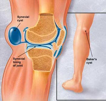
the knee

Figure 121. Lateral view of Baker�s cyst.
the knee

Figure 122. Sagittal image of Baker�s cyst on MRI.

Scan Setups

The following are HMSA suggestions for knee imaging. Knee protocols should be designed to yield diagnostic images of the menisci, bones, articular cartilage, and all ligamentous structures of the knee. While many radiologists may require additional imaging of the ACL, protocols that are designed for optimal imaging of the cartilage and menisci should also produce adequate images of the ACL. Always check with your radiologist for his/her imaging preferences.

Axial Scans

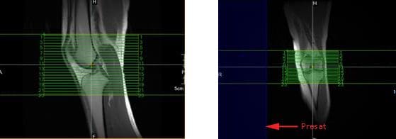
When positioning axial slices for the knee, sagittal and coronal images can be used to insure inclusion of all pertinent anatomy. The slices should extend superiorly to include the entire patella, and inferiorly to include the tibial tuberosity and patellar tendon insertion. A presat can be placed over the unaffected lower extremity to reduce the possibility of wrap-around artifact, as seen in the coronal image in Figure 139.
the knee

Figure 139. Axial slice setup using sagittal and coronal images.

Coronal Scans

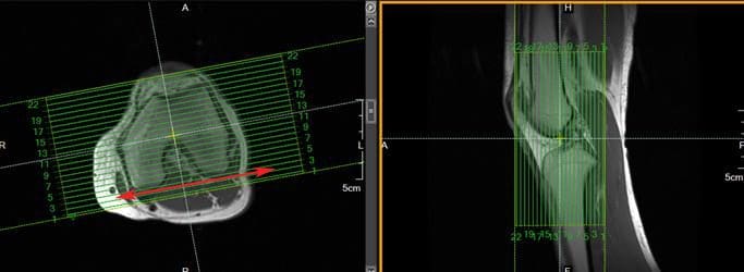
Coronal slices of the knee should include the anatomy from the posterior femoral condyles to the anterior portion of the patella. Visualize a line connecting the lateral and medial condyles of the femur. Typically, the coronal slices are angled so that they are parallel to that line, as seen in the axial image in Figure 140.
the knee

Figure 140. Coronal slice setup using axial and sagittal images.

Sagittal Scans

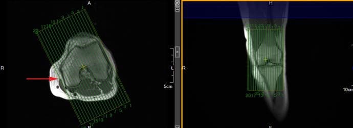
Sagittal slices should include the anatomy from the medial condyle to the lateral condyle. The slice group may be angled per your radiologist�s preference, but should remain perpendicular to the coronal slices. Typically, the slice group is angled so that it is parallel to the medial border of the femoral condyle, as seen in the axial image in Figure 141.
the knee

Figure 141. Sagittal slice setup using axial and coronal images.

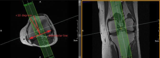
In addition to routine oblique sagittal images, some radiologists prefer an additional sagittal scan of the ACL with thin slices and high spatial resolution. Axial and coronal images can be used for slice setup. Referenced literature recommends that the angle of the slice group should not exceed 10� from a line drawn perpendicular to the bicondylar line (line that connects the posterior femoral condyles), as seen in Figure 142.
the knee

Figure 142. Sagittal ACL slice setup using axial and coronal images.

 

blank
References:

Kapit, Wynn, and Lawrence M. Elson. The Anatomy Coloring Book. New York: HarperCollins, 1993.

Hip Anatomy, Function, and Common Problems. (Last updated 28July2010). Retrieved from http://healthpages.org/anatomy-function/hip-structure-function-common-problems/

Cluett, J. M.D. (Updated 22May2012). Labral Tear of the Hip Joint. Retrieved from http://orthopedics.about.com/od/hipinjuries/qt/labrum.htm

Hughes, M. D.C. (15July2010). Diseases of the Femur Bone. Retrieved from http://www.livestrong.com/article/175599-diseases-of-the-femur-bone/

A Patient�s Guide to Perthes Disease of the hip. (n.d.). Retrieved from http://www.orthopediatrics.com/docs/Guides/perthes.html

Hip Injuries and Disorders. (Last reviewed 10February2012). Retrieved from http://nlm.nih.gov/medlineplus/hipinjuriesanddisorders.html

Ligament of head of femur. (Updated 20December2011). Retrieved from http://en.wikipedia.org/wiki/Ligament_of_head_of_femur

Ewing�s sarcoma. (Last modified 06January2012). Retrieved from http://en.wikipedia.org/wiki/Ewing%27s_sarcoma

Hip Anatomy. (n.d.). Retrieved from http://www.activemotionphysio.ca/Injuries-Conditions/Hip

Iliotibial Band Friction Syndrome. (n.d.). Retrieved from http://www.physiotherapy-treatment.com/iliotibial-band-friction-syndrome.html

Snapping hip syndrome. (Last modified 09November2011). Retrieved from http://en.wikipedia.org/wiki/Snapping_hip_syndrome

Sekul, E. (Updated 03February2012). Meralgia Paresthetica. Retrieved from http://emedicine.medscape.com/article/1141848-overview

Yeomans, S. D.C. (Updated 07July2010). Sciatic Nerve and Sciatica. Retrieved from http://www.spine-health.com/conditions/sciatica/sciatic-nerve-and-sciatica

Mayo Clinic staff. (26July2011). Meralgia paresthetica. Retrieved from http://www.mayoclinic.com/health/meralgia-paresthetica/DS00914

Deep Vein Thrombosis (DVT)-Blood Clots in the Legs. (n.d.). Retrieved from http://catalog/nucleusinc.com/displaymonograph.php?MID=148

Petersilge, C. M.D. (03May2000). Chronic Adult Hip Pain: MR Arthrography of the Hip. Retrieved from http://radiographics.rsna.org/content/20/suppl_1/S43.full

Acetabular branch of medial circumflex femoral artery. (Last modified 17November2011). Retrieved from http://en.wikipedia.org/wiki/Acetabular_branch_of_medial_circumflex_femoral_artery

Cluett, J. M.D. (Updated 26March2011). Hip Bursitis. Retrieved from http://orthopedics.about.com/cs/hipsurgery/a/hipbursitis.htm

Steinbach, L. M.D., Palmer, W. M.D., Schweitzer, M. M.D. (10June2002). Special Focus Session MR Arthrography. Retrieved from http://radiographics.rsna.org/content/22/5/1223.full

Schueler, S. M.D., Beckett, J.M.D., Gettings, S.M.D. (Last updated 05August2010). Ischial Bursitis/Overview. Retrieved from http://www.freemd.com/ischial-bursitis/overview.htm

Hwang, B., Fredericson, M., Chung, C., Beaulieu, C., Gold, G. (29October2004). MRI Findings of Femoral Diaphyseal Stress Injuries in Athletes. Retrieved from http://www.ajronline.org/content/185/1/166.full.pdf

The Femur (Thigh Bone). (n.d.). Retrieved from http://education.yahoo.com/reference/gray/subjects/subject/59

Norman, W. PhD, DSc. (n.d.). Joints of the Lower Limb. Retrieved from http://home.comcast.net/~wnor/lljoints.htm

Femur. (Last modified 24September2012). Retrieved from http://en.wikipedia.org/wiki/Femur

Wheeless, C. III, M.D. (Last updated 25April2012). Ligaments of Humphrey and Wrisberg. Retrieved from http://wheelessonline.com/ortho/ligaments_of_humphrey_and_wrisberg

Muscle Strains in the Thigh. (Last reviewed August2007). Retrieved from http://orthoinfo.aaos.org/topic.cfm?topic=A00366

Shiel, W. Jr., M.D. (Last reviewed 23July2012). Hamstring Injuries. Retrieved from http://www.medicinenet.com/hamstring_injury/article.htm

Hamstring Muscle Injuries. (Last reviewed July 2009). Retrieved from http://orthoinfo.aaos.org/topic.cfm?topic=a00408

Knee. (Last modified 19September2012). Retrieved from http://en.wikipedia.org/wiki/Knee

DeBerardino, T. M.D. (Updated 30March2012). Quadriceps Injury. Retrieved from http://emedicine.medscape.com/article/91473-overview

Kan, J.H. (n.d.). Osteochondral Abnormalities: Pitfalls, Injuries, and Osteochondritis Dissecans. Retrieved from http://www.arrs.org/shopARRS/products/s11p_sample.pdf

Nerves of the Lower Limb. (Last updated 30March2006). Retrieved from http://download.videohelp.com/vitualis/med/lowrnn.htm

The Adductor Canal. (Last updated 30March2006). Retrieved from http://download.videohelp.com/vitualis/med/addcanal.htm

Nabili, S. M.D. (n.d.). Varicose Veins & Spider Veins. Retrieved from http://www.medicinenet.com/varicose_veins/article.htm

Basic Venous Anatomy. (n.d.). Retrieved from http://vascular-web.com/asp/samples/sample104.asp

Femoral nerve. (Last modified 23September2012). Retrieved from http://en.wikipedia.org/wiki/Femoral_nerve

Peron, S. RDCS. (Last modified 16October2010). Anatomy � Lower Extremity Veins. Retrieved from http://www.vascularultrasound.net/vascular-anatomy/veins/lower-extremity-veins

Medical Multimedia Group, L.L.C. (n.d.). Knee Anatomy. Retrieved from http://www.eorthopod.com/content/knee-anatomy

Knee Joint Anatomy, Function and Problems. (Last updated 06July2010). Retrieved from http://healthpages.org/anatomy-function/knee-joint-structure-function-problems/

Coronary ligament of the knee. (Last modified 09May2010). Retrieved from http://en.wikipedia.org/wiki/Coronary_ligament_of_the_knee

Walker, B. (n.d.). Patellar Tendonitis Treatment � Jumper�s Knee. Retrieved from http://www.thestretchinghandbook.com/archives/patellar-tendonitis.php

Osgood-Schlatter disease. (Last reviewed 12November2010). Retrieved from http://www.ncbi.nlm.nih.gov/pubmedhealth/PMH0002238/

Grelsamer, R. M.D. (n.d.). The Anatomy of the Patella and the Extensor Mechanism. Retrieved from http://kneehippain.com/patient_pain_anatomy.php

Oblique popliteal ligament. (Last modified 24March2012). Retrieved from http://en.wikipedia.org/wiki/Oblique_popliteal_ligament

Shiel, W. Jr., M.D. (Last reviewed 27July2012). Chondromalacia Patella (Patellofemoral Syndrome). Retrieved from http://www.medicinenet.com/patellofemoral_syndrome/article.htm

Knee. (Last modified 19September2012). Retrieved from http://en.wikipedia.org/wiki/Knee

Mosher, T. M.D. (Last updated 11April2011). MRI of Knee Extensor Mechanism Injuries Overview of the Knee Extensor Mechanism. Retrieved from http://emedicine.medscape.com/article/401001-overview

Carroll, J. M.D. (December 2007). Oblique Menisco-meniscal Ligament. Retrieved from http://radsource.us/clinic/0712

DeBerardino, T. M.D. (Last updated 30March2012). Medial Collateral Knee Ligament Injury. Retrieved from http://emedicine.medscape.com/article/89890-overview#a0106

Farr, G. (Last updated 31December2007). Joints and Ligaments of the Lower Limb. Retrieved from http://becomehealthynow.com/article/bodyskeleton/951/

Knee anatomy overview. (02March2008). Retrieved from http://www.kneeguru.co.uk/KNEEnotes/node/741

Dixit, S. M.D., Difiori, J. M.D., Burton, M. M.D., Mines, B. M.D. (15January2007). Management of Patellofemoral Pain Syndrome. Retrieved from http://www.aafp.org/afp/2007/0115/p194.html

Knee Muscles. (Last updated 05September2012). Retrieved from http://www.knee-pain-explained.com/kneemuscles.html

Popliteus muscle. (Last updated 20February2012). Retrieved from http://en.wikipedia.org/wiki/Popliteus_muscle

Kneedoc. (10February2011). Nerves. Retrieved from http://thekneedoc.co.uk/neurovascular/nerves

Wheeless, C. III, M.D. (Last updated 15December2011). Popliteal Artery. Retrieved from http://wheelessonline.com/ortho/popliteal_artery

The Popliteal Artery. (n.d.) Retrieved from http://education.yahoo.com/reference/gray/subjects/subject/159

Knee bursae. (Last updated 09May2012). Retrieved from http://en.wikipedia.org/wiki/Bursae_of_the_knee_joint

Hirji, Z., Hunjun, J., Choudur, H. (02May2011). Imaging of the Bursae. Retrieved from http://www.ncbi.nlm.nih.gov/pmc/articles/PMC3177464/

Kimaya Wellness Limited. (n.d.). Organ>Popliteal Artery. Retrieved from http://kimayahealthcare.com/OrganDetail.aspx?OrganID=103&AboutID=1

Total Vein Care. (Last updated 24February2012). Varicose Vein Anatomy and Function for Patients. Retrieved from http://www.veincare.com/education/

Tibia. (Last updated 01April2012). Retrieved from http://en.wikipedia.org/wiki/Tibia

Norkus,S., Floyd, R. (Published 2001). The Anatomy and Mechanisms of Syndesmotic Ankle Sprains. Retrieved from http://www.ncbi.nlm.nih.gov/pmc/articles/PMC155405/

Soleus muscle. (Last updated 10April2012). Retrieved from http://en.wikipedia.org/wiki/Soleus_muscle

Achilles Tendinitis. (Last reviewed June2010). Retrieved from http://orthoinfo.aaos.org/topic.cfm?topic=A00147

Wheeless, C. III,M.D. (Last updated 11April2012). Sural Nerve. Retrieved from http://wheelessonline.com/ortho/sural_nerve

Medical Multimedia Group, L.L.C. (Last updated 26July2006). Ankle Syndesmosis Injuries. Retrieved from http://www.orthogate.org/patient-education/ankle/ankle-syndesmosis-injuries.html

Cluett, J. M.D. (Last updated 16September2008). Exertional Compartment Syndrome. Retrieved from http://orthopedics.about.com/od/overuseinjuries/a/compartment.htm

Leg Veins (Thigh, Lower Leg) Anatomy, Pictures and Names. (Last updated 21November2010). Retrieved from http://www.healthype.com/leg-veins-thigh-lower-leg-anatomy-pictures-and-names.html

Cluett, J.M.D. (Last updated 6October2009). Stress Fracture. Retrieved from http://orthopedics.about.com/cs/otherfractures/a/stressfracture.htm

Ostlere, S. (1December2004). Imaging the ankle and foot. Retrieved from http://imaging.birjournals.org/content/15/4/242.full

Inverarity, L. D.O. (Last updated 23January2008). Ligaments of the Ankle Joint. Retrieved from http://physicaltherapy.about.com/od/humananatomy/p/ankleligaments.htm

Golano, P., Vega, J., DeLeeuw, P., Malagelada, F.,Manzanares, M., Gotzens, V., van Dijk, C. (Published online 23March2010). Anatomy of the ankle ligaments:a pictorial essay. Retrieved from http://www.ncbi.nlm.nih.gov/pmc/articles/PMC2855022/

Numkarunarunrote, N., Malik, A., Aguiar, R.,Trudell, D., Resnick, D. (11October2006). Retinacula of the Foot and Ankle: MRI with Anatomic Correlation in Cadavers. Retrieved from http://www.ajronline.org/content/188/4/W348.full

Medical Multimedia Group, L.L.C. (n.d.). A Patient�s Guide to Ankle Anatomy. Retrieved from http://www.eorthopod.com/content/ankle-anatomy

The Anterior Tibial Artery. (n.d.). Retrieved from http://education.yahoo.com/reference/gray/subjects/subject/160

Foot and Ankle Anatomy. (Last updated 28July2011). Retrieved from http://northcoastfootcare.com/pages/Foot-and-Ankle-Anatomy.html

Donnelly, L., Betts, J., Fricke, B. (1July2009). Skimboarder�s Toe: Findings on High-Field MRI. Retrieved from http://www.ajronline.org/content/184/5/1481.full

Foot. (Last updated 28August2012). Retrieved from http://en.wikipedia.org/wiki/Foot

Wiley, C. (n.d.). Major Ligaments in the Foot. Retrieved from http://www.ehow.com/list_6601926_major-ligaments-foot.html

Turf Toe: Symptoms, Causes, and Treatments. (Last reviewed 9August2012). Retrieved from http://www.webmd.com/fitness-exercise/turf-toe-symptoms-causes-and-treatments

Cluett, J. M.D. (Last updated 02April2012). Turf Toe. Retrieved from http://orthopedics.about.com/od/toeproblems/p/turftoe.htm

Neurology and the Feet. (n.d.) Retrieved from http://footdoc.ca/www.FootDoc.ca/Website%20Nerves%20Of%20The%20Feet.htm

The Veins of the Lower Extremity, Abdomen, and Pelvis. (n.d.). Retrieved from http://education.yahoo.com/reference/gray/subjects/subject/173

Corley, G., Broderick, B., Nestor, S., Breen, P., Grace, P., Quondamatteo, F., O�Laighin, G. (n.d.). The Anatomy and Physiology of the Venous Foot Pump. Retrieved from http://www.eee.nuigalway.ie/documents/go_anatomy_of_the_plantar_venous_plexus_manuscript.pdf

Morton�s neuroma. (Last modified 8August2012). Retrieved from http://en.wikipedia.org/wiki/Morton%27s_metatarsalgia

References For Anatomy Pics:

Figures 1, 5, 6, 24- http://www.orthopediatrics.com/docs/Guides/perthes.html

Figures 2, 3, 11, 12, 14, 15, 16, 18, 23, 25- http://www.activemotionphysio.ca/Injuries-Conditions/Hip/Hip-Anatomy/a~299/article.html

Figure 4- http://hipkneeclinic.com/images/uploaded/hipanatomy_xray.jpg

Figures 7, 8, 9- http://hipfai.com/

Figure 10- http://en.wikipedia.org/wiki/File:Ewing%27s_sarcoma_MRI_nci-vol-1832-300.jpg

Figure 13- http://www.chiropractic-help.com/Patello-Femoral-Pain-Syndrome.html

Figure 17- http://www.thestretchinghandbook.com/archives/ezine_images/adductor.jpg

Figure 19- http://media.summitmedicalgroup.com/media/db/relayhealth-images/hipanat.jpg

Figures 20-22- http://www.ajronline.org/content/182/1/137.full.pdf+html

Figure 43, 44- http://radiographics.rsna.org/content/20/suppl_1/S43.full

Figure 45- http://www.exploringnature.org/db/detail.php?dbID=24&detID=2768

Figures 46-48- http://www.ajronline.org/content/185/1/166.full.pdf

Figure 49- http://arrs.org/shopARRS/products/s11p_sample.pdf

Figure 50- http://www.thestretchinghandbook.com/archives/medial-collateral-ligament.php

Figures 51, 52- http://www.radsource.us/clinic/0712

Figures 53, 54- http://www.osteo-path.co.uk/BodyMap/Thighs.html

Figure 55- http://www.ncbi.nlm.nih.gov/pmc/articles/PMC1963576/

Figure 56- http://legacy.owensboro.kctcs.edu/gcaplan/anat/Notes/API%20Notes%20M%20%20Peripheral%20Nerves.htm

Figure 57- http://www.keywordpictures.com/keyword/lateral%20cutaneous%20nerve%20of%20thigh/

Figure 58- http://home.comcast.net/~wnor/postthigh.htm

Figure 59- http://becomehealthynow.com/glossary/CONG437.htm

Figure 60- http://fitsweb.uchc.edu/student/selectives/Luzietti/Vascular_pvd.htm

Figure 61- http://www.fashion-res.com/peripheral-vascular-disease-with-stenting-in-the/

Figure 62- http://www.wpclipart.com/medical/anatomy/blood/femoral_artery_and_branches_in_leg.png.html

Figure 63- http://www.globalteleradiologyservices.com/Deep_Vein_Thrombosis_Overview.htm

Figure 64- http://www.vascularultrasound.net/vascular-anatomy/veins/lower-extremity-veins

Figure 82- http://www.jeffersonhospital.org/diseases-conditions/knee-ligament-injury.aspx?disease=658f267f-75ab-4bde-8781-f2730fafa958

Figure 83- http://javierjuan.ifunnyblog.com/anatomybackofknee/

Figure 84- http://www.kneeandshouldersurgery.com/knee-disorders/tibial-osteotomy.html

Figure 85- http://www.disease-picture.com/chondromalacia-patella-physical-therapy/

Figure 86- http://www.eorthopod.com/content/bipartite-patella

Figure 87- http://www.orthogate.org/patient-education/knee/articular-cartilage-problems-of-the-knee.html

Figure 88- http://www.webmd.com/pain-management/knee-pain/menisci-of-the-knee-joint

Figure 89- http://sumerdoc.blogspot.com/2008_07_01_archive.html

Figure 90- http://www.concordortho.com/patient-education/topic-detail-popup.aspx?topicID=55befba2d440dc8e25b85747107b5be0

Figure 91- http://trialx.com/curebyte/2011/08/16/pictures-for-chondromalacia-patella/

Figure 92- http://radiopaedia.org/images/1059

Figure 93- http://radiologycases.blogspot.com/2011/01/osgood-schlatter-disease.html

Figure 94- http://www.physioquestions.com/2010/09/07/knee-injury-acl-part-i/

Figure 95- http://www.jeffersonhospital.org/diseases-conditions/knee-ligament-injury.aspx?disease=4e3fcaf5-0145-43ea-820f-a175e586e3c8

Figures 96, 97- http://radiology.rsna.org/content/213/1/213.full

Figures 98-101- http://appliedradiology.com/Issues/2008/12/Articles/Imaging-the-knee–Ligaments.aspx

Figure 102- http://radiopaedia.org/images/408156

Figure 103- http://aftabphysio.blogspot.com/2010/08/joints-of-lower-limb.html

Figures 104, 105- http://www.radsource.us/clinic/0310

Figure 106- http://nwrunninglab.com/patellar-tendonitis.html

Figure 107- http://www.aafp.org/afp/2007/0115/p194.html

Figure 108- http://www.reboundsportspt.com/blog/tag/knee-pain

Figure 109- http://www.norwellphysicaltherapy.com/Injuries-Conditions/Knee/Knee-Issues/Quadriceps-Tendonitis-of-the-Knee/a~1803/article.html

Figure 110- http://kneeguru.co.uk/KNEEnotes/node/479

Figure 111- http://www.magicalrobot.org/BeingHuman/2010/03/fascia-bones-and-muscles

Figure 112- http://home.comcast.net/~wnor/postthigh.htm

Figures 113, 115, 157-159- http://ipodiatry.blogspot.com/2010/02/anatomy-of-foot-and-ankle_26.html

Figure 114- http://medchrome.com/basic-science/anatomy/the-knee-joint/

Figure 116- http://www.sharecare.com/question/what-are-varicose-veins

Figure 117- http://mendmyknee.com/knee-and-patella-injuries/anatomy-of-the-knee.php

Figures 118-120- http://www.ncbi.nlm.nih.gov/pmc/articles/PMC3177464/

Figure 121- http://www.riversideonline.com/health_reference/Disease-Conditions/DS00448.cfm

Figure 122- http://arthritis.ygoy.com/2011/01/01/what-is-an-arthritis-knee-cyst/

Figure 143- http://usi.edu/science/biology/mkhopper/hopper/BIOL2401/LABUNIT2/LabEx11week6/tibiaFibulaAnswer.htm

Figure 144- http://web.donga.ac.kr/ksyoo/department/education/grossanatomy/doc/html/fibula1.html

Figure 145- http://becomehealthynow.com/popups/ligaments_tib_fib_bh.htm

Figure 146- http://www.parkwayphysiotherapy.ca/article.php?aid=121

Figure 147- http://aidmyankle.com/high-ankle-sprains.php

Figure 148- http://legsonfire.wordpress.com/what-is-compartment-syndrome/

Figures 149, 152- http://www.stepbystepfootcare.ca/anatomy.html

Figures 150, 151- http://www.gla.ac.uk/ibls/US/fab/tutorial/anatomy/jiet.html

Figure 153- http://www.athletictapeinfo.com/?s=tennis+leg

Figure 154- http://radsource.us/clinic/0608

Figure 155- http://www.eorthopod.com/content/achilles-tendon-problems

Figure 156- http://achillesblog.com/assumptiondenied/not-a-rupture/

Figure 181- http://www.orthopaedicclinic.com.sg/ankle/a-patients-guide-to-ankle-anatomy/

Figure 182- http://www.activemotionphysio.ca/article.php?aid=47

Figure 183- http://www.ajronline.org/content/193/3/687.full

Figures 184, 186- http://www.eorthopod.com/content/ankle-anatomy

Figure 185- http://www.crossfitsouthbay.com/physical-therapy/learn-yourself-a-quick-anatomy-reference/ankle/

Figures 187, 227- http://www.activemotionphysio.ca/Injuries-Conditions/Foot/Foot-Anatomy/a~251/article.html

Figure 188- http://inmotiontherapy.com/article.php?aid=124

Figures 189, 190- http://home.comcast.net/~wnor/ankle.htm

Figure 191- http://skillbuilders.patientsites.com/Injuries-Conditions/Ankle/Ankle-Anatomy/a~47/article.html

Figure 192- http://metrosportsmed.patientsites.com/Injuries-Conditions/Foot/Foot-Anatomy/a~251/article.html

Figure 193- http://musc.edu/intrad/AtlasofVascularAnatomy/images/CHAP22FIG30.jpg

Figure 194- http://musc.edu/intrad/AtlasofVascularAnatomy/images/CHAP22FIG31B.jpg

Figure 195- http://veinclinics.com/physicians/appearance-of-vein-disease/

Figure 196- http://mdigradiology.com/services/interventional-services/varicose-veins.php

Figure 216- http://kidport.com/RefLib/Science/HumanBody/SkeletalSystem/Foot.htm

Figure 217- http://www.joint-pain-expert.net/foot-anatomy.html

Figure 218- http://www.thetoedoctor.com/turf-toe-symptoms-and-treatment/

Figures 219, 220- http://radsource.us/clinic/0303

Figure 221- http://www.ajronline.org/content/184/5/1481.full

Figure 222- http://www.answers.com/topic/arches

Figure 223- http://www.mayoclinic.com/health/medical/IM00939

Figure 224- http://radsource.us/clinic/0904

Figure 225- http://www.ortho-worldwide.com/anfobi.html

Figure 226- http://www.coringroup.com/lars_ligaments/patientscaregivers/your_anatomy/foot_and_ankle_anatomy/

Figure 228- http://www.stepbystepfootcare.ca/anatomy.html

Figure 229- http://iupucbio2.iupui.edu/anatomy/images/Chapt11/FG11_18aL.jpg

Figure 230- http://www.ajronline.org/content/184/5/1481.full.pdf

Figure 231- http://metrosportsmed.patientsites.com/Injuries-Conditions/Foot/Foot-Anatomy/a~251/article.html

Figure 232- http://www.painfreefeet.com/nerve-entrapments-of-the-leg-and-foot.html

Figures 233, 234- http://emedicine.medscape.com/article/401417-overview

Figure 235- http://web.squ.edu.om/med-Lib/MED_CD/E_CDs/anesthesia/site/content/v03/030676r00.HTM

Figure 236- http://www.nysora.com/peripheral_nerve_blocks/classic_block_tecniques/3035-ankle_block.html

Figure 237- http://ultrasoundvillage.net/imagelibrary/cases/?id=122&media=464&testyourself=0

Figure 238- http://www.joint-pain-expert.net/foot-anatomy.html

Figure 239- http://jap.physiology.org/content/109/4/1045.full

Figure 240- http://microsurgeon.org/secondtoe

Figure 241- http://elu.sgul.ac.uk/rehash/guest/scorm/406/package/content/common_iliac_veins.htm

Close Accordion
MRI To Evaluate Lumbar Posterior Ligament Complex Post Trauma

MRI To Evaluate Lumbar Posterior Ligament Complex Post Trauma

The importance of Magnetic Resonance Imaging to evaluate the integrity of the lumbar posterior ligament complex post trauma.

Abstract: Posterior ligamentous complex(PLC), consisting of the supraspinous ligament, interspinous ligament, ligamentum flavum, and the facet joint capsules is thought to contribute significantly to the stability of the lumbar spine. There has been much debate on whether Magnetic Resonance Imaging(MRI) is specific and sensitive in diagnosing pathology to the PLC. The objective is to determine the necessity of MRI imaging for evaluating the integrity of the lumbar posterior ligament complex post trauma.

Key Words: Magnetic Resonance Imaging(MRI), interspinous ligament, posterior ligament complex, low back pain, ligament laxity, electromyography, impairment rating

A 41-year-old male, presented to my office for an examination with complaints of low back pain with numbness, tingling and weakness into the left lower extremity after he was the restraint driver in a motor vehicle collision approximately three and a half months� post trauma.�He�rated the pain as a�3/10 on a visual analog scale with 10/10 being the worst and the pain and noted the pain as being�present most of the time.� He stated that he was on pain killers daily and this helped manage his daily activities. Without pain killers his pain levels are rated 8/10 being present most of the time. The pain killers stated by the patient are Oxycodone and Naproxen.
He�reported that the pain would be aggravated by activities which required excessive standing, repetitive bending, and lifting. He further noted that in the morning the pain was increased and his left leg would be numb and weak for about the first hour.

The patient stated that his care to date had been managed by a pain management clinic and that he had minimal improvement with treatment which has included physical therapy and massage therapy. He reported the pain clinic next recommended steroid injections which he refused. He states there has been was no imaging ordered and that an Electromyography(EMG) had been performed. He was told the test was negative for pathology.

Prior History: No significant medical history was reported.
Clinical Findings:�The patient is 6�0� and weighs 210 lbs.

Physical Exam Findings:

Cervical Spine:
Cervical spine range of motion is full and unrestricted. Maximum cervical compression is negative. Motor and other regional sensory exam are unremarkable at this time.

Thoracic Spine:
Palpation of the thoracic spine region reveals taught and tender fibers in the area of the bilateral upper and mid thoracic musculature. Thoracic spine range of motion is restricted in flexion, extension, bilateral lateral flexion, and bilateral rotation. Regional motor and sensory exam are unremarkable at this time.

Lumbar Spine:
Palpation of the lumbosacral spine region reveals taught and tender fibers in the area of the lumbar paraspinal musculature. Lumbar spine range of motion is limited in flexion, extension, bilateral lateral flexion and bilateral rotation. Extension restriction is due to pain and spasm. Straight leg raise causes pain at approximately 50 degrees when testing either side in the left low back. There is no radicular symptomatology down the leg. Kemp�s maneuver recreates pain in the L4 region on the left. No radicular symptoms are noted. The patient is able to heel and toe walk. Regional motor and sensory exam is unremarkable at this time other than L4, L5 and S1 dermatomes having decreased sensation with light touch.

Muscle testing of the upper and lower extremities was tested at a 5/5 with the exception of the left quadricep tested at a 4/5.� The patient�s deep tendon reflexes of the upper and lower extremities were tested including triceps, biceps, brachioradialis, patella, and Achilles and all were tested at 2+ bilaterally except the left patellar reflex was 1+.

RANGES OF MOTION EVALUATION

All range of motions are based on the�American Medical Association�s Guides to the Evaluation of Permanent Impairment, 5th�Edition1�and performed by a dual inclinometer for the lumbar spine.

�� Range of Motion������Normal�������� Examination�������� % Deficit

Flexion 60 48 20
Extension 25 12 52
Left Lateral Flexion 25 16 36
Right Lateral Flexion 25 18 28

An MRI was ordered to rule out gross pathology.

Imaging:

walia201.png

A lumbar MRI reveals;
1)��� Mild disc bulges at T11-T12, T12-L1, L1-L2 and L5-S1
2)��� Low disc signals indicative of disc desiccation at T11-T12, T12-L1, L1-L2, L2-L3, L3-L4 and L4-L5
3)��� Retrolisthesis of 2mm at L3-L4
4)��� Mild ligamentous hypertrophy at L1-L2, L2-L3, L3-L4, L4-L5 and L5-S1
5)��� L4-L5 has a Grade 1-2 tear of the interspinous ligament with mild inflammation
6)��� L5-S1 has a Grade 1 interspinous ligament tear with mild inflammation

After reviewing the MRI I ordered lumbar x-rays to rule out ligament laxity.

X-RAY STUDIES

Lumbar x-rays reveal the following:
1)��� Left lateral tilt
2)��� Retrolisthesis at L1 of 3mm
3)��� Retrolisthesis at L2 of 3mm
4)��� Combined excessive translation of 4mm of L1 during flexion-extension
5)��� Combined excessive translation of 4mm of L2 during flexion-extension
6)��� Excessive translation of L3 in extension posteriorly of 2.5mm
7)��� Decreased disc space at L5-S1

Chiropractic care was initiated. The patient was placed on an initial care plan of 2-3x/week for 3 months and then a recommended break in care for one month so the patient could be evaluated for permanency while he was not care dependent.

At maximum medical improvement, he had continued low back pain rated 4/10, continued numbness and tingling into his left leg and left quadricep weakness rated 4/5. He does not need pain killers for pain management anymore. He continues chiropractic care every two weeks to manage his symptoms.

Conclusion:
In this specific case, pathology to the posterior ligament complex diagnosed on MRI lead to the x-ray finding of excessive translation at L1-L2 and L2-L3. The patient was given a permanent impairment rating of 22% based on my interpretation of the American Medical Association�s Guides to the Evaluation of Permanent Impairment, 5th�Edition1. The interspinous ligament tears at the L4-L5 and L5-S1 level would not have been diagnosed without the MRI.

There has been much debate on whether MRI imaging has a role in evaluating lumbar PLC. MRI is a powerful diagnostic tool that can provide important clinical information regarding the condition of the PLC. Useful sequences for spinal MRI in trauma include sagittal and axial T1-weighted images, T2-weighted FSE, fat-saturated T2-weighted FSE, and STIR sequences to highlight bone edema.2�Ligamentous injuries are best identified on T2-weighted images with fat saturation because the ligaments are thin and bonded on either side by fat, which can appear as hyperintense on both T1 and T2 images.3�T1-weighted images are inadequate in isolation for identifying ligamentous injuries.4�

The diagnostic accuracy for MRI was reported for both supraspinous ligament and interspinous ligament injury with a sensitivity of 89.4% and 98.5%, respectively, and a specificity of 92.3% and 87.2% in 35 patients.5
For patients with persistent symptoms after trauma an MRI may be indicated to evaluate posterior ligamentous complex integrity.

Competing Interests:� There are no competing interests in the writing of this case report.

De-Identification: All of the patient�s data has been removed from this case.

References:
1. Cocchiarella L., Anderson G. Guides to the Evaluation of Permanent Impairment, 5th Edition, Chicago IL, 2001 AMA Press.
2. Cohen, W.A., Giauque, A.P., Hallam, D.K., Linnau, K.F. and Mann, F.A., 2003. Evidence-based approach to use of MR imaging in acute spinal trauma.�European journal of radiology,�48(1), pp.49-60.
3. Terk, M.R., Hume-Neal, M., Fraipont, M., Ahmadi, J. and Colletti, P.M., 1997. Injury of the posterior ligament complex in patients with acute spinal trauma: evaluation by MR imaging.�AJR. American journal of roentgenology,�168(6), pp.1481-1486.
4. Saifuddin, A., Green, R. and White, J., 2003. Magnetic resonance imaging of the cervical ligaments in the absence of trauma.�Spine,�28(15), pp.1686-1691.
5. Haba H, Taneichi H, Kotani Y, et al. Diagnostic accuracy of magnetic resonance imaging for detecting posterior ligamentous complex injury associated with thoracic and lumbar fractures.�J Neurosurg. 2003; 99(1 Suppl):20-26.

SaveSave

Syndesmosis: A Lifetime Of Pain?

Syndesmosis: A Lifetime Of Pain?

I have been travelling through Athens and now Istanbul. My 11 year old is a Percy Jackson nut and has been filling me in with the who’s who of Greek mythology and I am learning Latin words every day. Quite an education!

I looked up the word syndesmosis and the Latin translation is “(New Latin, from Greek sundesmos) bond, ligament, from sundein, meaning to bind together”. As sports injury professionals, we know syndesmosis to be the joint articulation between the tibia and the fibula bones around the ankle. These two bones are �bound’ together with very firm and strong ligaments.

Syndesmosis comes to mind after I saw a girl sprain a syndesmosis at the Archaeological Museum in Istanbul today. This poor girl was preoccupied by the hundreds of cats and kittens running all over the place and did not see the uneven cobblestones on which she placed her foot. At the same time, she turned to change direction. This is a common mechanism of injury for a syndesmosis – a forced dorsiflexion and rotation on a fixed foot.

Rehab Masterclass Issue 140 Of Sports Injury Bulletin


Of all the ankle injuries, injury to the syndesmosis is the biggest pest to sports physios and the like. And unlike simple garden variety ankle sprains that heal quickly, the syndesmosis takes a LONG time to heal properly. If you deal with athletes that are susceptible to syndesmosis sprains, I’m sure you will agree that these are harder injuries to manage because of the severe consequences if done badly.

I go into a fair bit of detail in my Sports Injury Bulletin piece about syndesmosis injuries, detailing how they happen, how to identify them and then manage them. What I would like to highlight here are the implications of mismanaging a syndesmosis sprain.

In the current issue of The Journal of Sports and Physical Therapy, a group of Japanese researchers discovered that individuals who had chronic ankle instability (CAI) had a distal fibula that was positioned more lateral compared with healthy individuals with no CAI. In effect, those who had suffered serious syndesmosis injuries in the past and ended up with a wider distance between the fibula and the tibia, suffered more ongoing ankle pain than those without a tibfib separation.

Research shows that even a 1mm displacement of the talus within the mortise (due to a wider placed fibula) can reduce the contact area in the talocrural joint by 42% (Ramsey and Hamilton 1976). Mismanaged syndesmosis injuries, resulting in an excessive amount of opening, can lead to early onset arthritic changes and chronic ankle instability. The talus bone bounces around in the now wider tibfib articulation.

A Widening Of The Fibula Is Due To One Of The Following:


Poor initial management, whereby the athlete is allowed to weight bear too early and this weight bearing forces the fibula away from the tibia as the syndesmosis ligaments are trying to heal.

The degree of damage is so severe that proper tightening of these ligaments is not possible without surgical intervention such as a screw or similar being placed between the two bones to �force’ them together.

The key for a sports injury practitioner, is to properly identify a regular ankle sprain from a more serious syndesmosis injury. If you get this part wrong and allow the athlete to get back to weight bearing too early, then expect some complaints about a chronically painful ankle some time down the track.


Call Today!

Kobayashi et al (2014). �Fibular malalignment in individuals with chronic ankle instability.’ JOPST. 44(11); pp 841-910.

Ramsey and Hamilton (1976). J Bone and J Surgery Am. 58(3); 356-357.

Mastodon