For individuals experiencing pelvic pain, it could be a disorder of the pudendal nerve known as pudendal neuropathy or neuralgia that leads to chronic pain. The condition can be caused by pudendal nerve entrapment, where the nerve becomes compressed or damaged. Can knowing the symptoms help healthcare providers correctly diagnose the condition and develop an effective treatment plan?
Pudendal Neuropathy
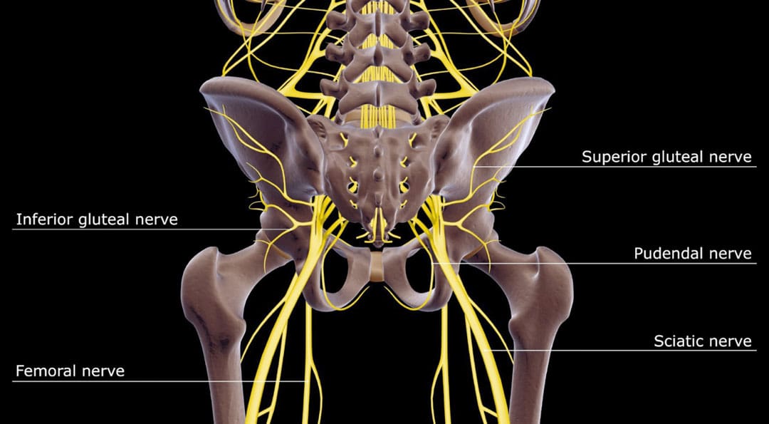
The pudendal nerve is the main nerve that serves the perineum, which is the area between the anus and the genitalia – the scrotum in men and the vulva in women. The pudendal nerve runs through the gluteus muscles/buttocks and into the perineum. It carries sensory information from the external genitalia and the skin around the anus and perineum and transmits motor/movement signals to various pelvic muscles. (Origoni, M. et al., 2014) Pudendal neuralgia, also referred to as pudendal neuropathy, is a disorder of the pudendal nerve that can lead to chronic pelvic pain.
Causes
Chronic pelvic pain from pudendal neuropathy can be caused by any of the following (Kaur J. et al., 2024)
Excessive sitting on hard surfaces, chairs, bicycle seats, etc. Bicyclists tend to develop pudendal nerve entrapment.
Trauma to the buttocks or pelvis.
Childbirth.
Diabetic neuropathy.
Bony formations that push against the pudendal nerve.
Thickening of ligaments around the pudendal nerve.
Symptoms
Pudendal nerve pain can be described as stabbing, cramping, burning, numbness, or pins and needles and can present (Kaur J. et al., 2024)
In the perineum.
In the anal region.
In men, pain in the scrotum or penis.
In women, pain in the labia or vulva.
During intercourse.
When urinating.
During a bowel movement.
When sitting and goes away after standing up.
Because the symptoms are often hard to distinguish, pudendal neuropathy can often be hard to differentiate from other types of chronic pelvic pain.
Cyclist’s Syndrome
Prolonged sitting on a bicycle seat can cause pelvic nerve compression, which can lead to chronic pelvic pain. The frequency of pudendal neuropathy (chronic pelvic pain caused by entrapment or compression of the pudendal nerve) is often referred to as Cyclist’s Syndrome. Sitting on certain bicycle seats for long periods places significant pressure on the pudendal nerve. The pressure can cause swelling around the nerve, which causes pain and, over time, can lead to nerve trauma. Nerve compression and swelling can cause pain described as burning, stinging, or pins and needles. (Durante, J. A., and Macintyre, I. G. 2010) For individuals with pudendal neuropathy caused by bicycling, symptoms can appear after prolonged biking and sometimes months or years later.
Take breaks at least 20–30 seconds after each 20 minutes of riding.
While riding, change positions frequently.
Stand up to pedal periodically.
Take time off between riding sessions and races to rest and relax the pelvic nerves. 3–10 day breaks can help in recovery. (Durante, J. A., and Macintyre, I. G. 2010)
If pelvic pain symptoms are barely starting to develop, rest and see a healthcare provider or specialist for an examination.
Seat
Use a soft, wide seat with a short nose.
Have the seat level or tilted slightly forward.
Seats with cutout holes place more pressure on the perineum.
If numbness or pain is present, try a seat without holes.
Bike Fitting
Adjust the seat height so the knee is slightly bent at the bottom of the pedal stroke.
The body’s weight should rest on the sitting bones/ischial tuberosities.
Keeping the handlebar height below the seat can reduce pressure.
The Triathlon bike’s extreme-forward position should be avoided.
A more upright posture is better.
Mountain bikes have been associated with an increased risk of erectile dysfunction than road bikes.
Shorts
Wear padded bike shorts.
Treatments
A healthcare provider may use a combination of treatments.
The neuropathy can be treated with rest if the cause is excessive sitting or cycling.
Injury Medical Chiropractic and Functional Medicine Clinic care plans and clinical services are specialized and focused on injuries and the complete recovery process. Our areas of practice include Wellness and nutrition, Chronic Pain, Personal Injury, Auto Accident Care, Work Injuries, Back Injury, Low Back Pain, Neck Pain, Migraine Headaches, Sports Injuries, severe sciatica, Scoliosis, Complex Herniated Discs, Fibromyalgia, Chronic Pain, Complex Injuries, Stress Management, and Functional Medicine Treatments. If the individual requires other treatment, they will be referred to a clinic or physician best suited for their condition, as Dr. Jimenez has teamed with the top surgeons, clinical specialists, medical researchers, therapists, trainers, and premiere rehabilitation providers.
Pregnancy and Sciatica
References
Origoni, M., Leone Roberti Maggiore, U., Salvatore, S., & Candiani, M. (2014). Neurobiological mechanisms of pelvic pain. BioMed research international, 2014, 903848. doi.org/10.1155/2014/903848
Durante, J. A., & Macintyre, I. G. (2010). Pudendal nerve entrapment in an Ironman athlete: a case report. The Journal of the Canadian Chiropractic Association, 54(4), 276–281.
Chiaramonte, R., Pavone, P., & Vecchio, M. (2021). Diagnosis, Rehabilitation and Preventive Strategies for Pudendal Neuropathy in Cyclists, A Systematic Review. Journal of functional morphology and kinesiology, 6(2), 42. doi.org/10.3390/jfmk6020042
For individuals who suffer from migraine headaches, can incorporating physical therapy help decrease pain, improve mobility, and manage future attacks?
Migraine Physical Therapy
Cervicogenic migraine headaches can cause pain, limited motion, or confusing symptoms like dizziness or nausea. They may originate from the neck or cervical spine and be called cervicogenic headaches. A chiropractic physical therapy team can assess the spine and offer treatments that help improve mobility and decrease pain. Individuals may benefit from working with a migraine physical therapy team to perform treatments for specific conditions, quickly and safely relieving pain and returning to their previous level of activity.
Cervical Spine Anatomy
The neck is comprised of seven stacked cervical vertebrae. The cervical vertebrae protect the spinal cord and allow the neck to move through:
Flexion
Extension
Rotation
Side bending
The upper cervical vertebrae help support the skull. There are joints on either side of the cervical level. One connects to the back of the skull and allows motion. This suboccipital area is home to several muscles that support and move the head, with nerves that travel from the neck through the suboccipital area into the head. The nerves and muscles in this area may be a source of neck pain and/or headaches.
Symptoms
Sudden motions can trigger symptoms of cervicogenic migraine, or they may come on during sustained neck postures. (Page P. 2011) The symptoms are often dull and non-throbbing and may last several hours to days. Symptoms of cervicogenic migraine headache may include:
Pain on both sides of the back of the head.
Pain in the back of the head that radiates to one shoulder.
Pain on one side of the upper neck that radiates to the temple, forehead, or eye.
Pain in one side of the face or cheek.
Reduced range of motion in the neck.
Sensitivity to light or sound
Nausea
Dizziness or vertigo
Diagnosis
Tools a physician may use may include:
X-ray
MRI
CT scan
Physical examination includes neck range of motion and palpation of the neck and skull.
When first visiting a physical therapist, they will go through medical history and conditions, and questions will be asked about the onset of pain, symptom behavior, medications, and diagnostic studies. The therapist will also ask about previous treatments and review medical and surgical history. Components of the evaluation may include:
Palpation of the neck and skull
Measures of neck range of motion
Strength measurements
Postural assessment
Once the evaluation is completed, the therapist will work with the individual to develop a personalized treatment program and rehabilitation goals. Various treatments are available.
Exercise
Exercises to improve neck motion and decrease pressure on cervical nerves may be prescribed and may include. (Park, S. K. et al., 2017)
Cervical rotation
Cervical flexion
Cervical side bending
Cervical retraction
The therapist will train the individual to move slowly and steadily and avoid sudden or jerky movements.
Postural Correction
If forward head posture is present, the upper cervical spine and the suboccipital area could compress the nerves that travel up the back of the skull. Correcting posture may be an effective strategy for treatment and can include:
Performing targeted postural exercises.
Utilizing a supportive neck pillow for sleep.
Using a lumbar support when sitting.
Kinesiology taping may help increase tactile awareness of back and neck position and improve overall postural awareness.
Heat/Ice
Heat or ice may be applied to the neck and skull to help decrease pain and inflammation.
Heat can help relax tight muscles and improve circulation and may be used before performing neck stretches.
Massage
If tight muscles are limiting neck motion and causing head pain, a massage can help improve mobility.
A special technique called suboccipital release loosens the muscles that attach the skull to the neck for improved motion and decreased nerve irritation.
Manual and Mechanical Traction
Part of the migraine physical therapy plan may involve mechanical or manual traction to decompress the neck’s discs and joints, improve motion in the neck, and decrease pain.
Joint mobilizations may be used to improve neck motion and manage pain. (Paquin, J. P. 2021)
Electrical Stimulation
Electrical stimulation, like electro-acupuncture or transcutaneous neuromuscular electrical stimulation, may be used on the neck muscles to decrease pain and improve headache symptoms.
Therapy Duration
Most migraine physical therapy sessions for cervicogenic headaches last about four to six weeks. Individuals may experience relief within a few days of starting therapy, or symptoms may come and go in different phases for weeks. Some experience continued migraine headache pain for months after starting treatment and use techniques they learned to help control symptoms.
Injury Medical Chiropractic and Functional Medicine Clinic specializes in progressive therapies and functional rehabilitation procedures focused on restoring normal body functions after trauma and soft tissue injuries. We use Specialized Chiropractic Protocols, Wellness Programs, Functional and integrative Nutrition, Agility and mobility Fitness Training, and Rehabilitation Systems for all ages. Our natural programs use the body’s ability to achieve specific measured goals. We have teamed up with the city’s premier doctors, therapists, and trainers to provide high-quality treatments that empower our patients to maintain the healthiest way of living and live a functional life with more energy, a positive attitude, better sleep, and less pain.
Chiropractic Care For Migraines
References
Page P. (2011). Cervicogenic headaches: an evidence-led approach to clinical management. International journal of sports physical therapy, 6(3), 254–266.
Headache Classification Committee of the International Headache Society (IHS) (2013). The International Classification of Headache Disorders, 3rd edition (beta version). Cephalalgia : an international journal of headache, 33(9), 629–808. doi.org/10.1177/0333102413485658
Rana M. V. (2013). Managing and treating headache of cervicogenic origin. The Medical clinics of North America, 97(2), 267–280. doi.org/10.1016/j.mcna.2012.11.003
Park, S. K., Yang, D. J., Kim, J. H., Kang, D. H., Park, S. H., & Yoon, J. H. (2017). Effects of cervical stretching and cranio-cervical flexion exercises on cervical muscle characteristics and posture of patients with cervicogenic headache. Journal of physical therapy science, 29(10), 1836–1840. doi.org/10.1589/jpts.29.1836
Paquin, J. P., Tousignant-Laflamme, Y., & Dumas, J. P. (2021). Effects of SNAG mobilization combined with a self-SNAG home-exercise for the treatment of cervicogenic headache: a pilot study. The Journal of manual & manipulative therapy, 29(4), 244–254. doi.org/10.1080/10669817.2020.1864960
Can knowing the serving size help lower sugar and calories for individuals who enjoy eating dried fruits?
Dried Fruits
Dried fruits, like cranberries, dates, raisins, and prunes, are great because they last a long time and are healthy sources of fiber, minerals, and vitamins. However, dried fruits contain more sugar and calories per serving because they lose volume when dehydrated, allowing more to be consumed. This is why the serving size matters to ensure one does not overeat.
Serving Size
Fruits are dried in dehydrators or left in the sun to dehydrate naturally. They are ready once most of the water has disappeared. The loss of water decreases their physical size, which allows individuals to eat more, increasing sugar and calorie intake. For example, around 30 grapes fit in a single measuring cup, but 250 raisins can fill one cup once dehydrated. Nutritional information for fresh and dried fruit.
Thirty raisins have 47 calories and under 10 grams of sugar.
Grapes’ natural sugar content varies, so different types can be subject to nutritional value assessments.
Some fruits, like cranberries, can be very tart, so sugar or fruit juices are added during drying.
Ways to Use
Fresh fruit may be higher in certain vitamins, but mineral and fiber content are retained during drying. Dried fruits are versatile and can be made part of a healthy, balanced diet that can include:
Lightly sweeten oatmeal with a small serving of dried fruits for a hearty and healthy breakfast.
Salads
Toss dark, leafy greens, fresh apple slices, dried cranberries or raisins, and cheeses.
Main Course
Use dried fruit as an ingredient in savory entrees.
Protein Bar Substitutes
Raisins, dried blueberries, apple chips, and dried apricots are convenient and last longer than fresh fruit, making them perfect when protein bars are unavailable.
At Injury Medical Chiropractic and Functional Medicine Clinic, our areas of practice include Wellness & Nutrition, Chronic Pain, Personal Injury, Auto Accident Care, Work Injuries, Back Injury, Low Back Pain, Neck Pain, Migraine Headaches, Sports Injuries, Severe Sciatica, Scoliosis, Complex Herniated Discs, Fibromyalgia, Chronic Pain, Complex Injuries, Stress Management, Functional Medicine Treatments, and in-scope care protocols. We focus on what works for you to achieve improvement goals and create an improved body through research methods and total wellness programs.
For individuals wanting to improve core stability, can using the right size exercise or stability ball help improve workouts and achieve goals?
Exercise Stability Ball
An exercise ball, stability ball, or Swiss ball is a piece of fitness equipment used in gyms, Pilates and yoga studios, and HIIT classes. (American Council on Exercise. 2014) It is inflated with air to supplement bodyweight workouts or improve posture and balance. It can also be used as a chair. They add a core stability challenge to almost any exercise (American Council on Exercise, N.D.) Getting the appropriate exercise ball size and firmness for your body and purpose will ensure an optimal workout.
Size
The exercise ball size should be proportional to individual height.
Individuals should be able to sit on the ball with their legs at a 90-degree angle or slightly more, but not less.
The thighs should be parallel to the ground or angled slightly down.
With the feet flat on the floor and the spine straight, not leaning forward, backward, or sideways, the knees should be even with or slightly lower than the hips.
Getting the right exercise ball for weight is also important. Individuals who are heavy for their height may need a larger ball to keep the knees and legs at the correct angle. It is recommended to check the weight rating of the ball, its durability, and its high burst resistance before buying.
Inflation
Individuals want a little give on the ball’s surface for exercise. When sitting on the exercise stability ball, body weight should create a little seat and provide more stability. More importantly, it allows sitting evenly on the ball, which is essential for exercising with proper spinal alignment. (Rafael F. Escamilla et al., 2016) Inflation is a matter of preference, but the more inflated the ball is, the more difficult it will be to balance the body, whether sitting or in other positions. It is recommended not to over-inflate the ball at the risk of bursting. The ball may require reinflation occasionally, so many are sold with a small pump for this purpose.
Exercises and Stretches
Exercise balls are highly versatile, inexpensive, and easy-to-use workout tools. They are beneficial for improving core strength and stability. Ways to be used include:
Target exercises for core activation and strengthening.
At Injury Medical Chiropractic and Functional Medicine Clinic, we focus on what works for you and strive to create fitness and better the body through research methods and total wellness programs. These natural programs use the body’s ability to achieve improvement goals and athletes can condition themselves to excel in their sport through proper fitness and nutrition. Our providers use an integrated approach to create personalized programs, often including Functional Medicine, Acupuncture, Electro-Acupuncture, and Sports Medicine principles.
Escamilla, R. F., Lewis, C., Pecson, A., Imamura, R., & Andrews, J. R. (2016). Muscle Activation Among Supine, Prone, and Side Position Exercises With and Without a Swiss Ball. Sports health, 8(4), 372–379. doi.org/10.1177/1941738116653931
Footwear can cause lower back pain and problems for some individuals. Can understanding the connection between footwear and back problems help individuals find the right shoes to maintain back health and relieve pain?
Footwear Back Pain
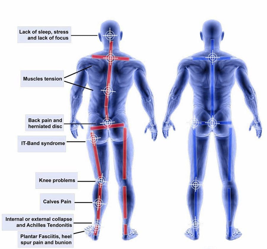
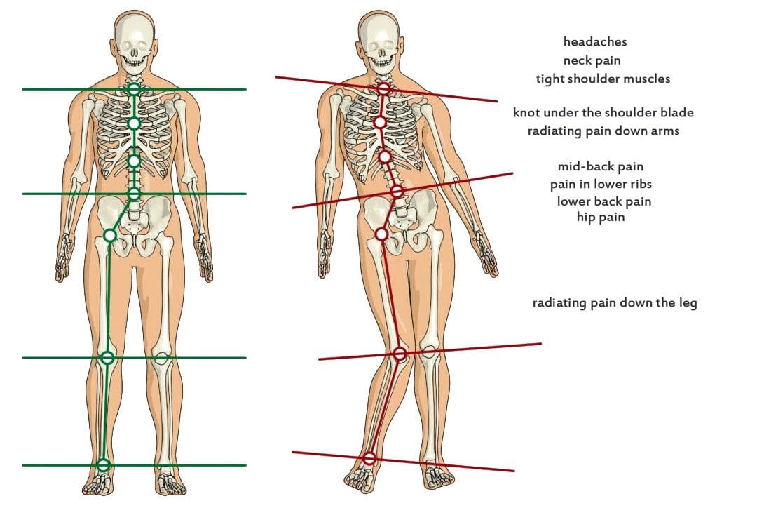
The back provides the strength for physical activities. Back pain affects daily life and can have various causes. Unhealthy posture, walking, twisting, turning, bending, and reaching can contribute to back problems that result in pain. According to the CDC, 39% of adults report living with back pain (Centers for Disease Control and Prevention, 2019). Improper footwear can also contribute to back pain. Selecting footwear carefully can help bring pain relief and help maintain spinal health. Individuals can enjoy less pain and manage symptoms by choosing shoes that maintain spinal alignment and protect the feet from blunt impact.
Understanding the Back Pain-Footwear Connection
Improper footwear could be the cause of lower back pain. What impacts the bones at the bottom of the neuromusculoskeletal system radiates upward and affects the spine and back muscles. What footwear is used travels upward, impacting gait, posture, spinal alignment, and more. When back problems originate from the feet, these are biomechanical issues. Biomechanics means how the bones, joints, and muscles work together and how changes in external forces impact the body.
Movement
When the feet impact the ground, they are the first extremities to absorb shock for the rest of the body. Individuals will start to walk differently if they have a problem or change in their feet. Wearing shoes with improper support can increase the wear and tear on the muscles and joints, leading to awkward and unnatural movement. For example, consider the difference between standing on tiptoes in high heels and the natural flat-footed state. Well-cushioned shoes help absorb impact and lessen pain sensations. The pressures on each of the joints shift balance, which causes instability problems with less pressure on some and more on others. This creates an imbalance that leads to pain and joint conditions.
Posture
Maintaining a healthy posture is another factor in preventing or alleviating back pain. With the right footwear, the body can maintain a healthier stance and the right curvature throughout the spine, and it helps distribute the weight evenly. This results in decreased stress on ligaments, muscles, and joints. (Harvard Health Publishing. 2014) It’s recommended to see an orthopedist to get to the root of an individual’s condition. For some, a herniated disc, sciatica, automobile collision, fall, unhealthy ergonomics, or a combination, as well as other underlying issues, may be contributing to their back pain.
Shoe Types and Their Impact on The Back
How various shoes impact posture, potentially causing or relieving back pain.
High Heels
High heels can definitely contribute to back pain. They change body posture, causing a domino effect on the spine. The body’s weight is shifted to increase pressure on the balls of the feet, and the spine’s alignment becomes altered. High heels also affect how the ankles, knees, and hips move when walking, balance, and how the back muscles operate, all of which can worsen back pain.
Flat Shoes
Flat shoes may not be the best choice for spinal health. If they lack arch support, they can cause the foot to roll inward, known as pronation. This can contribute to misalignment, which can strain the knees, hips, and lower back. However, they can be a decent choice if they provide arch support. When wearing flat shoes with healthy support, the weight is distributed evenly on the feet and the spine. This helps maintain correct posture, which can help prevent and/or alleviate back pain.
Sneakers, Tennis, and Athletic Shoes
Sneakers, tennis, and athletic shoes can relieve back pain with thorough cushioning and support. Choosing the right ones involves determining the activity that will be done in them. There are tennis, running, basketball, pickleball, skating shoes, and more. Research what features will be needed for the sport or activity. This could include:
Heel cups
Insole cushioning
Wide base
Other features to meet individual foot needs.
It is recommended that athletic shoes be changed every 300 to 500 miles of walking or running or with any signs of unevenness when placed on a flat surface, as worn-out soles and degraded materials can increase the risk of injury and back pain. (American Academy of Podiatric Sports Medicine, 2024). If a certain pair puts the legs, hips, or ankles into an unnatural position or impedes regular movement, it may be time to replace them.
Choosing the Right Shoes
The ideal solution for choosing shoe wear is to get a gait analysis and a review of how you walk and run. Various healthcare professionals may offer this service to tailor each individual’s search for the right shoes for back pain. In gait analysis, individuals are asked to run and walk, sometimes on camera, while a professional notes physical tendencies, like when the foot hits the ground and whether it rolls inward or outward. This provides data on affected posture, movement, pain levels, how much arch support is needed, and what type to wear to help prevent back pain. Once the analysis is complete, it will guide you on what to look for, such as what level of arch support, heel height, or material is best for you.
Injury Medical Chiropractic and Functional Medicine Clinic specializes in progressive, cutting-edge therapies and functional rehabilitation procedures focused on clinical physiology, total health, practical strength training, and complete conditioning. We focus on restoring normal body functions after trauma and soft tissue injuries. We use Specialized Chiropractic Protocols, Wellness Programs, Functional and integrative Nutrition, Agility and mobility Fitness Training, and Rehabilitation Systems for all ages. Our programs are natural and use the body’s ability to achieve specific measured goals rather than introducing harmful chemicals, controversial hormone replacement, unwanted surgeries, or addictive drugs. We have teamed up with the city’s premier doctors, therapists, and trainers to provide high-quality treatments that empower our patients to maintain the healthiest way of living and live a functional life with more energy, a positive attitude, better sleep, and less pain.
For individuals who are getting into exercise, fitness, and physical activity, can knowing how glycogen works help in workout recovery?
Glycogen
When the body needs energy, it draws on its glycogen stores. Low-carbohydrate, ketogenic diets and intense exercise deplete glycogen stores, causing the body to metabolize fat for energy. Glycogen is supplied through carbohydrates in an individual’s diet and is used to power the brain, physical activity, and other bodily functions. The molecules made from glucose are mainly stored in the liver and muscles. What is eaten, how often, and the activity level influence how the body stores and uses glycogen. Restoring glycogen after physical activity or working out is a vital part of the recovery process. The body can quickly mobilize glycogen from these storage sites when it needs fuel. Eating enough carbohydrates to reach health goals and activity levels is essential for success.
What Is It
It is the body’s stored form of glucose or sugar.
It is stored in the liver and muscles.
It is the body’s primary and preferred energy source.
It comes from carbohydrates in foods and drinks.
It is made from several connected glucose molecules.
Production and Storage
Most carbohydrates eaten are converted to glucose, which becomes the body’s main energy source. However, when the body doesn’t need fueling, the glucose molecules become linked chains of eight to 12 glucose units, forming a glycogen molecule.
Process Triggers
Eating a carbohydrate-containing meal will raise blood glucose levels in response.
Increasing glucose signals the pancreas to produce insulin, a hormone that helps the body’s cells take up glucose from the bloodstream for energy or storage.
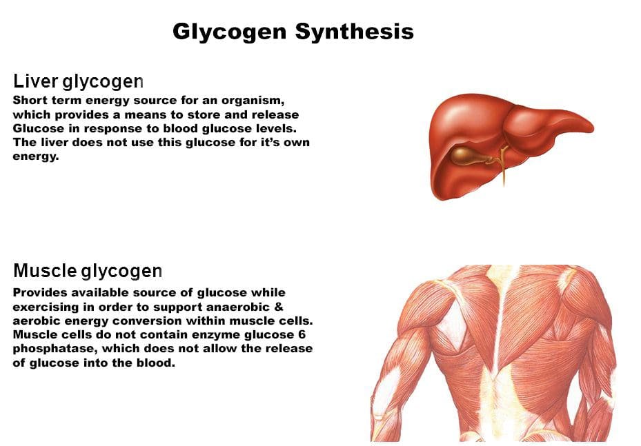
Insulin activation causes the liver and muscle cells to produce an enzyme called glycogen synthase, which links glucose chains together.
With enough glucose and insulin, glycogen molecules can be delivered to the liver, muscles, and fat cells for storage.
Since most glycogen is found in the muscles and liver, the amount stored in these cells varies depending on activity level, how much energy is burned at rest, and the foods eaten. The muscles primarily use glycogen stored in the muscles, while glycogen stored in the liver is distributed throughout the body, mainly to the brain and spinal cord.
Body Usage
The body converts glucose to glycogen through a process called glycogenesis. During this process, various enzymes help the body break down glycogen in glycogenolysis so the body can use it. The blood has a set amount of glucose ready to go at any given time. The insulin levels also drop when the level begins to decline, either from not eating or burning glucose during exercise. When this happens, an enzyme known as glycogen phosphorylase starts breaking the glycogen down to supply the body with glucose. Glucose from liver glycogen becomes the body’s primary energy. Short bursts of energy use glycogen, whether during sprints or heavy lifting. (Bob Murray, Christine Rosenbloom, 2018) A carbohydrate-rich pre-workout drink can provide energy to exercise longer and recover quicker. Individuals should eat a post-workout snack with a balanced amount of carbohydrates to replenish glycogen stores. The brain also uses glucose for energy, with 20 to 25% of glycogen going toward powering the brain. (Manu S. Goyal, Marcus E. Raichle, 2018) Mental sluggishness or brain fog can develop when not enough carbohydrates are consumed. When glycogen stores are depleted through exercise or insufficient carbs, the body can feel fatigued and sluggish and perhaps experience mood and sleep disturbances. (Hugh S. Winwood-Smith, Craig E. Franklin 2, Craig R. White, 2017)
Diet
What foods are eaten and how much physical activity an individual does also influence glycogen production. The effects can be acute if one follows a low-carb diet, where carbohydrates, the primary source of glucose synthesis, are suddenly restricted.
Fatigue and Brain Fog
When first starting a low-carb diet, the body’s glycogen stores can be severely depleted and individuals may experience symptoms like fatigue and brain fog. (Kristen E. D’Anci et al., 2009)
The symptoms begin to subside once the body adjusts and renews its glycogen stores.
Water Weight
Any amount of weight loss can have the same effect on glycogen stores.
Initially, individuals may experience a rapid drop in weight.
Over time, weight may plateau and possibly increase.
The phenomenon is partly due to glycogen composition, which is also water. Rapid glycogen depletion at the onset of the diet triggers the loss of water weight. Over time, glycogen stores are renewed, and the water weight returns. When this happens, weight loss can stall or plateau. Fat loss can continue despite the short-term plateau effect.
Exercise
If undertaking a strenuous exercise routine, there are strategies to help avoid decreased performance that may be helpful:
Carbo-loading
Some athletes consume excessive amounts of carbohydrates before working out or competing.
Extra carbohydrates provide plenty of fuel.
The method has fallen out of favor as it can lead to excess water weight and digestive issues.
Glucose Gels
Energy gels containing glycogen can be consumed before or as needed during an event to increase blood glucose levels.
For example, energy chews are effective supplements for runners to help increase performance during extended runs.
Low-Carb Ketogenic Diet
Eating a diet high in fat and low in carbohydrates can put the body in a keto-adaptative state.
In this state, the body begins to access stored fat for energy and relies less on glucose for fuel.
At Injury Medical Chiropractic and Functional Medicine Clinic, our providers use an integrated approach to create personalized care plans for each individual, often including Functional Medicine, Acupuncture, Electro-Acupuncture, and Sports Medicine principles. Our goal is to restore health and function to the body.
Sports Nutrition and Sports Dietician
References
Murray, B., & Rosenbloom, C. (2018). Fundamentals of glycogen metabolism for coaches and athletes. Nutrition reviews, 76(4), 243–259. doi.org/10.1093/nutrit/nuy001
Goyal, M. S., & Raichle, M. E. (2018). Glucose Requirements of the Developing Human Brain. Journal of pediatric gastroenterology and nutrition, 66 Suppl 3(Suppl 3), S46–S49. doi.org/10.1097/MPG.0000000000001875
Winwood-Smith, H. S., Franklin, C. E., & White, C. R. (2017). Low-carbohydrate diet induces metabolic depression: a possible mechanism to conserve glycogen. American journal of physiology. Regulatory, integrative and comparative physiology, 313(4), R347–R356. doi.org/10.1152/ajpregu.00067.2017
D’Anci, K. E., Watts, K. L., Kanarek, R. B., & Taylor, H. A. (2009). Low-carbohydrate weight-loss diets. Effects on cognition and mood. Appetite, 52(1), 96–103. doi.org/10.1016/j.appet.2008.08.009
For individuals who are dealing with back pain and problems, could knowing how to improve and maintain intervertebral disc health help alleviate symptoms?
Intervertebral Disc Health
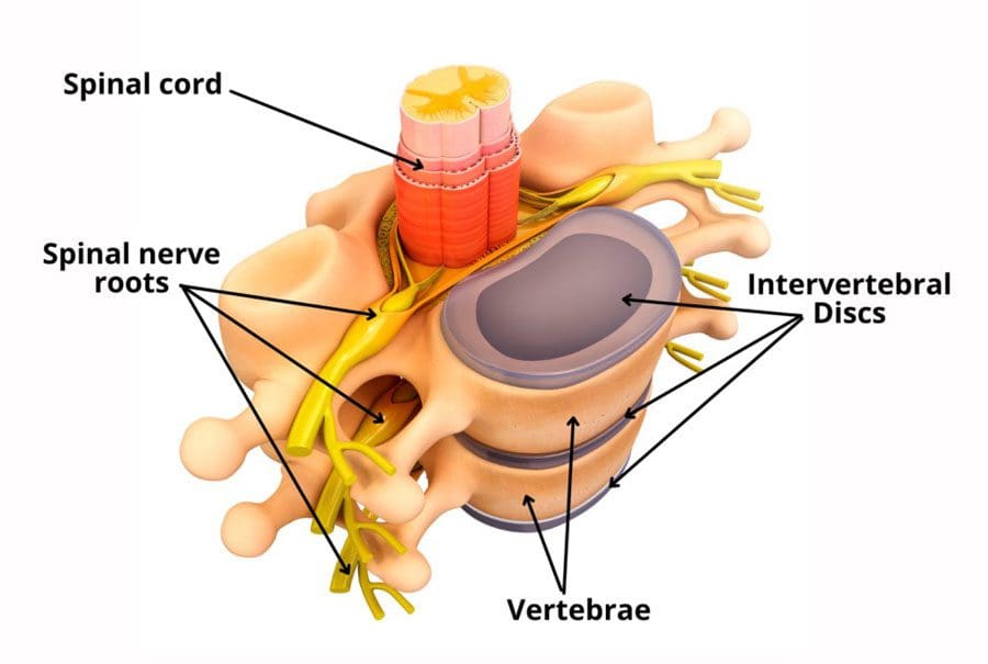
The spinal column comprises 24 movable bones and 33 bones called vertebrae. The vertebral bones are stacked on top of each other. The intervertebral disc is the cushioning substance between the adjacent bones. (Dartmouth. 2008)
Bones
The vertebral bones are small and round in an area called the vertebral body. In the back is a bony ring from which protrusions extend and arches and pathways are formed. Each structure has one or more purposes and includes: (Waxenbaum JA, Reddy V, Williams C, et al., 2023)
Stabilizing the spine.
Providing a space for the connective tissue and back muscles to attach.
Providing a tunnel for the spinal cord to pass through cleanly.
Providing a space where nerves exit and branch out to all areas of the body.
Structure
The intervertebral disc is the cushioning that sits between the vertebrae. The design of the spine allows it to move in various directions:
Flexion or bending
Extension or arching
Tilting and rotation or twisting.
Powerful forces act upon and influence the spinal column to produce these movements. The intervertebral disc absorbs shock during movement and protects the vertebrae and spinal cord from injury and/or trauma.
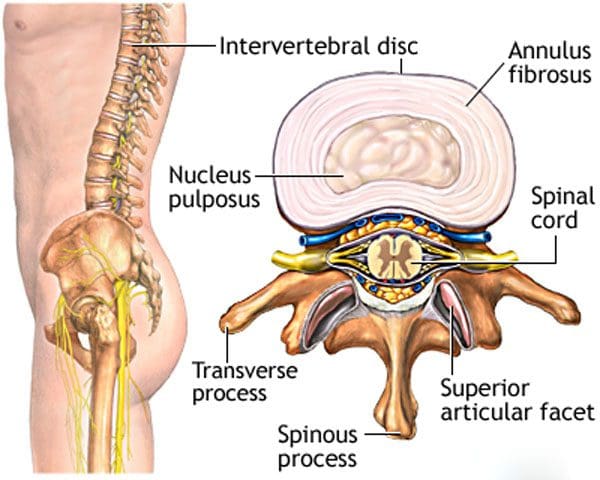
Ability
On the outside, strong woven fiber tissues form an area called the annulus fibrosis. The annulus fibrosis contains and protects the softer gel substance in the center, the nucleus pulposus. (Y.S. Nosikova et al., 2012) The nucleus pulposis provides shock absorption, flexibility, and pliability, especially under pressure during spinal movement.
Mechanics
The nucleus pulposus is a soft gel substance located in the center of the disc that allows elasticity and flexibility under stress forces to absorb compression. (Nedresky D, Reddy V, Singh G. 2024) The swivel action alters the tilt and rotation of the vertebra above and below, buffering the effects of spinal motion. The discs swivel in response to the direction the spine moves. The nucleus pulposus is made mostly of water, which moves in and out through small pores, acting as byways between the vertebra and disc bone. Body positions that load the spine, like sitting and standing, push the water out of the disc. Lying down on the back or in a supine position facilitates water restoration into the disc. As the body ages, the discs lose water/dehydrate, leading to disc degeneration. The intervertebral disc has no blood supply, which means that for a disc to receive necessary nutrition and for waste removal, it must rely on water circulation to stay healthy.
Care
Some ways of maintaining intervertebral disc health include:
Paying attention to posture.
Changing positions frequently throughout the day.
Exercising and moving around.
Applying correct body mechanics to physical activities.
Sleeping on a supportive mattress.
Drinking plenty of water.
Eating healthy.
Maintaining a healthy weight.
Drinking alcohol in moderation.
Quitting smoking.
At Injury Medical Chiropractic and Functional Medicine Clinic, we treat injuries and chronic pain syndromes by improving an individual’s ability through flexibility, mobility, and agility programs tailored for all age groups and disabilities. Our chiropractic team, care plans, and clinical services are specialized and focused on injuries and the complete recovery process. Our areas of practice include Wellness & Nutrition, Acupuncture, Chronic Pain, Personal Injury, Auto Accident Care, Work Injuries, Back Injury, Low Back Pain, Neck Pain, Migraine Headaches, Sports Injuries, Severe Sciatica, Scoliosis, Complex Herniated Discs, Fibromyalgia, Chronic Pain, Complex Injuries, Stress Management, Functional Medicine Treatments, and in-scope care protocols. If other treatment is needed, individuals will be referred to a clinic or physician best suited to their injury, condition, and/or ailment.
Beyond the Surface: Understanding the Effects of Personal Injury
Waxenbaum, J. A., Reddy, V., Williams, C., & Futterman, B. (2024). Anatomy, Back, Lumbar Vertebrae. In StatPearls. www.ncbi.nlm.nih.gov/pubmed/29083618
Nosikova, Y. S., Santerre, J. P., Grynpas, M., Gibson, G., & Kandel, R. A. (2012). Characterization of the annulus fibrosus-vertebral body interface: identification of new structural features. Journal of anatomy, 221(6), 577–589. doi.org/10.1111/j.1469-7580.2012.01537.x
IFM's Find A Practitioner tool is the largest referral network in Functional Medicine, created to help patients locate Functional Medicine practitioners anywhere in the world. IFM Certified Practitioners are listed first in the search results, given their extensive education in Functional Medicine


 Shoe Types and Their Impact on The Back
 Ability


