Hypothyroidism is a disease caused when the thyroid gland doesn’t produce enough thyroid hormones. There are treatments available, however, one is used more commonly than others: thyroid hormone replacement treatment.
What is thyroid hormone replacement treatment?
To understand the purpose of thyroid hormone replacement treatment, you need to comprehend the interaction of T4 and T3, both essential thyroid hormones which are often affected with hypothyroidism or other thyroid diseases.
T3 and T4
The full name of T3 is triiodothyronine, and T4’s full name is tetraiodothyronine or thyroxine. T3 and T4 control the metabolism of your body. Then your metabolism slows down if you do not have enough of these. Your metabolic rate dictates how quickly food is processed by you, how fast your heart beats, how fast it is possible to think as well as how much heat your body generates. Essentially, T3 and T4 are in control of how energy is used by your body.
However, T3 and T4 aren’t equivalent in power; T3 is the more active hormone of both. Taking T4 hormone is considered the standard treatment for hypothyroidism while T3 is stronger. The cause of this is due to the fact that the majority of the T3 in our bodies used to become T4. They provide an iodine atom up to socialize with these cells when T4 hormones come into contact with other cells in the blood vessels. If T4 loses an iodine atom, it becomes T3.
When this T4 into T3 conversion occurs, T3 then communicates the metabolic “message” into the other cells throughout the body. Of taking only T4 therapy, the advantage is that you’re letting your body to perform some of the activities which is taking T4 and changing it. The half life of T4 is also longer than the T3 (7 times versus 24 hours), that means that it is going to remain for a longer period in your body after ingestion.
Thyroid Hormone Replacement Therapy Objective
If you’re prescribed a type of thyroid hormone replacement therapy, the objective is to compensate for the lack of hormone secreted from the thyroid gland. You may have a dose of T4 from a taken pill.
But it’s important to see that every patient’s treatment is different. There is no cookie-cutter dosage or therapy plan in regards to thyroid hormone replacement treatment. The way the hormones are absorbed by the human body, together with the quantity is varied. Your treatment plan will be individualistic. As such, you should expect a certain amount of experimentation when it comes to locating form and the dose of treatment that works best for you.
Though artificial T4 supplements would be the most prescribed type of thyroid hormone replacement therapy, there are a variety of forms, including monster thyroid supplements. Synthetic T3 is sometimes given after thyroid surgery, when awaiting the ablation in case of cancer as part of treatment in certain conditions.
Thyroid hormone replacement treatment is a really individualized therapy procedure, and it’s highly effective when prescribed correctly. The goal of thyroid hormone replacement treatment, in most cases, would be to normalize your thyroid gland (TSH) levels. Your healthcare professional and you will go over what treatment choice will alleviate your symptoms that are hypothyroid, letting you live a normal life once more.
The scope of our information is limited to chiropractic and spinal injuries and conditions. To discuss options on the subject matter, please feel free to ask Dr. Jimenez or contact us at 915-850-0900 .�
By Dr. Alex Jimenez
Additional Topics: Wellness
Overall health and wellness are essential towards maintaining the proper mental and physical balance in the body. From eating a balanced nutrition as well as exercising and participating in physical activities, to sleeping a healthy amount of time on a regular basis, following the best health and wellness tips can ultimately help maintain overall well-being. Eating plenty of fruits and vegetables can go a long way towards helping people become healthy.
Hypothyroidism is evaluated and diagnosed by a physician, your primary care doctor or an endocrinologist. Many factors, signs, and symptoms are taken into consideration when hypothyroidism is diagnosed.
How is hypothyroidism diagnosed?
A diagnosis is reached after a thorough review of the patient’s symptoms, family and medical history, risk factors, physical examination, and effectively, a blood test. There are many types of blood tests, which the most authoritative one is known as the TSH test (thyroid-stimulating hormone). However, in some cases, healthcare professionals may refer patients to receive a total T4 or T4, free T4 index, or even thyroxine to aid in the diagnosis.
Why Hypothyroidism is not Diagnosed on Symptoms Alone
Lots of the signs of hypothyroidism are fairly frequent complaints found in people with a normally functioning thyroid gland, so it can be tough to decipher if the symptoms are linked to the thyroid gland. Among the best ways to find out whether your symptoms might be related to a thyroid condition is to consider how long you have been experiencing them. For example, have you felt cold when others were warm? Did you just begin to notice decreased energy? It might be associated with a thyroid issue if you are beginning to notice new signs and symptoms. But only a specialized healthcare professional (eg, endocrinologist) can diagnose a thyroid issue.
Medical and Family History
It is important to give your doctor as many details as you can about your own personal medical history, in addition to family history (eg, mom had eczema). Make sure you talk about:
Your overall state of health, particularly any changes you’ve noticed on your general well-being.
Your family’s health history, especially if a near relative was diagnosed with hypothyroidism (or any other thyroid-related issues).
Whether you’ve ever had thyroid surgery, or radiation into your own neck to deal with cancer.
Any medications you could be taking that could cause hypothyroidism (eg, amiodarone, lithiumion, interferon alpha, interlukin-2, or even earlier chemotherapy).
Physical Evaluation
Your doctor will perform a thorough examination and look for physical signs of hypothyroidism, such as:
Proof of dry skin
Swelling around the eyes and legs
Slower reflexes
Slower heartbeat
Blood Tests
Hypothyroidism can be diagnosed using different blood tests such as:
TSH Evaluation
A thyroid-stimulating hormone or TSH is a blood test that measures the amount of T4 (thyroxine) that the thyroid gland has been indicated to create. In case you have an abnormally significant degree of TSH, it might indicate you have hypothyroidism.
T4 (thyroxine) Evaluation
The thyroid gland produces T4 (thyroxine). The T4 along with the free T4 index are blood tests which, in conjunction with a TSH test, can let your doctor know your thyroid is functioning.
The adrenal gland tells the thyroid how much thyroxine to produce through signaling by TSH. There are cells from the pituitary gland that determine what your body’s “set point” is. Your collection point is that the normal array of TSH as determined by your thyroid gland that your body needs.
As blood flows throughout the pituitary gland, the very same cells detect if there are sufficient T4 levels in the body. The pituitary sends the amount of TSH into the thyroid to maintain levels in the standard range in case your T4 amount is sufficient. If your level is too low, the pituitary sends TSH outside telling the thyroid to make more T4. In case your T4 level is too high, the pituitary sends TSH that is less out, then telling the thyroid to make less T4.
Normal and Abnormal TSH Ranges
0.4 mU/L to 4.0 mU/L is considered the reference array (there may be slight variation depending on the laboratory), and people that have a normally functioning thyroid gland usually fall within this range.
If TSH measures > 4.0 mU/L, a second evaluation (T4) is done to verify the results. TSH p4.0/mU/L using a very low T4 level indicates hypothyroidism.
If your TSH is > 4.0 mU/L along with your T4 level is normal, this may prompt your physician to test your serum anti-thyroid peroxidase (anti-TPO) antibodies. When these antibodies are found, it may signal an autoimmune thyroid disease, which is a risk factor for developing hypothyroidism. In case you have those anti-bodies, your doctor will perform and TSH test at least once each year.
An easy way to remember how the thyroid works is to think about supply and demand. The TSH rises as the T4 level drops. The TSH drops as the T4 level rises. But not everyone with hypothyroidism has elevated levels of TSH. If the pituitary is not working properly, perhaps it does not send out regular TSH levels. But if the quantity of TSH is off, the thyroid will not make the perfect quantity of T4. This is rare and is called secondary or central hypothyroidism.
The scope of our information is limited to chiropractic and spinal injuries and conditions. To discuss options on the subject matter, please feel free to ask Dr. Jimenez or contact us at 915-850-0900 .
By Dr. Alex Jimenez
Additional Topics: Wellness
Overall health and wellness are essential towards maintaining the proper mental and physical balance in the body. From eating a balanced nutrition as well as exercising and participating in physical activities, to sleeping a healthy amount of time on a regular basis, following the best health and wellness tips can ultimately help maintain overall well-being. Eating plenty of fruits and vegetables can go a long way towards helping people become healthy.
Genetic: Integrative and functional medicine came to the forefront for many medical practitioners and patients alike when they
became dissatisfied with traditional medicine�s sole focus on what was considered �science-based� treatment approaches. Traditional medicine�s viewpoint of dealing with symptoms in isolation from the rest of a patient�s body, mind, and spirit can be too confining when it comes to certain conditions.
This evolution to a more function-centered approach as opposed to a disease-centered way of seeing the whole person has led to improved healthcare. It also looks at prevention, not simply illness and at living in a healthy state, not simply disease-free.
What Is Integrative & Functional Medicine?
Practitioners of integrative and functional medicine take into consideration genetic, environmental, and lifestyle issues when listening to their patients describe the symptoms plaguing them. Their inclusion of these issues makes the process more of a natural medicine approach.
With the dramatic increase in chronic illness conditions and the lack of training traditional physicians have in dealing with these conditions, the move into integrative and functional medicine is needed.
Many of these chronic illness conditions have a genetic component that, along with environmental and lifestyle factors, lead to serious limitations on people�s lives. This shows the importance of the individual biochemical and genetic aspects of each person on his or her health.
This other approach in medicine realizes the necessity of considering nutrition, exercise, diet, and genetics in evaluating and remediating chronic illness conditions. The use of genetic testing in integrative and functional medicine is one way to take all of these factors into account.
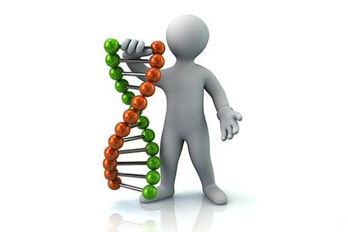
SNPs & Integrative & Functional Medicine
Upon completion of the mapping of the human genome, we know there are 20-25,000 genes in each genome. With this knowledge came the information that there are over 80 million variants in the human genome.
These variants are comprised in part of single nucleotide polymorphisms (SNPs) and deletions or insertions in the genome. It is these SNPs that provide significant health information to providers of integrative and functional medicine to prevent or alleviate chronic illness conditions.
Knowing the presence of and placement of SNPs through genetic point mutation testing allows evaluation of the susceptibility to develop many of the chronic illness conditions that affect people today. In addition, this kind of testing helps pinpoint relevant SNPs and their corresponding metabolic markers in individuals.
Testing of this kind provides targeted interventions through the use of traditional medicine approaches as well as supplementation through integrative and functional medicine approaches. Monitoring of individuals� progress is also made easier with genetic testing by measuring metabolic markers found in the original tests over a period of time.
Individual monitoring of this type is necessary when this kind of personalized intervention and supplementation is used. If there is an overload of either medications or supplementations, there can be an impact on the performance of metabolic processes that can lead to side effects. These side effects can influence functions and responses, such as the immune response.
Individual SNPs will determine how well medications and supplements are working.
Genetic Testing In Relation To Diet & Weight Loss
Integrative and functional medicine practitioners not only deal with illness, they also provide health and wellness evaluations. Current research has shown how important a role genetics plays in the prevention of many chronic health conditions.
Genetic testing can show vulnerabilities to conditions and suggest options for individuals. This kind of testing can also provide valuable information concerning how individuals can respond to different attempts to live more healthy lives.
Genetic testing has been shown to be effective in several areas: diet, eating behavior traits, nutritional needs, exercise, body and weight, and metabolic health. For each of these areas, there are certain genetic markers that can provide information regarding how genetics will affect each of these areas.
Diet
People are seemingly obsessed with weight. How to lose it and keep it off, how to re-distribute it to look more attractive. Professionals in integrative and functional medicine are approached regularly for help in this area.
Everyone knows it�s hard for some people to lose weight on any kind of diet, while others can lose weight any time they want. It�s not just due to lack of willpower that people don�t lose the weight they want. It may also be due to genetics.
Research has shown about 88 percent of people have bodies that resist burning fat through low-intensity exercise. Most people will gain weight if they eat almost any carbs (about 45 percent of people) or almost any fat (about 39 percent of people).
The reason for this is a diet and type of exercise matched to specific genotype lead to weight loss. These diets and exercise types are not the same for everyone.
For example, let�s look at adrenoceptor Beta 3 (ADRB3) with an SNP on rs4994. There are different variations of this gene. If you are either an AA or TT genotype, you have what is called a genetic privilege and just about any kind of exercise will work for you. On the other hand, if you don�t have either of these AA or TT genotypes, this is a genetic disprivilege and only a high-intensity type exercise will help you lose weight.
Further analysis of other genes and SNPs can tell you the type of diet, either low carb or low fat, that will work best for you. In fact, using a diet matched to your genetics can result in a loss of two and half times as much weight as a diet not matched to genetics.
In addition to choosing the right diet to lose weight, choosing the right diet may also help you avoid developing a chronic health condition. Research has shown diet to be implicated in many chronic illness conditions, so genetic testing to determine your specific vulnerability to illnesses and your response to particular foods may help prevent them.
Knowing your predisposition to illnesses can lead to targeted dietary and lifestyle changes that may modify any existing conditions and help prevent future developments. Future research may bring more information regarding bioavailable components in foods that can aid in alleviating health issues.
COMT & CYP19 Genes
Research has identified certain genes that work together and appear to show that some people retain fat regardless of, or in spite of, exercise.
In one study, researchers found two genes, COMT and CYP19 that appeared to be involved in patterns of fat loss and exercise. Having one CYP19 gene and variants of that gene did not affect fat, intra-abdominal fat, or total fat. However, having two of these genes seemed to be related to slightly more decrease in body mass index and significantly more decrease in total fat and percentage of body fat.
The researchers also found that having one genotype of the COMT gene and one copy of the CYP19 gene seemed related to significant loss of BMI, total fat, and percentage of body fat.
Why and how these genes and combinations work isn�t known yet. More research is needed to determine this. Other research suggests women with a specific CYP19 variant may also have increased levels of estradiol and estrone which may make it harder for them to lose fat through exercise.
Environmental Factors
Weight loss or gain is not solely at the mercy of your genetics however. A combination of genetics and environment is likely behind your success or failure regarding your weight loss attempts.
The thinking of professionals is divided on the subject of genetics versus environment/lifestyle choices. One set of these professionals regards environment to be the telling component. They point to the teaching over the years that food is a reward for good performance at anything. This, combined with constant reminders about food that are around us all the time, makes it hard for some people to lose weight and/or keep it off.
Others believe losing weight and keeping it off are more related to biological functions. They have found people to be metabolically different after losing up to ten percent of their body weight. Their brains also seem to respond to food differently. The emotional response to food is greater, but the brain regions that deal with food restraint are less active. This sets up the person to regain the weight lost.
Further research into why people lose weight and maintain that loss will be needed. Some of that research has to be on the genetic basis of weight loss.
Eating Behavior
Integrative and functional medicine practitioners view eating behavior as important for overall health.�These behaviors include snacking behavior, feelings of satiety, craving for sweets, desire for food or certain foods, and the disinhibition of eating.
Nutrigenetics and nutrigenomics are two new fields of study related to how genes affect our diet and how our diet affects genes, respectively. Obesity, cancer, and heart disease are three of the health conditions most investigated in these two new fields.
One study involving these new fields showed the bitter taste gene receptor hTAS2R38 to be involved in tasting glucosinolates, found in some fruits and vegetables. Three genotypes in this gene receptor have been identified: PAV/PAV, PAV/AVI, and AVI/AVI.
Those individuals with PAV/PAV are said to be supertasters. They are very sensitive to bitter tastes in some foods and in some man-made compounds used in research. People with PAV/AVI are considered medium tasters. They can taste bitter in the research compounds, but not as much as the supertasters. Individuals with AVI/AVI are labeled non-tasters. They don�t taste bitter in the research compounds.
While it�s difficult to completely understand why these differences occur, it does appear they can make a difference in people�s diets. It could be that people who taste bitter greatly or somewhat will avoid certain vegetables that contain this bitter taste. Vegetables like kale and broccoli have this taste.
In this way, genetics have a significant influence on eating behavior.
Research indicates taste is only one of the ways genetics affects eating behavior. Caloric intake, meal size, and frequency of eating also appear to be affected. People�s desire for fats, carbohydrates, or proteins also may be influenced by genetics.
Research has found apolipoprotein A-II (APOA2) to be implicated in this kind of desire. Three variants in this gene, TT, TC, and CC, have been isolated as factors affecting the choice of fats, carbs, and proteins. One study showed both men and women who had the recessive CC chose more fat and protein and fewer carbs than either of the T alleles. The CC group ate about 200 more calories than the other group and tended to develop obesity more frequently.
It appears that APOA2 may affect not only food choices but also feelings of satiety.
Nontasters seem to prefer and seek out fats and flavors, so dieting may be more difficult for them to stick with and lose weight. Supertasters, on the other hand, enjoy a variety of foods, especially those that are spicy and robust. This may help them with diets.
Understanding the factors that appear to influence eating behaviors has gained importance with the tremendous increase in obesity in the U.S. and around the world, along with diabetes and cardiovascular disease. Eating behavior must be seen as a complex inter-relationship among psychological, cultural, physical, and genetic factors that influence the choice of foods, the amount of food intake, caloric intake, and timing of meals.
Regulating Eating Behavior
Clearly, taste affects food choices as seen in the discussion above. Another of the bitter receptors, TAS2R5, may also assist in regulating eating behavior. Alcohol dependence has been associated with an SNP in this receptor, along with another receptor, TAS2R16. These research findings seem to indicate variants in the TAS2R gene to be associated with ingestive behavior.
Genetic influence over meal amounts, how often people eat, and the timing of meals is a new area of study and may involve digestive neuroendocrine hormones such as CCK, leptin, and ghrelin. Studies are underway investigating the effects of these hormones on pathways that influence eating behavior.
A gene with a strong association with the risk of obesity, FTO, appears to contribute to obesity by downregulating leptin production in adipocytes. Adiposity and satiety appear to be associated with a fairly common variant, rs9939609. One study showed the A allele of rs9939609 to influence post-meal feelings of satiety and possibly to influence the excess caloric intake seen in men and women with high BMIs.
A gene involved in the detoxification of nutrients during digestion, AKR1B10, also appears to play a role in influencing human eating behavior.
Nutritional Needs & Genetic Testing
Another area in which integrative and functional medicine practitioners use genetic testing is in�determining nutritional needs of their patients. As we have seen previously, genetic variants have an effect on taste and thus on nutrition. When people choose foods that �fit� their tastes but are short on nutrients, their health suffers. People also appear to have genetic responses to some supplements, such as some of the B vitamins and vitamin C.
The impact of nutrition is a lifetime factor, and practitioners of integrative and functional medicine evaluate nutritional needs closely. Any genetic variant that leads to abnormal nutritional requirements would likely be incompatible with survival. For example, miscarriage is more likely in a woman whose fetus has two alleles that negatively affect the use of any given nutrient than a woman whose fetus just has the common functional variants.
Several studies have isolated genes and alleles that affect nutrients and their utilization. For example, an SNP (Ala222Val) in the methylenetetrahydrofolate reductase (MTHFR) gene leads to a significant alteration in folate metabolism, increasing the risk of neural tube defects (NTDs) and cardiovascular disease, but lowering the risk of colon cancer. Increasing folate intake lowers the risks of developing serious health conditions.
Research has found other SNPs that alter homocysteine metabolism and folate uptake and transport. SNPs in enzymes that affect utilization and metabolism of vitamin B12 seem to be associated with NTDs and the possible development of Down syndrome and colon cancer.
SNPs in the vitamin D receptor may be associated with asthma in both children and adults. Lipid pathways, alcohol metabolism, and lactose metabolism appear to be affected by SNPs in other genes, also. A beneficial effect of these SNPs in the ancestors of certain ethnic groups or ancestral subpopulations may have been present, even though they tend to carry the risk of an adverse outcome today.
Environmental changes have been shown to bring a previously silent allele into a role as a disease allele. The aldolase B enzyme metabolizes fructose and was silent even with a high number of polymorphisms. In recent times, when fructose was added to foods as a sweetener, the polymorphisms began presenting as disease alleles.
Integrative and functional medicine professionals can use this information to guide their patients into more healthy lives.
Genetic Testing & Exercise
Integrative and functional medicine also uses genetic testing to determine the best types of exercise for different people and to explore the likelihood of injuries of several kinds in athletes. This latter area of research and clinical practice can help reduce the number and severity of athletic injuries for adult and child athletes.
While there have been some gene variants associated with athletic ability, none have been shown to be predictive to any degree. Research in this area is promising for decreasing serious injury in young athletes. But to date, little scientific information regarding a genetic variation in young athletes is available.
Genetic testing as a way of choosing which athlete to select for a particular sport is increasing. However, little evidence has been found to show it is more accurate than traditional ways of selecting candidates. The ethics of this kind of testing for young athletes has been brought into question.
ACE Genes
Two genes and the SNPs associated with them have been examined in several population samples and thus have robust findings. The ACE I/D polymorphism was first found to be associated with human performance several years ago. This gene is part of the renin-angiotensin system that controls blood pressure through its effect on the regulation of body fluid levels.
The ACE I allele lowers ACE activity in serum and tissue. The D allele increases ACE activity in serum and tissue. The ACE I/I genotype has been shown over and over again to indicate performance endurance and greater efficiency in exercise. The ACE DD genotype has been shown to indicate strength and power performance levels.
This ACE I/D genotype does not appear to have predictive ability in Kenyan athletes, suggesting the confounding influence of ethnicity or geography.
ACTN3 Gene
The ACTN3 is strongly associated with the protein alpha-actinin-3. This protein is involved exclusively in fast type II muscle fibers that are used in explosive activities. SNP R577X indicates a stop codon at position 577 rather than an arginine (R). An R allele puts athletes at an advantage in power sports. A study of the ACTN3 R577X variant in elite European athletes showed those in power event to be 50 percent less likely to have the XX variant and those involved in endurance events to be 1.88 times more likely to have the XX variant. For world-class endurance athletes, the odds of having the XX variant were 3.7 times larger when compared with lower-level athletes. It appears the ACTN3 gene is more important at the upper levels of sports.
While research shows the effects of the ACTN3 gene on athletic performance, especially in higher class athletes, the effects in the general population were negligible. It is unclear just what the association of this gene in the general population and choice of athletic activities in this population might be.
Resistance to injury and the ability to recover from injuries are also very important factors not only in professional sports but also for the general population. The emphasis on physical activity currently seen in the culture increases the risk of injury and the need for information regarding recovery.
Concussions and tendinopathies have been studied fairly extensively. Information on these two growing areas of injury among young athletes has been valuable for integrative and functional medicine specialists.
These two areas are important due to the long-lasting effects of both on young athletes. Research and clinical practice have shown the effects of concussion to linger into old age where they can increase the cognitive decline normally seen at that time of life.
APOE4 Gene
A better understanding of the genetic aspects of injury and recovery can help practitioners of integrative and functional medicine to both protect those young athletes at risk for injury and to better treat those who suffer injuries.
Regarding concussion, the gene most studied is APOE and its three alleles. The APOE e4 allele has been implicated in the development of Alzheimer�s Disease. This allele has been studied recently to determine its association, if any, with concussion risk and outcomes of traumatic brain injury. To date, the results are not clear.
Some findings have shown people with the e4 allele to have less favorable outcomes from traumatic brain injuries and boxers with this allele had higher chronic brain injury scores. These findings are consistent with e4 being a risk allele. However, one study of college athletes with the e4 allele did not find them to be more likely to suffer a concussion. Another study showed the e4 allele was not associated with poorer head trauma outcomes in children.
Another APOE variant, G-219T, has been linked with increased risk of concussion in athletes. Those athletes with the TT genotype compared to those with the GG genotype had a risk of concussion three times larger. A weak association was found in that same study between the tSer53Pro polymorphism in MAPT, the tau-protein encoding gene, and risk of concussion.
Collagen Genes, Integrative &Functional Medicine
Collagen is the primary component of tendons and ligaments, thus it is connected very closely with research into tendinopathies. It is no surprise that two variants in genes coding for collagen (COL1A1 and COL5A1) have been shown to suggest increased risk of injury to tendons. MMP3, a gene associated with connective tissue wound repair and the gene encoding TNC, an extracellular matrix protein, have also been implicated in increased risk of tendinopathies.
These are preliminary studies that need replication and further study to validate the findings.
Genetic Testing & Metabolic Health
Metabolic syndrome and metabolic health have been studied extensively due to metabolic syndrome being a major risk factor for the development of diabetes mellitus 1 and cardiovascular disease. Genetic and environmental factors interrelate in a complex fashion to bring about this condition. A cluster of metabolic abnormalities, including hypertension, dyslipidemia, abdominal obesity, insulin resistance, and impaired glucose tolerance make up metabolic syndrome.
All of the components of metabolic syndrome are highly heritable. Studies have shown links between metabolic syndrome and genes such as PPARg, adiponectin, CD36, and beta receptors.
There has been a considerable investigation into the heritability of metabolic syndrome. One study involved over 2,200 individuals in over 500 family groups. It was the first to identify major genes influencing metabolic syndrome.
Chromosome 3q27 was significantly linked to six factors involved in metabolic syndrome: weight, leptin, insulin, waist circumference, hip circumference, and insulin/glucose ratio. Chromosome 17p12 was strongly linked to plasma leptin levels.
Another study evaluated over 200 SNPs in 110 genes for their effects on coronary artery disease, highly implicated in metabolic syndrome. SNPs in eight of these genes showed association with metabolic syndrome: LDLR, GBE1, IL1R1, TGFB1, IL6, COL5A2, SELE and LIPC.
These genes are described below:
LDLR: Low Density Lipoprotein Receptor gene. It is strongly involved in the homeostasis of cholesterol. Hypercholesterolemia in families has been linked to mutations of this gene.
GBE1: Glycogen Branching Enzyme gene. It is involved in coding the glycogen branching enzyme which aids in glycogen synthesis. Branching of these chains allows a great number of glycosyl units to be stored in a molecule of glycogen.
IL1R1: Interleukin 1 Receptor, Type 1. Interleukin 1 is made up of two proteins, IL1-alpha and IL1-beta, and is a mediator of inflammation.
TGFB1: Transforming Growth Factor, Beta 1. This gene encodes the peptide involved in many functions in cells. Apoptosis may result due to dysregulation of the activation of this gene.
IL6: Interleukin 6 gene. It is a cytokine that regulates the immune response by activating a cell surface signaling assembly. Its production by neoplastic cells has been implicated in the growth of a number of cancers.
COL5A2: Collagen, Type V, Alpha 2. Mutations in the gene may bring on weakened connective tissue throughout the body.
SELE: Selectin E gene. May be involved in the pathogenesis of atherosclerosis.
Some of the more common inherited metabolic conditions include:
Lysosomal storage disorders. These can result in the buildup of toxic substances inside lysosomes in the cells.
Glycogen storage conditions. Sugar storage problems can lead to weakness, low blood sugar, and muscle pain.
Mitochondrial disorders: Can lead to muscle damage.
Peroxisomal disorders: Can lead to a buildup of toxic products of metabolism.
Metal metabolism disorders: Special proteins control levels of trace metals in the blood. A malfunction in these proteins caused by genetic metabolism disorders can lead to toxic levels of metals in the body.
Symptoms of genetic metabolism disorders include:
Low energy levels
Decreased appetite
Abdominal pain
Weight loss
Jaundice
Seizures
From this list of symptoms, it�s easy to see the relationship�of metabolic syndrome and adrenal fatigue. Practitioners of integrative and functional medicine will be faced with patients who present with adrenal fatigue and these similar symptoms. This makes it important for them to understand at least the basics behind Adrenal Fatigue Syndrome (AFS).
Adrenal Fatigue Syndrome
Feelings of fatigue and lethargy are presented more and more frequently in health care professionals� offices. Combined with concentration difficulties, sleep problems, inability to lose weight, feeling your brain is in a fog, fatigue, and lethargy may point to AFS as the basic issue.
AFS is a constellation of many nonspecific symptoms that can become debilitating. The onset of the symptoms is slow and can be missed by traditionally trained professionals.
The symptoms of AFS result from�the body�s normal response to stress�from any source. The hypothalamic-pituitary-adrenal (HPA) axis is set into motion, releasing hormones and other chemicals that are designed to deal with stress. At the end of the axis are the adrenal glands that secrete cortisol, the stress fighting hormone. The purpose of this hormone is to limit the effects of stress on the body.
In normal circumstances, once the stress ceases, the cortisol levels decline and the adrenals get a chance to recover. However, in our stress-filled culture, the stresses continue. This puts the demand on the adrenals at an extreme level. At some point, the adrenals are no longer able to secrete cortisol, which results in damage to the body from the effects of stress.
Levels of inflammation and an increased immune response results. Inflammation has been implicated in many chronic illness conditions. It is at this point that the body begins breaking down from the accumulation of symptoms such as fatigue, brain fog, insulin resistance, and increasing inflammation.
NeuroEndoMetabolic (NEM) Response
The traditional medical viewpoint of addressing individual symptoms and/or organs when working to alleviate illness conditions is simply too mechanistic. A more comprehensive viewpoint is needed in order to effectively deal with symptoms of AFS. The NEM model is such a viewpoint.
The model says it is important to consider organ systems operating in an interrelationship in which whatever affects one organ system affects others as well. In this regard, it is in line with�the integrative and functional medicine viewpoint.
The NEM model is a functional approach that looks at interactions between the individual�s environment and the gastrointestinal, endocrine, and metabolic organ systems, among others. This allows a healthcare practitioner to find the root causes, triggers, immediate causes, and genetic factors involved in a person�s illness condition.
This is a much more comprehensive approach to alleviating people�s symptoms and illness conditions.
Increasing and unrelenting stress is a part of our culture that is detrimental to the health of every individual. The metabolic component of the NEM model added to the neuroendocrine aspect helps professionals to see how localized organ-specific responses and systemic responses are necessary for successfully dealing with stress.
The metabolic component of our stress response is very subtle in the early stages. But the derangements of our metabolism worsen as time goes on and stress doesn�t stop. By the time the stress response reaches stage 3 or 4, these derangements can become debilitating. At the severe stage, they can lead to hypersensitivity to supplements and to paradoxical reactions.
Very significant and debilitating symptoms begin arising. Often, these lead the person to be bed-ridden due to their severity.
AFS & Genetics
A question integrative and functional medicine experts and those who suffer from AFS all want to know is: Can you inherit AFS?
Before answering that question, you need to understand even if you have a gene or several genes that are involved in a health condition like AFS, it doesn�t mean you will automatically get that condition. Before genes can do anything, either positive or negative, to your health, they have to get the signal to �switch on.�
One good thing about that signal is you have quite a bit of control over it. Scientists and researchers have discovered environment, choices you can make, exert significant control over whether genes are turned on or off. This is called gene expression.
Can you choose to switch specific genes on or off? That�s beyond us at this point. What you can do is make good lifestyle choices, good exercise choices, good diet choices and either activate or de-activate genes in this way. Genetic testing as seen in integrative and functional medicine practices is a way to determine your choices in many areas. Which diet works best for you and what exercises will best benefit you can be answered through this kind of testing.
Answering the specific question posed above, �Can you inherit AFS?�, is a complicated process.
Two genes with significant involvement in this answer are MTHFR and COMT. Both are involved with methylfolate. People with mutations in MTHFR don�t have enough methylfolate leading to less adrenaline because of interference in the methylation process. Methylation aids in the production of adrenaline and other hormones.
The other gene, COMT, is involved in the production of hormones and chemicals in the body. Low levels of methylfolate with this gene leads to lower levels of epinephrine and higher levels of norepinephrine.
The lack of methylfolate with both of these genes, especially MTHFR, leads to feelings of fatigue.
When your body is stricken by stress, both your adrenals and MTHFR are affected. This leads to the fatigue felt by those of you who suffer from AFS. The enzyme that produces dopamine and serotonin is also dependent on methylation to work right. Low levels of methylfolate can lead to low levels of both of these neurochemicals which can then lead to low energy and fatigue.
What Can You Do To Improve Energy Levels?
There are some things you can do to aid in increasing energy and improving the work of the two genes mentioned, MTHFR and COMT.
Balance your blood sugar levels by eating three or four small meals per day. These meals should include good grains like quinoa or rice, good carbs, and vegetables. You can add protein from fish or free-range chicken.
Supplements can help support your adrenal glands and the methylation process also. Vitamin B1, B2, and B6 will help. There are usually no side effects from vitamin B1, but if you should begin feeling any itching, notice any rashes, or have trouble breathing, contact your healthcare professional immediately.
Side effects from B2 are also rare. Very yellow urine will be seen, but this is not serious. If you do have any rashes, breathing trouble, or itching, contact your physician at once.
Taken in large doses for a long time, B6 can cause side effects. Headache, nausea, and drowsiness are enough to contact your healthcare professional at once.
Some people try taking methylfolate (5-MTHF), but this is a labor-intensive effort and could bring on some serious side effects if your body is not ready for it. If your body gets overwhelmed by the 5-MTHF, you can feel headaches, irritability, anxiety, and heart palpitations. Get medical help right away for these side effects.
Despite advance testing, it is important to remember that tests are simply data points of alert. A clinical decision should be made after a detailed consideration of the history and state of the body. A shotgun approach to treating abnormal laboratory values is a common clinical mistake and can lead to negative clinical outcomes.
Conclusion
The mapping of the human genome has provided an opportunity for researchers and clinicians alike to consider the roles genes play in health and wellness. Discovering the presence and effects of single nucleotide polymorphisms (SNPs) has increased not only our knowledge of how genes affect health, but also has given us tools to use in preventing and remediating many chronic illness conditions.
Integrative and functional medicine practitioners have been among the professionals to use this information in a practical sense. Whether AFS can be inherited is yet to be seen. Clinically, we do see a strong correlation from one generation to the next.
Genetic testing to examine the working of MTHFR and COMT may be of some help. Diet and supplements can also increase your chances of these two genes working correctly and alleviating some of the symptoms of AFS.
Because genetic testing is still in the very early phase of development, it is important to take all data points with the right perspective and refrain from treating abnormal laboratory numbers while the root cause of the problem can be masked.
� Copyright 2017 Michael Lam, M.D. All Rights Reserved.
Hyperthyroidism and hypothyroidism can cause a variety of muscle or joint-related symptoms. Both hypothyroidism and hyperthyroidism are proven to cause what are known as myopathies, the clinical term for diseases which affect your skeletal muscles. Skeletal muscles are the muscles connected to your bones. A good illustration of skeletal muscles are the quadriceps in your thighs or your biceps in the arm.
How does thyroid disease cause muscle and joint pain?
Myopathies most frequently are seen in what are called the proximal muscles. These are the muscles which are closest to the center of the human body, like the thigh or shoulder. In myopathies brought on by inflammation or metabolic conditions like autoimmune thyroid disorder, white blood cells can attack parts of your muscle and the surrounding blood vessels. Levels of certain biochemical substances may end up accumulating on your muscles, leading to pain or weakness. Different thyroid diseases can also be associated with particular types of muscle and joint issues.
Muscle and Joint Disease With Hypothyroidism
Hypothyroidism could lead to a number of muscular and joint-related symptoms. These signs and symptoms involves muscles that are currently pressing on your nerves, or fluid retention, which leads to the swelling of muscles. Some of the issues seen include:
General muscle fatigue and pain, such as muscle cramps and muscular stiffness
General joint pain, achiness, and stiffness, known as “arthropathy”
Tendonitis in your arms or arms
Carpal tunnel syndrome, which involves tingling, pain, fatigue, achiness, or numbness on your torso, fingers, or forearms. It is due to swelling of membranes which compress a nerve in your forearm
Tarsal tunnel syndrome, similar to carpal tunnel syndrome, causes discomfort, tingling, burning, and other discomfort in the arch of your foot, the base of your foot, and can extend into your toes
Frozen shoulder, also called adhesive capsulitis, causes pain, limited movement, and stiffness
Joint and Muscle Pain With Hyperthyroidism
In hyperthyroidism, such as Graves’ disease, you might experience muscle weakness and tiredness, known as hyperthyroid myopathy. Pain in muscles is much less prevalent in hyperthyroidism. Some people with hyperthyroidism really get rid of muscle strength and tone, a procedure that can be known as “muscle wasting.” Some common complaints include:
Difficulty climbing stairs
Difficulty holding or gripping objects with your palms
Trouble reaching your arms over the head
In some cases, the muscles changed can include those that help you swallow, so you could have any hoarseness or difficulty swallowing.
When the Pain Does Not Go Away
Typically, the majority of these symptoms and ailments generally resolve for the most part with appropriate treatment of your thyroid illness. When muscle and joint pain does not go away with Appropriate thyroid treatment, however, it is time to ask several questions:
If you are hypothyroid, are you getting adequate and suitable treatment? In other words, is your therapy “optimized” or are you undertreated? Fulfilling with a demand for extra T3, or Resolving thyroid hormone replacement, may have to resolve your muscle and joint pain.
If you’re receiving optimum thyroid therapy, and still enduring muscle and joint issues, in the event you get a referral to a rheumatologist for additional evaluation and possible treatment? A thorough test can be provided by A trained rheumatologist for fibromyalgia and arthritis. Rheumatologists cure various pain disorders, some autoimmune conditions, arthritis, fibromyalgia and tendonitis, and are experts in muscle and joint issues. To find a rheumatologist in your area, check the American College of Rheumatology’s Doctor Directory.
Have you ever been evaluated for fibromyalgia? Interestingly, upon the topic of fibromyalgia, some practitioners believe that fibromyalgia is manifestation of hypothyroidism or a symptom of. Fibromyalgia is a syndrome that has tender points within the entire body, with fatigue and weakness.
If you look into other remedies? Some patients with chronic muscle and joint pain have had success with therapies, such as massage, acupuncture, and myofascial treatment. Concerning nutritional supplements, researchers at the National Institutes of Health’s National Institute of Arthritis and Musculoskeletal and Skin disorders have found that glucosamine and chondroitin “may have some efficacy against the indicators of osteoarthritis.”
The scope of our information is limited to chiropractic and spinal injuries and conditions. To discuss options on the subject matter, please feel free to ask Dr. Jimenez or contact us at 915-850-0900 .�
By Dr. Alex Jimenez
Additional Topics: Wellness
Overall health and wellness are essential towards maintaining the proper mental and physical balance in the body. From eating a balanced nutrition as well as exercising and participating in physical activities, to sleeping a healthy amount of time on a regular basis, following the best health and wellness tips can ultimately help maintain overall well-being. Eating plenty of fruits and vegetables can go a long way towards helping people become healthy.
Could there be a connection between thyroid disease and your joint pain? Yes, but, fortunately, a variety of treatment therapies and remedies may help ease the pain, improving your overall health and wellness.
Why does thyroid disease lead to joint pain?
Your thyroid gland secretes hormones that regulate metabolism, the body’s way of converting the food you consume into energy. People with hypothyroidism have an underactive thyroid, which means not one of those metabolism-controlling hormones is properly produced. “Any disturbance in the way that you burn energy may impact how your muscles feel,” says R. Mack Harrell, MD, president-elect of the American Association of Clinical Endocrinologists and an endocrinologist at Memorial Regional Hospital in Hollywood, Fla.. Individuals with complex hypothyroidism may find that fluid builds in joints that causes swelling which contributes to pain because their metabolism slows down.
These measures may help you reduce that pain that’s keeping you down.
Explore Other Sources of Joint Pain
Hypothyroidism most commonly happens when your immune system mistakes your thyroid for an enemy, which interferes with its ability to make the appropriate amount of thyroid hormone. In the same way, rheumatoid arthritis (RA) is another disorder that causes your immune system to go off-track, your body strikes your joints and connective tissues, and it can be very debilitating. You are more prone to the other if you’ve got any of these autoimmune disorders, although doctors aren’t certain why. It can be difficult to tell if one or both are causing your joint pain. “We can mend hypothyroidism with the proper dose of hormone,” Dr. Harrell says. “And therapies are offered for RA. Either way, finding a proper cure for your pain and the origin is the first step to feeling better.”
Measure Up to Low-Impact Aerobics
Twenty to 60 minutes of near-daily weightlifting, really any exercise that gets your heart pumping, will help speed up your metabolism and counter weight gain, a frequent hypothyroidism symptom and also a contributor to joint pain. But if you’ve got joint or knee pain, choose aerobics to prevent further joint pain. A stationary bike at the gym is easier on the knees. Swimming is the excellent exercise, the water buoys your body and cushions joints.
Strengthen your Muscles
Power or weight-training exercises build muscle mass, which uses more calories than fat even at rest. This can ease the strain and promotes weight loss. Stronger muscles also help safeguard joints. For instance, the muscles which support the knees are developed by strengthening exercises like lunges, squats, and leg lifts. Start slow with 15 repetitions of each exercise, says Igor Klibanov, a personal coach in Toronto, creator of Fitness Solutions Plus, and writer of “Unlimited Progress: The Way To Unlock Your Body’s Potential.” Build around 3 sets of 15 reps each.
Get Plenty of Sleep
“Sleep is the time for muscles and joints to recuperate,” Klibanov states. “If you’re not sleeping well, you’re not recovering as quickly as you may be.” When you’re sleep deprived, what’s more, you are very likely to crave crap and relaxation foods that can promote weight reduction, which adds stress to your joints and increases joint pain. Aim for seven to eight hours of quality sleep each evening.
Stick to a Nutritious Diet
Change the crap food which can cause weight gain with choices that improve your health. For instance, add fatty fish to your diet. It is a fantastic supply of omega-3 fatty acids, known to decrease inflammation. Fatty fish such as mackerel, salmon, and tuna have the highest amounts of omega-3’s. Also be sure to have lots of fresh fruit and vegetables high in antioxidants, which may counter inflammation.
Practice Yoga
Yoga poses are an excellent way to provide relief to joint pain whilst also increasing flexibility. For shoulder pain, look for poses that open your chest, such as this pose. Stretch your arms over your head, as you inhale. Clasp your hands together and then turn up your palms toward the ceiling. Drop your shoulders and straighten up as if pushing through your head. Hold for 30 minutes. Release your hands, bringing them down behind you. Clasp your hands behind your back and lift your arms. Hold for another 30 seconds.
Do Not Let Fatigue Win
Fatigue is among the most common hypothyroidism symptoms. You are going to benefit from exercise because it is going to increase your metabolism and help you keep flexibility despite joint and muscle pain, even though you might feel listless. If you’re too exhausted to complete a exercise routine, break it up into several bouts, even 10 minutes will help. Also, relaxation and stretching exercises within 2 hours of bedtime may help you sleep better, Klibanov says.
Meditate for Stress Relief
With a chronic condition like hypothyroidism, everyday can be stressful, and that anxiety can actually promote pain and tension. That’s why it’s important to find ways to decrease anxiety, like the practice of meditation. This kind of meditation teaches you you can distract yourself from what’s bothering you by refocusing your attention, often on your breathing. A little study in the “Journal of Neuroscience” in April 2011 discovered that mindful meditation can reduce your sensitivity to pain.
The scope of our information is limited to chiropractic and spinal injuries and conditions. To discuss options on the subject matter, please feel free to ask Dr. Jimenez or contact us at 915-850-0900 .�
By Dr. Alex Jimenez
Additional Topics: Wellness
Overall health and wellness are essential towards maintaining the proper mental and physical balance in the body. From eating a balanced nutrition as well as exercising and participating in physical activities, to sleeping a healthy amount of time on a regular basis, following the best health and wellness tips can ultimately help maintain overall well-being. Eating plenty of fruits and vegetables can go a long way towards helping people become healthy.
Gluten Free: During a visit to my orthopedist I made a confession: �I stopped eating gluten and�this might sound a little crazy, but�a lot of my joint pain disappeared.
She smiled broadly and said, �You�re not the first person to say that.�
I stopped eating gluten because couple of friends suggested it might relieve some unexplained symptoms I was experiencing, like fatigue and mild joint pain. I had strong doubts, but my primary care doctor and I had run out of ideas (I was waiting to see a specialist), so I figured I had nothing to lose.
More recently, medical experts have begun to acknowledge the connection between gluten and joint pain described as non-pathologic (unrelated to disease).
Both my orthopedist and primary care provider agree that my gluten-free diet is probably keeping my joint pain and other
Before you throw away your pasta and cereal in search of joint pain relief, consider these factors:
Going gluten free isn�t for everyone.�
Whole grains are a recommended part of a healthy diet. No research suggests everyone should start eating a gluten free diet. But for people experiencing painful joint inflammation, eliminating gluten and other �pro-inflammatory� foods may be one treatment approach to consider.
Food products labeled �gluten free� aren�t necessarily healthy.�
It�s almost always better to eat whole foods as opposed to processed foods that are gluten-free, but still full of sugar or saturated fats. For example, skip the gluten-free sugar cereal and make yourself a bowl of gluten-free oatmeal or a fruit smoothie for breakfast.
Eating a gluten-free diet isn�t a magic bullet.�
Adopting other healthy habits, such as making time for exercise, is essential to eliminating joint pain.
A health professional can help.It�s always a good idea to tell yourdoctor about lifestyle changes, including achange in diet. A doctor may refer you to a registered dietician who can recommend certain foods, helping ensure you get enough nutrients and fiber in your gluten-free diet.
You might experience gluten withdrawal.Many people report that their inflammatory symptoms initially got worse after starting their gluten free diet. This withdrawal stage can last days or even weeks, so you may not want to go gluten free right before a big event, like a vacation, holiday, or the start of a newjob.
No single treatment or lifestyle habit can eliminate the symptoms of arthritis, but going gluten-free may be an option worth trying as part of your overall treatment plan.
If you have fatigue and mysterious pain in your joints, muscles, and ligaments that seems to come out of nowhere, you are not alone: many people throughout the world face such issues, which are often disabling. Thousands of people per year visit their doctor in hopes of isolating the cause of such pain; most of them have tried traditional and over-the-counter remedies for joint pain to no avail.
Joint pain can be caused by a variety of accidents or existing conditions: it can be the consequence of a fall, structural issues, twisted ligaments, pulled muscles, or an underlying inflammatory condition, among other possibilities. These obvious causes are easily diagnosed by conventional methods. However, pain can also appear spontaneously, with no apparent cause and clean medical workup, making the etiology uncertain. Such pain can be associated with Adrenal Fatigue.
Migratory Pain
For those who suffer from Adrenal Fatigue, some of this pain might be migratory. Migratory pain is a type of pain that moves throughout the body with no discernible pattern. One day you may feel pain in the right side of your body, but the next day you may feel it in the left side. All too often, this type of mobile pain of unknown origin comes with underlying symptoms of Adrenal Fatigue and does not usually respond to typical remedies for joint pain, baffling doctors and many other medical practitioners.
After telling your doctor about the pain you are experiencing, he or she will likely perform a variety of tests, perhaps including an x-ray scan. More often than not, the results may seem perfectly normal, and yet the pain persists. Your doctor may decide that you have fibromyalgia, however, your pain may actually be due to Adrenal Fatigue. In those with Adrenal Fatigue Syndrome (AFS), the body is in a state of tiredness caused by advanced and chronic stress. This stress strains the adrenal glands and�disrupts the NeuroEndoMetabolic (NEM) stress response, which is the body�s main mechanism of dealing with stress.
NEM & Remedies For Joint Pain
The NEM stress response is a complex system in which organs and bodily systems work together to protect the body from excessive stress. The system includes six types of stress responses: inflammatory, neuro-active, cardiac, hormonal, metabolic, and detoxifying. Together, these responses work to restore the body�s normal function during times of heavy stress. It is important that remedies for joint pain do not disrupt this complex system.
The�adrenal glands are the main control�center for stress responses outside the nervous system. Your body has two adrenal glands, which are about the size of a walnut, located directly above the kidneys. They control your body�s responses by secreting cortisol, a hormone that helps your body cope with stress. Properly functioning adrenal glands are a keystone to overall health and wellbeing. Due to today�s high-stress society, however, this natural defense can easily become disrupted, allowing toxins to accumulate and do great damage to the body. Excessive and chronic stress can overburden the adrenal glands, inhibiting hormone output and causing the body�s natural coping mechanisms to fail.
As stress and fatigue advance, new symptoms and ailments associated with Adrenal Fatigue will emerge. Early stage symptoms include low blood pressure, insomnia, and lethargy; advanced stage symptoms include anxiety, panic disorders, heart palpitations, low libido, hypersensitivities to medication, and food sensitivities. All of these symptoms can negatively affect your daily life. Eventually, as the NEM stress response fails, even the smallest bodily stresses can seem unbearable because your body�s natural coping mechanisms have been slowed and overloaded.
The Detoxification & Inflammation Circuits
The liver is�the body�s primary detoxification organ, aided by the extracellular matrix. A buildup of toxins and metabolites will therefore occur when the liver slows down to conserve energy. This is the body�s way of conserving the nutritional reserves it has left. As your body slows down, your liver becomes more sluggish and levels of toxins and metabolites increase, often leading to inflammation. These toxins accumulate and are not eliminated efficiently. This accumulation causes many other problems because the blood circulates these metabolites throughout the body constantly and rapidly, with a one-minute cycle.
Some of these metabolites can be quite toxic to the body. These metabolites may trigger inflammation, which in turn can trigger pain. Upon reaching the joints, these metabolites may become �stuck,� meaning they are slow to move through the joints and muscles. If your joints or muscles are already inflamed, the toxins and metabolites will further irritate the muscles, causing additional inflammation.
The Inflammation Circuit consists of the gut, microbiome, and immune system.�The gastrointestinal tract and microbiome�play an important role in breaking down and absorbing metabolites. If you are constantly eating unhealthy foods that your body can�t handle, you may trigger inflammatory responses. If you are constipated, and food is rotting in your intestines for long periods of time, then you are at higher risk of inflammation. This inflammation causes pain in random places because of the buildup of metabolites in the bloodstream. This also slows the immune system, because it has to deal with the extra toxins, compounding the inflammation.
If you experience migrating pain, this is an important clue that the cause of the pain may be metabolic, rather than structural (such as the strain of a ligament or muscle, which is usually more confined to a certain area). If you experience a dull to slightly severe pain of unknown origin that seems to migrate throughout the body and no one can seem to give you a direct answer as to the cause, you may be suffering from Adrenal Fatigue Syndrome. Consider metabolites, examine your stress levels, and investigate your diet, including supplements. In rare cases, some medications and supplements�even those often used as a remedies for joint pain�can trigger inflammation that is fundamentally caused by Adrenal Fatigue.�Pain medications may help temporarily, but they tend to hide the underlying condition, and can cause collateral damage. Remember, pain is a sign of an underlying problem. Suppressing or ignoring pain can cause long-term damage if the cause is not addressed.
Remedies For Joint Pain: Conclusion
Joint pain of unknown origin can cause a myriad of debilitating problems, including the additional stress of trying to find effective remedies for joint pain. It can be a scary and confusing time, especially when test results show no abnormalities and your doctor can�t figure out what�s wrong. It�s important to find and address the cause of the inflammation. If you experience other concurring symptoms similar to those of Adrenal Fatigue, find a practitioner who can support your NEM stress response. Proper restorative strategies will help your body cope with both the stress and the pain.
IFM's Find A Practitioner tool is the largest referral network in Functional Medicine, created to help patients locate Functional Medicine practitioners anywhere in the world. IFM Certified Practitioners are listed first in the search results, given their extensive education in Functional Medicine


 People are seemingly obsessed with weight. How to lose it and keep it off, how to re-distribute it to look more attractive. Professionals in integrative and functional medicine are approached regularly for help in this area.
 Research has identified certain genes that work together and appear to show that some people retain fat regardless of, or in spite of, exercise.
 Integrative and functional medicine practitioners view eating behavior as important for overall health.�
 Research indicates taste is only one of the ways genetics affects eating behavior. Caloric intake, meal size, and frequency of eating also appear to be affected. People�s desire for fats, carbohydrates, or proteins also may be influenced by genetics.
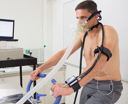 Integrative and functional medicine also uses genetic testing to determine the best types of exercise for different people and to explore the likelihood of injuries of several kinds in athletes. This latter area of research and clinical practice can help reduce the number and severity of athletic injuries for adult and child athletes.
 The ACTN3 is strongly associated with the protein alpha-actinin-3. This protein is involved exclusively in fast type II muscle fibers that are used in explosive activities. SNP R577X indicates a stop codon at position 577 rather than an arginine (R). An R allele puts athletes at an advantage in power sports. A study of the ACTN3 R577X variant in elite European athletes showed those in power event to be 50 percent less likely to have the XX variant and those involved in endurance events to be 1.88 times more likely to have the XX variant. For world-class endurance athletes, the odds of having the XX variant were 3.7 times larger when compared with lower-level athletes. It appears the ACTN3 gene is more important at the upper levels of sports.
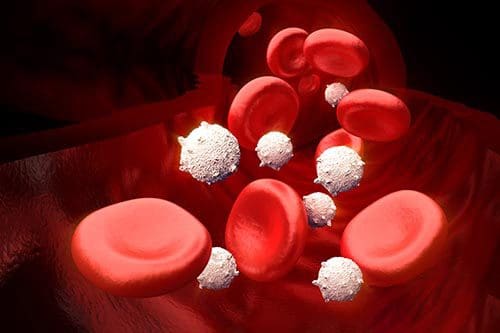 Metabolic syndrome and metabolic health have been studied extensively due to metabolic syndrome being a major risk factor for the development of diabetes mellitus 1 and cardiovascular disease. Genetic and environmental factors interrelate in a complex fashion to bring about this condition. A cluster of metabolic abnormalities, including hypertension, dyslipidemia, abdominal obesity, insulin resistance, and impaired glucose tolerance make up metabolic syndrome.
 Feelings of fatigue and lethargy are presented more and more frequently in health care professionals� offices. Combined with concentration difficulties, sleep problems, inability to lose weight, feeling your brain is in a fog, fatigue, and lethargy may point to AFS as the basic issue.
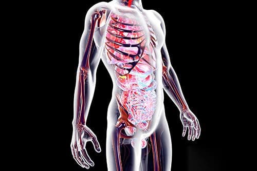 Increasing and unrelenting stress is a part of our culture that is detrimental to the health of every individual. The metabolic component of the NEM model added to the neuroendocrine aspect helps professionals to see how localized organ-specific responses and systemic responses are necessary for successfully dealing with stress.
 The mapping of the human genome has provided an opportunity for researchers and clinicians alike to consider the roles genes play in health and wellness. Discovering the presence and effects of single nucleotide polymorphisms (SNPs) has increased not only our knowledge of how genes affect health, but also has given us tools to use in preventing and remediating many chronic illness conditions.


 Joint pain of unknown origin can cause a myriad of debilitating problems, including the additional stress of trying to find effective remedies for joint pain. It can be a scary and confusing time, especially when test results show no abnormalities and your doctor can�t figure out what�s wrong. It�s important to find and address the cause of the inflammation. If you experience other concurring symptoms similar to those of Adrenal Fatigue, find a practitioner who can support your NEM stress response. Proper restorative strategies will help your body cope with both the stress and the