As one of the stabilizers for the body, the knees are located between the thighs and legs, allowing flexion and extension. The knees help the hips by supporting the upper body’s weight and allowing the legs to move from one place to another without feeling pain. The knee has various muscles and ligaments surrounding the knee joint, allowing the leg to be bent when active. One of the muscles is located behind the knee, known as the popliteus, and supports the legs. However, minor injuries or actions can affect the knees causing the joint to be in a “lock” position and develop myofascial trigger points that can induce muscle spasms in the knees. Today’s article focuses on the popliteus muscle, how knee pain is associated with trigger points, and how to manage knee pain through various treatments. We refer patients to certified providers that incorporate multiple methods in the lower body extremities, like knee pain treatments correlating to myofascial trigger points, to aid many people dealing with pain symptoms along the popliteus muscles. We encourage and appreciate each patient by referring them to associated medical providers based on their diagnosis, especially when appropriate. We understand that education is an excellent source to asking our providers intricated questions at the patient’s request. Dr. Alex Jimenez, D.C., only utilizes this information as an educational service. Disclaimer
What Is The Popliteus Muscle?
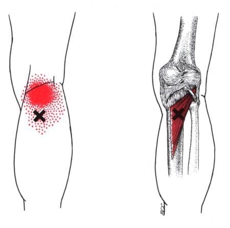
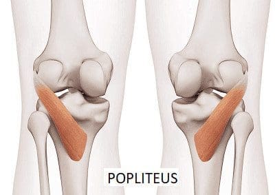
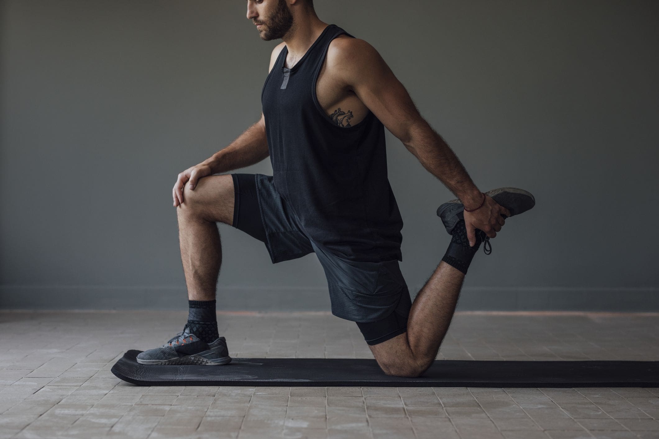
Have you been dealing with pain behind your knees? Do you have issues bending your knees when climbing up or down the stairs? Or do your back knee muscles start to twitch uncontrollably, causing muscle spasms? Many knee issues correlate with various factors that can affect the popliteus muscle and develop trigger points. The popliteus is a small muscle with a very important job as it is a major stabilizing muscle to the knees. The popliteus muscle originates from the lateral side of the femur and inserts itself into the posterior surface of the tibia. Some attachments are between the popliteus and lateral meniscus, allowing the knees to be in motion and providing flexion without pain and entrapment. Additional studies reveal that when a person exercises, the popliteus’s basic function helps bring about and maintain internal rotation of the tibia on the femur. The popliteus also helps prevent the foot from external rotation and allows the individual to stand correctly. However, injuries to the knee could overstretch the popliteus muscle and cause mobility issues to the knee flexion.
Knee Pain Associated With Trigger Points
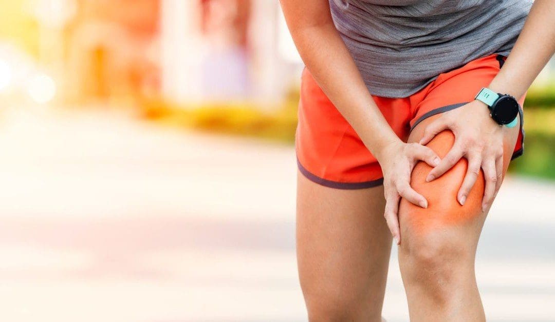
When dealing with knee pain, it could often be a joint disorder like osteoarthritis or a musculoskeletal condition like sciatica pain associated with the knee. These issues could be due to normal factors like constantly sitting down or bending down to lift heavy objects that cause the knees to buckle. However, when the popliteus muscle has been continuously overused from being bent, it can form tiny nodules known as trigger points to cause knee pain. Studies reveal that trigger points on the muscles surrounding the knee are often ignored during a clinical diagnosis. Trigger points cause referred pain to the surrounding muscles, accompanied by various sensory sensations like heaviness, tingling, and hypersensitivity to the popliteus muscle. In “Myofascial Pain and Dysfunction,” written by Dr. Travell, M.D. stated that one of the chief complaints that many patients often talk to their doctors about is the pain they feel in the back of their knees when they are in a crouch position. The book also states when normal actions like running or twisting have overloaded the popliteus muscle, it can cause trauma or strain to the popliteus muscle and tear the posterior cruciate ligament to the knees.
How To Find Trigger Points In The Popliteus- Video
Have you been having knee issues that make walking difficult for a long period? Do you feel like your knees are locking up constantly? What about feeling unstable when standing or carrying objects around? These issues that affect the knees are associated with trigger points along the popliteus muscles. The popliteus muscle is small, located at the back of the knees, and assists with knee flexion. When the popliteus muscle becomes overused, it can cause trigger points to form and cause knee issues. Studies reveal that various issues, like tendon injuries, are associated with repetitive mechanical stresses that can cause degenerative knee lesions. Any trauma or muscle strain can affect the knee’s function of flexing and bending without pain for trigger points to form along the popliteus muscles. The video above focuses on the popliteus muscle, where the trigger points are located, and where the referred pain patterns are situated in the knees. On the bright side, all is not lost, as various treatments offer ways to manage knee pain associated with trigger points.
Managing Knee Pain Through Various Treatments
When it comes to knee pain, many individuals will apply an ice or heat compress to allow the surrounding muscles to relax while reducing the pain and swelling. Other individuals use over-the-counter medicines to eliminate the pain for a few hours. While these work at managing knee pain, various treatments target trigger points and can help improve flexion mobility back to the knees. Studies reveal that muscle stretching on the popliteus muscle contributes to joint position sense to knee joint stability and function. Stretching the popliteus muscles can reduce the pain in the back of the knee while elongating the muscle fibers to manage trigger points from forming again. Other treatments that people can do to avoid trigger points from returning is to avoid walking or running in a lateral sloped area to prevent the knees from locking up. Incorporating these treatments to prevent knee issues and allow the knee to function properly.
Conclusion
The knees are one of the stabilizers in the body that are located between the thighs and legs, allowing flexion and extension. As a small muscle located in the back of the knees, the popliteus stabilizes the knees and enables them to be in motion without pain. However, when the popliteus muscle becomes overstretched and overused, it can develop trigger points in the popliteus that invoke referred pain to the surrounding muscles and cause the knees to lock up. To that point, it causes the body to be unstable and mimics knee pain issues. Fortunately, trigger points are treatable through various treatments that help relieve the pain and reduce the trigger points from returning. When these treatments are utilized on the knees, the surrounding muscles regain flexion mobility in the lower body.
References
English, S, and D Perret. “Posterior Knee Pain.” Current Reviews in Musculoskeletal Medicine, U.S. National Library of Medicine, 12 June 2010, https://www.ncbi.nlm.nih.gov/pmc/articles/PMC2941578/.
Ghaffarinejad, Farahnaz, et al. “Effect of Static Stretching of Muscles Surrounding the Knee on Knee Joint Position Sense.” British Journal of Sports Medicine, U.S. National Library of Medicine, Oct. 2007, https://www.ncbi.nlm.nih.gov/pmc/articles/PMC2465159/.
Hyland, Scott, and Matthew Varacallo. “Anatomy, Bony Pelvis and Lower Limb, Popliteus Muscle.” In: StatPearls [Internet]. Treasure Island (FL), StatPearls Publishing, 6 June 2022, https://www.ncbi.nlm.nih.gov/books/NBK526084/.
Mann, R A, and J L Hagy. “The Popliteus Muscle.” The Journal of Bone and Joint Surgery. American Volume, U.S. National Library of Medicine, Oct. 1977, https://pubmed.ncbi.nlm.nih.gov/908724/.
Sánchez Romero, Eleuterio A, et al. “Prevalence of Myofascial Trigger Points in Patients with Mild to Moderate Painful Knee Osteoarthritis: A Secondary Analysis.” Journal of Clinical Medicine, U.S. National Library of Medicine, 7 Aug. 2020, https://www.ncbi.nlm.nih.gov/pmc/articles/PMC7464556/.
Travell, J. G., et al. Myofascial Pain and Dysfunction: The Trigger Point Manual: Vol. 2:the Lower Extremities. Williams & Wilkins, 1999.
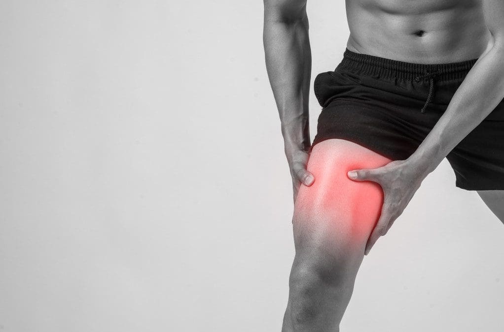
Many individuals utilize their lower muscles to move around and stay active as each muscle does its job and allows mobility to the hips and thighs. In sports, the thigh muscles are utilized constantly to extend the legs and bend the knees, allowing a powerful force to win any sports competition. At the same time, various sports injuries can occur to the hips, thighs, and legs and can affect the muscles causing pain and discomfort to the lower extremities. A hamstring injury is one of the most common injuries that can affect the thighs, which can cause many athletes to be taken out of their favorite sport to recover from the injury. Today’s article looks at the hamstring muscle, how trigger points correlate with a hamstring strain, and how various stretches can reduce muscle strain on the hamstrings. We refer patients to certified providers who incorporate multiple methods in the lower body extremities, like upper thigh and hip pain treatments correlating to myofascial trigger point pain, to aid individuals dealing with pain symptoms along the hamstring muscles. We encourage and appreciate patients by referring them to associated medical providers based on their diagnosis, especially when appropriate. We understand that education is an excellent solution to asking our providers complex questions at the patient’s request. Dr. Jimenez, D.C., utilizes this information as an educational service only. Disclaimer
What Are The Hamstring Muscles?
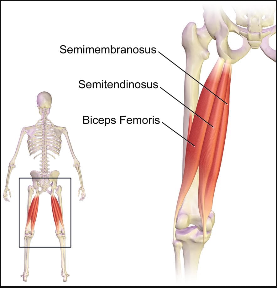
Do you experience pain in the back of your upper thigh? When walking from one place to another, do you hear a popping sound in the back of your thigh? Or are you dealing with muscle tenderness in the back of your upper thigh? Many of these symptoms correlate with issues affecting the hamstrings causing trigger points to affect the upper thighs. As one of the most complex muscles comprising three muscles (semitendinosus, semimembranosus, biceps femoris), the hamstrings play a crucial part in daily activities. From simple actions like standing to explosive movements like sprinting or jumping, the hamstrings are known as posterior thigh muscles that begin from the pelvis and run behind the femur bone and cross the femoroacetabular and tibiofemoral joints. The hamstring muscles in the body play a prominent role in hip extension and is a dynamic stabilizer of the knee joint. To that point, the hamstring muscles are the most susceptible muscle that succumbs to injuries that can lead to disability in the legs and affect daily activities.
Hamstring Strain & Trigger Points
Since the hamstrings are the most susceptible muscles that can succumb to injuries, it takes a while for the muscle to heal, depending on the severity of the damage. Studies reveal that the hamstrings can occur injuries when a person is running or sprinting due to their anatomic arrangement, which causes the muscles to strain. To that point, depending on how much force has impacted the hamstrings, the injuries can lead to 3 of the following:
Grade 1: Mild pain or swelling (no loss of function)
Grade 2: Identifiable partial tissue disruption with moderate pain and swelling (minimal loss of function)
Grade 3: Complete disruption of the tissue with severe pain and swelling (total loss of function)
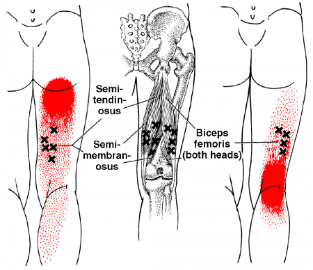
The pain that patients experience can be painful when walking, causing them to limp. In “Myofascial Pain and Dysfunction,” written by Dr. Janet G. Travell, M.D., stated that when patients are dealing with pain in their hamstrings, it could potentially be associated with trigger points along the three muscles, causing pain and disability in the upper thighs. The book also mentioned that when trigger points affect the hamstrings, it can lead to muscle inhibition, compromising hip stability. Another issue that trigger points associated with hamstring strain causes in the body are that when individuals are sitting down are likely to experience posterior pain in the buttock, upper thighs, and back of the knees. Luckily, there are various ways to reduce the pain along the hamstring muscles.
Trigger Point Of The Week: Hamstrings- Video
Have you dealt with pain along the back of your upper thighs? Does it feel uncomfortable when you are sitting down? Or do your hamstrings ache or feel tight after running for a long period? People dealing with issues in their hamstrings could be dealing with muscle strain associated with trigger points. The hamstring muscles play a vital role in the body as it allows the individual to walk, run, bend the knees and even extend the legs. The hamstring muscles are also the most susceptible to injury, causing disability to the legs. Studies reveal that trigger points associated with the hamstring muscles can lead to soreness or irritability in the muscle fibers that may interfere with the biomechanics and normal functioning of the lower limbs. The video above explains where the hamstrings are located and how the trigger points can cause referred pain to the hamstrings. To that point, trigger points can affect a person’s ability to walk and affect the surrounding muscles in the lower body while mimicking other chronic conditions.
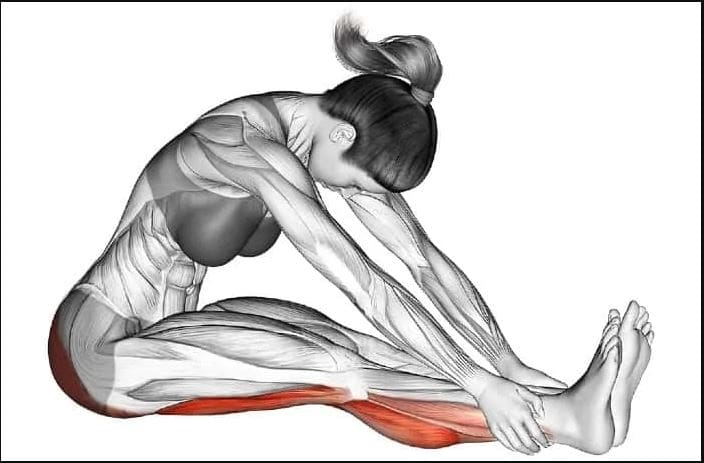
Various Stretches To Reduce Muscle Strain On The Hamstrings
When the hamstrings become injured, the healing rate usually depends on how severe the injury is in the hamstrings. If a hamstring injury is mild, the tears or strains can heal within about three to eight weeks, and if the hamstring injury is severe, the tears or strains could be long as three months. When the hamstrings are tense and on the verge of tearing, many people should stop overusing the muscle. Various stretches can reduce muscle strain on the hamstrings and relieve tension from the hamstrings to allow mobility back to the legs. Studies reveal that manual ischemic compression on the upper thigh muscles can significantly reduce pain in the lower limbs. This allows the individual to manage the trigger points associated with the hamstrings and reduce the chances of them re-occurring in the legs.
Conclusion
As the most important muscle in the lower body extremities, the hamstrings play a crucial part in the body as they allow the individual to walk, run, and stand without feeling pain. However, even though they are important muscles, they are susceptible to injuries. When the hamstrings become injured, the recovery process varies depending on the severity and can develop trigger points along the muscle fibers. To that point, it causes referred pain along the upper thigh muscle and affects a person’s ability to walk. Fortunately, incorporating various stretches to the hamstrings can alleviate the pain and reduce the trigger points from re-occurring in the muscle. This allows mobility back to the legs, and many individuals can resume their daily activities.
References
Esparza, Danilo, et al. “Effects of Local Ischemic Compression on Upper Limb Latent Myofascial Trigger Points: A Study of Subjective Pain and Linear Motor Performance.” Rehabilitation Research and Practice, Hindawi, 4 Mar. 2019, https://www.ncbi.nlm.nih.gov/pmc/articles/PMC6425406/.
Poudel, Bikash, and Shivlal Pandey. “Hamstring Injury – Statpearls – NCBI Bookshelf.” In: StatPearls [Internet]. Treasure Island (FL), StatPearls Publishing, 28 Aug. 2022, https://www.ncbi.nlm.nih.gov/books/NBK558936/.
Rodgers, Cooper D, and Avaias Raja. “Anatomy, Bony Pelvis and Lower Limb, Hamstring Muscle.” In: StatPearls [Internet]. Treasure Island (FL), StatPearls Publishing, 29 Jan. 2022, https://www.ncbi.nlm.nih.gov/books/NBK546688/.
Thummar, Ravindra C, et al. “Association between Trigger Points in Hamstring, Posterior Leg, Foot Muscles and Plantar Fasciopathy: A Cross- Sectional Study.” Journal of Bodywork and Movement Therapies, U.S. National Library of Medicine, 7 Aug. 2020, https://pubmed.ncbi.nlm.nih.gov/33218537/.
Travell, J. G., et al. Myofascial Pain and Dysfunction: The Trigger Point Manual: Vol. 2:the Lower Extremities. Williams & Wilkins, 1999.
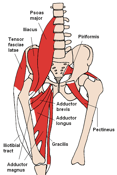
The hips and thighs have a working relationship as their jobs are to maintain stability for the legs and pelvis while supporting the upper body’s weight. These two body groups have various muscles, tendons, and nerves that have specific jobs that allow mobility to the lower body. Many athletes in multiple sports events use their thighs to exert a huge amount of power to be the best. This is due to the adductor muscles in the thighs that allow the athlete to win the event. These adductor muscles are voluminous in size and can become overstretched if the muscles have been worked out too much or injuries have caused dysfunction in the surrounding muscles, causing mobility issues. To that point, the adductor muscles will develop myofascial trigger points and cause hip and thigh pain. Today’s article looks at the two adductor muscles (Longus and Magnus), how myofascial trigger points affect the adductor muscles, and available treatments to manage hip adductor trigger points. We refer patients to certified providers who incorporate multiple methods in the lower body extremities, like thigh and hip pain treatments correlating to myofascial trigger point pain, to aid individuals dealing with pain symptoms along the adductor muscles. We encourage and appreciate patients by referring them to associated medical providers based on their diagnosis, especially when appropriate. We understand that education is an excellent solution to asking our providers complex questions at the patient’s request. Dr. Jimenez, D.C., utilizes this information as an educational service only. Disclaimer
Adductor Longus & Adductor Magnus
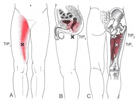
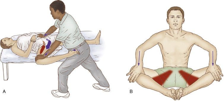
Have you been dealing with groin pain located near your thighs? Do you feel muscle tenderness or stiffness when stretching your inner thighs? Or have you been feeling unstable in your hips or thighs when walking? Many individuals, especially athletes and older adults, could be experiencing myofascial trigger points associated with groin pain along their adductor muscles. The thighs contain several muscles and functions that allow many people to bend and extend their knees and hips. The adductor muscles allow the legs to move inward toward one another. The adductor muscles have five muscles: magnus, brevi, longus, pectineus, and gracilis. These muscles enable functionality to the thighs and hips, and we will look at two adductor muscles in the inner thighs. The long adductor muscle is a large, fan-shaped muscle that starts from the superior aspect of the pubis bone and travels down to connect at the thigh bone. Studies reveal that the adductor longus is a long and thin muscle with many actions for the thighs, including external/lateral rotation and thigh flexion.
Now the adductor Magnus is a large triangular-shaped muscle of the inner thighs that are important for thigh and hip function and stabilizing the pelvis. Studies reveal that even though the adductor Magnus is a large muscle in the inner thighs, its primary function is to allow the thigh to move in a larger range of motion without any pain inflicted on the thigh muscles. However, the adductor muscle can succumb to various issues affecting the thighs and groin regions of the body that can be overstretched and strain the body.
Myofascial Trigger Points Affecting The Adductor Muscles
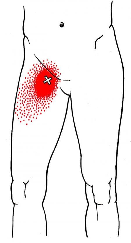
Groin pain is a multi-factorial pain issue that affects the lower limbs, and its often due to muscle strain in the inner thigh muscles. This pain increases during vigorous activities and when there is a sudden twist in the hips. When the adductor muscles suddenly change in motion when the body is active, they can be overstretched and correlate to myofascial trigger points that can affect the inner thigh and groin regions. According to “Myofascial Pain and Dysfunction,” by Dr. Travell, M.D., patients with active myofascial trigger points in the two adductor muscles (Longus and Magnus) would become frequently aware of the pain in their groin and medial thigh. When the adductor muscles have myofascial trigger points in the inner thigh, diagnosing is difficult since the individual thinks they are suffering from groin pain when the pain is in their inner thighs. To that point, studies reveal that many individuals participating in various sports would suffer from groin pain due to myofascial trigger points affecting the adductor muscles. Luckily, there are multiple treatments to reduce the pain in the adductor muscles.
Hip Adductors: Trigger Point Anatomy- Video
Have you been dealing with groin pain when you are walking? What about experiencing unquestionable thigh pain that affects your daily activities? Or does stretching your inner thigh muscles seem difficult, causing muscle tenderness? Many of these symptoms correlate with groin pain associated with myofascial trigger points affecting the adductor muscles in the inner thighs. The adductor muscles allow mobility function to the thighs and enable the hips to have a wide range of motion. When the adductor muscles are overstretched due to a sudden change of hip rotation or injury has occurred on the thighs can lead to referred pain in the groin and inner thighs and develop myofascial trigger points. The video above shows where the trigger points are located in the hip adductor muscles. The video also explains where the pain is localized in the adductor muscles and the symptoms it produces that can affect the lower body extremities. Fortunately, even though diagnosing myofascial trigger points are a bit challenging, available treatments can manage trigger points along the hip adductors.
Available Treatments To Manage Hip Adductor Trigger Points
When myofascial trigger points affect the hip adductor muscles, many individuals complain about stiffness in their inner thighs and how they feel miserable when they don’t have mobility from their thighs and hips. As stated earlier, trigger points are a bit challenging when diagnosed, but they are treatable when doctors examine patients dealing with myofascial pain in their hips and thigh muscles. Once the diagnosis is complete, doctors work with pain specialists who can locate the trigger points and devise a treatment plan to relieve the pain. Available treatments like trigger point injections can minimize the pain and reduce the chances of trigger points returning. Other available therapies like exercising or stretching, especially for the hips and thighs. Specific exercises for the hips and thigh muscles can help strengthen the adductor muscles from suffering pain and can help reduce the pain symptoms. Another treatment is applying moist heat on the hip adductor muscles to release the tension from the tight muscles and allow mobility back to the hip adductors.
Conclusion
The adductor muscles work with the hips and thighs to allow a wide range of motions and extension to the knees and hips. The hips and the thighs allow stability to the lower body and support the weight to the upper body. When injuries or sudden changes start to affect the adductor muscles, it can lead to symptoms of groin pain associated with myofascial trigger points. Myofascial trigger points produce tiny nodules in the affected muscle that causes referred pain to the muscle group. When this happens, it causes the body to be dysfunctional and can affect a person’s mobility to function in the world. Luckily myofascial trigger points are treatable through various techniques and treatments that can reduce the chances of trigger points from re-occurring in the body.
References
Jeno, Susan H, and Gary S Schindler. “Anatomy, Bony Pelvis and Lower Limb, Thigh Adductor Magnus Muscle.” In: StatPearls [Internet]. Treasure Island (FL), StatPearls Publishing, 1 Aug. 2022, https://www.ncbi.nlm.nih.gov/books/NBK534842/.
Sedaghati, Parisa, et al. “Review of Sport-Induced Groin Injuries.” Trauma Monthly, Kowsar, Dec. 2013, https://www.ncbi.nlm.nih.gov/pmc/articles/PMC3864393/.
Simons, D. G., and L. S. Simons. Myofascial Pain and Dysfunction: The Trigger Point Manual: Vol. 2:the Lower Extremities. Williams & Wilkins, 1999.
Takizawa, M, et al. “Why Adductor Magnus Muscle Is Large: The Function Based on Muscle Morphology in Cadavers.” Scandinavian Journal of Medicine & Science in Sports, U.S. National Library of Medicine, 27 Apr. 2012, https://pubmed.ncbi.nlm.nih.gov/22537037/.
van de Kimmenade, R J L L, et al. “A Rare Case of Adductor Longus Muscle Rupture.” Case Reports in Orthopedics, Hindawi Publishing Corporation, 2015, https://www.ncbi.nlm.nih.gov/pmc/articles/PMC4397006/.
The hips and the thighs have an established relationship where mobility and stability play a part in the body’s lower extremities. The lower extremities’ main job is to support the upper body’s weight while stabilizing the hips and allowing movement from the thighs to the legs and feet. When it comes to the thighs in the lower body, the various muscle surrounds the thighs and skeletal joints to allow the legs to move from one place to another. One of the muscle groups in the thighs is known as the quadriceps femoris. This muscle group is activated when a person is in motion and can succumb to injuries from trauma or normal factors. When this happens, issues like myofascial pain syndrome can affect the thigh muscle and cause referred pain to travel to the knees. Today’s article focuses on the quadriceps femoris, how myofascial pain syndrome is associated with thigh pain, and trigger point therapy on the quadriceps. We refer patients to certified providers who incorporate multiple methods in the lower body extremities, like thigh and hip pain treatments correlating to myofascial pain, to aid individuals dealing with pain symptoms along the quadriceps for muscle. We encourage and appreciate patients by referring them to associated medical providers based on their diagnosis, especially when appropriate. We understand that education is an excellent solution to asking our providers complex questions at the patient’s request. Dr. Jimenez, D.C., utilizes this information as an educational service only. Disclaimer
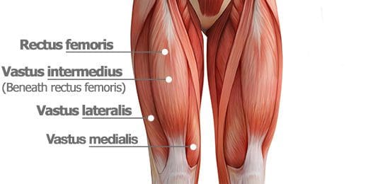
What Are The Quadriceps Femoris?
Have you been dealing with knee issues when you are walking? What about muscle tenderness or soreness in your thighs? Or have you been experiencing knee complaints when you are running? These areas of complaint are correlated with trigger points associated with thigh pain along the quadriceps femoris. As one of the most voluminous muscles in the human body, the quadriceps femoris is a group of muscles predominant in the thighs and is extraordinarily important. This muscle group is essential for daily activities like climbing the stairs or getting up from a seated position, allowing repercussions on the knees and hip joints. The quadriceps femoris consist of four thigh muscles to allow extension to the knees:
Vatus medialis
Vatus lateralis
Vatus intermedius
Rectus femoris
Studies reveal that these four different muscles fuse to form the quadricep tendon and stabilize the patella and thigh flexion at the hips and knee extension. This muscle group is highly important for athletes participating in sports events but can succumb to injuries through muscle strain.
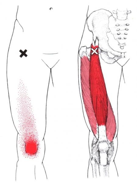
Myofascial Pain Syndrome Associated With Tigh Pain
When the thigh muscles, especially the quadriceps femoris, can be overstretched and overused when in motion. Thigh pain is nothing to be alarmed about in its acute form; however, it can develop small nodules along the four muscle fibers that can cause referred pain to the hips and knees. To that point, it can correlate through quadriceps muscle strain to the thighs. Studies reveal that normal factors like kicking, jumping, or a sudden change of direction of running can potentially cause the muscle fibers to be overstretched and develop pain due to localized swelling corresponding to loss of motion from myofascial pain syndrome.
In “Myofascial Pain and Dysfunction,” written by Dr. Janet G. Travell, M.D., the book states that myofascial pain syndrome can invoke referred pain to the affected muscle or muscle group, causing the body to be dysfunctional. Myofascial pain syndrome associated with thigh pain can be managed through various treatments and could allow mobility back to the thighs, legs, knees, and hips. The book even mentions how the four muscles in the quadriceps femoris cause different pain issues in various body parts due to myofascial pain syndrome. For the rectus femoris, many people would complain about knee pain and weakness when climbing stairs. The vatus medialis would initially produce a toothache-like pain deep within the knee joint, often misinterpreted as joint inflammation. The vatus intermedius causes many individuals to have difficulty fully straightening their knees and causes them to develop buckling knee syndrome. And finally, the vatus lateralis could cause many individuals to complain about feeling pain when walking and that the pain is being distributed on the lateral aspect of the thigh, including the knees.
Trigger Point Therapy: Stretching The Quadriceps- Video
Have you been dealing with pain in your thighs and knees? Do you find it difficult to climb up or down the stairs? Or have you been experiencing inflammation in your knee joints? All these symptoms that you are experiencing in your thighs, knees, and hips correlate with trigger points created by myofascial pain syndrome affecting the quadriceps femoris. The quadriceps femoris is a voluminous group of muscles that allows the individual to do daily activities like climbing up or down the stairs, running, jumping, and getting up from a seated position. When various issues can cause the quadricep femoris to become overstretched and overused, it could develop myofascial pain syndrome/trigger points along the muscle fibers to mimic knee pain and cause dysfunction in knee mobility. Even though myofascial pain syndrome is poorly diagnosed, individuals can manage it through various treatments that target myofascial trigger pain. The video above explains where the quadriceps femoris muscles are located on the thigh and where the trigger points are in the muscle fibers. The video also provides various stretching techniques on the quadriceps to reduce pain-like symptoms along the thighs.
Trigger Point Therapy On The Quadriceps
When it comes to releasing myofascial pain syndrome on the quadriceps, treatments like dry needling, acupuncture, or manual stretching can help loosen and lengthen the quadricep muscles from becoming shorten and can reduce myofascial trigger points from causing more issues on the knees and thighs. At the same time, treatment alone can only go so far in rehabilitation unless the person dealing with myofascial pains syndrome associated with thigh pain do some corrective actions to prevent trigger points from reproducing on the quads. Actions like:
Avoid prolonged sitting
Stretching the quads as part of your warm-up
Sleeping with a pillow between the knees
These actions allow the quadriceps to relax and prevent pain-like issues from affecting the knees. To that point, these actions can help many individuals have mobility back to their legs and allow them to bend their knees without feeling pain.
Conclusion
The quadriceps femoris consists of four thigh muscles that fuse to enable mobility functions in the knees without pain. As the most voluminous muscle group in the body, the quadriceps femoris allows the thighs to function when in motion and allow the knees to extend. When various issues cause the quadriceps femoris muscles to be overstretched, it can develop trigger points/myofascial pain syndrome that mimics knee pain and can affect how a person is walking. Thankfully, various treatments specializing in myofascial pain syndrome can reduce the pain symptoms from the quadriceps femoris and bring back knee mobility to the legs.
References
Bordoni, Bruno, and Matthew Varacallo. “Anatomy, Bony Pelvis and Lower Limb, Thigh Quadriceps Muscle.” In: StatPearls [Internet]. Treasure Island (FL), StatPearls Publishing, 10 May 2022, https://www.ncbi.nlm.nih.gov/books/NBK513334/.
Kary, Joel M. “Diagnosis and Management of Quadriceps Strains and Contusions.” Current Reviews in Musculoskeletal Medicine, Humana Press Inc, 30 July 2010, https://www.ncbi.nlm.nih.gov/pmc/articles/PMC2941577/.
Rozenfeld, Evgeni, et al. “The Prevalence of Myofascial Trigger Points in Hip and Thigh Areas in Anterior Knee Pain Patients.” Journal of Bodywork and Movement Therapies, U.S. National Library of Medicine, 14 May 2019, https://pubmed.ncbi.nlm.nih.gov/31987560/.
Simons, D. G., and L. S. Simons. Myofascial Pain and Dysfunction: The Trigger Point Manual: Vol. 2:the Lower Extremities. Williams & Wilkins, 1999.
Waligora, Andrew C, et al. “Clinical Anatomy of the Quadriceps Femoris and Extensor Apparatus of the Knee.” Clinical Orthopaedics and Related Research, Springer-Verlag, Dec. 2009, https://www.ncbi.nlm.nih.gov/pmc/articles/PMC2772911/.
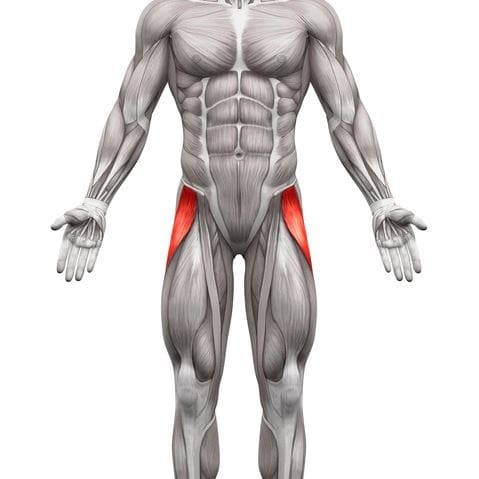
When many individuals begin to work out or start training for an event, they incorporate various muscles to give optimal output and strength when doing a set of exercises. Many athletes or individuals trying to train for an event or to better themselves have to do a pre-workout routine involving various stretches to warm up the muscles before the actual workout and do stretches post-workout again. This ensures that the muscles are ready to give it their all when a person is working out. The body has various parts with different functions and jobs that help the body’s motor function. The upper body has the shoulders, arms, hands, elbows, neck, head, and chest to allow movements and stability. At the same time, the lower body has the hips, low back, thighs, legs, knees, pelvis, and feet to support the upper body’s weight and stabilize the lower extremities from collapsing. When various factors affect the body, it can lead to dysfunction and causes referred pain to different body locations that can mask chronic conditions. Today’s article looks at one of the lower body muscles located at the inner thighs, known as the pectineus muscle, how trigger point pain affects the inner thighs, and various stretches to strengthen the hip adductors. We refer patients to certified providers who incorporate multiple methods in the lower body extremities, like thigh and hip pain treatments correlating to trigger point pain, to aid individuals dealing with pain symptoms along the pectineus muscle. We encourage and appreciate patients by referring them to associated medical providers based on their diagnosis, especially when appropriate. We understand that education is an excellent solution to asking our providers complex questions at the patient’s request. Dr. Jimenez, D.C., utilizes this information as an educational service only. Disclaimer
What Is The Pectineus Muscle?
Have you been experiencing pain in your inner thighs? Do you find it difficult to play various sports? Do you feel tenderness or soreness in your thighs or near your groin? Most of these symptoms are associated with trigger point pain along the pectineus muscles that affect the thighs. The pectineus is part of the anterior thigh muscles that extend the leg to the knee joint. The pectineus works with another muscle known as the sartorius and a muscle group known as the quadriceps femoris. The pectineus muscle is responsible for flexion, adduction, and medial rotation since it is a hip adductor for the thighs. This muscle is important for various sports activities like running, skating, soccer, or basketball and can become overused due to overstretching the legs too far, thus developing trigger points in the pectineus muscle.
Trigger Point Pain Affecting The Inner Thighs
When athletes overuse their legs and overstretch the pectineus muscle, it can cause issues with the thighs, hips, and legs’ mobility causing referred pain to the lower body. This is known as trigger point pain and can be challenging when diagnosing where the pain is located. Studies reveal that trigger point pain affecting the inner thighs, especially the pectineus muscle, can mimic groin and hip pain, causing various symptoms in the lower extremities. The multiple symptoms can include:
Weak adductor muscles
Muscle fatigue
Decreased range of motion
Leg-length discrepancy
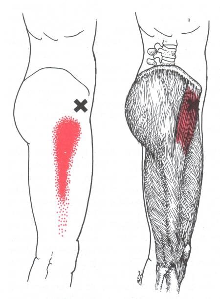
Various reasons can lead to the development of trigger point pain associated with the inner thighs along the pectineus; according to “Myofascial Pain and Dysfunction,” written by Dr. Janet G. Travell, M.D., stated that when patients are dealing with pectineus trigger points would complain about the referred pain surrounding the muscle but not the muscle itself. The book also mentioned that nerve entrapment could also be an issue since trigger points like to mimic other chronic conditions. Trigger points along the pectineus muscle can also develop associated with hip joint diseases like advanced osteoarthritis.
Treating Trigger Points In Hip Adductors- Video
Are you experiencing issues when moving around constantly? Do you experience pain in your inner thighs and hips? Or do you have difficulty rotating your thighs or hips? If you have been dealing with these issues throughout your entire life, it could be due to your pectineus muscles being affected by trigger points along your inner thighs. Trigger points (myofascial pain syndrome) develop tiny nodules along the muscle fibers, causing referred pain to the surrounding muscles that can cause dysfunction in the lower extremities. Studies reveal that myofascial trigger points can cause the affected muscles to be intensely sensitive and irritable, predominantly near the reflex muscle. To that point, it causes hip and thigh disability in the lower body. Fortunately, there are ways to reduce the pain and manage the trigger point pain along the pectineus muscle, as shown in the video above. The hip adductor muscles are being stretched and treated for trigger point pain and allowing mobility back to the hips and inner thighs.
Various Stretches To Strengthen Hip Adductor
Since the pectineus muscle is part of the hip adductor muscles, various stretches can reduce the chances of trigger points from future development while minimizing the pain that it is causing along the surrounding muscles. Studies reveal that multiple exercises and stretches for the pectineus muscle can help with hip flexion and stabilization. These stretches can help stretch and strengthen the hip adductor muscles while preventing groin pain associated with trigger points. Incorporating these stretches before and after a workout can reduce trigger points and allow hip mobility and thigh rotation back to the legs. This ensures that the trigger points along the pectineus muscle are managed, and the individual doesn’t have to suffer from referred pain issues on the thighs and can move around without pain.
Conclusion
As part of the hip adductor muscles, the pectineus is a small muscle that extends the leg to the knees and allows the thighs to flex, adduct, and rotate without pain. This muscle is important for many athletes participating in sports and can be easily overstretched to cause referred pain around the thighs. To that point, it can develop trigger points along the pectineus muscles can correlate to groin pain in the lower extremities. All is not lost, as various stretches and exercises can strengthen the hip adductor muscles and improve thigh and hip mobility. This allows athletes and individuals to continue playing the sport they enjoy.
References
Giphart, J Erik, et al. “Recruitment and Activity of the Pectineus and Piriformis Muscles during Hip Rehabilitation Exercises: An Electromyography Study.” The American Journal of Sports Medicine, U.S. National Library of Medicine, July 2012, https://pubmed.ncbi.nlm.nih.gov/22523373/.
Khan, Ayesha, and Abdul Arain. “Anatomy, Bony Pelvis and Lower Limb, Anterior Thigh Muscles.” In: StatPearls [Internet]. Treasure Island (FL), StatPearls Publishing, 10 June 2022, https://www.ncbi.nlm.nih.gov/books/NBK538425/.
Kiel, John, and Kimberly Kaiser. “Adductor Strain.” In: StatPearls [Internet]. Treasure Island (FL), StatPearls Publishing, 21 June 2022, https://www.ncbi.nlm.nih.gov/books/NBK493166/.
Simons, D. G., and L. S. Simons. Myofascial Pain and Dysfunction: The Trigger Point Manual: Vol. 2:the Lower Extremities. Williams & Wilkins, 1999.
Wada, Juliano T, et al. “An Anatomical Basis for the Myofascial Trigger Points of the Abductor Hallucis Muscle.” BioMed Research International, Hindawi, 22 Jan. 2020, https://www.ncbi.nlm.nih.gov/pmc/articles/PMC6998759/.
The lower body extremities help provide stability to the various body parts, including the hips, thighs, pelvis, legs, knees, and feet. The hips and thighs comprise multiple muscles and nerves that provide mobility to the lower half and allow the host to move around in different locations. While the hip muscles act on the thigh muscles at the hip joint and stabilize the pelvis, the thigh muscles allow the lower body to bend, flex and rotate while bearing most of the upper body’s weight and keeping alignment with the hips and legs. One of the thigh muscles is the sartorius muscle, and if it becomes overused and injured can lead to complications in the form of myofascial pain syndrome. Today’s article post examines the sartorius muscle, how myofascial trigger pain is associated with the sartorius, and the effectiveness of myofascial pain treatment on the thighs. We refer patients to certified providers who incorporate multiple methods in the lower body extremities, like thigh pain treatments correlating to myofascial pain syndrome, to aid individuals dealing with pain symptoms along the sartorius muscle. We encourage and appreciate patients by referring them to associated medical providers based on their diagnosis, especially when appropriate. We understand that education is an excellent solution to asking our providers complex questions at the patient’s request. Dr. Jimenez, D.C., utilizes this information as an educational service only. Disclaimer
What Is The Sartorius Muscle?
Are you experiencing pain in the upper, mid, or lower parts of your thighs? Do you have difficulty walking for long periods? Or do your knees hurt more than usual? Most of these issues correlate with myofascial trigger pain associated with the sartorius muscle. As the longest muscle that spans from the hips to the knee joints, the sartorius muscle, or the “tailor muscle,” serves as both a hip and knee flexor while working with other muscles that allow hip mobility. The sartorius shares its origin location with the TFL (tensor fascia latae) muscle at the anterior superior iliac spine and is responsible for internal rotation at the hips. In the book, “Myofascial Pain and Dysfunction,” the author Dr. Janet G. Travell, M.D., mentioned that the sartorius muscle assists the iliacus and the TFL muscles in hip flexion while assisting the short head of the bicep femoris in the knees for knee flexion, allowing the individual to walk for long distances. Even though this long muscle assists in hip and knee flexion, it can succumb to injuries and create issues with the hips and knees in the lower body.
Myofascial Trigger Pain Associated With The Sartorius Muscle
When traumatic forces or normal factors begin to affect the sartorius muscle, the surrounding muscles on the thighs and hips are also affected. The sartorius muscle allows the individual to move around and allows flexion to the hips and knees when injuries or the muscle is being overused; it can cause pain-like symptoms that correlate with hip and knee issues associated with myofascial trigger pain. Myofascial trigger pain along the sartorius muscle doesn’t usually occur in the muscle but can occur in conjunction with trigger point involvement in the surrounding muscles. Studies reveal that myofascial trigger pain is found in the hip muscles and can cause issues in the lumbopelvic-hip muscles of the lower body. This causes referred pain on the sartorius to be more diffused and superficial to the knees. When myofascial trigger pain is associated with the sartorius, many individuals often mistake it for knee pain. To that point, myofascial trigger pain could affect how a person walks and bends at the knees.
Anatomy & Palpation Of The Sartorius Muscle- Video
Are you experiencing issues when you are walking? Do your knees hurt constantly? Or are you experiencing tenderness or pain in your thighs? Most of these issues correlate with myofascial trigger pain associated with the sartorius muscle. The sartorius is a long muscle that connects the hips and spans to the knee joints to provide hip and knee flexion. The sartorius muscle works with the other muscles in the thighs and hips, allowing hip mobility and motor function to the legs. When multiple issues affect the sartorius and the surrounding muscles, it can develop into myofascial trigger pain and cause overlapping risk profiles to the knees and hips. To that point, it causes referred pain issues in the hips and knees, making the individual have difficulty walking from place to place. However, there are available treatments to reduce the pain in the hips and knees and manage the myofascial trigger pain from affecting the sartorius muscle on the thighs. The video above explains the anatomy of the sartorius muscle location and how palpation is used to locate the muscle to see if it is tight or could be affected by trigger points along the muscle fibers. This is one of the techniques that is used when a person is dealing with myofascial trigger pain associated with the sartorius muscle.
The Effectiveness Of Myofascial Pain Treatment On The Thighs
When a person is dealing with myofascial trigger pain in their thighs, and it is affecting the sartorius, many will often try to find available treatments to alleviate the pain. Treatments like dry needling are one of the various myofascial pain treatments that can reduce pain and related disability on the thighs, hips, and knees. Studies reveal that dry needling treatments can help manage knee pain syndrome associated with trigger points on the thighs. However, treatment alone can not be the only solution to reduce myofascial trigger pain in the thighs. Various hip stretches can loosen up tight hip flexors and help elongate the sartorius muscles to break up the nodules and improve mobility function to the hips and knees. People can even utilize self-ischemic compression to allow a more effective stretch on the sartorius muscle.
Conclusion
As the longest muscle in the thighs, the sartorius helps provide a service to hip and knee flexion while working with various muscles to keep the legs moving. When the sartorius muscles become overused and start to cause referred pain to the hips and knees, it can develop into myofascial trigger pain along the sartorius muscle. This can make many individuals believe they are suffering from knee pain when it’s their thigh muscle. However, myofascial trigger pain is treatable through treatments and corrective actions that people can incorporate into their daily activities to prevent pain from escalating and manage trigger points along the sartorius muscle. This can allow people to get back their mobility in their legs.
References
Rahou-El-Bachiri, Youssef, et al. “Effects of Trigger Point Dry Needling for the Management of Knee Pain Syndromes: A Systematic Review and Meta-Analysis.” Journal of Clinical Medicine, MDPI, 29 June 2020, https://www.ncbi.nlm.nih.gov/pmc/articles/PMC7409136/.
Samani, Mahbobeh, et al. “Prevalence and Sensitivity of Trigger Points in Lumbo-Pelvic-Hip Muscles in Patients with Patellofemoral Pain Syndrome.” Journal of Bodywork and Movement Therapies, U.S. National Library of Medicine, 15 Oct. 2019, https://pubmed.ncbi.nlm.nih.gov/31987531/.
Simons, D. G., and L. S. Simons. Myofascial Pain and Dysfunction: The Trigger Point Manual: Vol. 2:the Lower Extremities. Williams & Wilkins, 1999.
Walters, Benjamin B, and Matthew Varacallo. “Anatomy, Bony Pelvis and Lower Limb, Thigh Sartorius Muscle.” In: StatPearls [Internet]. Treasure Island (FL), StatPearls Publishing, 29 Aug. 2022, https://www.ncbi.nlm.nih.gov/books/NBK532889/.
The thighs in the lower half of the body work together with the hips to stabilize the legs when the body is in motion. The thighs and the hips also support the weight of the upper half of the body and are surrounded by muscles, ligaments, and nerve roots to supply blood and sensory-motor function to the legs. One of the thigh muscles that work with the hips is the tensor fasciae latae (TFL) muscle. When the thigh muscles are being overused or suffer from injuries, tiny nodules known as trigger points (myofascial pain syndrome) can affect a person’s ability to function worldwide. Today’s article examines what the tensor fasciae latae muscles do, how myofascial pain syndrome affects the thighs, and various stretches/techniques for the thighs. We refer patients to certified providers who incorporate multiple methods in the lower body extremities, like thigh pain treatments correlating to trigger points, to aid individuals dealing with pain symptoms along the tensor fasciae latae muscle. We encourage and appreciate patients by referring them to associated medical providers based on their diagnosis, especially when it is appropriate. We understand that education is an excellent solution to asking our providers complex questions at the patient’s request. Dr. Jimenez, D.C., utilizes this information as an educational service only. Disclaimer
What Does The Tensor Fasciae Latae Muscle Do?
Do you have difficulty walking for a long period? So you feel that your hips feel unstable when you move? Or do you feel radiating pain down from your thighs to your knees? Thigh pain associated with these symptoms can affect a person’s ability to move around from one location to another due to trigger points affecting the tensor fasciae latae muscle. The tensor fasciae latae (TFL) muscles are located at the proximal anterolateral thigh and originate from the anterior superior iliac spine. The TFL muscle is between the superficial and deep muscle fibers of the iliotibial (IT) band, as its attachment assists with knee flexion and lateral rotation. The TFL muscles also work together with the gluteus muscles in various hip movements. Studies reveal that the primary function of the TFL muscles is providing balance to the body’s weight and the non-weight-bearing leg to walk. The TFL muscles allow the individual to walk, run, and assist with movement and stabilization to the hips and knees without pain inflicted on the joints and muscles.
Myofascial Pain Syndrome Affecting The Thighs
Since the TFL muscles allow the person to walk and run, this muscle can become overused and strained through repetitive motions causing many issues to the hips, knees, and thighs. When these issues affect the TFL muscles, they can develop nodules along the muscle fibers known as trigger points or myofascial pain syndrome. Myofascial pain syndrome is a musculoskeletal disorder that can invoke referred pain in one location of the body while affecting the surrounding muscles in a different body location. Myofascial pain syndrome associated with the TFL muscles can cause issues to the hips, thighs, and knees while affecting a person’s ability to walk. Studies reveal that the prevalence of myofascial pain syndrome on the TFL muscles correlates to pain and disability in the thighs. When myofascial pain syndrome affects the TFL muscles, it can mimic chronic knee osteoarthritis.
Even though myofascial pain syndrome is challenging to diagnose, it is treatable through various stretches and techniques. In Dr. Janet G. Travell, M.D.’s book, “Myofascial Pain and Dysfunction,” it mentioned that when patients have active trigger points in their TFL muscles, they become aware of the referred pain affecting their hip joints and are unable to lie comfortably on their sides due to the body-weight pressure pressing on the affected TFL muscle. The book also points out that when pain is referred to from trigger points associated with the TFL muscles, it can be mistaken for pain in the glutes.
Trigger Point Of The Week: Tensor Fasciae Latae- Video
Have you been experiencing difficulty walking from one location to another? Do you feel pain in your thighs or knees? Or do you have a problem lying down on your side that is causing you pain? If you have been dealing with walking issues, it could be due to myofascial trigger pain in your tensor fasciae latae (TFL) muscles affecting your ability to walk. The TFL muscles help provide stability to the hips and thighs and assist with knee flexion and lateral rotation. This muscle also allows people to walk and run without any pain inflicted on the joints and muscles. When repetitive motions start to cause the TFL muscles to become overused and strained, it can lead to myofascial pain syndrome or trigger points developing, causing referred pain to the thighs. The video above explains where the TFL muscles are located and where the trigger points on the TFL muscles are causing pain to the thighs. Myofascial pain syndrome can mimic other chronic conditions like knee osteoarthritis, which causes pain and disability to the lower half of the body.
Various Stretches & Techniques For The Thighs
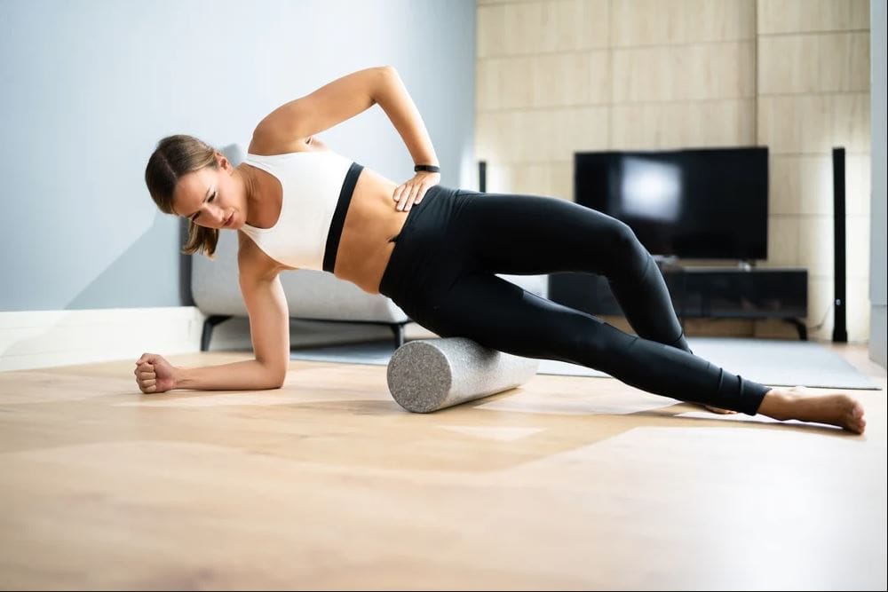
Now myofascial pain syndrome is challenging to diagnose in an examination due to the referred pain affecting one location of the body than the actual source of where the pain is coming. However, it is treatable through various techniques and stretches for the thighs to restore leg mobility. Studies reveal that direct stretching of the TFL (tensor fasciae latae) muscles can reduce long-term pain effects on the hips, thighs, and lower back and improve hip and thigh mobility. Various stretches like hip extensions and laterally rotating the hips can break the myofascial trigger points in the TFL muscle. Using a foam roller on the hips can gently stretch and loosen the muscle fibers on the TFL and help warm up the muscle before working out. Sitting down correctly in a chair can help the hips from causing more muscle strain to the thighs and prevent the TFL muscles from being shortened. Incorporating these stretches and techniques can improve hip and thigh mobility in the legs, allowing the individual to walk or run without pain.
Conclusion
The TFL (tensor fasciae latae) muscles are located on the proximal anterolateral thigh between the IT (iliotibial) band, which assists with knee flexion and lateral rotation. The TFL muscle also works with the gluteal muscles and allows the person to walk, run, and help with stability movement to the hips and knees with inflicted pain on the joints and surrounding muscles. When the TFL muscles become overused, they can develop myofascial trigger pain on the TFL, causing referred hip, knee, and thigh pain. This can cause the individual not to be able to walk for long periods and think they might have osteoarthritis in the knees. Fortunately, people can incorporate various stretches and techniques to reduce the pain in the thighs and hips while managing myofascial trigger pain along the TFL muscles. These various stretches and techniques allow mobility back to the hips and thighs so the individual can walk without pain.
References
Gottschalk, F, et al. “The Functional Anatomy of Tensor Fasciae Latae and Gluteus Medius and Minimus.” Journal of Anatomy, U.S. National Library of Medicine, Oct. 1989, https://www.ncbi.nlm.nih.gov/pmc/articles/PMC1256751/.
Ohtsuki, Keisuke. “A 3-Month Follow-up Study of the Long-Term Effects of Direct Stretching of the Tensor Fasciae Latae Muscle in Patients with Acute Lumbago Using a Single-Case Design.” Journal of Physical Therapy Science, The Society of Physical Therapy Science, May 2014, https://www.ncbi.nlm.nih.gov/pmc/articles/PMC4047246/.
Simons, D. G., and L. S. Simons. Myofascial Pain and Dysfunction: The Trigger Point Manual: Vol. 2:the Lower Extremities. Williams & Wilkins, 1999.
Sánchez Romero, Eleuterio A, et al. “Prevalence of Myofascial Trigger Points in Patients with Mild to Moderate Painful Knee Osteoarthritis: A Secondary Analysis.” Journal of Clinical Medicine, MDPI, 7 Aug. 2020, https://www.ncbi.nlm.nih.gov/pmc/articles/PMC7464556/.
Trammell, Amy P, et al. “Anatomy, Bony Pelvis and Lower Limb, Tensor Fasciae Muscle – NCBI Bookshelf.” In: StatPearls [Internet]. Treasure Island (FL), StatPearls Publishing, 8 Aug. 2022, https://www.ncbi.nlm.nih.gov/books/NBK499870/.
IFM's Find A Practitioner tool is the largest referral network in Functional Medicine, created to help patients locate Functional Medicine practitioners anywhere in the world. IFM Certified Practitioners are listed first in the search results, given their extensive education in Functional Medicine