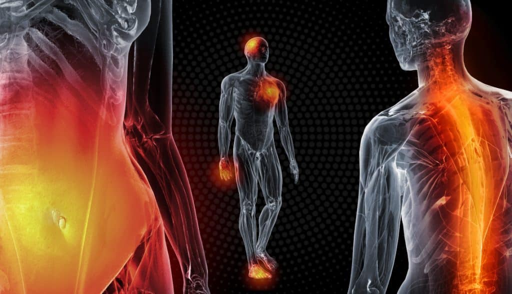
Around the world, many individuals will have some reaction to the foods they consume. This type of reaction can lead to a series of unwanted symptoms that affect not only the vital organs of the body but also the musculoskeletal system. This allergic reaction can cause inflammatory effects that lead to joint pain and swelling while affecting a person’s quality of life. Inflammation is the body’s natural defense response from the immune system to repair the body inside and out. When food allergies start to affect the entire body, it can cause the individual to be in constant pain, and many individuals will go to treatments to reduce the symptoms caused by food allergic reactions; however, the residual effects of the allergic reaction can still interfere with the body and affect the musculoskeletal system. Today’s article focuses on food allergies, how they are associated with inflammation in the musculoskeletal system, and how MET therapy can help relieve inflammation associated with food allergies. We utilize and provide valuable information about our patients to certified medical providers who use soft tissue stretching methods like MET to reduce inflammation associated with food allergies affecting the musculoskeletal system. We encourage patients by referring them to our associated medical providers based on their findings. We support that education is a marvelous way to ask our providers the most interesting questions at the patient’s acknowledgment. Dr. Alex Jimenez, D.C., incorporates this information as an educational service. Disclaimer
What Are Food Allergies?
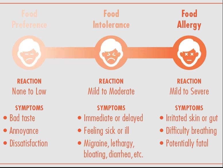
Have you been dealing with muscle swelling in different locations of your body? Do you see redness or feel a burning sensation in your muscles? Or do your muscles and joints feel achy throughout the day? Many of these pain-like symptoms are associated with inflammatory effects caused by food allergies. Research studies have revealed that food allergies are often defined as an immune reaction to food proteins that many individuals worldwide and, when indigested, are responsible for various symptoms that involve the skin, gastrointestinal tract, and respiratory tract. Many individuals would often confuse a food allergy with food intolerance since the musculoskeletal and gastrointestinal systems are caused by inflammation. Research studies have found that food intolerances are non-immunological responses that cause numerous symptoms and hypersensitivity to the body. Many factors correlate to food intolerances, and food allergies can affect the musculoskeletal system with pain-like symptoms like inflammation.
Food Allergies Associated With Inflammation In The Musculoskeletal System
When food allergies or food intolerances occur in the body, it can cause the individuals to have unwanted pain-like symptoms to cause inflammation to appear in the body. When it comes to inflammation in the body, it is produced by the immune system. It helps repair old cells and attack foreign invaders affecting the musculoskeletal system. In “Clinical Applications of Neuromuscular Techniques,” Dr. Leon Chaitow, N.D., D.O., and Dr. Judith Walker DeLany, L.M.T. stated that specific individualized pathophysiological responses exist to many foods and liquids that are being taken accounted for a significant amount of overlapping symptoms being produce. The book also states that it includes pain and discomfort to the musculoskeletal system. To that point, to figure out these presenting symptoms, whatever allergic pathogen is being derived from the food itself could be the result. Additional studies mentioned that food allergies and tolerances are sometimes not established when inflammation from the GI tract and causing pain-like symptoms associated with the musculoskeletal system. Fortunately, there are various treatments to reduce the effects of food allergies and intolerances while restoring the musculoskeletal system.
The Benefits Of A Healthy Diet & Chiropractic Care-Video
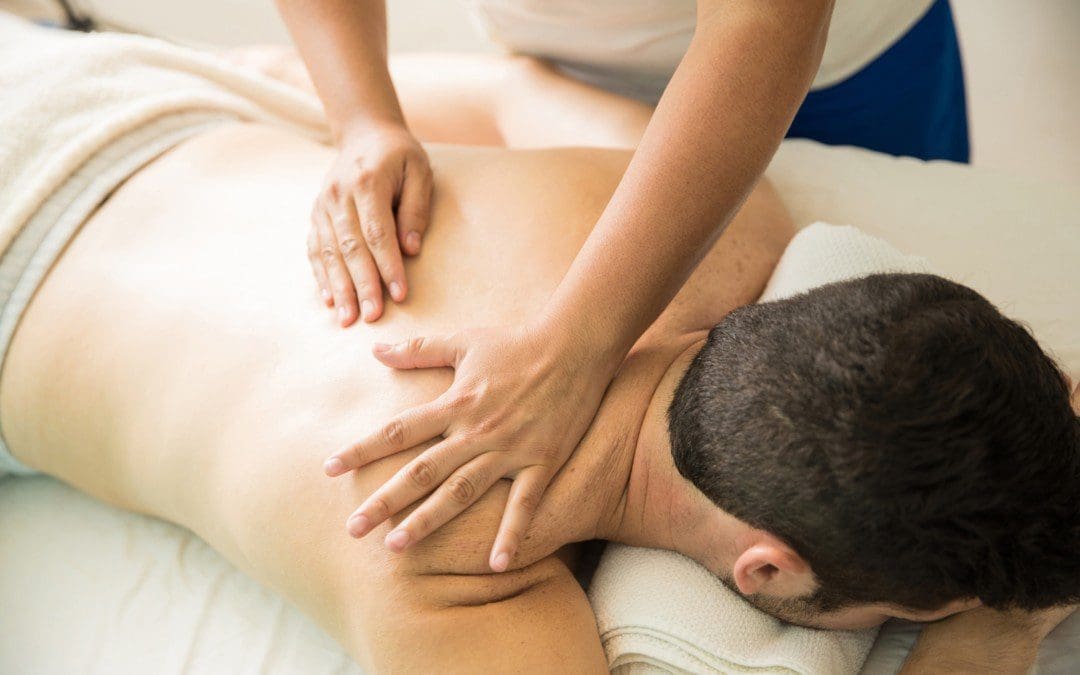
Have you been dealing with gut inflammation that is affecting your musculoskeletal system? Are you experiencing aches and pains throughout your entire body? Or do you have joint issues that are causing you limited mobility? Many of these issues are associated with food intolerances and allergies that can cause inflammation of the musculoskeletal system leaving the individual in pain. Fortunately, there are many ways to reduce the effects of inflammation associated with food allergies. The video above explains how eating the right foods while being considerate of food allergies can be combined with treatment like chiropractic care, which can help reduce inflammation while realigning the body through manual manipulation. Chiropractors also use soft tissue techniques like MET to help regain joint mobility and reduce the effects of inflammation-causing muscle and joint stiffness.
MET Therapy Relieving Inflammation Associated With Food Allergies
Therapies like soft tissue massages, massage therapy, physical therapy, or chiropractic care all work together with having a nutritional diet plan to prevent flare-ups from food allergies and intolerances. Research studies have found that MET helps stretch the affected muscles induced by inflammation associated with food allergies. This technique allows the body to naturally heal itself and prevent inflammation from exceeding more into the body. Combined with anti-inflammatory foods, many individuals know what food they can and can not consume. Additionally, it allows them to be more mindful of their bodies and sends them on the right track of their health and wellness journey.
Conclusion
Overall, many individuals often confuse food allergies and food intolerances, which can cause the musculoskeletal system to be dealing with symptoms of inflammation and pain. Since inflammation is the body’s natural defense system, it is important to be mindful of what is consumed to prevent overlapping risk profiles from causing muscle and joint pain. Luckily, numerous treatments are available to reduce the effects of chronic inflammation associated with food allergies and help the body naturally heal itself. Combining treatments like MET and a healthy nutritional diet can help the body reduce the effects of inflammation from affecting the musculoskeletal system while also allowing the individual to make smart choices in their health and wellness journey.
References
Chaitow, Leon, and Judith Walker DeLany. Clinical Applications of Neuromuscular Techniques. Churchill Livingstone, 2003.
Lopez, Claudia M, et al. “Food Allergies – Statpearls – NCBI Bookshelf.” In: StatPearls [Internet]. Treasure Island (FL), StatPearls Publishing, 31 Jan. 2023, https://www.ncbi.nlm.nih.gov/books/NBK482187/.
Ohtsuka, Yoshikazu. “Food Intolerance and Mucosal Inflammation.” Pediatrics International : Official Journal of the Japan Pediatric Society, U.S. National Library of Medicine, 2015, https://pubmed.ncbi.nlm.nih.gov/25442377/.
Sbardella, Silvia, et al. “Muscle Energy Technique in the Rehabilitative Treatment for Acute and Chronic Non-Specific Neck Pain: A Systematic Review.” Healthcare (Basel, Switzerland), U.S. National Library of Medicine, 17 June 2021, https://www.ncbi.nlm.nih.gov/pmc/articles/PMC8234422/.
Tuck, Caroline J, et al. “Food Intolerances.” Nutrients, U.S. National Library of Medicine, 22 July 2019, https://www.ncbi.nlm.nih.gov/pmc/articles/PMC6682924/.
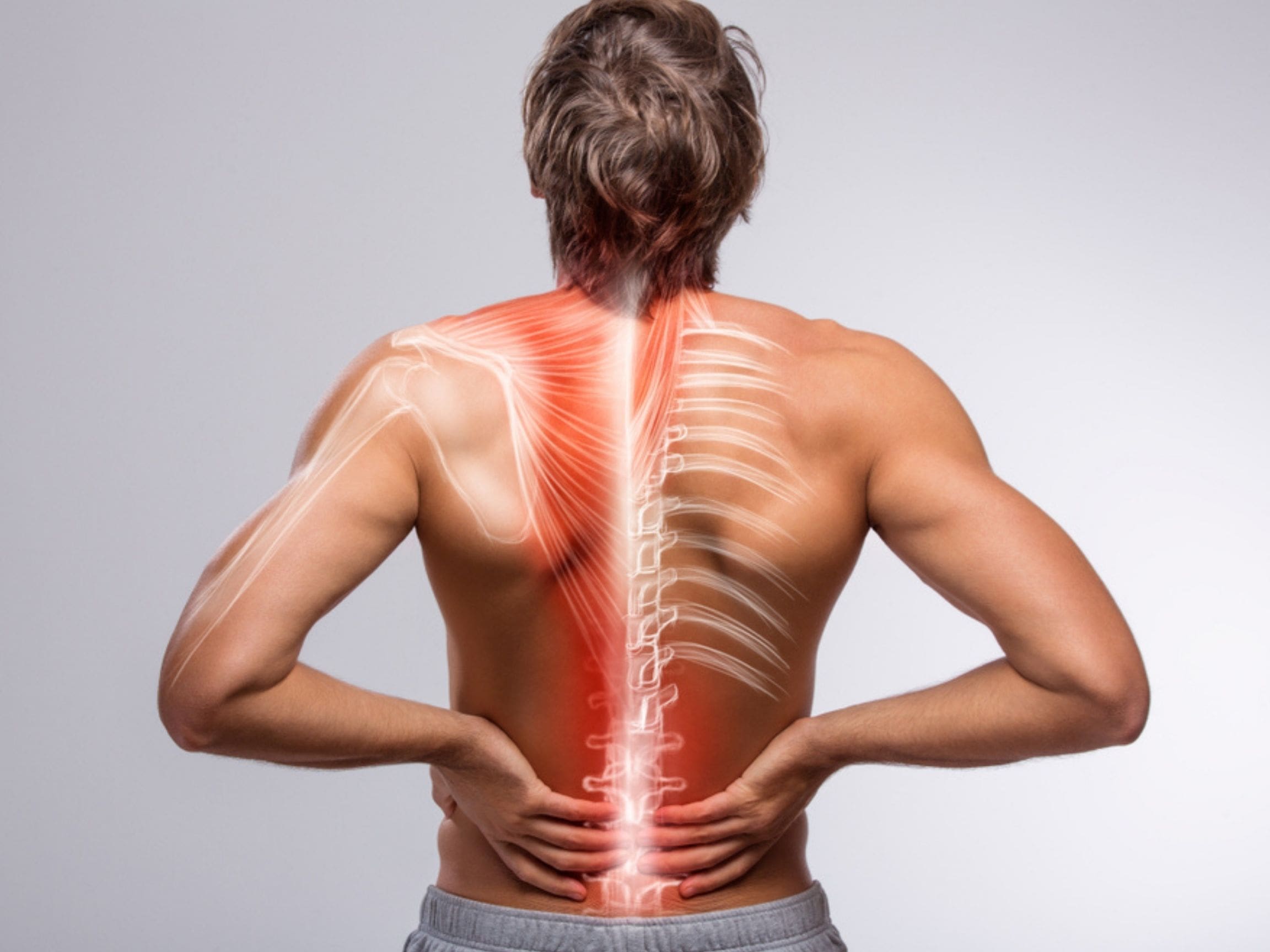
When the body sustains an injury or a virus, the immune system springs into action by sending out cytokines to the affected area and causes a process known as inflammation. Inflammation in the body is good for eliminating numerous pathogens, damaged cellular structures, viruses, or infections. Inflammation causes the affected area to be warm to the touch and causes swelling that will decrease in the recovery state. However, inflammation in the body can be positive and negative depending on the severity of the injury. When inflammation is in its chronic form, it can cause muscle and tissue damage to the musculoskeletal system while being associated with other conditions in the body’s system, like the gastrointestinal, nervous, and reproductive systems. In today’s article, we will focus on how chronic inflammation affects the body’s muscles and how the MET technique can help relieve muscle inflammation in soft tissues. We utilize valuable information about our patients to certified medical providers who use methods like MET combined with manual stretching therapy to reduce inflammation associated with musculoskeletal conditions. We encourage patients by referring them to our associated medical providers based on their findings. We support that education is a marvelous way to ask our providers the most interesting questions at the patient’s acknowledgment. Dr. Alex Jimenez, D.C., incorporates this information as an educational service. Disclaimer
How Inflammation Works In The Body
Have you been experiencing pain in different muscle areas in your body? What about dealing with aches and pains in the morning? Or do your muscles feel tight and hurt more when you bend to pick up an object? Many of these issues affecting the musculoskeletal system are associated with inflammation. As stated earlier, inflammation can positively and negatively impact the body depending on the severity of the injury. Inflammation is a natural healing process for the body that is characterized by redness, swelling, and heat to the soft tissues so that they can be repaired within a few days to weeks. Studies reveal that when the body deals with various pathogenic factors, the immune system releases inflammatory cytokines to the pathogenic factor and starts to heal the affected area.
Inflammation helps the sprained muscle tissues heal naturally and can be healed within 2-3 days to a few weeks in its acute stage. However, when inflammation is in a chronic state of the body, it causes damage to not only the affected muscle and tissue areas but can even affect the surrounding ligaments, joints, and vital organs. A great example is when a person has a sprained ankle, the affected area swells up, becomes red, and is tender to the touch when its acute inflammation. For chronic inflammation, the body is in constant pain that can be slow and long, and it takes several months to years to heal. Another great example would be gut disorders associated with chronic muscle inflammation.
What Does Chronic Inflammation Do To The Body’s Muscles?
So what does chronic inflammation do to the body’s muscles, and how does it affect the surrounding structures? According to research studies, chronic inflammation has been associated with musculoskeletal disorders that can cause an impact in reducing muscle strength and muscle mass. When this happens, it can cause the muscles to weaken and generate the surrounding muscle groups to compensate by working harder. This leads to misalignment in the body and causes overlapping risk profiles in the various muscle groups. In “Clinical Applications of Neuromuscular Techniques,” Dr. Leon Chaitow, N.D., D.O., and Dr. Judith Walker DeLany, L.M.T., stated that numerous factors associated with inflammation could impact how a person sleeps, eat, and function throughout the day. The book also noted that when pathogens can disrupt the natural cycling between the defensive and repair modes of inflammation correlating with the immune system can be disrupted in ill health. A chronic inflammatory cytokine shift could lock the body into a pro-inflammatory state.
Reducing Inflammation In The Body- Video
Have you been dealing with constant inflammation in your muscles? Do you feel muscle weakness or strain when you are in motion? Or do other portions of your body starts to feel aches or pains? Many of these issues are associated with chronic inflammation affecting the musculoskeletal system. Inflammation is part of the immune system’s natural defense that sends cytokines to the affected area and starts the healing process. Inflammation has two patterns: acute and chronic. Acute inflammation is associated with sprains from a twisted ankle or wrist, a sore throat, or an infection. While at the same time, chronic inflammation is associated with muscle pain, sleep disorders, or rashes that affect the skin. When the body is suffering from chronic inflammation, it can be due to many overlapping factors that can cause the body to be in pain. Luckily numerous treatments can help the body and reduce the effects of inflammation. The video above explains how incorporating an anti-inflammatory diet and chiropractic care can help reduce chronic inflammation effects in the body.
The MET Technique & Muscle Inflammation
Regarding chronic muscle inflammation, the musculoskeletal system deals with numerous symptoms like pain, stiffness, and weakness associated with various pathological factors. Chronic inflammation can develop from insufficient sleep, eating high-cholesterol foods, not getting enough exercise, and being stressed, which can affect the body and its systems. Fortunately, numerous available treatments can reduce inflammation. Studies reveal that treatments like MET therapy can help reduce pain correlated with muscle inflammation and increase the range of motion to the joints. The body can reduce the effects of chronic inflammation and keep it under control when MET therapy is combined with an anti-inflammatory diet. Also, practicing mindfulness can help reduce the stress contributing to chronic inflammation, which can help relax the body and naturally heal itself. Making these small changes can benefit many individuals trying to be healthier.
Conclusion
When it comes to inflammation in the musculoskeletal system, it can lead to overlapping risk profiles that can cause the body to be misaligned and have many pain-like symptoms that can make a person’s life miserable. Since inflammation is part of the immune system’s natural response to fight off infections, incorporating anti-inflammatory diets and MET stretching can help reduce the effects of inflammation and help the body heal itself from the inside out.
References
Chaitow, Leon, and Judith Walker DeLany. Clinical Applications of Neuromuscular Techniques. Churchill Livingstone, 2003.
Chen, Linlin, et al. “Inflammatory Responses and Inflammation-Associated Diseases in Organs.” Oncotarget, U.S. National Library of Medicine, 14 Dec. 2017, https://www.ncbi.nlm.nih.gov/pmc/articles/PMC5805548/.
Thomas, Ewan, et al. “The Efficacy of Muscle Energy Techniques in Symptomatic and Asymptomatic Subjects: A Systematic Review.” Chiropractic & Manual Therapies, U.S. National Library of Medicine, 27 Aug. 2019, https://www.ncbi.nlm.nih.gov/pmc/articles/PMC6710873/.
Tuttle, Camilla S L, et al. “Markers of Inflammation and Their Association with Muscle Strength and Mass: A Systematic Review and Meta-Analysis.” Ageing Research Reviews, U.S. National Library of Medicine, 26 Sept. 2020, https://pubmed.ncbi.nlm.nih.gov/32992047/.
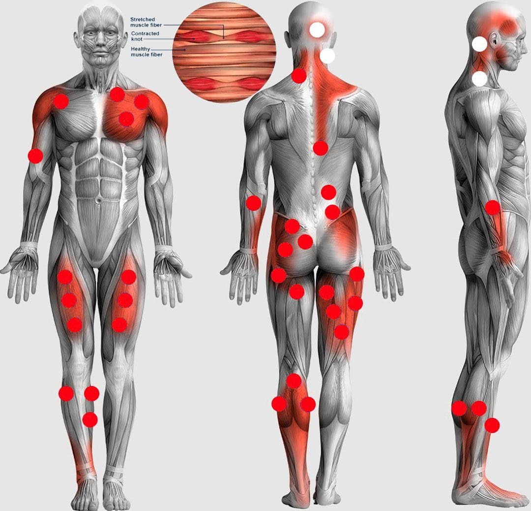
Muscle knots or trigger points are tissues/segments of muscle fibers stuck in a contracted state and balled up or become entangled. To the touch, they can feel like small bumps, nodules, or knots. They are primarily found in muscles, but they can also be found in tendons, fascia, periosteum, and ligaments. Tight muscles can limit flexibility, increase discomfort and pain symptoms, and cause further injury that can develop into chronic conditions. Trigger points can develop in all muscles and multiple muscles simultaneously. The Injury Medical Chiropractic and Functional Medicine Team can create a personalized treatment plan for trigger point alleviation.
Muscle Knots Trigger Points
When muscle fibers are stuck in contraction, blood circulation decreases in and around the affected area, and the necessary nutrients and oxygen cannot be delivered. The excessive accumulation of the chemicals – acetylcholine, and calcium leads to a lack of oxygen in the area, which causes muscle fiber contraction and spasms. As the muscle fibers overly contract, they form a knot. Waste materials build up in the fibers as the blocked circulation doesn’t allow removal. This irritates the trigger point, which reacts by sending out pain signals. The brain responds by telling the body not to use that muscle, causing the muscle to tighten, become weak, and lose range of motion. The other muscles have to work harder to compensate.
Trigger Point Types
Active and Latent
One type is an active trigger point.
The trigger point causes pain and discomfort even when the muscle or tissue rests.
The other type is latent, which means that the point does not cause pain or is sometimes even noticed until pressure is applied to the point or area.
Because key trigger point muscles are weakened, the muscles around that point need to work harder, causing multiple knots to form.
Referred pain happens when a satellite trigger point causes sensations in a different area from the point itself.
Causes
The main causes include the following:
Direct Trauma
Muscle trauma or injury can cause fibers not to heal properly, causing segments to stay deprived of oxygen and knotted.
Excessive and Extended Exercising
During exercise/physical activity, profound muscle strain and injury can occur from pushing too hard for too long without proper recovery and repair.
Maintaining Healthy Posture
Unhealthy postures can cause tension, stiffness, and aches and pains.
Stress and Fatigue
Emotional and physical stress takes a toll on the body, which can result in excessive muscle contraction.
Most of the time, it happens subconsciously without realizing that the body is tensing up.
This is why engaging in relaxation activities, including therapeutic massage, is important.
Inactivity
Lack of physical activity and a sedentary lifestyle can cause muscle spasms and over-contracting, leading to muscle knots.
Medical Conditions
Conditions that affect muscles, like arthritis and fibromyalgia or medications, can cause muscle spasms and the development of trigger points.
Chiropractic and Massage Therapy
Chiropractic and therapeutic massage is an effective treatment to relieve muscle knots and symptoms and restore the body to optimal function. Massage uses various techniques to increase blood circulation, break down inflexible scar tissue and stretch and loosen muscles. Pain and discomfort decrease by allowing the muscles to move and restoring blood flow.
Massage Therapy Chiropractic Care
References
Barbero, Marco, et al. “Myofascial pain syndrome and trigger points: evaluation and treatment in patients with musculoskeletal pain.” Current Opinion in Supportive and palliative care vol. 13,3 (2019): 270-276. doi:10.1097/SPC.0000000000000445
Cheung, Karoline, et al. “Delayed onset muscle soreness: treatment strategies and performance factors.” Sports medicine (Auckland, N.Z.) vol. 33,2 (2003): 145-64. doi:10.2165/00007256-200333020-00005
Money, Sarah. “Pathophysiology of Trigger Points in Myofascial Pain Syndrome.” Journal of Pain & palliative care pharmacotherapy vol. 31,2 (2017): 158-159. doi:10.1080/15360288.2017.1298688
Moraska, Albert F et al. “Responsiveness of Myofascial Trigger Points to Single and Multiple Trigger Point Release Massages: A Randomized, Placebo-Controlled Trial.” American Journal of physical medicine & Rehabilitation vol. 96,9 (2017): 639-645. doi:10.1097/PHM.0000000000000728
Weerapong, Pornratshanee, et al. “The mechanisms of massage and effects on performance, muscle recovery, and injury prevention.” Sports medicine (Auckland, N.Z.) vol. 35,3 (2005): 235-56. doi:10.2165/00007256-200535030-00004
When it comes to the body, the lower portion has three compartments of muscles that work together to provide stability and mobility to the host when they are in motion. The anterior, posterior, and lateral compartments have numerous muscles, tissues, and ligaments that support the spine and allow the musculoskeletal system to do various movements without pain. When normal factors affect the body, it can cause symptoms of overlapping risk profiles that can lead to musculoskeletal pain disorders associated with pain-like symptoms in the joints and muscles. Today we will look at one of the muscle compartments known as the postural muscles, how postural pain affects the body, and how manual therapy combined with the MET technique can improve the postural muscles. We mention valuable information about our patients to certified medical providers who use methods like the MET combined with manual therapy to reduce pain-like symptoms associated with musculoskeletal disorders. We encourage patients by referring them to our associated medical providers based on their findings. We support that education is a marvelous way to ask our providers the most interesting questions at the patient’s acknowledgment. Dr. Alex Jimenez, D.C., incorporates this information as an educational service. Disclaimer
What Are The Postural Muscles?
Are you experiencing muscle stiffness in your lower back? What about aches and pain in your shoulders and neck? Or have you noticed your legs feel heavy after sitting down for a long time? Many of these issues are associated with the postural muscles that are causing pain to the musculoskeletal system. So what are the postural muscles in the musculoskeletal system? Well, they are the core muscles that are deep within the abdomen, pelvis, and back. Research studies reveal that the curvature of the spine (cervical, thoracic, and lumbar regions) communicates and works with the central nervous system and musculoskeletal system to provide balance, support, and resistance against pressure on the body. The postural muscles are important in the body as they ensure the host has perfect posture when walking, sitting, or standing. Additional studies mentioned that good postural and stability control are the fundamentals for motor skills. When a person has good postural control, it can help give them a stable gait when walking. However, as the body ages, the postural muscles can become weak and lead to muscle strain while affecting the joints and tendon structures.
The Effects Of Postural Pain In The Body
So what happens to the body when dealing with pain in the postural muscles, and how does it affect one’s posture? Research studies reveal that reducing back muscle endurance from prolonged sitting, slouching, or constantly looking down can cause muscle strain symptoms in the postural muscles. To that point, it can lead to increased development of low back pain and lumbar discomfort in the joints. As stated earlier, other factors like slouching, prolonged sitting, and constantly looking down can affect the lower back, shoulders, and neck. The various muscles that make up the shoulders and neck would become strained and tensed, leading to shoulder and neck pain that can cause discomfort. When these overlapping symptoms affect the body, it can cause many overlapping symptoms that can overlap and cause the individual to be miserable.
How Neck Injuries Affect The Body- Video
Have you been experiencing any muscle strain in your neck, shoulders, or lower back? Does stretching bring temporary relief? Or have you been dealing with heaviness in your legs? Many of these factors are associated with weak postural muscles that can contribute to poor posture, low back and neck pain, and other musculoskeletal issues. The video above explores the common causes and symptoms of neck injuries and how treatments like chiropractic care can help alleviate the symptoms. Treatments like chiropractic care utilize manual manipulation and various techniques to help realign the body and reduce any musculoskeletal disorders associated with pain. Chiropractic care is non-invasive and works with other medical professionals to restore the body naturally.
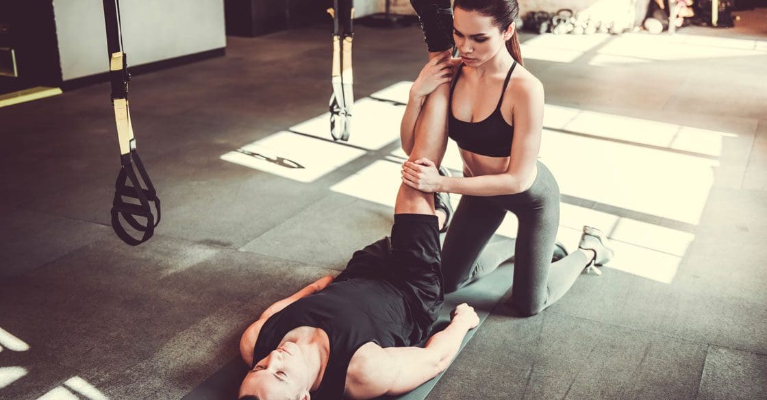
Manual Therapy & MET On Postural Muscles
So what can one do when dealing with postural pain and trying to find relief? Many individuals go to treatments like chiropractic care, which can allow the body to be realigned and restored naturally. Treatments like chiropractic care can help restore good posture in the spine while relieving and reducing excess pain and strain on the various muscles, tendons, and joints. Additionally, chiropractors use techniques like the MET technique to reduce stress on the soft tissues and restore the joint’s range of motion. According to the book, “Clinical Applications of Neuromuscular Techniques,” Leon Chaitow N.D., D.O., and Judith Walker DeLany L.M.T., stated that muscle restoration is accompanied by biomechanical solutions and strategies that are introduced to the body and become a key focus on the muscles that required strengthening, enhancing, and improve breathing and posture function. When therapists like chiropractors and massage therapists use the MET technique, the affected muscles can be stretched and strengthened while restoring the structural and functional imbalances the body has endured. This can help improve postural muscles while allowing the body to heal naturally. This allows the individual to be mindful of how they present themselves with good posture.
Conclusion
Overall, the body requires the postural muscles to help stabilize and keep the body mobile. When musculoskeletal disorders associated with pain started to affect the muscles through bad posture or other normal factors, it can cause these muscles to be weak and develop musculoskeletal conditions that can lead many individuals to constant pain. Luckily therapies like chiropractic care combined with the MET technique allow the affected muscles to be stretched and strengthened. This allows the body to be realigned and restored naturally. Incorporating stretching combined with chiropractic care can help many individuals be mindful of their posture and enable them to continue their health and wellness journey without pain.
References
Carini, Francesco, et al. “Posture and Posturology, Anatomical and Physiological Profiles: Overview and Current State of Art.” Acta Bio-Medica : Atenei Parmensis, U.S. National Library of Medicine, 28 Apr. 2017, https://www.ncbi.nlm.nih.gov/pmc/articles/PMC6166197/.
Chaitow, Leon, and Judith Walker DeLany. Clinical Applications of Neuromuscular Techniques. Churchill Livingstone, 2003.
Jung, Kyoung-Sim, et al. “Effects of Prolonged Sitting with Slumped Posture on Trunk Muscular Fatigue in Adolescents with and without Chronic Lower Back Pain.” Medicina (Kaunas, Lithuania), U.S. National Library of Medicine, 23 Dec. 2020, https://www.ncbi.nlm.nih.gov/pmc/articles/PMC7822118/.
Ludwig, Oliver, et al. “Neuromuscular Performance of Balance and Posture Control in Childhood and Adolescence.” Heliyon, U.S. National Library of Medicine, 31 July 2020, https://www.ncbi.nlm.nih.gov/pmc/articles/PMC7398941/.
The various muscle groups in the body allow the host to move around and function through many actions without feeling any discomfort or pain. The body has two sections: upper and lower portions that have different functions, from turning the neck from side to side to allowing the legs to enable the body to move around. When various issues or factors begin to affect the body over time, like muscle injuries or normal factors like poor posture and prolonged sitting, it causes overlapping risk profiles that can lead to chronic musculoskeletal conditions. When musculoskeletal disorders affect the body, it can lead to muscle and joint pain that can cause misalignment in the spine and cause the muscle fibers to become short and tense. Luckily there are available treatments that allow the body to realign itself and stretch those short muscles. Today’s article looks at how muscle pain affects the body and how different variations of the MET technique are used to reduce and stretch muscle pain. We mention and provide valuable information about our patients to certified medical providers who use techniques like the MET and therapy for individuals with muscle strain associated with body pain. We give encouragement to patients by referring them to associated medical providers based on their diagnostic findings. We support that education is a marvelous way to ask our providers the most interesting questions at the patient’s acknowledgment. Dr. Alex Jimenez, D.C., incorporates this information as an educational service. Disclaimer
How Does Muscle Pain Affect The Body?
Have you been dealing with muscle strain or pain in different body areas? Are you experiencing any referred pain in other body locations? Or are your muscles feeling extremely tight that it is causing you pain? When the body is dealing with various issues that are causing the muscle fibers to be tensed, it can lead to muscle pain and cause many people to suffer. Studies reveal that muscle pain is caused when painful conditions like mechanical forces, ischemia, and inflammation stimulate the body’s free nerve endings. Many of these factors also correlate with musculoskeletal disorders like fibromyalgia and myofascial pain that can develop trigger points (palpable, small nodules) in the muscle fibers to cause the muscles to become stiff and contract. Additional studies also reveal that when the muscles begin to cramp up, especially the calves, it can become extremely painful and involuntary as it affects the entire muscle group, the muscle itself, or any selected muscle fibers. This causes the individual to be in pain in an acute setting as the muscle relaxes; however, if the muscle fibers are still in constant contraction, it can lead to chronic issues that affect the muscle group.
Overcoming Pain With Chiropractic Care-Video
Regarding muscle pain in the body, studies reveal that the nociceptive nerve endings in the muscles and tissue fibers can cause the neuron signals from the central nervous system to become hyperexcitable, and hyperactivity can lead to muscle pain. This causes the muscle group and the surrounding muscles to tense and invokes pain when in motion. To that point, it can cause the individual dealing with muscle pain to try and find various treatments to alleviate the pain and continue their lives. When it comes to pain can be relieved through treatments like chiropractic care and massage therapy to reduce the effects of the muscle pain associated with musculoskeletal disorders. The video above explains how treatments like chiropractic care incorporate different techniques to realign the body from subluxation and help stretch the tight, short muscles using manual manipulation and the MET technique.
Variations Of The MET Technique
When the muscles in the musculoskeletal system are dealing with pain in different locations or one location in the body, it can cause the individual to be in constant pain. Luckily treatments like chiropractic care are non-invasive and therapeutic as they utilize different techniques to realign the spine and stretch the tight muscle groups. In “Clinical Applications of Neuromuscular Techniques,” written by, Leon Chaitow, N.D., D.O., and Judith Walker DeLany, L.M.T., stated that when muscle pain affects tone muscle or causes structural changes to the joint’s ROM (range of motion) can lead to shortness and stress to the muscle group. Studies reveal that MET is a stretching technique therapists use to contract the affected muscle in a precisely controlled direction voluntarily. Many stretch variations of the MET technique allow the muscles to be stretched, strengthening and improving local circulation while mobilizing joint restriction. Down below are some of the variations of stretching techniques with MET.
Isometric Contraction: Reciprocal Inhibition In Acute Setting
The isometric contraction technique is used for reciprocal inhibition in an acute setting where the affected muscles are dealing with symptoms of muscle spasms. The isometric contraction allows the therapist to help relax acute muscular spasms and mobilize restricted joints while preparing the joints for manipulation.
Starting point: When acute muscles or joint problems affect the body’s functionality, therapists must commence an easy restriction barrier.
Modus Operandi: The affected muscles are used in an isometric contraction, allowing the short muscle to relax.
Forces: The therapist and individual forces are matched and involve 20% of the individual’s strength to increase no more than 50%.
Duration: Initially7-10 seconds while increasing up to 20 seconds.
Action following contraction: The area of the muscle and joint are taken to a new restricted barrier without stretching after complete relaxation. Therapists should perform the movement to a new restricted barrier on exhalation.
Repetitions: Repeat three to five times until no further gain in the range of motion is possible.
Isometric Contraction: Post-isometric Relaxation In Chronic Setting
The isometric contraction technique is used for post-isometric relaxation in a chronic setting where the muscles are severely contracted. The isometric contraction technique is known as post-facilitation stretching, where therapists stretch chronic or subacute restricted, fibrotic, contracted soft tissues or muscle tissues affected by myofascial trigger point pain.
Starting point: Short of resistance barrier
Modus Operandi: The affected muscles are used in the isometric contraction that allows the shortened muscles to relax and let an easier stretch.
Forces: Both the therapist and individual forces match and increase about 30% of the patient’s strength and increase to 50% of contractions up to 20 seconds.
Duration: Initially 7-10 seconds and increasing up to 20 seconds.
Action following contraction: The rest period is 5 seconds so the body can relax completely before being stretched, and during exhalation, the muscle goes through a painless, new restriction barrier position that is held for at least 10-60 seconds.
Repetitions: Repeat three to five times.
Isotonic Eccentric Contraction
The isotonic eccentric contraction is an isolytic technique to strengthen weak postural muscles that are tensed or tight from prolonged sitting or poor posture.
Starting point: At the restriction barrier
Modus Operandi: When the muscle is contracted and prevented, the therapist uses this technique to slowly overcome and reverse the contracting muscle so it can be stretched to full resting length.
Forces: Therapists use greater forces on the individual and build up subsequent contractions for the affected muscle (*Utilize this stretch on individuals who are not dealing with osteoporotic issues in their muscles and joints)
Duration: Five to seven seconds
Repetitions: Repeat three to five times if discomfort is not excessive.
Isokinetic
The isokinetic technique is a combination of isotonic and isometric contractions that many pain specialists like chiropractors and massage therapists use to tone weakened musculature, build strength in all the surrounding muscles that are involved in a particular joint function, and help train and balance the effects on the body’s muscle fibers.
Starting point: Easy mid-range position
Modus Operandi: The individual uses moderate resistance as the therapist puts the joint through a rapidly full range of movement as this technique is different than simple isotonic exercises, and resistance varies from each person. This technique progressively increases as the procedure progress.
Forces: The therapist uses moderate forces to prevent movement from the individual and then progresses to full forces.
Duration of contraction: Up to four seconds.
Conclusion
Different stretching techniques in MET therapy allow the affected muscles to be stretched and lengthened while reducing muscle pain affecting a body’s location. It is important to be mindful of the movements that can cause the muscles to be overstretched or cramped up, which can cause pain. Treatments incorporating these various stretching techniques allow the affected muscles to relax and restore naturally.
Chaitow, Leon, and Judith Walker DeLany. Clinical Applications of Neuromuscular Techniques. Churchill Livingstone, 2003.
Faqih, Anood I, et al. “Effects of Muscle Energy Technique on Pain, Range of Motion and Function in Patients with Post-Surgical Elbow Stiffness: A Randomized Controlled Trial.” Hong Kong Physiotherapy Journal : Official Publication of the Hong Kong Physiotherapy Association Limited = Wu Li Chih Liao, U.S. National Library of Medicine, June 2019, https://www.ncbi.nlm.nih.gov/pmc/articles/PMC6467834/.
Gregory, Nicholas S, and Kathleen A Sluka. “Anatomical and Physiological Factors Contributing to Chronic Muscle Pain.” Current Topics in Behavioral Neurosciences, U.S. National Library of Medicine, 2014, https://www.ncbi.nlm.nih.gov/pmc/articles/PMC4294469/.
Mense, Siegfried. “The Pathogenesis of Muscle Pain.” Current Pain and Headache Reports, U.S. National Library of Medicine, Dec. 2003, https://pubmed.ncbi.nlm.nih.gov/14604500/.
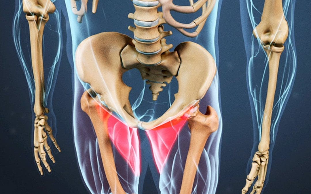
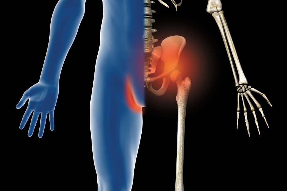
The muscles surrounding the hips in the lower extremities provide stability to the lumbar spine and pelvis while allowing mobility, flexibility, and rotation to the groin, legs, and thighs. The muscles surrounding the groin consist of three large muscle groups: the abdominals, iliopsoas, and adductors, which have a casual relationship with the various ligaments and soft tissues that can be succumbed to injuries or other symptoms that can lead to pain and misalignment to the body. This happens to many individuals who participate in sports or frequently exercise, which can cause strain near the groin and the surrounding muscles. Today’s article focuses on the causes of a groin strain, how it affects the hips and pelvis, and how available treatments can help reduce groin strain. We mention and provide valuable information about our patients to certified medical providers who use techniques like the MET and therapy for individuals with a groin strain and causing mobility issues when functioning. We give encouragement to patients by referring them to associated medical providers based on their diagnostic findings. We support that education is a marvelous way to ask our providers the most interesting questions at the patient’s acknowledgment. Dr. Alex Jimenez, D.C., incorporates this information as an educational service. Disclaimer
The Causes Of Groin Strain
Have you experienced any mobility issues when walking? Do you feel pain near your hips or groin? Or do you feel muscle weakness in your thighs when exercising? Many of these issues are associated with a muscle strain near the groin affecting your lower body. The muscles surrounding the groin allow the legs and thighs to move around in flexion, rotation, and extension without feeling pain. However, when a person starts to feel pain radiating around their groin, it can become an issue if not taken care of. Studies reveal that some of the causes, like adductor strain or injuries from physical activities, can cause the muscle fibers in the three muscle groups of the groin to be in pain. In “Clinical Applications of Neuromuscular Techniques,” authors Leon Chaitow and Judith Walker DeLany, stated that chronic joint and soft tissue conditions predate presenting acute symptoms to the groin muscle regions. To that point, if a person has predisposed injuries in the groin muscles, it can affect how a person walks and functions when in motion, potentially leading to issues affecting the hips and pelvis.
How Groin Strain Affect The Hips & Pelvis
Studies reveal multiple pathologies that can coexist in the hips and pelvis that could cause similar symptoms to the hips and pelvis that can correlate with several organ systems like the gut and reproductive system that causes referred pain to the groin. Additional studies also mentioned that groin pain is widely known as an issue among professional and amateur athletes. They could experience different symptoms and injuries from groin pain associated with the hips and pelvis. An example would be if an individual is participating in a sports event, they could be experiencing myofascial trigger points related to the groin, affecting the pelvis and hips. Some of the symptoms associated with groin strain include:
Stiffness
Swelling
Muscle weakness
Groin discomfort
Bruising around the area
Leg discomfort when flexing
Walking issues
Lower stomach or back symptoms
Many of these symptoms that affect the hips and pelvis can lead to many issues that cause people to be in constant pain or discomfort until they find relief.
Chiropractic Care: The Ancient Healing Art for Modern Life- Video
Have you been experiencing mobility issues? What about pain in your hips, pelvis, or groin? Or do you often feel discomfort when stretching? Many of these issues are associated with groin strain along the hips and pelvic region. Groin strain occurs in the three large muscle groups: the abdominals, iliopsoas, and adductors, when they are overstretched and cause referred pain to the hips and pelvis. This causes many individuals to have instability and mobility issues. Some of the present symptoms that occur to these muscles include:
Pain in active movement
Pain during palpation
Localized swelling
Pain while stretching the muscle-tendon
When pain is causing issues in the groin, many individuals will go to available treatments to help reduce the pain and regain mobility when exercising. One of the available treatments is chiropractic care. The video above shows how chiropractic care is incorporated into modern life as it uses non-invasive hands-on treatment to realign the body and help lengthen the affected muscles.
Available Treatments To Reduce Groin Strain
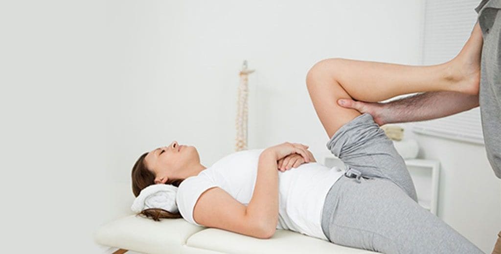
Numerous treatments can help reduce groin strain from affecting the lower extremities and help bring mobility back to the surrounding muscles. Many individuals would utilize ice packs, resting and elevating the leg to prevent future injuries from re-occurring. However, if left untreated, it can worsen and lead to chronic conditions. Luckily treatments like chiropractic care combined with soft tissue stretching techniques like MET (muscle energy techniques) can help to mobilize restricted joints and relax acute muscular spasms or contractions while preparing the joint for manipulation. A chiropractor will utilize these techniques to realign the spine from subluxation and loosen stiff joints and surrounding muscles that have been affected. Chiropractors will also inform their patients to incorporate various exercises and stretches to help strengthen the affected muscle group and become more aware of their bodies.
Conclusion
Overall, the various muscles, tendons, and ligaments surrounding the lower extremities, like the thighs, legs, and groin, allow mobility, flexibility, and rotation to the lower portion of the body. When injuries begin to affect these extremities, it can lead to referred pain to the hips and pelvis, causing groin strain and invoking pain. If not treated right away, it can develop into chronic conditions that cause mobility and stability issues in the lower body. The upper body would have to compensate for the pain causing subluxation to the spine. Luckily, treatments like chiropractic care combined with stretching techniques like MET can help reduce the pain in the groin muscles while lengthening the short muscles. These techniques allow mobility back to the lower extremities and realign the body so the individuals can be pain-free.
References
Bisciotti, Gian Nicola, et al. “Groin Pain Syndrome: An Association of Different Pathologies and a Case Presentation.” Muscles, Ligaments and Tendons Journal, U.S. National Library of Medicine, 20 Oct. 2015, https://www.ncbi.nlm.nih.gov/pmc/articles/PMC4617224/.
Chaitow, Leon, and Judith Walker DeLany. Clinical Applications of Neuromuscular Techniques. Churchill Livingstone, 2003.
Kiel, John, and Kimberly Kaiser. “Adductor Strain.” In: StatPearls [Internet]. Treasure Island (FL), StatPearls Publishing, 21 June 2022, https://www.ncbi.nlm.nih.gov/books/NBK493166/.
Tyler, Timothy F, et al. “Groin Injuries in Sports Medicine.” Sports Health, U.S. National Library of Medicine, May 2010, https://www.ncbi.nlm.nih.gov/pmc/articles/PMC3445110/.
With the body being a complex machine with various muscle groups and sections that work to keep the body mobile, it is important to know that weak muscles in the upper and lower portions of the body can cause unwanted pain-like symptoms that can lead to dysfunction over time. When numerous environmental factors and habits affect the muscle groups, it can lead to overlapping risk factors that cause tightness in the affected muscles and lead to injuries. In the lower portions of the body, the hips, thighs, hamstrings, and glute muscles help stabilize the pelvis region. When these factors start to cause issues with these muscles, it can lead to injuries and problems for those muscle groups. Today’s article will examine how hamstring injuries occur, how it affects the lower body, and how treatments and techniques like MET (muscle energy techniques) are utilized to relieve hamstring injuries. We mention valuable information about our patients to certified medical providers who provide therapy techniques like the MET and care treatment for individuals with hamstring injuries associated with the lower body portions. We give encouragement to patients by referring them to our associated medical providers based on their diagnostic findings. We provide the support that education is a spectacular way when asking our providers the most helpful questions at the patient’s acknowledgment. Dr. Alex Jimenez, D.C., utilizes this information as an educational service. Disclaimer
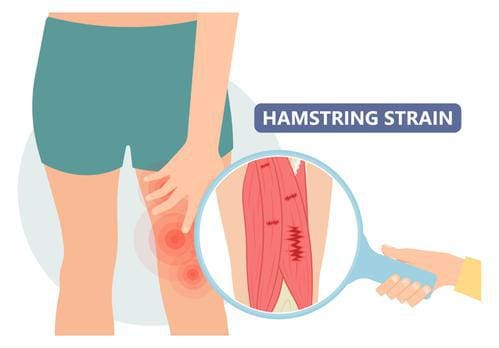
How Do Hamstring Injuries Occur?
Have you noticed that your hamstrings are feeling tight? Are you constantly sitting down for an extended period? Or are you experiencing low back pain that is affecting your hamstrings? Many individuals will usually experience low back pain along the thigh with associated symptoms of muscle weakness that causes the muscle fibers to be tighter and sore. When the muscle fibers are tight consistently, it causes the back of the leg muscles uncomfortable and can make movement difficult. Studies reveal that the back of the leg muscles or hamstrings is highly susceptible to injuries, especially in athletes. The hamstring muscles comprise three major muscles in the posterior location of the thigh. When a person is overstretching the hamstrings or having muscle tightness from being sedentary can cause these injuries and discomfort to the lower extremities. Additional research studies mentioned that hamstring injuries could range from acute muscle strain to chronic proximal hamstring tendinopathy associated with muscle ruptures.
How Does It Affect The Lower Body?
Since the hamstring muscles succumb to injuries from overstretching or becoming weak, how would it affect the lower body and cause mobility issues? Well, when the hip flexors or the hamstrings become tight and tense, it can cause an altercation to the pelvis region and cause spinal misalignment. To that point, it can lead to muscle stiffness and pain in the hamstrings while correlating to low back pain and can cause the individual to be confused as they think it is sciatica instead of a hamstring injury. Studies reveal in “Clinical Applications of Neuromuscular Techniques,” written by Leon Chaitow, N.D., D.O., and Judith Walker DeLany, L.M.T., states that when there is a range of other biomechanical features that could be predisposed to hamstring injuries that can cause a chain of reactions that can involve not only the hamstrings but the toes, the spine, the trunk, and the upper extremities. Losing the ability to function in the lower extremities can cause dysfunction, muscle weakness, and instability in an individual.
Natural Healing: Chiropractic Care for Injury Recovery- Video
Have you been experiencing stiffness or pain in your hamstrings? What about feeling discomfort in one side of your hips and glutes? Or are you experiencing muscle strain? Many of these issues are correlated with hamstring injuries that can cause muscle weakness and instability in the body. Fortunately, when dealing with a hamstring injury, techniques like gentle stretching and warming up the muscles allow the injury to recover and bring relief. Another way a person can get pain relief from a hamstring injury is through chiropractic care. Chiropractic care can help relieve the problem without using medications, injections, or surgery through spinal manipulation and incorporating various techniques to stretch the stiff muscles and realign the body. The video above explains how chiropractic care is utilized for injury recovery.
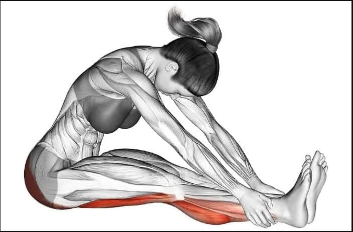
Treatments For Hamstring Injuries
When dealing with hamstring injuries, it is important to rest to prevent future injuries from occurring and incorporate gentle stretches of the targeted muscles to avoid cramping and pain. If gentle stretching doesn’t bring relief, then it is recommended to have a personalized treatment and program with a chiropractor. A chiropractor uses hands-on therapy to loosen and relieve tense muscles, thus reinforcing flexibility and range of motion back to the hamstrings. Chiropractic care also incorporates stretching techniques like MET to improve hamstring flexibility. Studies reveal that the MET technique uses soft tissue mobilization to increase the hamstring’s ROM (range of motion) while bringing mobility back to the hips and reducing pain. Furthermore, these stretches and treatments can help decrease instability and allow the individual to be pain-free.
Conclusion
The hamstrings are located along the back of the thigh and just below the knees, as they can succumb to injuries due to overstretching or other factors that cause symptoms of pain and weakness. Hamstring injuries are common and can range from acute to chronic, depending on the injury. Many people with hamstring injuries often deal with overlapping issues of sciatica and low back pain that can lead to instability in the lower body. Luckily, different treatments and stretching techniques can help lengthen the hamstring muscles, promote flexibility back to the hamstrings, and cause relief to the affected muscle.
References
Chaitow, Leon, and Judith Walker DeLany. Clinical Application of Neuromuscular Techniques. Churchill Livingstone, 2002.
Chu, Samuel K, and Monica E Rho. “Hamstring Injuries in the Athlete: Diagnosis, Treatment, and Return to Play.” Current Sports Medicine Reports, U.S. National Library of Medicine, 2016, https://www.ncbi.nlm.nih.gov/pmc/articles/PMC5003616/.
Gunn, Leanna J, et al. “Instrument-Assisted Soft Tissue Mobilization and Proprioceptive Neuromuscular Facilitation Techniques Improve Hamstring Flexibility Better than Static Stretching Alone: A Randomized Clinical Trial.” The Journal of Manual & Manipulative Therapy, U.S. National Library of Medicine, Feb. 2019, https://www.ncbi.nlm.nih.gov/pmc/articles/PMC6338275/.
Poudel, Bikash, and Shivlal Pandey. “Hamstring Injury – Statpearls – NCBI Bookshelf.” In: StatPearls [Internet]. Treasure Island (FL), StatPearls Publishing, 28 Aug. 2022, https://www.ncbi.nlm.nih.gov/books/NBK558936/.
IFM's Find A Practitioner tool is the largest referral network in Functional Medicine, created to help patients locate Functional Medicine practitioners anywhere in the world. IFM Certified Practitioners are listed first in the search results, given their extensive education in Functional Medicine