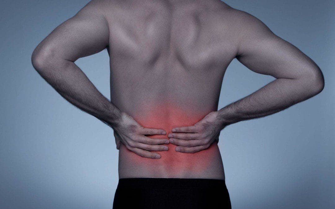
In individuals with discogenic low back pain, how does incorporating decompression reduce muscle strain in the back?
Introduction
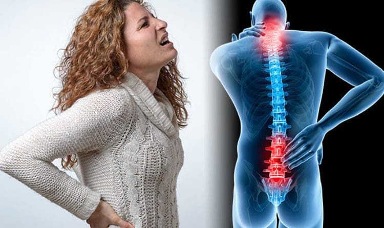
When it comes to low back pain, many people often complain that the surrounding muscles will ache constantly, and there is no relief from their primary doctors. Muscle strain associated with low back pain is one of the pain-like symptoms that many individuals experience when normal or traumatic factors start to cause issues in the lower back region of the body. When people begin to make constant repetitive motions correlating with normal daily activities like heavy lifting objects, poor posture, or stepping wrong, it can cause micro-tears to the surrounding muscles and the spinal discs in the lumbar region. When the spinal discs degenerate over time and have been under constant pressure, it can aggravate the surrounding nerve roots causing pain-like problems to the surrounding muscles, ligaments, and tissues, leading to musculoskeletal disorders corresponding with discogenic low back pain. Pain affecting the lower back can lead to a life of disability and make a person feel miserable. To that point, many individuals will seek non-surgical treatment to reduce discogenic pain associated with the low back and can find the relief they have sought. Today’s article examines how discogenic low back pain causes low back pain and how non-surgical treatments like decompression reduce discogenic low back pain and restore muscle strength. Additionally, we communicate with certified medical providers who incorporate our patient’s information to reduce muscle strain correlating with discogenic low back pain. We also inform them that decompression can help mitigate the pain-like symptoms associated with degenerated discs affecting the lower back region. We encourage our patients to ask amazing questions while looking for education from our associated medical providers about their low back issues. Dr. Jimenez, D.C., incorporates this information as an educational service. Disclaimer
Discogenic Low Back Pain Causing Muscle Strain
Do you often experience a pinched nerve or muscle strain in your lower back that hurts when standing? Do you feel symptoms of muscle spasms in your lower back or behind your legs? Or do you and your loved ones feel numbness or tingling sensations in your back, legs, and feet after sitting down excessively? These pain-like issues are associated with discogenic low back pain, which can lead to the development of disability in many people. Discogenic low back pain is developed when the intervertebral (spinal) disc degenerates over time and can contribute to disability. (Mohd Isa et al., 2022) When there are structural changes to the spinal disc that causes the degeneration to progress, it can lead to dysfunction and instability in the lumbar spine. The spinal discs in the spine have the primary job of absorbing the unwanted pressure load that the body is experiencing. Over time though, the spinal disc can degenerate and crack under pressure, leading to discogenic low back pain. Discogenic low back pain can lead to increased pain in the lower back region’s paraspinal muscles and muscle atrophy, inflammation, and muscle strain in the lower back muscles and lumbar spinal discs. (Huang et al., 2022) When the spinal disc is under constant pressure, the inflammatory cytokines can induce nerve ingrowth, structural and biomechanical changes, and a release of pain factors to contribute to the effects of discogenic low back pain. (Lyu et al., 2021) When people are dealing with discogenic low back pain associated with muscle strain, it can make them miss out on their daily activities.
From Injury To Recovery With Chiropractic-Video
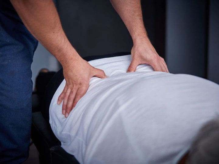
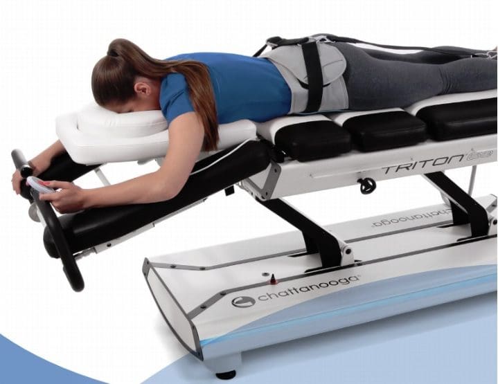
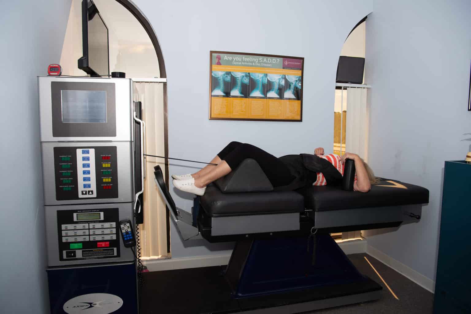
When many individuals are experiencing discogenic low back pain, it can be difficult for pain specialists and doctors to identify the underlying source of pain since it is a multifactorial musculoskeletal disorder. (Fujii et al., 2019) However, numerous ways exist to reduce the pain and allow the individual to return to their daily routines. Non-surgical treatments are an excellent way to minimize the pain-like symptoms associated with discogenic low back pain. Treatments like decompression therapy and chiropractic care can create a happy experience for many individuals dealing with discogenic low back pain as it is safe, cost-effective, and gentle on the spine. Decompression can help reduce the pain in the posterior segment of the lumbar spine while relaxing the surrounding muscles and ligaments and pulling the affected disc back to its original position. (Choi et al., 2022) This creates negative pressure on the spinal column and increases disc height on the spine, which allows the fluids and nutrients to flood back into the spine and rehydrate the disc. Decompression therapy can also be combined with chiropractic care, as the spine can be manipulated mechanically or manually to allow the body to realign itself. This, in turn, promotes the body’s natural healing properties to work its magic and provide relief. The video explains how these treatments can positively impact many suffering individuals and help them regain their health.
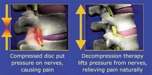
Decompression Reducing Discogenic Low Back Pain
Decompression therapy allows the individuals to be strapped into a traction machine in a supine position and gently pulls the spine to enable the affected disc to return to the spine and lay off the pressure on the aggravating nerve root that is causing muscle strain. This causes the intervertebral disc space to change through negative pressure, which allows the height to increase in the intervertebral height without stimulating the surrounding fibers around the disc. (Oh et al., 2019) This allows the facet joints in the spine to be realigned, allowing them to be in their moveable location back to the spine to alleviate pain, thus restoring normal posture and activating body functions. When individuals incorporate decompression therapy consecutively, it can minimize the pain caused by discogenic low back pain and allows the individual to have a personalized plan to ensure the pain doesn’t return. (Macario et al., 2008)
Restoring Muscle Strength In The Low Back
Decompression therapy allows the affected muscle to be stretched gently, which can be strengthened through other treatments like physical therapy. This can effectively reduce discogenic low back pain associated with the affected discs and positively influence spinal mobility and muscle strength. (Wang et al., 2022) Even though degeneration in the spinal disc is a natural process, it is important to be mindful of the body to prevent pain-like symptoms from occurring and causing issues to the back. Decompression therapy can positively influence many individuals looking to regain their health and reduce the pain they are experiencing from discogenic low back pain so they can return to their daily activities.
References
Choi, E., Gil, H. Y., Ju, J., Han, W. K., Nahm, F. S., & Lee, P. B. (2022). Effect of Nonsurgical Spinal Decompression on Intensity of Pain and Herniated Disc Volume in Subacute Lumbar Herniated Disc. International Journal of Clinical Practice, 2022, 6343837. https://doi.org/10.1155/2022/6343837
Fujii, K., Yamazaki, M., Kang, J. D., Risbud, M. V., Cho, S. K., Qureshi, S. A., Hecht, A. C., & Iatridis, J. C. (2019). Discogenic Back Pain: Literature Review of Definition, Diagnosis, and Treatment. JBMR Plus, 3(5), e10180. https://doi.org/10.1002/jbm4.10180
Huang, Y., Wang, L., Luo, B., Yang, K., Zeng, X., Chen, J., Zhang, Z., Li, Y., Cheng, X., & He, B. (2022). Associations of Lumber Disc Degeneration With Paraspinal Muscles Myosteatosis in Discogenic Low Back Pain. Front Endocrinol (Lausanne), 13, 891088. https://doi.org/10.3389/fendo.2022.891088
Lyu, F. J., Cui, H., Pan, H., Mc Cheung, K., Cao, X., Iatridis, J. C., & Zheng, Z. (2021). Painful intervertebral disc degeneration and inflammation: from laboratory evidence to clinical interventions. Bone Res, 9(1), 7. https://doi.org/10.1038/s41413-020-00125-x
Macario, A., Richmond, C., Auster, M., & Pergolizzi, J. V. (2008). Treatment of 94 outpatients with chronic discogenic low back pain with the DRX9000: a retrospective chart review. Pain Pract, 8(1), 11-17. https://doi.org/10.1111/j.1533-2500.2007.00167.x
Mohd Isa, I. L., Teoh, S. L., Mohd Nor, N. H., & Mokhtar, S. A. (2022). Discogenic Low Back Pain: Anatomy, Pathophysiology and Treatments of Intervertebral Disc Degeneration. Int J Mol Sci, 24(1). https://doi.org/10.3390/ijms24010208
Oh, H., Choi, S., Lee, S., Choi, J., & Lee, K. (2019). Effects of the flexion-distraction technique and drop technique on straight leg raising angle and intervertebral disc height of patients with an intervertebral disc herniation. Journal of Physical Therapy Science, 31(8), 666-669. https://doi.org/10.1589/jpts.31.666
Wang, W., Long, F., Wu, X., Li, S., & Lin, J. (2022). Clinical Efficacy of Mechanical Traction as Physical Therapy for Lumbar Disc Herniation: A Meta-Analysis. Comput Math Methods Med, 2022, 5670303. https://doi.org/10.1155/2022/5670303
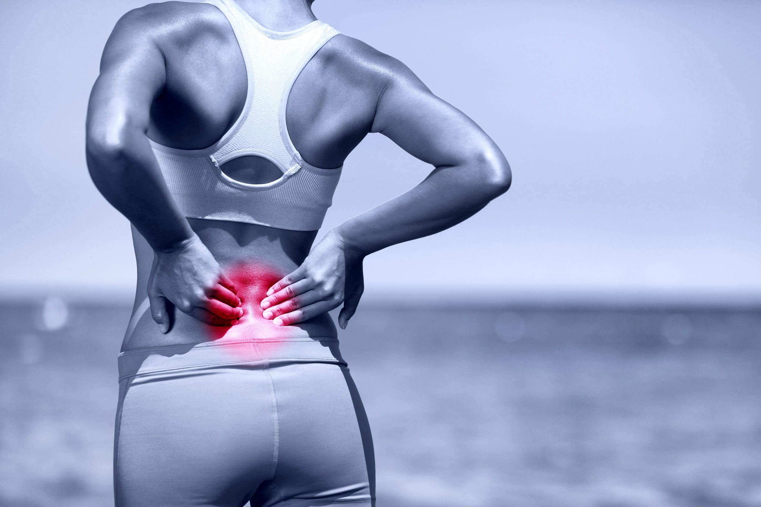
Can spinal decompression treatments be incorporated for individuals with lumbosacral pain and improve posture?
Introduction
Many individuals only realize they have poor posture once they perform a movement that causes pain in the lumbosacral region of their body. Have you or your loved ones experienced muscle aches or strains after relaxing in a weird position? Or do you begin to notice that you are hunched over when walking from one location to another? Many of these scenarios correspond to how we present ourselves with our posture. Our posture helps support the upper body’s weight while stabilizing the lower body through the spine and ensuring that our body is in an upright position when we are in motion. However, as we age, so do our bodies and spine, which then causes us to be in a hunched position, causing our posture to degenerate. This causes lumbosacral pain to develop along the body’s lower extremities, leading to overlapping risk profiles that cause mobility issues, poor posture, and disability if not treated right away. When this happens, the surrounding muscles, ligaments, and tissues around the lumbar spine will begin to develop pain-like symptoms and can make a person’s life miserable. Luckily there are various techniques and treatments to improve poor posture and reduce lumbosacral pain affecting many individuals. Today’s article looks at how lumbosacral pain affects a person’s posture and how spinal decompression and MET therapy can reduce lumbosacral pain and restore good posture. Additionally, we work hand-in-hand with certified medical providers who incorporate our patient’s information to treat and minimize lumbosacral pain associated with poor posture. We also inform them that spinal decompression combined with MET therapy can help with lumbosacral pain while improving good posture back to the body. We encourage our patients to ask profound questions while seeking education from our associated medical providers about their pain-like issues. Dr. Alex Jimenez, D.C., incorporates this information as an educational service. Disclaimer
Lumbosacral Pain Affects Posture
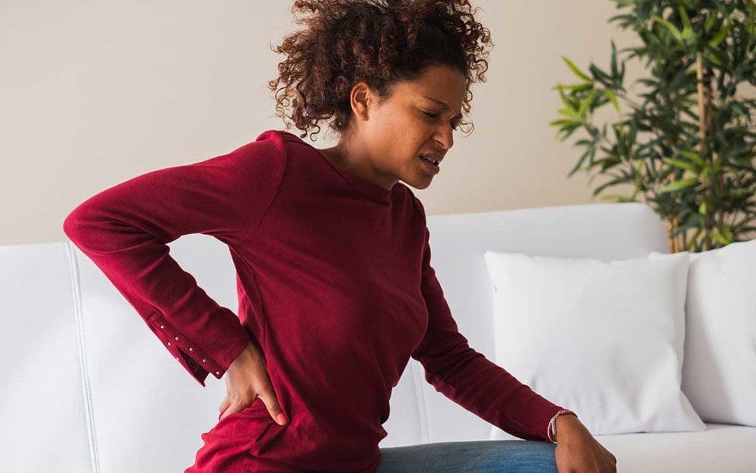
Have you noticed you are constantly slouching or being in a hunched position, only to feel muscle aches and strains in your lumbar-sacral region? Do you feel symptoms of tingling and numbness in your hips and buttock region after sitting down excessively? Or do you feel referred pain in one location and have to shift your weight to compensate for the pain? These pain-like symptoms and scenarios correspond to lumbosacral pain affecting your posture. The lumbosacral spine region has a natural curve that functions as a shock absorber to mitigate the body’s mechanical weight and stress when in motion. (Adams & Hutton, 1985) At the same time, it is susceptible to being constantly injured as the spinal disc is being compressed and can be herniated or damaged over time, manifesting into lumbosacral pain. So how does lumbosacral pain associate with poor posture? When individuals are dealing with low back problems associated with lumbosacral pain, the spinal disc in the lumbosacral region starts to have balancing issues when in motion. (Huang, Jaw, & Young, 2022) When people are dealing with difficulties balancing their gait cycle, it can cause their walking performance and postural control to be dysfunctional and cause the body to be misaligned, thus causing musculoskeletal issues to arise and affecting the lower body and its extremities. The surrounding muscles surrounding the lumbosacral region will begin to experience stiffness in the trunk region, which can cause musculoskeletal changes to the surrounding muscles when individuals start to be in an upright position. (Creze et al., 2019) When poor posture affects the trunk muscles, the surrounding accessory muscles start acting to compensate for the pain. To that point, lumbosacral pain associated with poor posture could lead to abdominal, low back, hip, and pelvic pain. However, many individuals can find various therapies and relieving techniques to improve posture, strengthen the surrounding muscles, and reduce pain-like symptoms.
Building A Stronger Body- Video
Many individuals can seek out various non-invasive therapies to alleviate the issue when it comes to improving posture and reducing lumbosacral pain. These therapies are cost-effective and personalized to the person’s pain. Treatments like chiropractic care and decompression can help restore good posture while realigning the body out of subluxation and help stretch the affected muscles. Coincidentally, non-surgical therapies can be combined with other treatments like physical therapy to strengthen the trunk muscles surrounding the lumbosacral region, thus reducing the load on the lumbosacral spine. (Callaghan, Gunning, & McGill, 1998) When people focus on their health and well-being, non-surgical therapies can provide a positive and safe experience with a team that can help reduce the pain the person has been dealing with their entire lives. The video above explains how these treatments work together to help you build a stronger body while revitalizing your energy and enhancing your health and wellness.
Spinal Decompression Reducing Lumbosacral Pain
When it comes to reducing lumbosacral pain associated with poor posture, many individuals can incorporate non-surgical treatments like spinal decompression and combine them with a personalized treatment plan to reduce the pain-like symptoms. What spinal decompression does to lumbosacral pain is that it helps mitigate intra-disc pressure while increasing disc space within the lumbosacral spinal region. (Amjad et al., 2022) Spinal decompression can help improve leg mobility and stretch out the affected muscles to kick-start the body’s natural healing process. Spinal decompression can even combine with physical therapy to help strengthen the lumbosacral region’s abdominal muscles and enable many individuals with poor posture to be mindful of how they present themselves.(Mielenz et al., 1997)
MET Therapy & Spinal Decompression Restoring Posture
When pain specialists like chiropractors and massage therapists incorporate spinal decompression treatment to reduce lumbosacral pain, they also utilize various techniques to strengthen the lumbosacral muscles to restore proper posture to the body. Many pain specialists use MET (muscle energy techniques) therapy to maintain while stretching the muscles and fascia in the affected areas. MET therapy combined with spinal decompression can help improve muscle shortness in the lumbar fascial tissue, improve posture, and even increase the lumbar and pelvic range of motion. (Tamartash & Bahrpeyma, 2022) These two non-surgical treatments can help many people by addressing their posture and movement dysfunction while strengthening their core stabilized muscles to reduce pain. (Norris & Matthews, 2008) Many individuals who want to regain their health and wellness can make small changes in their routine to improve their posture and be more mindful of their bodies to reduce the chances of lumbosacral pain returning.
Amjad, F., Mohseni-Bandpei, M. A., Gilani, S. A., Ahmad, A., & Hanif, A. (2022). Effects of non-surgical decompression therapy in addition to routine physical therapy on pain, range of motion, endurance, functional disability and quality of life versus routine physical therapy alone in patients with lumbar radiculopathy; a randomized controlled trial. BMC Musculoskelet Disord, 23(1), 255. https://doi.org/10.1186/s12891-022-05196-x
Callaghan, J. P., Gunning, J. L., & McGill, S. M. (1998). The relationship between lumbar spine load and muscle activity during extensor exercises. Phys Ther, 78(1), 8-18. https://doi.org/10.1093/ptj/78.1.8
Creze, M., Bedretdinova, D., Soubeyrand, M., Rocher, L., Gennisson, J. L., Gagey, O., Maitre, X., & Bellin, M. F. (2019). Posture-related stiffness mapping of paraspinal muscles. J Anat, 234(6), 787-799. https://doi.org/10.1111/joa.12978
Huang, C. C., Jaw, F. S., & Young, Y. H. (2022). Radiological and functional assessment in patients with lumbar spinal stenosis. BMC Musculoskelet Disord, 23(1), 137. https://doi.org/10.1186/s12891-022-05053-x
Mielenz, T. J., Carey, T. S., Dyrek, D. A., Harris, B. A., Garrett, J. M., & Darter, J. D. (1997). Physical therapy utilization by patients with acute low back pain. Phys Ther, 77(10), 1040-1051. https://doi.org/10.1093/ptj/77.10.1040
Norris, C., & Matthews, M. (2008). The role of an integrated back stability program in patients with chronic low back pain. Complement Ther Clin Pract, 14(4), 255-263. https://doi.org/10.1016/j.ctcp.2008.06.001
Tamartash, H., & Bahrpeyma, F. (2022). Evaluation of Lumbar Myofascial Release Effects on Lumbar Flexion Angle and Pelvic Inclination Angle in Patients with Non-Specific Low Back Pain. Int J Ther Massage Bodywork, 15(1), 15-22. https://doi.org/10.3822/ijtmb.v15i1.709
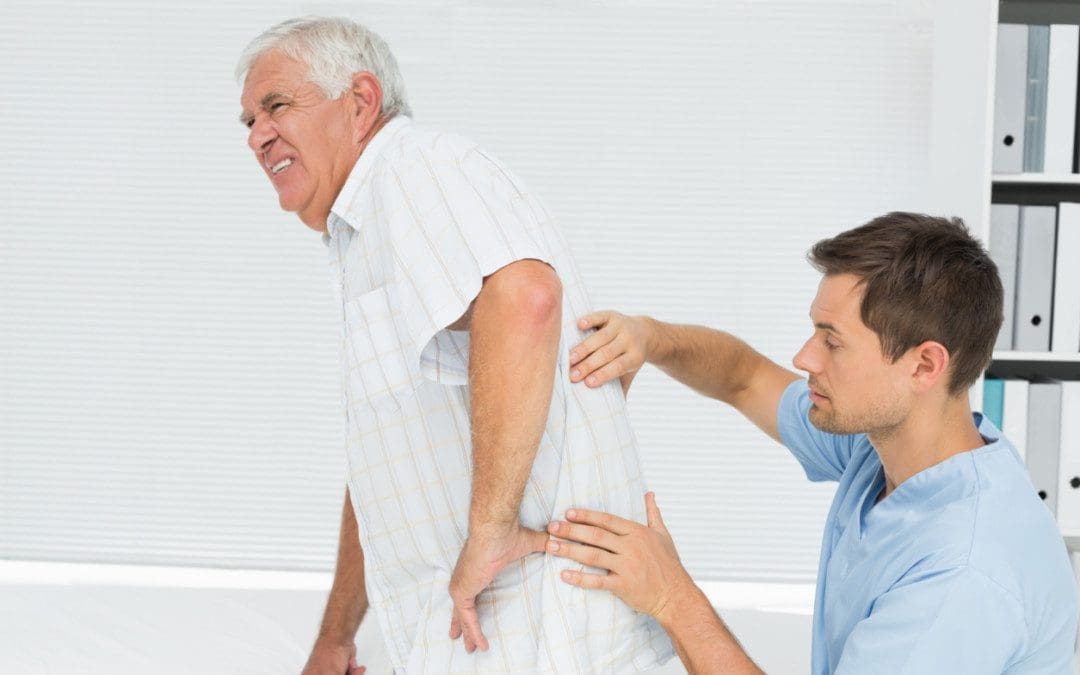
Can spinal decompression treat individuals with chronic low back pain to reduce joint arthritis and strengthen the surrounding muscles to restore lumbar mobility?
Introduction
When many individuals are dealing with pain in their lumbar region, more often than not, they believe that it’s the surrounding muscles that protect the spine that is being affected. However, that is only half of the problem. Do you or your loved ones often feel a warm sensation within your lower back, hips, and knees that radiates pain within your joints? Well, joint pain can correlate with low back pain in its chronic state. Since the body and spine can degenerate over time, it can cause the joints to wear and tear while rubbing against each other, causing joint arthritis to develop. When arthritic pain is associated with chronic low back pain, it can lead to overlapping risk profiles that can lead to a life of disability and make the individual miserable. Many pain-like symptoms correlating with chronic low back pain can develop over time and cause mobility and stability problems within the body. Fortunately, many non-surgical treatments can reduce the progression of joint arthritis and alleviate chronic low back pain. Today’s articles examine the correlation between joint arthritis and chronic low back pain while taking a look at how non-invasive treatments like spinal decompression can not only reduce chronic low back pain associated with joint arthritis but also restore lumbar mobility. Additionally, we work hand-in-hand with certified medical providers who incorporate our patient’s information to treat and reduce the progression of joint arthritis associated with low back pain. We also inform them that spinal decompression can help restore lumbar mobility while enhancing muscle strength back to the lumbar region. We encourage our patients to ask profound questions while seeking education from our associated medical providers about their pain-like issues. Dr. Alex Jimenez, D.C., incorporates this information as an educational service. Disclaimer
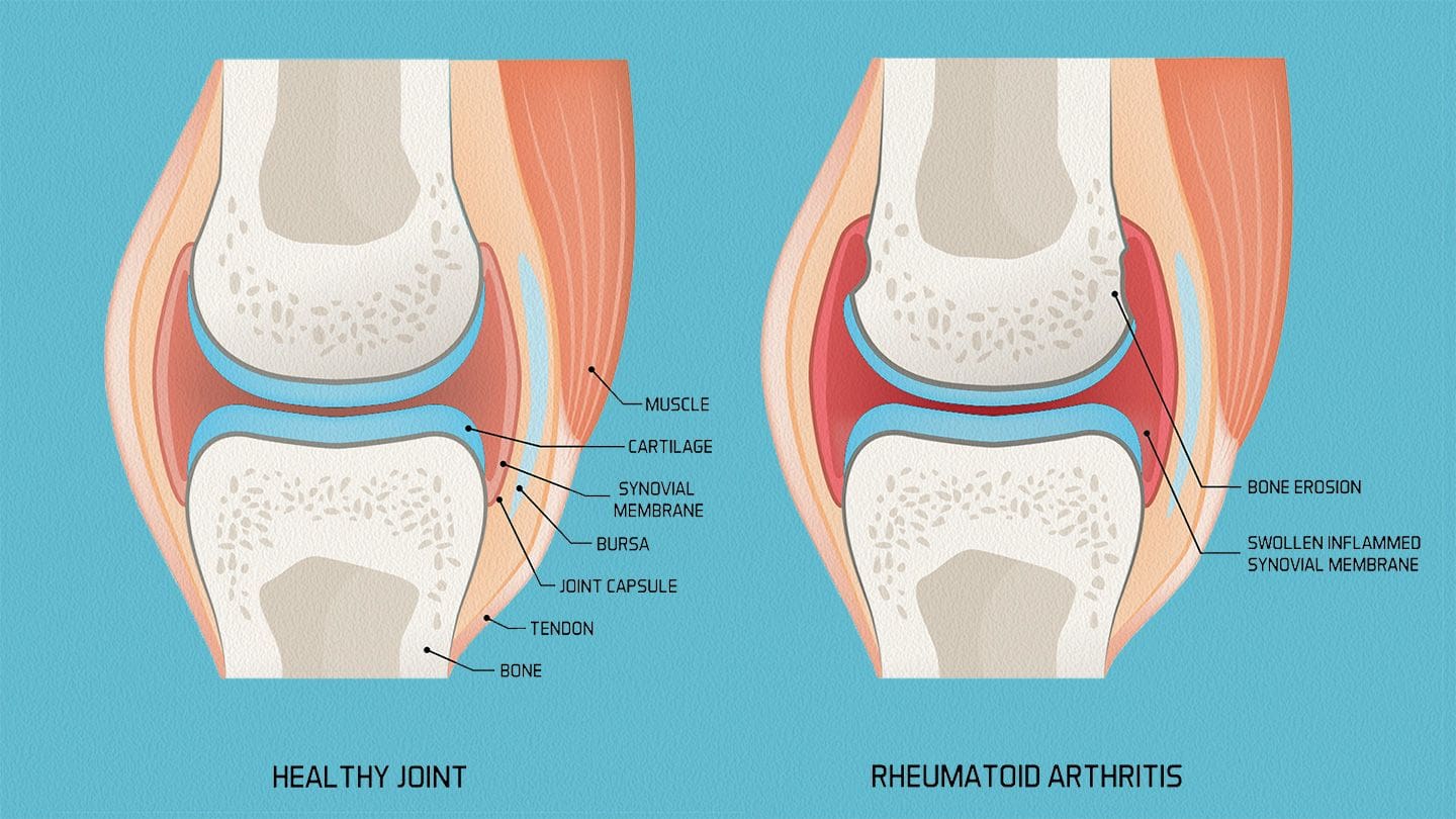
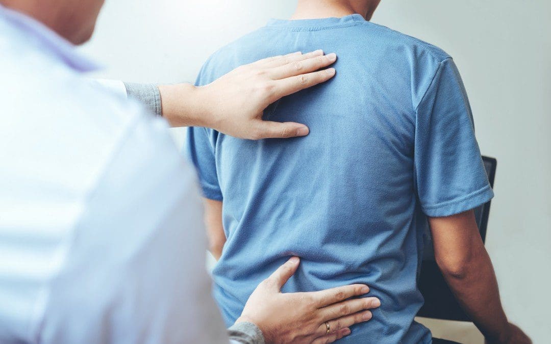
Joint Arthritis & Chronic Low Back Pain
Do you often experience stiffness in the morning that seems to go away after a few hours? Do you feel aches and pains at work, either at the desk or the requires heavy objects? Or do you feel your joints ache constantly that you are not getting enough sleep at night? These pain-like scenarios are associated with joint arthritis, which can develop into chronic low back pain. Many people know that the lumber spine and the lower extremities will experience high mechanical stress when the body is in an upright position without pain. As the lumbar spine and lower extremities begin to go through repetitive motions over time, that can cause the ligaments and surrounding muscles to have microtrauma tears, resulting in the development of joint arthritis, which can lead to inflammatory effects. (Xiong et al., 2022) Now inflammation in the body is beneficial and harmful depending on the severity within the affected area. Joint arthritis, especially spondylarthritis, is part of inflammatory diseases that affect the joint and the spine and can have various clinical manifestations. (Sharip & Kunz, 2020) The symptoms of joint arthritis include inflammatory pain in the affected area, joint stiffness and swelling, and muscle weakness. When dealing with inflammatory effects associated with joint arthritis, it can cause them to have a decreased quality of life, increase mortality, and become an economic burden. (Walsh & Magrey, 2021)
Now how does joint arthritis be associated with low back pain? When individuals start to make repetitive motions to their lumbar spine, it can lead to abnormal changes to the intervertebral discs. When unwanted pressure begins to compress the intervertebral disc constantly, it can cause wear and tear on the disc, causing them to crack and allowing the annular nociceptors to become over-sensitized. (Weinstein, Claverie, & Gibson, 1988) The affected disc then aggravates the surrounding nerve roots and muscles, causing low back pain. When individuals do their everyday normal, factors that cause degenerative changes to the intervertebral discs can lead to chronic low back pain. (Vernon-Roberts & Pirie, 1977) To that point, chronic low back pain associated with joint arthritis can become a chronic issue if not treated right away.
Arthritis Explained- Video
When reducing the effects of chronic low back pain associated with joint arthritis, many individuals seek treatments to relieve their pain-affected areas with a positive outcome. Non-surgical treatments could be the answer, combined with other therapies to reduce chronic low back pain. (Kizhakkeveettil, Rose, & Kadar, 2014) Non-surgical treatments can be customizable to the individual’s pain while being cost-effective. Many people with arthritic joints can benefit from non-surgical treatments as pain specialists like massage therapists and chiropractors can use various techniques to stretch out the affected muscles, increase the joint’s ROM (range of motions) and realign the body out of misalignment to promote the body’s natural healing process. The video above gives an overview of how arthritis can affect the joints, be associated with low back pain, and how these treatments can alleviate its symptoms through various techniques.
Spinal Decompression & Chronic Low Back Pain
Spinal decompression is a non-surgical therapy treatment that can help many individuals with chronic low back pain. Spinal decompression uses gentle traction on the lumbar spine to pull the spine, allowing the fluids and nutrients to flood back to the affected area and help the body naturally heal itself. When individuals start incorporating spinal decompression for their chronic low back pain, they will feel pressure off their spinal discs. (Ramos, 2004) When individuals begin to feel an improvement in their lumbar region after a few consecutive treatments, they will start to regain their lumbar mobility.
Spinal Decompression Restoring Lumbar Mobility
Spinal decompression can reduce the effects of chronic low back pain and restore lumbar mobility to the spine. Since spinal decompression uses gentle traction on the spine, the intervertebral disc will return to its original position, while the spinal cavity increases disc height. To that point, spinal decompression can cause individuals to improve mobility and cause them to return to their normal daily activities, as it correlates strongly with pain reduction. (Gose, Naguszewski, & Naguszewski, 1998) By incorporating spinal decompression as part of a routine, many individuals can regain their health without dealing with pain-like symptoms.
References
Gose, E. E., Naguszewski, W. K., & Naguszewski, R. K. (1998). Vertebral axial decompression therapy for pain associated with herniated or degenerated discs or facet syndrome: an outcome study. Neurol Res, 20(3), 186-190. https://doi.org/10.1080/01616412.1998.11740504
Kizhakkeveettil, A., Rose, K., & Kadar, G. E. (2014). Integrative therapies for low back pain that include complementary and alternative medicine care: a systematic review. Glob Adv Health Med, 3(5), 49-64. https://doi.org/10.7453/gahmj.2014.043
Ramos, G. (2004). Efficacy of vertebral axial decompression on chronic low back pain: study of dosage regimen. Neurol Res, 26(3), 320-324. https://doi.org/10.1179/016164104225014030
Vernon-Roberts, B., & Pirie, C. J. (1977). Degenerative changes in the intervertebral discs of the lumbar spine and their sequelae. Rheumatol Rehabil, 16(1), 13-21. https://doi.org/10.1093/rheumatology/16.1.13
Walsh, J. A., & Magrey, M. (2021). Clinical Manifestations and Diagnosis of Axial Spondyloarthritis. J Clin Rheumatol, 27(8), e547-e560. https://doi.org/10.1097/RHU.0000000000001575
Xiong, Y., Cai, M., Xu, Y., Dong, P., Chen, H., He, W., & Zhang, J. (2022). Joint together: The etiology and pathogenesis of ankylosing spondylitis. Front Immunol, 13, 996103. https://doi.org/10.3389/fimmu.2022.996103
For many individuals with low back pain, how does spinal decompression alleviate muscle stress as part of initial treatment?
Introduction
Many working individuals know that low back pain is a common problem that causes them to go to their primary doctor to get examined and miss out on work. Low back pain is a multifactorial musculoskeletal condition that causes overlapping risk profiles to affect the body’s lower extremities. Low back pain can correlate with musculoskeletal disorders like sciatica, abdominal pain, leg pain, and DDD (degenerative disc disease). It can range from acute to chronic, depending on the severity the individual is dealing with when it comes to pain. At the same time, low back pain can be non-specific or mechanical as the surrounding muscles, soft tissues, joints, and ligaments are affected and dealing with symptoms of muscle strain, unwanted pressure on the joint, causing stress, and muscle aches. Since the body and the spine naturally age, it causes more stress on the lower back as individuals begin to hunch over more when walking or carrying heavy objects, which causes more strain on the back muscles, leading them to a life of disability. Luckily, non-surgical treatments have become more available to reduce the effects of muscle stress associated with low back pain and alleviate the pain-like symptoms in the lumbar spine. Today’s article focuses on how numerous factors can cause muscle stress to the lower back and how spinal decompression can reduce its effects while alleviating low back pain. At the same time, we work hand-in-hand with certified medical providers who incorporate our patient’s information to treat and mitigate muscle stress on the lower back. We also inform them that non-surgical treatments like spinal decompression can help alleviate residual pain-like symptoms associated with low back pain and help them return to their daily activities. We encourage our patients to ask profound questions while seeking education from our associated medical providers about their pain-like issues. Dr. Alex Jimenez, D.C., incorporates this information as an educational service. Disclaimer
How Factors Cause Muscle Stress To The Low Back
Do you feel gradual or consistent pain in your lower back after carrying a heavy object from one location to another? Are you constantly taking medication for your low back pain to finish the workday? Or do you feel pain in your sciatic nerve that gets aggravated when you are in motion, and you feel relief when resting? Around the world, many people have dealt with low back pain and its associated symptom at some point. Since low back pain can be either specific or non-specific, pain can come from spinal issues that cause referred pain to a different body location or normal repetitive factors that cause discomfort to the surrounding soft tissues, muscles, and ligaments. Some symptoms correlating with low back pain include progressive motor or sensory issues, urinary retention, abnormal neurologic issues, spinal misalignment, or soft tissue abnormalities. (Will, Bury, & Miller, 2018) When many working individuals are dealing with non-specific low back pain, the surrounding soft tissues and muscles can become weak and overworked, which causes overlapping risk profiles and results in the development of low back pain.
Many working individuals with demanding jobs, whether physical or sedentary, will often strain their lumbar region from lifting/carrying heavy objects or being hunched over constantly at the computer. When a person continues to put repetitive stress on the surrounding muscles, it can cause chronic pain over time and become a major cause of work loss. (Becker & Childress, 2019) Low back pain can cause the individual to be more stressed since they are missing out on work. Low back pain can also be due to lumbar instability from the intervertebral disc and surrounding muscles and ligaments under constant pressure. (Hauser et al., 2022) Since the body and spine age over time naturally, many individuals dealing with low back pain will begin to feel their joints and muscle structures loosen over time, leading to musculoskeletal symptoms that prevent the spine from destabilizing. This can cause the individual to limit their ability to participate in daily activities and even reduce their quality of life. Luckily, non-surgical treatments can reduce the effects of low back pain while revitalizing the lumbar region so many people can continue their daily activities pain-free.
From Injury To Recovery-Video
When treating and reducing low back pain, many pain specialists like chiropractors and massage therapists can incorporate non-invasive treatments to ease low back pain. Non-invasive treatments like chiropractic care and spinal decompression can help reduce muscle stress on the lumbar region with mechanical or manual spinal manipulation and help relieve the affected lumbar area. Now spinal decompression and chiropractic care have a wonderful relationship as they use negative pressure on the intervertebral disc to increase nutrient flow back to the spine and help kick-start the body’s natural healing process. (Schimmel et al., 2009) These treatments are not only non-invasive, but they are also safe and cost-effective while being customizable to the person’s pain. The video above explains how these treatments can reduce soft tissue injuries and the effects of musculoskeletal pain on the lumbar region.
Spinal Decompression Reducing Muscle Stress From Low Back Pain
Non-surgical treatments like spinal decompression are incorporated into a person’s personalized health plan to reduce the effects of low back pain. Spinal decompression uses gentle traction on the spine to diminish the compressive load on the intervertebral disc to reduce herniation while stretching the lumbar spinal muscles and ligaments to decrease muscle spasms. (Sari et al., 2005) When individuals start to feel relief in their lower back due to spinal decompression as they begin to feel general improvement in the lumbar region. (Borman, Keskin, & Bodur, 2003) Since spinal decompression uses gentle traction on the spine, this gentle force can relieve radicular symptoms associated with the lower extremities. (Krause et al., 2000) Additionally, the effects of spinal decompression can be a positive experience for many individuals dealing with low back pain. (Pellecchia, 1994) When it comes to reducing low back pain, utilizing non-surgical treatments like spinal decompression can help improve mobility back to the individual and reduce the pain they were experiencing before. When people begin to think about their health and wellness, they can experience relief from the pain and get back to normalcy.
References
Becker, B. A., & Childress, M. A. (2019). Nonspecific Low Back Pain and Return To Work. American Family Physician, 100(11), 697-703. https://www.ncbi.nlm.nih.gov/pubmed/31790184
Borman, P., Keskin, D., & Bodur, H. (2003). The efficacy of lumbar traction in the management of patients with low back pain. Rheumatol Int, 23(2), 82-86. https://doi.org/10.1007/s00296-002-0249-0
Hauser, R. A., Matias, D., Woznica, D., Rawlings, B., & Woldin, B. A. (2022). Lumbar instability as an etiology of low back pain and its treatment by prolotherapy: A review. J Back Musculoskelet Rehabil, 35(4), 701-712. https://doi.org/10.3233/BMR-210097
Krause, M., Refshauge, K. M., Dessen, M., & Boland, R. (2000). Lumbar spine traction: evaluation of effects and recommended application for treatment. Man Ther, 5(2), 72-81. https://doi.org/10.1054/math.2000.0235
Sari, H., Akarirmak, U., Karacan, I., & Akman, H. (2005). Computed tomographic evaluation of lumbar spinal structures during traction. Physiother Theory Pract, 21(1), 3-11. https://www.ncbi.nlm.nih.gov/pubmed/16385939
Schimmel, J. J., de Kleuver, M., Horsting, P. P., Spruit, M., Jacobs, W. C., & van Limbeek, J. (2009). No effect of traction in patients with low back pain: a single centre, single blind, randomized controlled trial of Intervertebral Differential Dynamics Therapy. Eur Spine J, 18(12), 1843-1850. https://doi.org/10.1007/s00586-009-1044-3
How efficient is spinal decompression to alleviate pain-like symptoms in many individuals with low back pain?
Introduction
Low back pain is a common condition that affects many people worldwide. It can cause discomfort and prevent individuals from returning to their normal routines. The pain can be specific or non-specific, depending on the severity of the symptoms. It can also be associated with other musculoskeletal conditions, such as sciatica, DDD, and osteoarthritis, affecting the spine’s mobility and stability. Fortunately, treatments are available to reduce the pain and associated symptoms while relieving the lumbar spinal region. At the same time, working with certified medical providers who use our patients’ information to treat individuals experiencing pain-like symptoms associated with low back pain. We inform them that non-surgical treatments like decompression can help reduce the progression of low back pain and its associated pain-like symptoms. At the same time, we also explain to them how adding decompression to their routine can alleviate the pain-like symptoms. We encourage our patients to ask essential questions while seeking education from our associated medical providers about their situation. Dr. Alex Jimenez, D.C., provides this information as an educational service. Disclaimer
The Burden Of Low Back Pain
Do you often experience muscle stiffness and aches from excessive sitting at your desk job? Do you feel excruciating pain in your hips and low back after a long day of moving heavy objects? Or do you feel constant pain after moving from one location to another, only to find relief when resting? Many of these scenarios are a normal routine associated with low back pain for many working people. Since low back pain is common worldwide, it can greatly impact many individuals trying to alleviate it. For many individuals dealing with low back pain, it can be an economic burden, and it causes unnecessary stress to the working individual. (Maetzel & Li, 2002) When a person is dealing with low back pain and is working, it can lead to work disability which then cascades to frequent doctor visits to missing time off work to eventually short-term disability. At the same time, low back pain has many potential, either normal or traumatic factors affecting the region of the lower back, hips, and buttocks. Many individuals with low back pain noticed the corresponding relationship between the genetic and environmental factors that often correspond to where the pain is localized. (Manek & MacGregor, 2005)
Low back pain can often develop at an early age and be associated with serious pathologies (Jones & Macfarlane, 2005) that can correlate with environmental factors that lead to spinal misalignment or subluxation. Low back pain covers a huge spectrum of pain-like conditions that are frequently overlapped and can be prone to different stressors that can impact the body while becoming a challenge to diagnose when getting treated. (Knezevic et al., 2021) However, many individuals with low back pain can find relief from not only the economic burden of low back pain but also find the right therapies to alleviate pain-like symptoms associated with low back pain from their bodies.
Body In Balance- Video
When it comes to treating low back pain, many individuals will go to their primary doctors for a physical examination to determine where the pain radiates in the lumbar region. (Chou, Qaseem, et al., 2007) At the same time, the individual’s primary doctor should assess what factors are the causes for the development of low back pain and come up with a customized treatment plan to reduce the pain from the lumbar region while also working with other medical professionals to kick start the person’s health and wellness journey. Numerous non-surgical treatments are cost-effective, safe, and non-pharmacological when alleviating low back pain. They can provide positive, beneficial results after a few sessions of treatments to reduce low back pain. Non-surgical treatments like chiropractic care, massage therapy, spinal decompression, and physical therapy can help relieve many individuals with low back pain. The video above explains how these treatments, combined with the right tools, can help bring balance back into the body.
The Efficacy Of Spinal Decompression
When reducing the effects of low back pain, non-surgical treatments are cost-effective and safe for many individuals with low back pain. Many non-surgical therapies are efficient for chronic or acute low back pain and can be combined with numerous therapies to reduce the chances of low back pain from returning in the future. (Chou, Huffman, et al., 2007) Non-surgical treatments like spinal decompression are efficient for low back pain as it helps stretch the tight, shortened muscles in the lumbar region and reduce any pain-like symptoms associated with low back pain.
How Spinal Decompression Alleviates Low Back Pain
Now how would spinal decompression alleviate low back pain? Well, since low back pain has many factors that can contribute to its development, one of these factors that can contribute to low back pain is compressed intervertebral discs. When the spinal discs are compressed, they can become herniated under pressure and aggravate the spinal nerve root, leading to low back pain symptoms. With spinal decompression, individuals will be strapped to a traction machine and have their bodies gently pulled to reduce pressure off the aggravated nerve and pull the disc back into its original position. To that point, spinal decompression allows mobility back to the lower extremities and improves stability in the lumbar region. (Meszaros et al., 2000) Additionally, decompression can help reduce the effects of low back pain, whether done manually or through a traction machine that allows negative pressure into the spine while allowing the body to heal itself and rehydrate the spinal discs naturally. (Macario et al., 2008) With spinal decompression combined with other therapies, many people can return to work pain-free while being more mindful of how they listen to their bodies.
References
Chou, R., Huffman, L. H., American Pain, S., & American College of, P. (2007). Nonpharmacologic therapies for acute and chronic low back pain: a review of the evidence for an American Pain Society/American College of Physicians clinical practice guideline. Ann Intern Med, 147(7), 492-504. https://doi.org/10.7326/0003-4819-147-7-200710020-00007
Chou, R., Qaseem, A., Snow, V., Casey, D., Cross, J. T., Jr., Shekelle, P., Owens, D. K., Clinical Efficacy Assessment Subcommittee of the American College of, P., American College of, P., & American Pain Society Low Back Pain Guidelines, P. (2007). Diagnosis and treatment of low back pain: a joint clinical practice guideline from the American College of Physicians and the American Pain Society. Ann Intern Med, 147(7), 478-491. https://doi.org/10.7326/0003-4819-147-7-200710020-00006
Jones, G. T., & Macfarlane, G. J. (2005). Epidemiology of low back pain in children and adolescents. Arch Dis Child, 90(3), 312-316. https://doi.org/10.1136/adc.2004.056812
Knezevic, N. N., Candido, K. D., Vlaeyen, J. W. S., Van Zundert, J., & Cohen, S. P. (2021). Low back pain. The Lancet, 398(10294), 78-92. https://doi.org/10.1016/s0140-6736(21)00733-9
Macario, A., Richmond, C., Auster, M., & Pergolizzi, J. V. (2008). Treatment of 94 Outpatients With Chronic Discogenic Low Back Pain with the DRX9000: A Retrospective Chart Review. Pain Practice, 8(1), 11-17. https://doi.org/10.1111/j.1533-2500.2007.00167.x
Maetzel, A., & Li, L. (2002). The economic burden of low back pain: a review of studies published between 1996 and 2001. Best Pract Res Clin Rheumatol, 16(1), 23-30. https://doi.org/10.1053/berh.2001.0204
Manek, N. J., & MacGregor, A. J. (2005). Epidemiology of back disorders: prevalence, risk factors, and prognosis. Curr Opin Rheumatol, 17(2), 134-140. https://doi.org/10.1097/01.bor.0000154215.08986.06
Meszaros, T. F., Olson, R., Kulig, K., Creighton, D., & Czarnecki, E. (2000). Effect of 10%, 30%, and 60% body weight traction on the straight leg raise test of symptomatic patients with low back pain. J Orthop Sports Phys Ther, 30(10), 595-601. https://doi.org/10.2519/jospt.2000.30.10.595
How does decompression alleviate sciatic nerve pain in many working individuals with lumbar disc degeneration?
Introduction
The discs between the spinal vertebrae act as a cushion when pressure is added to the body. These discs get compressed when the body is in motion. Our bodies and spines also age as we age, which causes the spinal intervertebral disc to degenerate over time. The outer part of the spinal disc may crack under pressure, causing the inner part to protrude and irritate the spinal nerve roots. This can cause sciatic nerve pain, which may result in radiating pain in the legs, buttocks, and lower back. This can cause mobility issues and disability, making it difficult for working individuals to perform their jobs. Non-surgical treatments, such as safe and gentle traction, can help alleviate sciatic nerve pain by reducing pressure on the spine and maintaining disc height. By slowing down the progression of lumbar disc degeneration, individuals can reduce the frequency of visits to their primary care doctors and avoid disability. Today’s article provides an overview of lumbar disc degeneration, its association with sciatic nerve pain, and the benefits of decompression in restoring disc height and reducing pain. As we work with certified medical providers who use our patients’ information to treat individuals experiencing sciatic nerve pain associated with lumbar disc degeneration. We inform them that non-surgical treatments like decompression can help reduce the progression of lumbar disc degeneration and reduce sciatic nerve pain. At the same time, we also explain to them how adding decompression to their routine can alleviate the pain-like symptoms. We encourage our patients to ask essential questions while seeking education from our associated medical providers about their situation. Dr. Alex Jimenez, D.C., provides this information as an educational service. Disclaimer
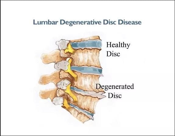
Lumbar Disc Degeneration Overview
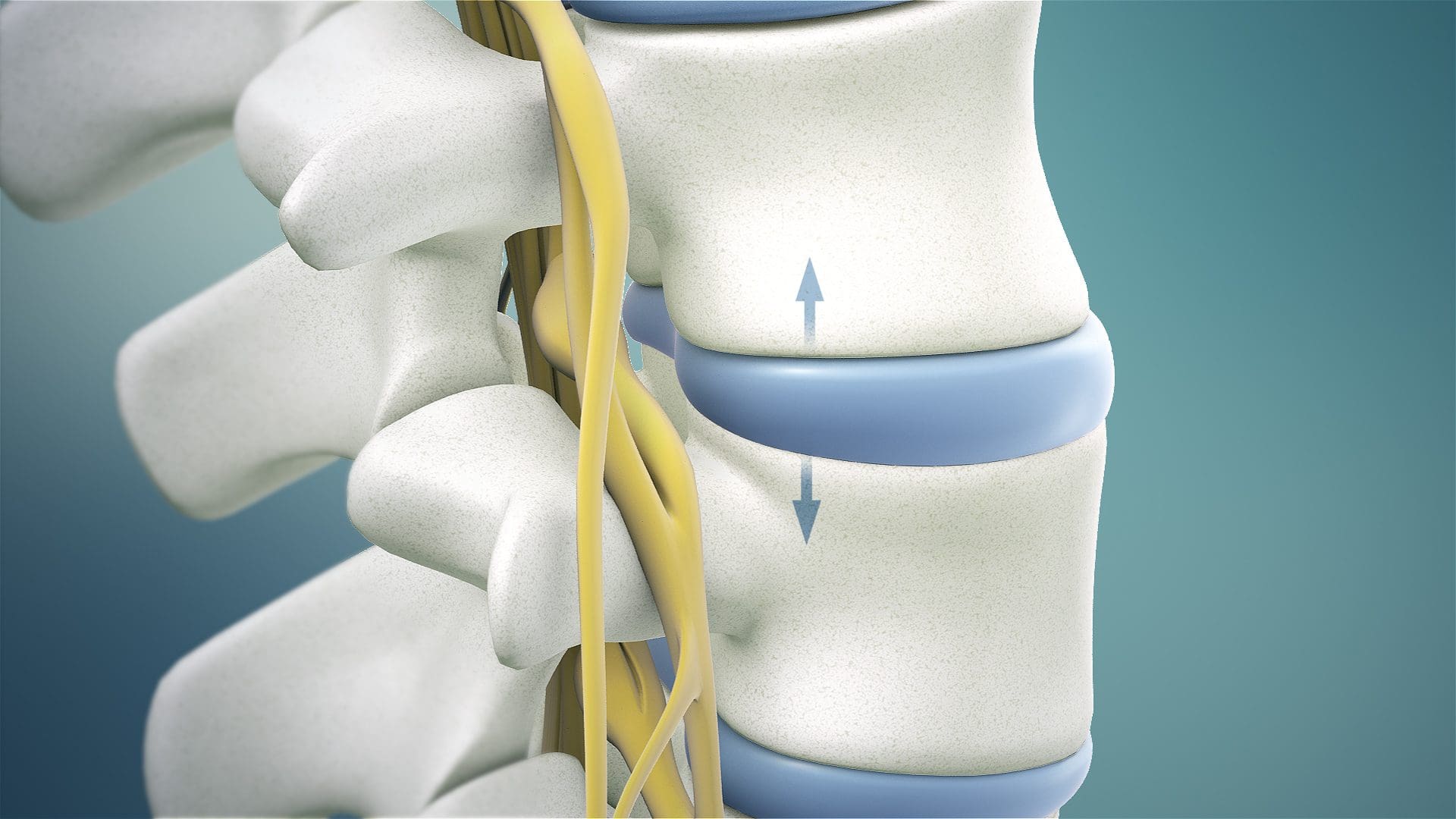
Have you begun to notice how your legs, buttocks, and feet are constantly experiencing tingling sensations that it makes it difficult to do everyday actions? Do you feel excruciating pain in your legs while standing, only to find relief from resting? Or do you notice that you are getting aches and muscle strains from normal motion like bending, twisting, and turning after waking up in the morning? Many individuals, especially older working adults, are experiencing these issues due to lumbar disc degeneration. Now lumbar disc degeneration usually occurs naturally as the body ages, which then causes the intervertebral disc to wear and tear constantly under pressure, then cracks and causes the disc to herniate. Since the intervertebral disc provides structural support and shock absorption against mechanical loads to the lumbar region, when normal or traumatic factors cause changes, it can cause dysfunction and instability in the lumbar spine. (Mohd Isa et al., 2022)
When the intervertebral discs begin to crack under pressure, it can cause pain-like symptoms affecting the upper and lower extremities. Additionally, lumbar disc degeneration can cause spinal flexibility to decrease and reduce the spine’s ROM (range of motion), which causes more stress on the surrounding muscles, ligaments, and tissues. Lumbar disc degeneration can cascade events from disc bulging to nerve root irritation. (Liyew, 2020) This means that when there are advanced degenerative changes to the lumbar facet joints and the surrounding soft tissues, it can cause the spinal canal to narrow and compress the adjacent nerve root. To that point, it can lead to reduce disc height and lead to sciatic nerve pain.
Lumbar Disc Degeneration Associated With Sciatica
Now how would lumbar disc degeneration be associated with sciatic nerve pain? When the intervertebral disc is being compressed under unwanted pressure, it can crack over time and herniate out of its original position, which then can press on the nerve root causing radiating pain to travel to the affected muscle area. Since the sciatic nerve is positioned in the lumbosacral region, it runs from the top of the gluteus muscles and down to the back of the hamstrings and calves. When unwanted pressure causes the intervertebral disc to herniate and start to affect the sciatic nerve, it causes a frequent symptom known as lumbar sciatica, where the herniated disc is compressing the sciatic nerve. (Zitouna et al., 2019) To that point, it can cause radiating, shooting pain down to the leg, making it difficult for individuals with demanding jobs to find relief. Since the lumbar intervertebral discs have a corresponding relationship with the central nervous system, the nerve roots that surround the spinal discs help with providing neuron signals to the adjacent muscles, which allows the arms, hands, legs, and feet to move. (Bogduk, Tynan, & Wilson, 1981) However, when the intervertebral discs are herniated, it can disrupt the neuron signaling to the muscles and cause referred pain to the lower or upper extremities. When this happens, many individuals opt to seek treatment.
Sciatica Secrets Revealed- Video
Many individuals dealing with sciatic nerve pain associated with lumbar disc degeneration will often find temporary relief to continue their work despite constant pain. This is due to the unwanted pressure that is causing an overload on the spinal disc to cause them to degenerate and invoke pain in the lower regions. At the same time, age and degenerative structural changes have a close relationship that can cause a greater effect on stress distribution to the lower back. (Adams, McNally, & Dolan, 1996) This leads to overlapping risk profiles contributing to sciatic nerve pain associated with lumbar disc degeneration due to normal and traumatic factors. However, many individuals don’t have to suffer from pain-like symptoms related to lumbar disc degeneration as there are numerous treatments to restore disc height and reduce sciatic nerve pain. Non-surgical treatments are great for many individuals looking for cost-efficiency and can be personalized to the person’s pain. (Louis-Sidney et al., 2022) Non-surgical treatments like chiropractic care, massage and physical therapy, and spinal decompression can help many individuals with lumbar disc degeneration associated with sciatic nerve pain. These treatments incorporate mechanical and manual manipulation of the spine to realign the body out of subluxation while incorporating various techniques to stretch out the soft tissues and muscles to strengthen their length and reduce nerve entrapment. The video above explains a bit more about how these treatments can restore mobility to the body, reduce inflammation, and relieve muscle tension caused by lumbar disc degeneration associated with sciatic nerve pain.
Spinal Decompression Restores Disc Height
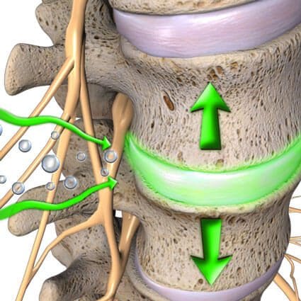
Now non-surgical treatments like spinal decompression can help reduce the progress of lumbar disc degeneration by restoring disc height. Spinal decompression uses gentle traction on the spine to allow the necessary nutrients, fluids, and blood to be reabsorbed back into the spine. It will enable the herniated disc to return to its original position and rehydrate it. (Yu et al., 2022) Since the spine and body age naturally, spinal decompression can restore disc height by creating negative pressure to regain mobility and stretch out the affected muscles surrounding the spine.
Spinal Decompression Reduces Sciatic Nerve Pain
Additionally, decompression can help reduce sciatic nerve pain as it can help the herniated disc alleviate the pressure on the sciatic nerve and can restore mobility to the body. Spinal decompression can become a positive outcome for many working individuals as it allows individuals to be more mindful of their habits that were the cause of the pain they were experiencing. (Brogger et al., 2018) With non-surgical treatments like spinal decompression, many individuals can become efficient with work while being more conscious about what factors contribute to the pain. This, in turn, allows them to focus on their well-being and strengthen their weak points with a personalized plan that will enable them to make small changes in their day-to-day lives and become pain-free after a few consecutive treatments.
References
Adams, M. A., McNally, D. S., & Dolan, P. (1996). ‘Stress’ distributions inside intervertebral discs. The effects of age and degeneration. J Bone Joint Surg Br, 78(6), 965-972. https://doi.org/10.1302/0301-620x78b6.1287
Bogduk, N., Tynan, W., & Wilson, A. S. (1981). The nerve supply to the human lumbar intervertebral discs. J Anat, 132(Pt 1), 39-56. https://www.ncbi.nlm.nih.gov/pubmed/7275791
Brogger, H. A., Maribo, T., Christensen, R., & Schiottz-Christensen, B. (2018). Comparative effectiveness and prognostic factors for outcome of surgical and non-surgical management of lumbar spinal stenosis in an elderly population: protocol for an observational study. BMJ Open, 8(12), e024949. https://doi.org/10.1136/bmjopen-2018-024949
Liyew, W. A. (2020). Clinical Presentations of Lumbar Disc Degeneration and Lumbosacral Nerve Lesions. Int J Rheumatol, 2020, 2919625. https://doi.org/10.1155/2020/2919625
Louis-Sidney, F., Duby, J. F., Signate, A., Arfi, S., De Bandt, M., Suzon, B., & Cabre, P. (2022). Lumbar Spinal Stenosis Treatment: Is Surgery Better than Non-Surgical Treatments in Afro-Descendant Populations? Biomedicines, 10(12). https://doi.org/10.3390/biomedicines10123144
Mohd Isa, I. L., Teoh, S. L., Mohd Nor, N. H., & Mokhtar, S. A. (2022). Discogenic Low Back Pain: Anatomy, Pathophysiology and Treatments of Intervertebral Disc Degeneration. Int J Mol Sci, 24(1). https://doi.org/10.3390/ijms24010208
Yu, P., Mao, F., Chen, J., Ma, X., Dai, Y., Liu, G., Dai, F., & Liu, J. (2022). Characteristics and mechanisms of resorption in lumbar disc herniation. Arthritis Res Ther, 24(1), 205. https://doi.org/10.1186/s13075-022-02894-8
Zitouna, K., Selmene, M. A., Derbel, B., Rekik, S., Drissi, G., & Barsaoui, M. (2019). An unexpected etiology of lumbosciatica. Tunis Med, 97(12), 1415-1418. https://www.ncbi.nlm.nih.gov/pubmed/32173813
In individuals with lumbosacral pain, how do cost-effective treatments compare to traditional care treatments affect muscle strain?
Introduction
The human spine is divided into three sections, which form an S-curve shape that supports the upper and lower body parts, maintaining good posture during movement. The spinal discs or intervertebral discs act as shock absorbers within each section of the spinal column. They help reduce axial overload and protect the spinal cord. The cervical, thoracic, and lumbar sections have specific roles in the upper and lower body parts, ensuring comfort and pain-free movement. However, many people engage in normal activities such as lifting improperly, sitting excessively, or carrying an unreasonable weight, leading to pain and disability over time without proper care. The lumbosacral region of the spine is the most commonly injured and is linked to low back pain. Lumbosacral pain can result from normal or traumatic factors, making individuals miss work or daily activities, leading to financial burdens when visiting a doctor. Symptoms associated with lumbosacral pain can cause referred pain to other parts of the body, leading individuals to think that the primary pain location is elsewhere. Fortunately, various cost-effective treatments can reduce the effects of lumbosacral pain and alleviate muscle strain in the lower back region. This article focuses on the many factors associated with lumbosacral pain, cost-effective treatments to reduce it, and the difference between traction and spinal decompression, which can alleviate muscle strain in the lumbosacral spinal region. As we work with certified medical providers who use our patients’ information to treat individuals experiencing lumbosacral pain and explain how combining non-surgical decompression as part of their routine can alleviate the pain-like symptoms affecting the lumbosacral region. We inform them about non-surgical treatments to ease lumbosacral pain while reducing muscle strain. We encourage our patients to ask essential questions while seeking education from our associated medical providers about their situation. Dr. Alex Jimenez, D.C., provides this information as an educational service. Disclaimer
Lumbosacral Pain Associated Factors
How many times a day have you been experiencing low back pain associated with lifting heavy objects? Do you feel muscle aches or strains in your lower back from excessing sitting from your job? Or do you feel pain in your lower back after a long day of work that feels better after sitting down? Many individuals don’t often realize that the pain they are feeling in their lumbosacral region could be due to the normal factors that are causing repetitive motions that are causing the spinal discs in the lumbosacral area to be compressed, damaged, or herniated. To that point, lumbosacral pain may correlate with low back pain. Since low back pain is mostly a non-specific issue, many working individuals with a sedentary desk job or an active job requiring physical exertion can be a clue to the causes of low back pain associated with lumbosacral pain. (See Tan & Kumar, 2021) Additionally, lumbosacral pain can cause the individual to have unwanted stress while undergoing treatment. The cost of treating lumbosacral pain associated with the low back can increase drastically.
The working individual would have to worry about the cost of traditional medical care and how to compensate for the lost wages to pay for the treatment. (Snook, 1988) This leads many individuals to continue working even when in excruciating pain by incorporating home treatments to reduce the pain temporarily. When the lumbosacral spine is dealing with pain, the nerve roots that surround the lumbosacral region will begin to go haywire, causing somato-visceral pain where the sensory signals cause symptoms of tingling and numbness to travel down to the legs, glutes, low back, and thighs. (Vaitkus & Sipylaite, 2021) Luckily, many individuals can be at ease in numerous ways. There are cost-effective treatments to reduce the pain-like issues associated with the lumbosacral region and alleviate the muscle strain caused by lumbosacral pain.
Can Core Exercises Erase Back Pain?-Video
Many individuals will look for home remedies to reduce the pain in the affected muscle area when treating lumbosacral pain associated with low back pain. Many people will opt for exercises, ice/hot packs, or massages to ease low back pain related to lumbosacral pain. (“Simple treatments best for acute low-back problems, say federal guidelines,” 1995) All these treatments are cost-effective and can be combined with non-surgical treatments to stretch the tight muscles, realign the spine, and help rehydrate the spinal discs back to the spine. The video above asks if core exercises can help ease back pain. The video details how weak core muscles correlate with lower back lumbosacral pain. Engaging the core during exercise can help stabilize the lumbosacral region while improving overall wellness.
When relieving lumbosacral pain, cost-effective non-surgical treatments can help many individuals find the relief they need. The effects of non-surgical treatments for the lumbosacral vertebrae apply various techniques to the spine by widening the spinal disc height, reducing muscle strain and spasms, and separating the vertebrae. (Colachis & Strohm, 1969) Many individuals have opted for these treatments because they are safe, cost-effective, and gentle on the spine. Since the spinal discs can be compressed due to unwanted axial load, spinal manipulation done by a chiropractor can realign the spine out of subluxation. (Cyriax, 1950) This allows the individual to feel instant relief and reduce the aggravated nerve roots from the lumbosacral spine. Other cost-effective treatments like traction therapy and spinal decompression can also alleviate lumbosacral pain that is causing the issue to many individuals.
Traction vs. Spinal Decompression
The difference between traction therapy and spinal decompression therapy varies within the individual and what their personalized treatment plan requires. Traction therapy incorporates half of the person’s body weight with additional weight to reduce nerve root compression and can be combined with other treatments like hot/cold therapies and electro-stimulation; combined with an exercise program can strengthen the weak muscles and reduce muscle strain. (Alrwaily, Almutiri, & Schneider, 2018)
With spinal decompression, many individuals will be strapped into a mechanical machine and feel a gentle pull on their spine. This creates negative pressure between the spine and allows the disc to lay off the aggravating nerve root and promote healing properties back to the disc. (Choi et al., 2022) Spinal decompression causes a direct distraction within the spinal segments with minimal discomfort to the individual. Both cost-effective treatments are suitable for individuals with lumbosacral pain along their spine as they can help relieve pain and reduce muscle strain along the lumbar region after a few sessions. Non-surgical treatments are beneficial for many individuals who are looking to take back their health and wellness without being in pain.
References
Alrwaily, M., Almutiri, M., & Schneider, M. (2018). Assessment of variability in traction interventions for patients with low back pain: a systematic review. Chiropr Man Therap, 26, 35. https://doi.org/10.1186/s12998-018-0205-z
Choi, E., Gil, H. Y., Ju, J., Han, W. K., Nahm, F. S., & Lee, P.-B. (2022). Effect of Nonsurgical Spinal Decompression on Intensity of Pain and Herniated Disc Volume in Subacute Lumbar Herniated Disc. International Journal of Clinical Practice, 2022, 6343837. https://doi.org/10.1155/2022/6343837
Colachis, S. C., Jr., & Strohm, B. R. (1969). Effects of intermittent traction on separation of lumbar vertebrae. Archives of Physical Medicine and Rehabilitation, 50(5), 251-258. https://www.ncbi.nlm.nih.gov/pubmed/5769845
See, Q. Y., Tan, J. B., & Kumar, D. S. (2021). Acute low back pain: diagnosis and management. Singapore Med J, 62(6), 271-275. https://doi.org/10.11622/smedj.2021086
Simple treatments best for acute low-back problems, say federal guidelines. (1995). Am J Health Syst Pharm, 52(5), 457. https://doi.org/10.1093/ajhp/52.5.457a
Vaitkus, A., & Sipylaite, J. (2021). Sensory Perception in Lumbosacral Radiculopathy with Radicular Pain: Feasibility Study of Multimodal Bedside-Suitable Somatosensory Testing. Acta Med Litu, 28(1), 97-111. https://doi.org/10.15388/Amed.2021.28.1.18
IFM's Find A Practitioner tool is the largest referral network in Functional Medicine, created to help patients locate Functional Medicine practitioners anywhere in the world. IFM Certified Practitioners are listed first in the search results, given their extensive education in Functional Medicine