El Paso Back Clinic Shockwave Therapy: A Non-Surgical Option for Chronic Pain
Why Real ESWT Matters for Deep Healing at an Integrative El Paso Back Clinic
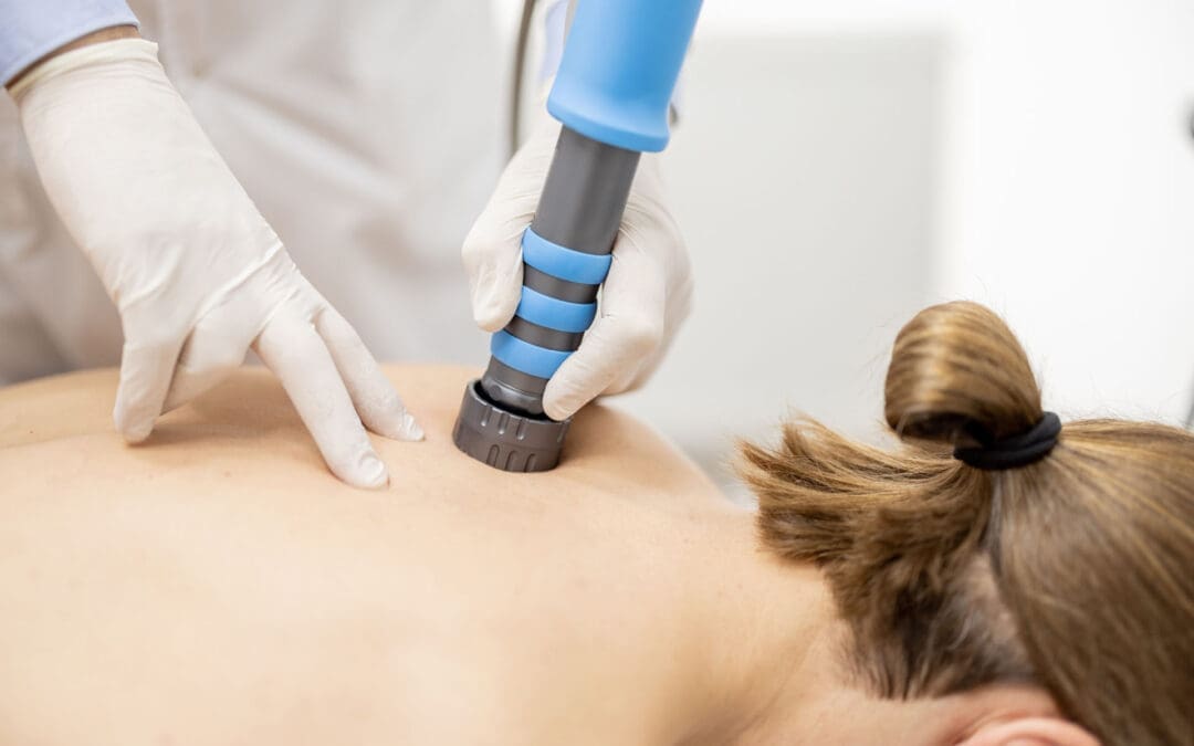
When people hear the term shockwave therapy, they often assume every machine is the same. It is not.
Some devices are true medical Extracorporeal Shockwave Therapy (ESWT) systems. Other devices are weaker radial pressure wave tools that are sometimes marketed as shockwave devices, even though they work differently. That difference matters if your goal is real tissue healing, not just short-term soreness relief. Mayo Clinic explains that focused shockwave (FSW) and radial pressure wave (RPW) are distinct waveforms, and only FSW is considered a “true shockwave” in a strict physical sense.
For a clinic like El Paso Back Clinic, where patients often come in with chronic pain, sports injuries, auto injuries, soft-tissue damage, and complex back conditions, the type of device and the treatment plan can make a big difference. The clinic’s site emphasizes multidisciplinary care, non-surgical recovery, and an integrative model that includes chiropractic, rehab, and functional medicine support.
This article explains, in plain language, what “real” shockwave therapy is, why focused shockwave is different from weaker devices, and how it fits into a complete recovery program in an integrative chiropractic setting.
What Is Real Shockwave Therapy?
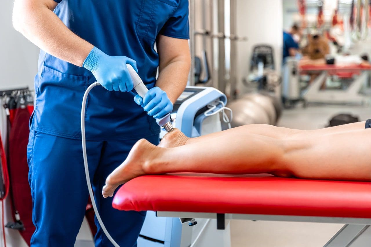
Extracorporeal Shockwave Therapy (ESWT) is a non-invasive treatment that sends acoustic energy (sound waves) into injured tissue from outside the body. It is used in musculoskeletal care to help reduce pain and support healing in stubborn injuries. UCHealth describes ESWT as a noninvasive option for people who have not responded well to more conventional treatments, noting that it delivers high-energy acoustic waves to injured areas.
Mayo Clinic also describes shockwave therapy as a growing tool in physical medicine and sports medicine, especially for tendon and fascia problems.
In simple terms
Shockwave therapy is used to help the body “restart” healing in tissue that has been painful or stuck for a long time, such as:
tendons
fascia
ligaments
some chronic soft-tissue injuries
certain bone healing problems (in selected cases)
Mayo Clinic lists many musculoskeletal uses, including plantar fasciitis, Achilles tendinopathy, patellar tendinopathy, and lateral epicondylitis (tennis elbow).
Not All “Shockwave” Machines Are the Same
This is the most important part of the topic.
Many clinics use the word shockwave, but there are two main categories of devices used in musculoskeletal care:
Focused Shockwave (FSW / F-ESWT)
Radial Pressure Wave (RPW / radial therapy)
Mayo Clinic clearly explains that these are different technologies and should not be treated as identical. In fact, Mayo states that only focused shockwave generates a true shockwave, while radial devices generate a radial pressure wave.
Why that matters
The difference is not just marketing. It affects:
how deep the energy goes
how precise the treatment is
how much energy reaches the target tissue
what conditions may respond best
If a patient has a deep tendon problem, scar tissue, or a stubborn chronic injury, the provider should know exactly what machine is being used and why.
Focused Shockwave vs. Radial Pressure Wave
Here is the practical difference in plain language.
Focused Shockwave (FSW)
Focused shockwave is designed to deliver energy to a specific target depth. It is more precise and is often the better choice when the provider wants to treat a deeper structure or a smaller, more exact area. Mayo Clinic notes that focused shockwave has different physical properties and can be used alone or in combination with radial treatment, depending on the condition.
Radial Pressure Wave (RPW)
Radial therapy spreads energy more broadly and is often more surface-level. Mayo Clinic explains that radial devices generate pressure waves and notes tissue penetration of about 4 to 5 cm in its 2022 discussion of radial ESWT.
That does not mean radial is “bad.” It means it is different. In many cases, radial therapy remains helpful. But if a clinic claims “shockwave” and the patient expects high-energy focused treatment, the patient should ask which device is being used.
Quick comparison
Focused shockwave
More precise targeting
True shockwave physics
Often used for deeper or more exact lesions
Better fit for some regenerative goals
Radial pressure wave
Broader spread
Pressure-wave technology
Often, more superficial or diffuse treatment
Can still be useful in the right case
Why Energy Dose Matters
Real ESWT is not just “machine on, machine off.” It is dosed.
One of the main ways clinicians describe ESWT dose is Energy Flux Density (EFD), and the standard unit is mJ/mm² (millijoules per square millimeter). A PubMed Central review explains that EFD is the professional parameter used to describe shockwave energy flow through tissue, and specifically notes the unit of measurement as mJ/mm².
This is important because:
stronger energy is not always better
tissue type matters
the diagnosis matters
different injuries need different treatment settings
A quality clinic should be able to explain the treatment plan in a way that matches your condition, rather than using the same approach for every patient.
Does Shockwave Therapy Create “Microtrauma”?
Many people explain shockwave therapy by saying it creates “microtrauma” that triggers healing. That is a common explanation, and Mayo Clinic Sports Medicine uses this language in a patient-friendly way, noting that acoustic waves can create microtrauma to help reinitiate a healing response in tendons.
That said, many experts also describe the process in a more modern way as mechanotransduction—meaning the waves create a mechanical signal that helps cells activate repair pathways. Mayo Clinic’s 2025 article also highlights mechanotransduction and regenerative effects like cellular signaling and neovascular changes.
A simple way to think about it
Shockwave therapy helps by:
stimulating local tissue response
improving healing signaling
reducing pain pathways over time
helping stubborn tissue become more “active” in repair
So the short answer is:
Yes, “microtrauma” is a common way to explain it.
But the bigger idea is that the shockwave creates a healing signal, not uncontrolled tissue damage.
FDA Regulation and Why It Matters
Another reason patients should ask questions is that regulatory status matters.
The FDA has approved/cleared specific extracorporeal shockwave devices for specific uses. For example, the FDA PMA listing for the OrthoSpec Extracorporeal Shock Wave Therapy device states that it is indicated for adults with proximal plantar fasciitis (with or without a heel spur) who have had symptoms for 6 months or more and have failed conservative treatment.
That helps patients understand two important points:
real ESWT is a recognized medical technology
device claims should match actual indications and training
If a clinic says “shockwave,” it is fair to ask:
What exact device is this?
Is it focused or radial?
Is it FDA-cleared/approved for a musculoskeletal indication?
These are smart questions, not rude questions.
Why Real ESWT Is Useful in an Integrative Chiropractic Clinic
Shockwave therapy can be very effective, but it works best when the diagnosis is correct, and the rest of the care plan supports healing.
That is where an integrative clinic model is helpful.
The El Paso Back Clinic describes on its website a multidisciplinary, non-surgical, and functional recovery approach that includes chiropractic care, rehab, and broader wellness support. It also describes care for back, auto, and sports injuries, tendinopathy-related issues, and chronic pain.
Why this pairing makes sense
Shockwave therapy targets soft tissue and the healing response.
Chiropractic and rehab help restore:
joint motion
spinal alignment
posture
movement control
load tolerance
When these are combined, the patient gets a more complete plan.
Example of an integrative recovery setup
A patient with chronic Achilles pain, plantar fasciitis, or post-accident scar tissue restriction may benefit from:
Focused shockwave or radial therapy (depending on the tissue depth and goal)
Chiropractic adjustments to improve joint mechanics
Mobility work to reduce compensation patterns
Strength training/rehab exercise to improve tissue tolerance
Lifestyle support (sleep, inflammation control, nutrition)
This is especially important for back and soft-tissue injuries, as pain often has multiple causes. The tissue may be irritated, but there may also be a movement issue, posture problem, or old compensation pattern keeping it from healing.
Clinical Observations in Dr. Alexander Jimenez’s Integrative Model
Public information on dralexjimenez.com and El Paso Back Clinic describes Dr. Alexander Jimenez as a Doctor of Chiropractic and board-certified Family Nurse Practitioner (DC, APRN, FNP-BC) who uses a multidisciplinary, integrative approach focused on non-surgical recovery, diagnostics, and personalized care.
His El Paso Back Clinic content also emphasizes:
advanced injury rehabilitation
chronic pain care
sports injury care
auto injury care
functional medicine support
team-based recovery planning
These clinic observations support the idea that shockwave therapy should not be used as a stand-alone “gadget” treatment. Instead, it fits best within a broader care plan that includes biomechanics, rehab, and whole-person recovery.
Why dual training matters in this setting
In a clinic model that blends chiropractic and nurse practitioner perspectives, the provider can often look at a case more completely, including:
musculoskeletal pain drivers
nerve irritation patterns
inflammation
healing delays
activity limitations
overall recovery readiness
That type of clinical reasoning is helpful when deciding whether a patient should receive:
focused shockwave
radial therapy
chiropractic and rehab only
imaging first
referral or co-management
What Conditions Often Respond to Shockwave Therapy?
Shockwave therapy is often used for chronic injuries that have not improved enough with standard care.
Mayo Clinic and UCHealth commonly describe these types of cases:
Plantar fasciitis
Tennis elbow (lateral epicondylitis)
Achilles tendinopathy
Patellar tendinopathy
Shoulder tendinopathy
Other chronic tendon or fascia pain problems
Mayo’s clinical articles also note that ESWT has roles in treating tendons, ligaments, fascia, and even in selected bone-healing situations.
It may be especially helpful when:
pain has lasted for months
the patient plateaued in regular therapy
surgery is being considered, but not yet desired
the injury is painful with loading (walking, running, lifting, gripping)
the provider wants a non-invasive option
How to Tell if a Clinic Is Offering “Real” Shockwave Therapy
Because the market uses confusing language, patients should ask direct questions before paying for treatment.
Ask these questions
Is this focused shockwave (FSW) or radial pressure wave (RPW)?
What condition are you treating, and why is this device the right choice?
How do you set the energy dose (EFD/mJ/mm2)?
How many sessions are usually recommended for my condition?
Will I also get rehab or movement treatment?
If my pain is deep, how will you target it?
Is the device FDA-cleared/approved for musculoskeletal use?
A strong clinic should be comfortable answering these questions in simple language.
Why Device Hype Alone Is Not Enough
Some clinics advertise shockwave therapy as a miracle treatment. That is not the best way to present it.
Shockwave therapy can be a powerful tool, but results depend on:
Even the best technology will not work well if the diagnosis is wrong or if the patient returns to the same harmful movement pattern right away.
This is one reason integrated care models, like the one described at El Paso Back Clinic and Dr. Jimenez’s clinical sites, can be so useful for complex injuries: patients receive more than one treatment option and more than one clinical lens.
Bottom Line: Focused ESWT Is the Better Choice for True Regenerative Shockwave Goals
If your goal is real regenerative shockwave therapy, focused shockwave (FSW/F-ESWT) is usually the benchmark because it is the true shockwave form and offers more precise targeting. Mayo Clinic makes this distinction very clearly.
Radial devices can still be helpful in many cases, but they are not the same technology. Patients should not be told they are identical.
For patients in El Paso dealing with:
chronic tendon pain
back-related soft tissue problems
sports injuries
accident-related soft tissue injury
stubborn pain that has not improved
An integrative clinic model like El Paso Back Clinic can be a strong fit because it combines:
non-invasive care
structural assessment
chiropractic and rehab
broader healing support
multidisciplinary planning
That is often what it takes to move from “temporary pain relief” to true recovery.
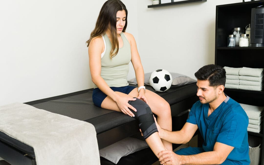
Common Fastpitch Softball Injuries and How El Paso Back Clinic’s Integrative Chiropractic Care Can Help
Fastpitch softball is a tough sport that asks a lot from players. Pitchers use the underhand windmill throw frequently, and everyone must move quickly and change direction quickly. This leads to pain in muscles and bones. The most common are overuse problems in the shoulder and elbow, like rotator cuff strains and UCL tears from all that pitching. Then there are sudden hurts, such as ACL tears in the knee, ankle sprains, and breaks from sliding, diving, or running into others. Players also deal with finger and hand issues, lower back pain, and concussions. At El Paso Back Clinic in El Paso, TX, they use integrative chiropractic care. This is a gentle, whole-body approach that includes spinal adjustments, muscle therapy, and rehab exercises. It addresses both acute injuries and the root causes of overuse. This care helps softball players heal faster, get stronger, and prevent re-injury. Led by Dr. Alexander Jimenez, DC, APRN, FNP-BC, the clinic focuses on athletes with personalized plans.
Common Injuries in Fastpitch Softball
Fastpitch softball can cause injuries due to its speed and repeated moves. Pitchers throw hard and often, putting stress on their arms. Other players dive, slide, and run, which can twist joints or cause impacts. Research shows shoulder and elbow overuse is the top issue for pitchers because of the windmill pitch (Rothman Orthopaedics, n.d.; Andrews Sports Medicine, n.d.). Lower-body problems result from quick stops and turns (Sports Medicine Clinics, 2025). Head injuries come from hits or crashes (Children’s Health, n.d.).
Here are some main overuse injuries:
Rotator cuff strains: Repeated throwing inflames the shoulder muscles, causing pain. This hits pitchers and throwers hard (Share UPMC, 2020; HDP Chiro, n.d.).
UCL tears: The elbow ligament gets stretched or torn due to the pitching force. Young players who overdo it are at risk (UC Health, n.d.; North Central Surgical, n.d.).
Sudden, acute injuries include:
ACL tears: Knee ligament rips during fast changes in direction. It can keep players out for months (Andrews Sports Medicine, n.d.; PubMed, n.d.).
Ankle sprains: Ankles twist while running or sliding into bases (Rock Valley PT, n.d.; Children’s Hospital, 2022).
Fractures: Breaks in fingers, hands, or wrists from dives or ball hits (Summit Orthopedics, 2022; Therapy Partners Group, n.d.).
Other common problems are:
Finger and hand injuries: From catching or batting (UC Health, n.d.).
Lower back pain: Caused by twisting or bad pitching form (North Central Surgical, n.d.; Share UPMC, 2020).
Concussions: Brain injuries from collisions or head hits (Children’s Health, n.d.; YouTube, n.d.).
These often stem from excessive play without breaks (PubMed, n.d.; PMC, n.d.). Strains and sprains are frequent in arms and legs (PMC, n.d.). To prevent them, use warm-ups, good technique, rest, and pitch limits (Rothman Orthopaedics, n.d.; UC Health, n.d.; NCYS, 2022).
Integrative Chiropractic Care at El Paso Back Clinic
At El Paso Back Clinic, integrative chiropractic care treats the whole body without surgery or meds. It’s holistic, meaning it looks at everything that affects health. The clinic combines chiropractic care with functional medicine and sports rehabilitation to address injuries and their causes (El Paso Back Clinic, n.d.; Integrative Chiro Center, n.d.). Dr. Alexander Jimenez and his team use evidence-based ways to help athletes.
Key parts of their care:
Spinal adjustments: These correct spinal misalignments to reduce pain, improve mobility, and support nerve function (Injury2Wellness, n.d.; SCUHS, n.d.).
Soft tissue therapy: Techniques such as massage reduce swelling and promote muscle healing (SCUHS, n.d.; Peoria Spine and Sport, n.d.).
Functional rehabilitation: Exercises build strength, balance, and flexibility to prevent re-injury (Push as RX, n.d.; Dallas Accident and Injury Rehab, n.d.).
The clinic also offers nutrition, stress management, and lifestyle tips to support full recovery (El Paso Back Clinic, n.d.). This differs from basic care by addressing root causes of softball injuries, such as poor posture or weak muscles (Chiropractic Sports Care, n.d.; El Paso Back Clinic, n.d.).
Benefits for Softball Players at El Paso Back Clinic
El Paso Back Clinic helps softball players recover quickly, play better, and avoid injuries. Their care corrects alignment and reduces inflammation to promote faster healing (SCUHS, n.d.). Players gain more power from balanced bodies, leading to stronger pitches and quicker moves (Dallas Accident and Injury Rehab, n.d.). Prevention is key—they spot problems early (Push as RX, n.d.; El Paso Back Clinic, n.d.).
Dr. Alexander Jimenez shares from his work: Overuse in softball causes inflammation and nerve issues. His methods, such as adjustments and nutrition, can help without surgery (Dr. Alexander Jimenez, n.d.; Dr. Alexander Jimenez LinkedIn, n.d.). He treats shoulders, knees, and backs with movement checks to stop repeats. This fits softball, where arm strain is common.
Benefits include:
Quicker recovery: Adjustments reduce pain and swelling so players return soon (Injury2Wellness, n.d.; SCUHS, n.d.).
Better performance: Stronger muscles and joints mean harder throws and faster runs (Dallas Accident and Injury Rehab, n.d.).
Injury prevention: Regular visits address imbalances, reducing overuse risk (El Paso Back Clinic, n.d.; Push as RX, n.d.).
Studies and videos support this. One shows that therapy for softball injuries is beneficial (YouTube, n.d.). At the clinic, athletes receive custom plans that include rehabilitation and education (El Paso Back Clinic, n.d.).
If you’re in El Paso or nearby, like Horizon City, contact El Paso Back Clinic today. Call +1-915-850-0900 or schedule an appointment. Locations include 11860 Vista Del Sol, Ste 128. Discover how Dr. Jimenez can help your game.
In the end, fastpitch softball risks injuries, but El Paso Back Clinic’s integrative care offers real help. It heals holistically and builds strength. Players stay on the field longer and stronger.
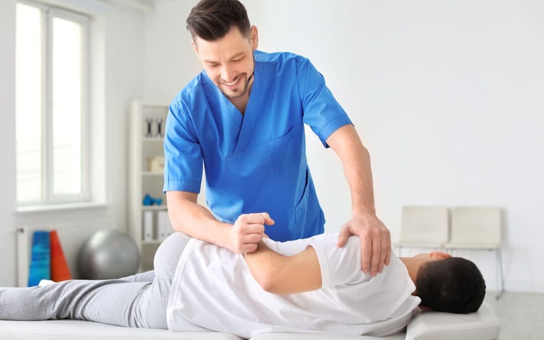
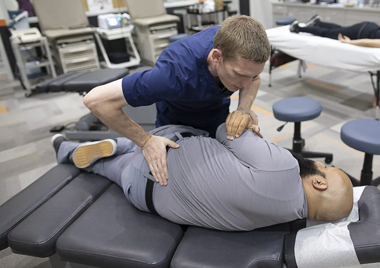
Optimal Joint Movement: Enhancing Mobility and Stability at El Paso Back Clinic
A chiropractor or Nurse Practitioner works with a patient in a rehabilitation center to improve joint mobility.
Optimal joint movement is essential for an active, pain-free life. At El Paso Back Clinic in El Paso, TX, we specialize in helping people achieve this through personalized chiropractic care. This article explains what optimal joint movement means, why it’s important, and how our clinic’s integrative approaches can restore it. Whether you’re dealing with back pain, sports injuries, or daily stiffness, our team, led by Dr. Alex Jimenez, DC, APRN, FNP-BC, uses spinal adjustments, rehabilitation, and functional medicine to get you moving better. Discover how we support joint health to improve function in everyday tasks and athletic pursuits.
Understanding Optimal Joint Movement
Optimal joint movement is the ability to move your joints through their full natural range of motion (ROM) smoothly, without pain, and with good control. It’s often referred to as high-quality mobility, blending flexibility with strength for daily activities and sports (University of Colorado Anschutz Medical Campus, n.d.).
At El Paso Back Clinic, we define it as moving joints efficiently while maintaining balance between mobility (active movement) and stability (joint control). This ensures muscles, ligaments, and tendons work together properly (National Academy of Sports Medicine, n.d.; Mainstay Medical, n.d.). For instance, a healthy shoulder should lift overhead to 180 degrees without strain, allowing you to reach shelves or throw a ball (Verywell Health, 2023a).
When injury or prolonged sitting disrupts this, mobility declines, leading to awkward movements elsewhere in the body (University of Colorado Anschutz Medical Campus, n.d.). Our clinic addresses this through holistic care, combining adjustments, soft-tissue therapy, and exercises to reduce inflammation and improve coordination.
Key Elements of Optimal Movement:
Full ROM: Joints reach their natural limits, like knee flexion to 140 degrees for squatting (The GO KNEE, n.d.).
Smooth Control: No jerking or pain, thanks to strong muscles and clear nerve signals.
Balance: Mobility for range, stability to prevent wobbles or injuries (ACE Fitness, n.d.a).
The Importance of Mobility and Stability Balance
At El Paso Back Clinic, we emphasize the balance between mobility and stability for peak performance. Mobility allows free movement, while stability keeps joints secure during activities (ACE Fitness, n.d.b). This synergy is key in our treatments.
Think of the body as a chain: Ankles and hips need mobility for steps, while knees and lower back provide stability (Motus Physiotherapy, n.d.; NASM, n.d.). If an ankle stiffens due to injury, the knee compensates, increasing the risk of pain (Physical Therapy at MJC, n.d.). Our chiropractic adjustments and rehab programs restore this chain, enhancing joint function.
Integrative care at our clinic—including spinal decompression and strength training—supports this balance, reducing the risk of injury and improving mobility (Peninsula Wellness Partners, n.d.).
Common Disruptions to Joint Mobility
Life factors can hinder optimal joint movement. Injuries cause swelling and tightness, limiting ROM (Frozen Shoulder Clinic, n.d.; Musculoskeletal Key, n.d.). A sedentary lifestyle, common in desk jobs, tightens muscles and stiffens joints (Dr. Ong Kee Leong, n.d.).
At El Paso Back Clinic, we see this in patients with back pain or sciatica, where poor posture leads to compensation and strain in other areas (OMassageT, n.d.). Aging, arthritis, or repetitive motions worsen it (Arthritis Foundation, n.d.; Chesapeake Regional, n.d.).
Typical Causes:
Trauma: Sprains create hard end-feels, stopping movement early (Physiopedia, n.d.c).
Inactivity: Shortens tissues, reducing flexibility (Dr Ong Kee Leong, n.d.).
Health Conditions: Arthritis limits ROM, causing bony sensations (Physiopedia, n.d.c).
Habits: Bad ergonomics unbalance the kinetic chain (OMassageT, n.d.).
Without correction, this increases fall risk and reduces quality of life. Our clinic’s diagnostic tools, such as digital X-rays, identify issues early.
Why Prioritize Optimal Joint Movement?
Good joint movement enhances everything from walking to sports. It prevents pain and boosts efficiency (OneStep, n.d.). At El Paso Back Clinic, we help athletes improve power and reduce injuries through better ROM (Activ Therapy, n.d.).
For daily life, it means easier tasks without fatigue (Baliston, n.d.). In walking, ankle flexion aids balance; poor ROM shortens strides (Baliston, n.d.). Our programs keep joints lubricated and muscles strong (Arthritis Foundation, n.d.).
At El Paso Back Clinic, maintenance starts with assessment. We measure ROM against norms using tools like goniometers (Physical Therapy at MJC, n.d.; Trainerize, n.d.). Then, we recommend exercises.
Regular activity, such as stretching, helps keep joints flexible (Arthritis Foundation, n.d.; Royal City Physiotherapy, n.d.). Our mobility drills focus on control for real-world use (Royal City Physiotherapy, n.d.).
Practical Tips:
Warm-Ups: Shoulder circles or ankle rolls (Chesapeake Regional, n.d.).
Stretching: Hold for 30 seconds on tight spots (Verywell Health, 2023a).
Strength Work: Squats for knee stability (ACE Fitness, n.d.b).
Activity: Low-impact, like swimming (Arthritis Foundation, n.d.).
Tools: Foam rollers for self-care (Muscle and Motion, n.d.).
Visit our East Side location for personalized plans.
Integrative Chiropractic Care at El Paso Back Clinic
Our clinic offers holistic chiropractic care to restore joint movement. Led by Dr. Alex Jimenez, we combine adjustments, therapy, and guidance (Peninsula Wellness Partners, n.d.; Evolved Health Chiropractic, n.d.).
Adjustments realign joints, easing inflammation and nerves (Rodgers Stein Chiropractic, n.d.a; Rodgers Stein Chiropractic, n.d.b). Soft tissue work and rehab build muscle support (Evolved Health Chiropractic, n.d.).
This approach enhances mobility, strengthens areas, and reduces risks (Core Integrative Health, n.d.; Duca Chiropractic, n.d.). Joint mobilization gently increases ROM (Smart Sports Medicine, n.d.).
Our Services:
Spinal Adjustments: Restore alignment for better ROM (Chiropractic Omaha, n.d.).
Functional Medicine: Addresses root causes, such as nutrition (TXMAC, n.d.).
Rehab: Exercises for long-term health (Duca Chiropractic, n.d.).
Clinical Insights from Dr. Alex Jimenez at El Paso Back Clinic
Dr. Alex Jimenez, DC, APRN, FNP-BC, heads El Paso Back Clinic, with over 30 years of experience in integrative care. At our facilities, he blends chiropractic, functional medicine, and rehab for joint issues (Jimenez, n.d.a; Jimenez, n.d.b).
His observations: Adjustments alleviate nerve-related issues, restoring ROM in cases of back pain or sciatica (Jimenez, n.d.a). Patients from accidents or sports regain mobility through tailored plans (Jimenez, n.d.a).
Dr. Jimenez focuses on root causes with nutrition and exercises, preventing surgery (Jimenez, n.d.b). For hips or knees, agility programs balance mobility and stability (Jimenez, n.d.a). Our holistic model empowers patients and aligns with evidence supporting better function (Jimenez, n.d.b).
At El Paso Back Clinic, optimal joint movement is achievable with our expert care. Balance mobility and stability to overcome disruptions. Visit elpasobackclinic.com or our El Paso locations for help from Dr. Jimenez’s team.
Anterior Hip and Leg Muscles: What They Are, What They Do, and Why They Hurt
A woman holds her aching anterior hip.
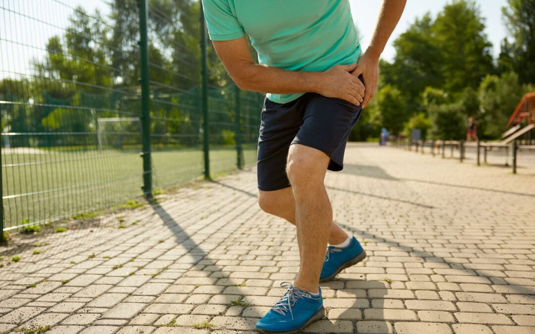
Pain in the front of the hip (often felt in the hip crease or groin area) and the front of the thigh is very common. It can show up when you stand up from a chair, climb stairs, run, kick, or even after sitting for a long time. The tricky part is this: front-hip pain is not always “just a tight hip flexor.” Sometimes it’s a muscle or tendon problem, but it can also be related to the hip joint, the pelvis, or the lower back.
This guide is written for everyday people in El Paso who want clear answers, plus a practical explanation of how an integrative chiropractic approach can help reduce pain and prevent flare-ups.
At El Paso Back Clinic, Dr. Alexander Jimenez and the team often observe a pattern: tight, overworked hip flexors, underactive glutes, and poor pelvic control—especially in people who sit a lot, train hard, or are recovering after an accident.
What “anterior hip and leg muscles” means
“Anterior” means the front side. The anterior hip and leg muscles are basically your “go-forward” and “stand-tall” muscles. They help you:
Lift your knee (hip flexion)
Step forward when walking or running
Stabilize your pelvis so your lower back doesn’t overwork
Straighten your knee (knee extension)
Control your leg when you climb stairs or squat
When these muscles get overloaded, they can feel tight, sore, weak, or sharp—depending on the cause.
The main anterior hip muscles (your hip flexors)
Hip flexors are not one muscle. They’re a group that works together.
Key hip flexor muscles
Iliopsoas (iliacus + psoas): the classic “deep hip flexor”
Rectus femoris: part of the quadriceps, crosses the hip and the knee
Sartorius: a long, strap-like muscle across the front of the thigh
Tensor fasciae latae (TFL): supports hip flexion and pelvic control
Pectineus (often grouped with hip flexors in clinical discussions)
Why iliopsoas matters so much
The iliopsoas helps:
Lift the thigh toward the trunk
Support the hip joint and pelvis
Add stability near the lumbar spine/pelvis connection
At El Paso Back Clinic, iliopsoas overuse is commonly discussed among athletes and active individuals who engage in sprinting, jumping, kicking, or repeated hip flexion.
The anterior thigh muscles (front of the thigh)
The main anterior thigh group is the quadriceps. They’re designed to extend the knee and help control motion during walking, stairs, squats, and landing.
Quadriceps muscles
Rectus femoris
Vastus medialis
Vastus lateralis
Vastus intermedius
The anterior thigh compartment is also supplied and controlled by key anatomical structures, such as the femoral nerve (often described as the L2–L4 roots) and the femoral artery system. That’s one reason pain patterns can sometimes feel confusing—muscles, nerves, and joints all influence the sensation you feel.
Why the anterior hip and leg muscles sometimes hurt sometimes
There are a few “big buckets” that explain most front-hip and front-thigh pain.
You’re asking the muscles to do too much, too often (overuse)
Overuse happens when the workload increases faster than your tissues can adapt. Common triggers include:
Sudden jump in running miles
More hills or speed work than usual
Lots of kicking (soccer, martial arts)
Heavy squats/lunges with poor control
Repetitive direction changes (basketball, football)
Overuse can irritate:
The muscle belly (soreness, tightness)
The tendon (tendinopathy-like pain)
The hip flexor attachment area near the front of the hip
Prolonged sitting keeps hip flexors in a “shortened” position
Sitting puts the hips into flexion. Over time, many people notice:
Hip tightness when standing up after sitting
A “pinchy” feeling in the front of the hip
Low back stiffness that shows up with hip tightness
Dr. Jimenez has emphasized in his recent writing that prolonged sitting can contribute to tight hip flexors and poor movement patterns, and that short movement breaks, along with targeted mobility work, can help many people feel better.
The hip flexors can be tight because other muscles are not doing their job
This is one of the most common “root causes” in stubborn cases:
Weak or underactive glutes
Weak deep core stabilizers
Limited hip mobility (the hip joint doesn’t move well)
Pelvic control issues (pelvis tips forward, rotates, or drops during gait)
El Paso Back Clinic explains that when the glutes weaken from inactivity and prolonged sitting, the hips and pelvis can become less stable and shift out of alignment, thereby increasing stress on surrounding tissues.
Sometimes the pain is not in the hip flexor at all
A major clinical point from family medicine guidelines is that hip pain often groups into:
Anterior (front)
Lateral (side)
Posterior (back)
…and the cause changes based on that pattern. Anterior hip pain may result from hip flexor injury, but it can also result from intra-articular hip joint problems (such as femoroacetabular impingement or labral pathology) or from referred pain.
A helpful “body map” concept is presented in educational videos that discuss what different hip pain locations can indicate, but a hands-on evaluation remains important when symptoms persist.
What the pain feels like: common patterns that guide the next step
These are not perfect rules, but they help you decide whether you’re dealing with a likely muscle/tendon issue or something deeper.
More likely muscle/tendon irritation (common hip flexor pattern)
Pain in the front hip crease
Worse with lifting the knee (stairs, marching)
Worse with running sprints, kicking, or hills
Tenderness in the front hip region
Feels tight after sitting
More likely hip joint involvement
Deep groin pain with hip rotation
Catching, clicking, locking, or “pinching”
Pain that persists despite basic stretching/rest
Range of motion feels blocked (especially flexion + rotation)
More likely low back/nerve referral
Front thigh pain plus low back symptoms
Numbness, tingling, and burning sensations
Symptoms that change with spine position
Why “stretching only” often fails
Stretching can feel good short-term, but it may not solve the real driver if the problem is:
Weak glutes and weak core control
A stiff hip joint or pelvic restriction
Poor movement strategy (how you squat, run, or stand)
A training load problem (too much too soon)
In other words, the hip flexors may be tight because they’re protecting you or compensating for something else.
How El Paso Back Clinic approaches anterior hip and leg pain
El Paso Back Clinic describes an integrative model that blends chiropractic care, rehabilitation concepts, and movement-based strategies, with a focus on mobility, flexibility, and the restoration of balanced function.
Here’s how that “integrative” approach commonly helps front-hip and front-thigh problems.
Identify the true driver (not just the sore spot)
A good evaluation typically includes:
History (training, sitting, injury, accident history)
Differentiation between hip joint vs. lumbar referral patterns
Dr. Jimenez has written about the importance of a structured hip evaluation to sort out the likely source of pain and match care to the pattern.
Restore joint motion and reduce protective “guarding”
When the pelvis/hip/lumbar spine isn’t moving well, the body often shifts load to the hip flexors and quads. Chiropractic-style care may focus on restoring smoother motion so the muscles stop overworking.
El Paso Back Clinic also discusses how muscle imbalance and chronic guarding can make it harder for muscles to “relax on their own,” especially after injuries.
Use soft tissue + targeted techniques to normalize muscle function
A common strategy is pairing hands-on care with neuromuscular techniques. El Paso Back Clinic specifically discusses assessing hip flexors with MET therapy (muscle energy technique) as part of reducing tightness and improving hip mobility.
Rebuild strength where it matters (glutes + core + hip control)
To prevent recurrence, the plan usually includes strengthening and control, especially:
Glute bridges and progressions
Hip abduction strength (side-lying or banded work)
Gradual reloading of hip flexors (instead of only stretching)
El Paso Back Clinic’s content repeatedly emphasizes that restoring balanced muscle function around the pelvis and hips supports daily movement and performance.
Practical tips you can start today (safe, simple, and realistic)
If your symptoms are mild and you’re not dealing with red flags, these are common first steps.
For desk workers and drivers (very common in El Paso)
Take 1–2 minute movement breaks every 30–60 minutes
Do a gentle hip flexor stretch (no sharp pinching)
Add a glute activation move (bridges or mini-band walks)
Keep your daily steps consistent (don’t go from 2,000 to 12,000 overnight)
For runners and athletes
Reduce aggravating volume for 1–2 weeks (not “stop forever,” just calm it down)
Avoid sprinting/kicking if it spikes sharp pain
Strengthen glutes and hip stabilizers 2–3x/week
Return to speed and hills gradually, not all at once
Quick self-check idea (mobility clue)
The Thomas Test is commonly used to screen for hip flexor tightness and may help distinguish whether the “tight feeling” is more iliopsoas- or quadriceps-based (rectus femoris). It’s not a diagnosis, but it can be a clue.
When you should get evaluated sooner rather than later
Don’t try to “stretch through it” if you have:
Severe pain after a fall or accident
Inability to bear weight
Fever or feeling unwell with hip pain
Worsening numbness/tingling or leg weakness
Persistent catching/locking and deep groin pain
A structured clinical examination is particularly important when hip pain may involve the hip joint or referral patterns.
The main takeaway
Your anterior hip and leg muscles—especially the hip flexors and quadriceps—are essential for walking, running, stairs, and posture. They often hurt because of:
Too much repeated load (overuse)
Too much sitting (hip flexors stay shortened)
Muscle imbalance (weak glutes/core causing hip flexors to overwork)
Hip joint or low back referral (pain “shows up” in the front)
An integrative chiropractic model—such as the one described in El Paso Back Clinic’s educational resources—focuses on identifying the underlying cause, restoring motion, improving muscle balance, and developing a plan to reduce the likelihood of recurrence.
How Integrative Chiropractic Care Prevents Future Injuries in Athletes Using Functional Movement Assessments
Sports: an athlete is in action on the field, ready to hit the ball during the game.
Athletes often push their bodies hard during training and competition. Small problems can build up over time and turn into painful injuries that force time off from sports. To catch these issues early, many athletes now ask for functional movement assessments as part of integrative chiropractic care. This method spots hidden imbalances like muscle tightness, weak spots, or stiff joints before pain starts. By addressing these problems with adjustments, soft-tissue work, and targeted exercises, practitioners help athletes stay healthy, move better, and avoid overuse injuries.
Functional movement assessments check how the body moves during everyday and sport-specific actions. These tests look at mobility, stability, balance, and coordination. Common movements include squats, lunges, reaching overhead, or stepping in different directions. The goal is to find areas where the body does not move smoothly or evenly. Even if nothing hurts yet, these assessments reveal subclinical imbalances—small issues that do not cause pain right away but can lead to bigger problems later.
Early detection of poor posture or uneven weight distribution
Spotting a limited range of motion in the hips, shoulders, or ankles
Identifying weak core or glute muscles that affect overall stability
Noting tight muscles that pull joints out of proper alignment
Integrative chiropractic care
Integrative chiropractic care combines spinal adjustments, soft-tissue therapies, and corrective exercises to effectively address these findings. Gentle adjustments move joints back into better positions, improving nerve signals and reducing pressure on surrounding tissues. Soft tissue work, such as massage or instrument-assisted techniques, loosens tight muscles and breaks up scar tissue. Corrective exercises then build strength and teach proper movement patterns. Together, these steps enhance nervous system function, optimize biomechanics, and stop the body from developing harmful compensation patterns.
The nervous system controls every muscle movement. When the spine or joints are misaligned, nerve messages can get disrupted. This leads to weaker muscle coordination or slower reaction times. Chiropractic adjustments help restore clear nerve pathways, so muscles fire at the right time and with the right force. Better biomechanics means joints move through their full, natural range without extra stress. This reduces wear and tear on knees, hips, shoulders, and the lower back.
Compensation patterns occur when one part of the body works harder to compensate for a weakness elsewhere. For example, tight hip flexors or a tilted pelvis in runners can cause the knees to track incorrectly, leading to pain or stress fractures over time. Faulty shoulder mechanics in swimmers or weightlifters can overload the rotator cuff. Integrative care addresses these root causes rather than just treating symptoms later.
Common subclinical imbalances identified through functional movement assessments include:
Muscle tension in the lower back or hamstrings that limits forward bending
Weak glute muscles that fail to stabilize the pelvis during running or jumping
Joint restrictions in the ankles that change walking or landing mechanics
Uneven shoulder mobility that affects throwing or overhead lifting
Poor core stability causes excessive arching in the lower back during lifts
By addressing these early, athletes lower their injury risk and maintain consistent training. Regular care also speeds recovery if minor issues arise, resulting in less downtime overall.
Practitioners often start with a thorough history and physical exam. They watch the athlete perform key movements and note any asymmetries or compensations. Based on the results, they create a personalized plan. Spinal adjustments realign the vertebrae to take pressure off nerves. Soft tissue therapies release tight fascia and muscles. Then, corrective exercises strengthen weak areas and retrain proper form. Over time, these steps improve balance, coordination, flexibility, and power output.
Key benefits of combining functional movement assessments with integrative chiropractic care:
Reduced chance of sprains, strains, tendonitis, and stress fractures
Improved joint mobility and muscle flexibility for better performance
Faster reaction times and coordination through better nerve function
Less inflammation and quicker recovery between workouts
Longer sports careers by preventing chronic overuse problems
Runners frequently show pelvic imbalances that tilt the hips and strain the iliotibial band or shins. Chiropractic adjustments and exercises that strengthen the glutes and core help keep the pelvis level, improving stride efficiency and cutting injury risk. Weightlifters with restricted shoulder mobility may compensate by excessively arching their backs, which can lead to low-back strain. Targeted soft tissue work and mobility drills correct this pattern before pain develops.
Football players and other contact-sport athletes benefit from regular checks of spinal alignment to better handle impacts. Swimmers gain from improved shoulder mechanics that prevent rotator cuff irritation. Weekend warriors who lift weights or cycle also see gains in endurance and reduced soreness. The approach works for athletes of all levels because it focuses on the root causes rather than waiting for symptoms.
Dr. Alexander Jimenez, DC, APRN, FNP-BC, brings valuable clinical observations to this field. As a chiropractor and board-certified family nurse practitioner with certifications in functional medicine, he emphasizes non-invasive, root-cause approaches. His work highlights how chiropractic adjustments, combined with functional assessments of mobility and biomechanics, help treat sports injuries, sciatica, and musculoskeletal imbalances. Dr. Jimenez observes that addressing nerve compression, inflammation, and movement dysfunction early—through adjustments, nutrition support, and tailored rehabilitation—enhances recovery and prevents recurrence in athletes and active individuals. His integrative practice in El Paso integrates chiropractic care with functional medicine to optimize performance, reduce chronic pain, and support long-term wellness.
This holistic view aligns with broader chiropractic principles that view the body as interconnected. When one area is restricted, it affects the whole kinetic chain. Integrative care breaks that cycle by restoring proper alignment and teaching sustainable movement habits.
Additional advantages athletes notice include:
Better posture during daily activities and sports
Enhanced proprioception (body awareness) for safer landings and cuts
Decreased muscle fatigue during long training sessions
Greater overall strength and power from efficient mechanics
Support for mental focus through reduced nagging discomfort
Preventing injuries this way also saves time and money by avoiding expensive treatments or missed competitions later. Many athletes report feeling stronger, more balanced, and more confident in their movements after consistent care.
To maintain results, athletes typically schedule regular visits. Frequency depends on training intensity, sport demands, and individual findings. Some come weekly during heavy training periods, while others maintain monthly check-ins. Between visits, they perform prescribed exercises at home or in the gym to reinforce new patterns.
Education plays a big role, too. Chiropractors teach proper warm-up routines, cool-down stretches, and body mechanics for specific sports. Nutritional guidance can sometimes complement care to support tissue repair and reduce inflammation. Collaboration with coaches, physical therapists, or trainers creates a complete support team.
In summary, functional movement assessments allow integrative chiropractic care to identify subclinical imbalances long before pain appears. Adjustments restore joint function, soft tissue therapies release restrictions, and corrective exercises build resilience. This combination enhances nervous system communication, optimizes biomechanics, and prevents compensation patterns that cause overuse injuries. Athletes—from runners dealing with pelvic tilts to lifters correcting shoulder mechanics—benefit by training more consistently, performing at higher levels, and enjoying longer, healthier careers. By addressing small issues proactively, this approach helps athletes stay in the game without painful interruptions.
Common Sports Injuries in El Paso and How El Paso Back Clinic Supports Full Recovery
Sports and physical activity are part of everyday life in El Paso. From running and weight training to football, soccer, and basketball, people of all ages stay active year-round. While this active lifestyle is healthy, it also leads to a high number of sports-related musculoskeletal injuries—especially when combined with the region’s heat, rough ground, and uneven terrain.
At El Paso Back Clinic, sports injury care focuses on restoring spinal alignment, joint mobility, muscle balance, and overall movement quality. When chiropractic care is combined with nurse practitioner (NP) support, athletes receive complete, coordinated care that promotes healing, performance, and long-term injury prevention.
Clinical observations from Dr. Alexander Jimenez, DC, APRN, FNP-BC, show that athletes recover more efficiently when spine health, joint mechanics, muscle function, and medical oversight are addressed together rather than separately.
Why Sports Injuries Are So Common in El Paso
El Paso presents unique physical challenges for athletes and active individuals. The environment itself can increase stress on the musculoskeletal system.
Common contributing factors include:
High temperatures, which increase fatigue and dehydration
Hard and uneven surfaces, stressing feet, ankles, knees, and hips
Year-round activity, limiting rest and recovery
High-impact sports, such as football and basketball
Repetitive movement patterns, common in running and training
When the spine and joints are not moving properly, the body compensates. Over time, these compensations increase injury risk and slow healing (NIAMS, n.d.).
Common Sports-Related Musculoskeletal Injuries Seen in El Paso
Sprains and Strains
Sprains and strains are among the most frequently treated injuries at El Paso Back Clinic.
Sprains affect ligaments
Strains affect muscles or tendons
Common areas include:
Ankles
Knees
Hamstrings
Lower back
These injuries often occur during quick movements, twisting, jumping, or improper warm-ups (Orthospine Centers, n.d.).
Knee Injuries (ACL, Meniscus, Runner’s and Jumper’s Knee)
Knee injuries are especially common in sports that involve cutting, jumping, or sudden stops.
Typical knee problems include:
ACL tears
Meniscus tears
Patellar tendonitis (jumper’s knee)
Runner’s knee
Misalignment in the spine, hips, or feet can increase stress on the knee joint, making chiropractic care an important part of recovery (Spectrum Therapy Consultants, n.d.).
Tendonitis and Overuse Injuries
Tendonitis develops when tendons are repeatedly stressed without enough recovery.
Common forms include:
Tennis elbow
Golfer’s elbow
Achilles tendonitis
Patellar tendonitis
These injuries often worsen slowly and are common in athletes who push through pain (Woodlands Sports Medicine, n.d.).
Shin Splints and Stress Fractures
Lower-leg injuries are common in runners and field athletes.
These include:
Shin splints
Foot stress fractures
Tibial stress injuries
Hard surfaces, worn footwear, and poor biomechanics increase the risk of these injuries (CTX Foot & Ankle, n.d.).
Hip Labral Tears
Hip labral tears affect the cartilage that stabilizes the hip joint.
Common symptoms include:
Deep hip or groin pain
Clicking or locking sensations
Reduced range of motion
These injuries are common among athletes who frequently twist, pivot, or sprint (Texas Spine Clinic, n.d.).
Rotator Cuff and Shoulder Injuries
Shoulder injuries often occur in athletes who lift, throw, or absorb contact.
Common issues include:
Rotator cuff strains or tears
Shoulder impingement
Joint instability
Shoulder pain is often linked to spinal and postural imbalances that chiropractic care addresses (Marque Medical, n.d.).
Lower Back Pain and Sciatica
Lower back pain is one of the most common complaints among athletes.
Contributing factors include:
Muscle strain
Core weakness
Poor posture
Spinal joint restrictions
When spinal alignment is compromised, nerve irritation such as sciatica may occur (Marque Medical, n.d.).
How Chiropractic Care at El Paso Back Clinic Helps Sports Injuries
Chiropractic care at El Paso Back Clinic focuses on restoring proper motion to the spine and joints. This allows the nervous system, muscles, and joints to work together efficiently.
Improving alignment reduces stress on injured tissues and supports natural healing (Vista Hills Chiropractic, n.d.).
Benefits of Chiropractic Care for Athletes
Athletes receiving chiropractic care often experience:
Reduced pain and stiffness
Improved joint mobility
Better balance and coordination
Faster recovery times
Lower risk of repeat injuries
Clinical experience shows that addressing spinal alignment early improves outcomes across many sports injuries (Jimenez, n.d.).
The Role of Nurse Practitioners in Integrated Sports Injury Care
Nurse practitioners (NPs) play an important role in sports injury management by providing medical oversight and coordination of care.
NPs may assist by:
Performing initial evaluations
Ordering diagnostic imaging (X-ray, MRI)
Managing pain and inflammation
Coordinating physical therapy
Monitoring healing progress
This medical support ensures injuries are accurately diagnosed and treated safely (NIAMS, n.d.).
Functional and Preventive Approach to Recovery
NPs often use a functional approach that looks beyond the injured area.
This includes evaluating:
Movement patterns
Training load
Nutrition and hydration
Sleep and recovery habits
Inflammation levels
Addressing these factors helps athletes heal fully and return stronger.
Coordinated Care: Chiropractic, NP, and Rehabilitation
One of the strengths of El Paso Back Clinic is coordinated care. Chiropractic care and NP oversight work together with rehabilitation to create a clear recovery plan.
A coordinated plan may include:
Chiropractic adjustments for alignment
Rehabilitation exercises for strength and stability
Medical monitoring for healing progress
Gradual return-to-sport planning
This team-based approach improves outcomes and reduces setbacks (Southwest Chiropractors, n.d.).
PRP Therapy and Advanced Recovery Options
For certain injuries, platelet-rich plasma (PRP) therapy may be recommended.
PRP may support healing for:
Tendon injuries
Ligament sprains
Knee injuries
Early osteoarthritis
NPs evaluate whether PRP is appropriate and coordinate care alongside chiropractic treatment and rehabilitation (Desert Institute of Sports Medicine, n.d.).
Clinical Example: Knee Injury Recovery at El Paso Back Clinic
Based on clinical observations from Dr. Jimenez, a typical knee injury recovery plan may include:
NP evaluation to diagnose the injury
Imaging to assess ligament or cartilage damage
Chiropractic care to improve spinal, hip, and knee alignment
Rehabilitation exercises to restore strength and stability
PRP therapy, when appropriate
Performance monitoring to prevent re-injury
This integrated approach supports long-term joint health and athletic performance.
Preventing Future Sports Injuries
Prevention is a major focus at El Paso Back Clinic.
Key strategies include:
Proper warm-ups and mobility work
Strengthening core and stabilizing muscles
Maintaining hydration in hot conditions
Correcting posture and movement patterns
Allowing adequate recovery time
Chiropractic and NP care help identify small problems before they become serious injuries (Texas Children’s Hospital, n.d.).
Long-Term Benefits of Integrated Sports Injury Care
Athletes who receive integrated care often experience:
Faster recovery
Fewer recurring injuries
Improved flexibility and strength
Better overall performance
Greater confidence in movement
Treating the spine, joints, muscles, and nervous system together leads to a more complete recovery.
Conclusion
Sports injuries are common in El Paso due to the climate, terrain, and high levels of physical activity. Injuries such as sprains, strains, knee injuries, tendonitis, back pain, and stress fractures can limit performance if not treated properly.
At El Paso Back Clinic, chiropractic care restores alignment and mobility, while nurse practitioners provide diagnostics, medical oversight, and coordinated treatment options. Together, this approach supports full recovery, injury prevention, and long-term performance.
Clinical experience from Dr. Alexander Jimenez shows that athletes recover best when care focuses on the whole musculoskeletal system—not just the painful area.
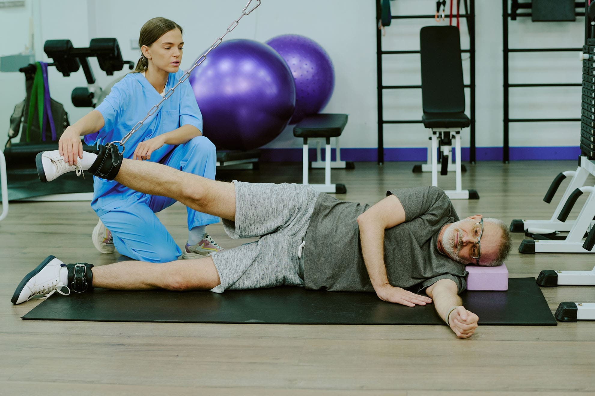
Make Your Health Goals Stick in 2026: How El Paso Back Clinic’s Integrative Team Supports Real Change
The patient uses a weight machine for injury rehabilitation under the supervision of a doctor of chiropractic and a nurse practitioner.
Most people don’t fail at New Year’s goals because they “don’t want it enough.” They fail because life gets busy, pain flares up, energy crashes, and stress piles on. When your body hurts or feels stiff, even simple plans—like walking more, lifting weights, or sleeping better—can feel harder than they should.
At El Paso Back Clinic, the goal is to make health changes easier to achieve and maintain through a team-based, integrative approach. That means bringing together the strengths of chiropractic care (movement, structure, mobility, and recovery) with the strengths of nurse practitioner care and wellness coaching (nutrition, sleep, stress, and whole-body support). The clinic describes this as a blend of injury care, wellness strategies, mobility programs, and integrated medicine designed to improve function and quality of life. El Paso Back Clinic® • 915-850-0900+2El Paso Back Clinic® • 915-850-0900+2
This kind of care supports common goals like:
increasing fitness and mobility
managing pain so you can stay active
improving energy and sleep
lowering stress and improving your stress response
“Integrative care” means your plan isn’t built around only one angle. Instead, it connects the pieces that usually get separated:
How you move
How you recover
How you eat
How you sleep
How you manage stress
How do you build habits that fit your real life
El Paso Back Clinic describes integrative chiropractic benefits as going beyond traditional adjustments by combining care approaches that support overall wellness and function. El Paso Back Clinic® • 915-850-0900
Why this matters for resolutions
Many resolutions are difficult to maintain because the plans ignore the real barriers. For example:
You want to exercise more—but your back pain spikes.
You want to lose weight—but your sleep is poor and your stress is high.
You want more energy—but your nutrition is inconsistent, and you’re not recovering.
An integrative plan helps because it aims to reduce the friction that makes healthy habits feel impossible.
The Team Approach: Chiropractor + Nurse Practitioner Mindset
Many clinics talk about how chiropractic care supports goals such as mobility, stress reduction, better sleep, and improved performance. gotcore.net+2Freedom Chiropractic+2 At El Paso Back Clinic, that support is often strongest when chiropractic care is paired with whole-person planning.
The chiropractor’s lane: move better with less strain
Chiropractic care commonly focuses on:
joint motion and spinal mechanics
posture and movement habits
mobility and flexibility
recovery support when you start working out again
helping reduce strain patterns that keep pain looping
The descriptions of services at El Paso Back Clinic emphasize spine-focused care and the restoration of function for back and musculoskeletal concerns. El Paso Back Clinic® • 915-850-0900+1
The NP/wellness lane: build a plan that supports your body from the inside out
A nurse practitioner and wellness-minded team approach can support:
nutrition planning that fits your schedule
sleep improvement routines
stress management strategies
health screening and medical risk review when appropriate
coaching that makes change more realistic to sustain
This matches the habit-focused guidance many health organizations recommend: set realistic goals, build routines, and avoid extreme “all at once” changes. Prism Health North Texas
Dr. Alexander Jimenez’s clinical observations (El Paso context)
Dr. Alexander Jimenez (DC, APRN, FNP-BC) frequently describes a dual-scope approach that connects biomechanics (how you move) with broader health planning (nutrition, functional assessments, and recovery strategies). His published clinic content also highlights the use of assessments and, when needed, imaging and integrated care planning to support recovery and function. LinkedIn+3El Paso, TX Doctor Of Chiropractic+3El Paso, TX Doctor Of Chiropractic+3
Why Resolutions Often Fail (And How an Integrative Plan Fixes That)
Here are common “resolution killers” and what a coordinated plan can do differently:
Pain blocks movement → Address mobility limits and movement mechanics so activity feels doable. National Spine & Pain Centers+1
Low energy → Improve sleep, nutrition consistency, and recovery structure. gotcore.net+1
Stress overload → Add stress skills and routines that calm the system and support follow-through. NIH News in Health+1
No accountability → Regular check-ins and plan adjustments keep you from quitting after a setback. drmmalone.com+1
A key idea in habit-based care is that early wins create a “positive feedback loop”—you feel better, so it becomes easier to keep going. drmmalone.com
1) Increase Fitness and Mobility (Without Getting Injured)
If your goal is to work out more, the priority is often moving well enough to train consistently.
Many chiropractic resources emphasize mobility, flexibility, and injury prevention as people increase activity at the start of the year. 5280 Balanced Health Center+2Freedom Chiropractic+2 El Paso Back Clinic also emphasizes flexibility, mobility, and agility programs to improve ability and quality of life. El Paso Back Clinic® • 915-850-0900
A simple evidence-based target
For general health, adults are commonly advised to aim for 150 minutes of moderate activity per week, plus 2 days of muscle-strengthening activities. CDC+1 That can be split into smaller chunks—like 30 minutes, 5 days a week.
What the integrative plan can look like
Assess mobility limits (hips, spine, shoulders) and address movement friction
Build a realistic weekly schedule
Progress intensity slowly, so you don’t crash or flare
Easy “start small” movement ideas:
10–20 minute walk after meals
2 strength sessions per week (basic full-body)
5-minute mobility routine daily
Progression rules that keep people consistent:
Add time before you add intensity
Keep at least 1–2 recovery days weekly
Measure consistency, not perfection
2) Manage Pain So You Can Stay Active
Pain goals often work better when you focus on function—not “zero pain tomorrow.” A pain-focused plan might aim to reduce flare-ups and increase what you can do safely. National Spine & Pain Centers
El Paso Back Clinic positions its care around helping people with frustrating injuries and chronic pain syndromes improve mobility and function. El Paso Back Clinic® • 915-850-0900
Practical pain goals that tend to stick
“Walk 20 minutes, 4 days/week without a flare.”
“Lift twice/week with pain staying under a 3–4/10.”
NP-style wellness support can focus on sleep, stress, consistency in nutrition, and pacing habits that support recovery. Prism Health North Texas+1
Helpful pacing ideas (simple but powerful):
Use shorter workouts more often
Stop just before your “flare threshold”
Build capacity gradually rather than “weekend warrior” bursts
3) Boost Energy the Smart Way
Energy is not just “motivation.” If you’re tired, your plan needs better recovery.
Many chiropractic sources link better sleep and reduced tension with feeling more capable and consistent over time. gotcore.net+1 El Paso Back Clinic also describes a wellness-focused approach aimed at improving energy, sleep, and overall function. El Paso Back Clinic® • 915-850-0900
It’s common to hear people say they want to “boost immunity.” A safe and practical way to think about this is:
You can support overall wellness by improving sleep, physical activity, and stress management—foundations that matter for health.
Regular physical activity is widely recommended for health. CDC
Mindfulness-based approaches have evidence supporting their effectiveness for stress, sleep, and pain management. NIH News in Health
So instead of chasing extreme detoxes or perfect diets, an integrative plan often focuses on steady basics:
sleep routine
movement most days
nutrition consistency
stress skills
That’s the kind of “quiet consistency” that makes resolutions last.
5) Lower Stress and Improve Stress Response
Stress shows up in the body: tight shoulders, headaches, jaw tension, shallow breathing, gut tension, and poor sleep.
Mindfulness-based treatments have evidence supporting reduced anxiety/depression symptoms and improved sleep, and may help people cope with pain. NIH News in Health Many chiropractic sources also connect care with stress reduction and better sleep as part of overall wellness. gotcore.net+1
Pick one main goal (fitness OR pain, energy, OR stress)
Add two support habits
Track consistency weekly
Adjust every 2–4 weeks
Examples of “support habits”:
protein at breakfast
20-minute walk 4x/week
5 minutes of mobility daily
bedtime routine 5 nights/week
A Simple 4-Week Plan (El Paso Back Clinic Style: Practical, Not Perfect)
This is a general example you can personalize with your provider team.
Week 1: Reduce friction
Identify mobility limits and pain triggers
Set one realistic activity goal
Begin a simple nutrition and sleep routine
Week 2: Build consistency
Add a second strength or mobility day
Keep intensity moderate
Track sleep and energy patterns
Week 3: Progress carefully
Increase walking time or training volume slightly
Add a stress routine you can repeat
Adjust the plan based on how your body responds
Week 4: Lock in your system
Keep what’s working
Simplify what isn’t
Create a “busy week version,” so you don’t fall off
This approach fits the clinic’s overall theme of improving function through mobility, recovery, and whole-person planning. El Paso Back Clinic® • 915-850-0900+1
When to Get Checked Right Away
If you have severe or unusual symptoms, don’t “push through.” Seek urgent medical care for red flags like:
chest pain, severe shortness of breath, fainting
sudden weakness, facial droop, confusion
loss of bowel/bladder control
fever with severe spine pain
major trauma with worsening symptoms
Bottom Line: Your Best Results Come From a Whole Plan
At El Paso Back Clinic, an integrative model supports real-life resolutions by combining:
IFM's Find A Practitioner tool is the largest referral network in Functional Medicine, created to help patients locate Functional Medicine practitioners anywhere in the world. IFM Certified Practitioners are listed first in the search results, given their extensive education in Functional Medicine