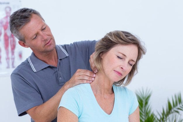
Back Pain Specialist: Mike Melgoza is a very active person who is always engaging in physical activity, as a result, he occasionally suffers from debilitating back pain symptoms. Mike Melgoza was struggling to sleep properly due to his symptoms of back pain before receiving chiropractic care with Dr. Alex Jimenez. Mike Melgoza has already started experiencing tremendous relief from his back pain and he highly recommends Dr. Alex Jimenez as the non-surgical choice for back pain.
Back Pain Specialist
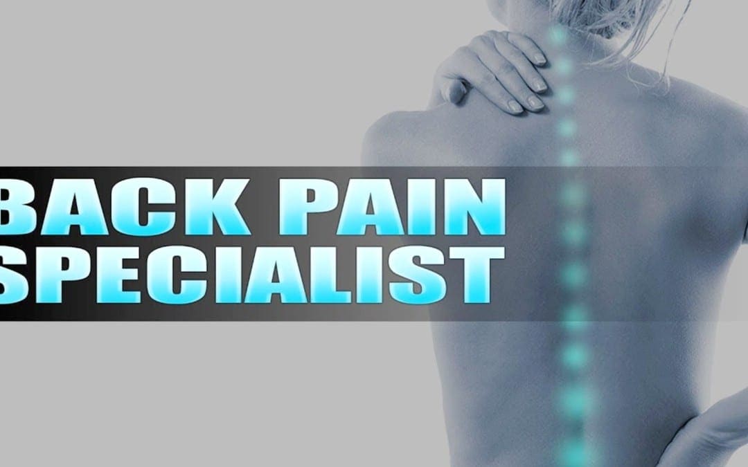
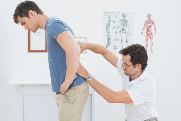
Back pain is one of the most common reasons people visit the doctor or miss work and it is also a leading cause of disability globally. The majority of people have back pain at least once throughout their lifetimes. Luckily, you can take steps to prevent or relieve back pain. If prevention fails, easy treatment and appropriate body mechanics frequently will heal your back in a few weeks and keep it operational for the long haul. Surgery is rarely required to treat back pain.
We are blessed to present to you�El Paso�s Premier Wellness & Injury Care Clinic.
As El Paso�s Chiropractic Rehabilitation Clinic & Integrated Medicine Center,�we passionately are focused treating patients after frustrating injuries and chronic pain syndromes. We focus on improving your ability through flexibility, mobility and agility programs tailored for all age groups and disabilities.
If you have enjoyed this video and/or we have helped you in any way please feel free to subscribe and share us.
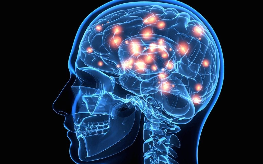
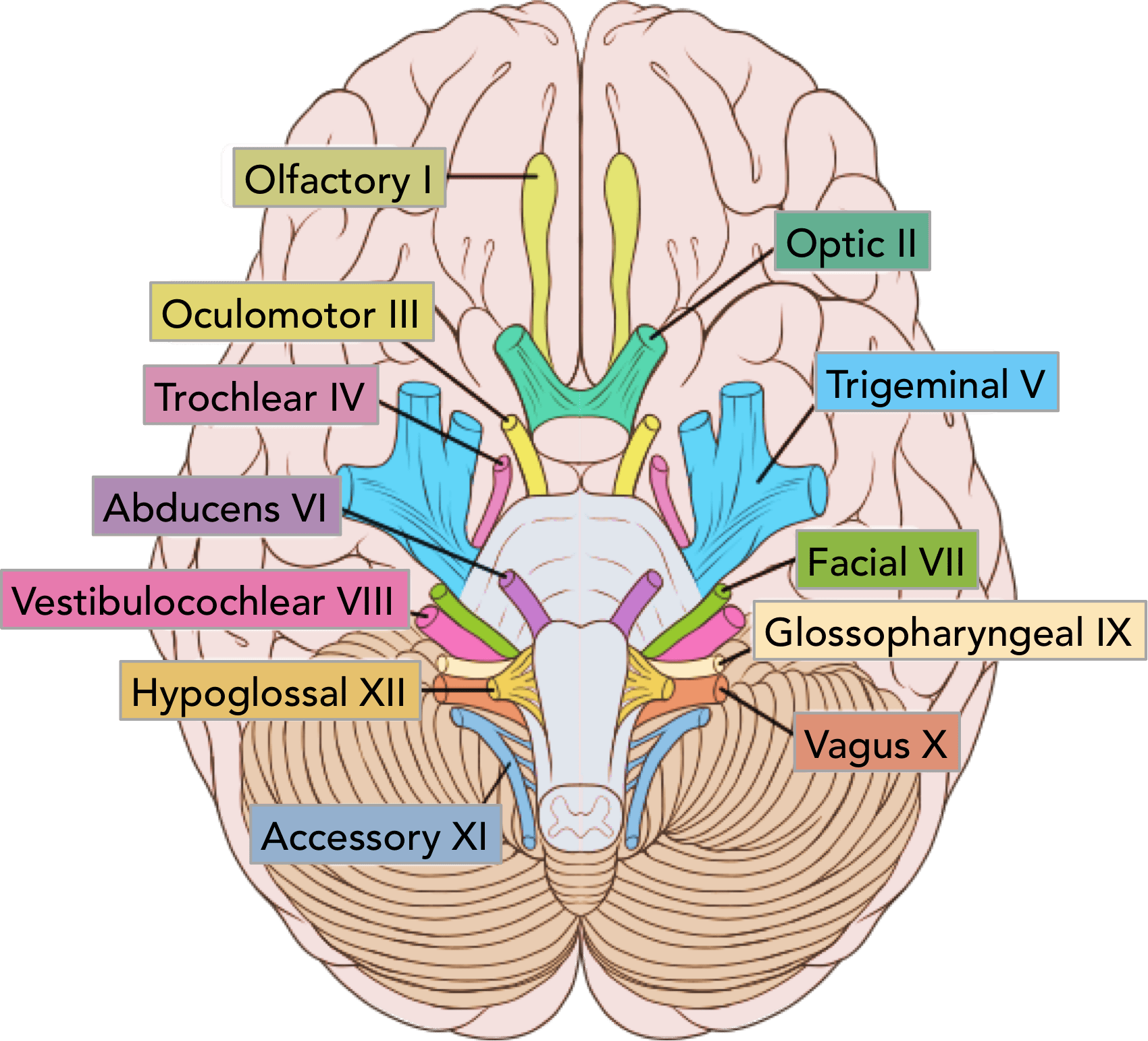
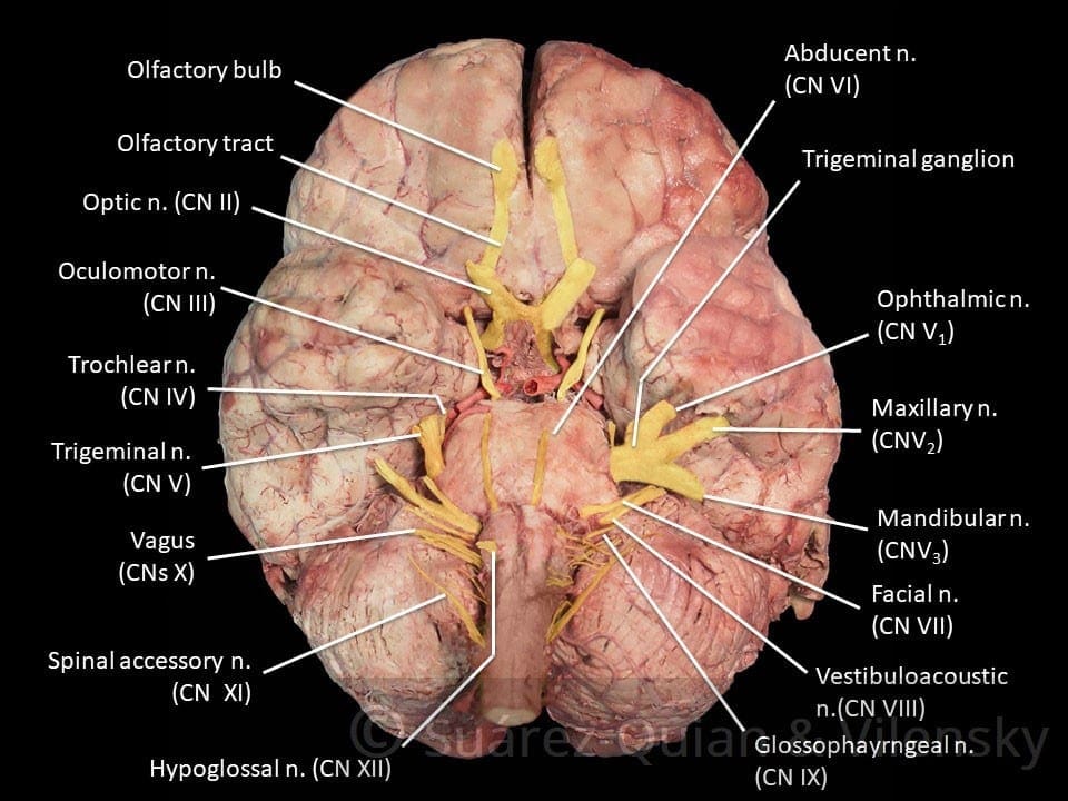
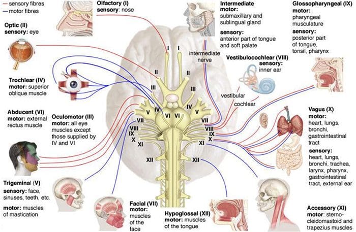
The cranial nerves are the nerves which come out straight from the brain, including the brainstem, in comparison to the spinal nerves, which come out from sections of the spinal cord. Of those, 10 out of 12 of these cranial nerves originate in the brainstem. Cranial nerves transfer information between the brain and parts of the human body, particularly to and from areas of the head and neck.
Spinal nerves exit from the spinal cord with the spinal nerve closest to the head (C1) exiting in the space above the first cervical vertebra. The cranial nerves, however, exit from the central nervous system above this region. Each cranial nerve is paired and is present on either side of the brain. Based on the definition in humans, there are twelve, sometimes thirteen, cranial nerve pairs, which have been assigned Roman numerals I-XII for identification, sometimes including cranial nerve zero as well. The numbering of the cranial nerves is based on the order in which they emerge from the brain, or from the front to the back of the brainstem.
The terminal nerves, olfactory nerves (I) and optic nerves (II) come out from the cerebrum, or forebrain, where the rest of the ten pairs of cranial nerves arise in the brainstem, which is the lower portion of the brain. The cranial nerves are considered components of the peripheral nervous system (PNS), though on a structural level, the olfactory, the optic and the trigeminal nerves are more accurately considered a portion of the central nervous system (CNS).
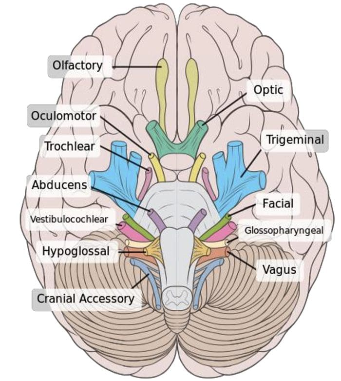
Most commonly, humans are believed to have twelve pairs of cranial nerves (I-XII). These include: the olfactory nerve (I), the optic nerve (II), the oculomotor nerve (III), the trochlear nerve (IV), the trigeminal nerve (V), the abducens nerve (VI), the facial nerve (VII), the vestibulocochlear nerve (VIII), the glossopharyngeal nerve (IX), the vagus nerve (X), the accessory nerve (XI), and the hypoglossal nerve (XII). There may be a thirteenth cranial nerve, known as the terminal nerve, or nerve N or O, which Is quite small and may or may not be functional in humans.
Anatomy of the Cranial Nerves
The cranial nerves are usually named according to their structure or function. For instance, the olfactory nerve (I) supplies smell, and the facial nerve (VII) supplies motor innervation to the face. Since Latin was the common language of the study of anatomy once the nerves were documented, recorded, and mentioned, many nerves maintain Greek or Latin names, including the trochlear nerve (IV), named based on its arrangement, as it supplies a muscle which attaches to a pulley (Greek: trochlea). The trigeminal nerve (V) is named based on its three components (Latin: trigeminus meaning triplets), and the vagus nerve (X) is known because of its wandering course (Latin: vagus).
In addition, cranial nerves are numbered according to their rostral-caudal, or front-back, position, when looking at the brain. If the brain is carefully removed from the skull, the nerves are typically visible in their numeric order, with the exception of the final nerve, the CN XII, which seems to come out from above, into the CN XI.
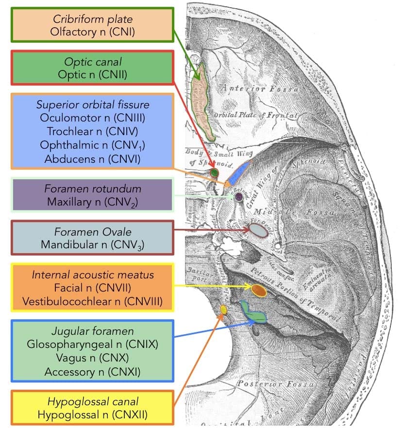
Cranial nerves have pathways within and away from the skull. The pathways inside the skull are known as “intracranial paths” and the pathways outside the skull are known as “extracranial pathways”. There are a number of holes in the skull known as “foramina”, by which the nerves may exit from the skull. All cranial nerves are paired, meaning that they can be found on both the left and right sides of the human body. The skin, muscles, or other structural function provided by a nerve on the same side of the human body as the side it originates from, is referred to as an ipsilateral function. In case the function is on the other hand from the origin of the nerve, then this is referred to as a contralateral function.
Location of the Cranial Nerves
After coming out from the brain, the cranial nerves from inside the skull must leave this bony structure in order to arrive to their destinations. Several of the cranial nerves pass through the foramina,�holes in the skull, as they journey to their destinations. Other nerves pass through bony canals, longer pathways enclosed by bone. The foramina and canals might contain more than just one cranial nerve, and may also include blood vessels. Below is a list of the twelve cranial nerves and a brief summary of their function.
The olfactory nerve (I), composed of many small separate nerve fibers, which passes through perforations from the cribiform plate component of the ethmoid bone. These fibers end in the upper part of the nasal cavity and also operate to communicate impulses containing information about scents or odors into the brain.
The optic nerve (II) passes through the optic foramen from the sphenoid bone in order to reach the eye. It communicates visual information to the brain.
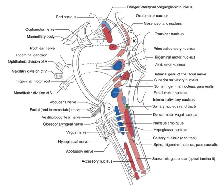
The oculomotor nerve (III), the trochlear nerve (IV), the abducens nerve (VI) and the ophthalmic division of the trigeminal nerve (V1) journey through the cavernous sinus to the superior orbital fissure, passing out of the skull into the orbit. These cranial nerves control the tiny muscles that move the eye and also offer sensory innervation to the eye and orbit.
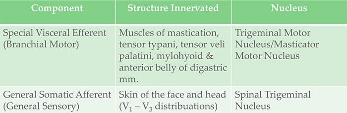
The maxillary division of the trigeminal nerve (V2) moves through the foramen rotundum from the sphenoid bone to supply the skin of the middle of the face.
The mandibular branch of the trigeminal nerve (V3) moves through the foramen ovale of the sphenoid bone to supply the lower face with sensory innervation. This nerve also extends to nearly all the muscles that control chewing.
The facial nerve (VII) and the vestibulocochlear nerve (VIII) both input the inner auditory canal in the temporal bone. The facial nerve subsequently extends to the side of the face using the stylomastoid foramen, also from the temporal bone. Its fibers then distribute to control and reach all of the muscles in charge of facial expressions. The vestibulocochlear nerve reaches the organs which control equilibrium and hearing in the temporal bone, and therefore doesn’t reach the outside surface of the skull.
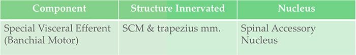
The glossopharyngeal (IX), the vagus nerve (X) and the accessory nerve (XI) all emerge from the skull via the jugular foramen to enter the neck. The glossopharyngeal nerve provides innervation to the upper throat and the back of the tongue, the vagus nerve offers innervation to the muscles at the voicebox, and proceeds down to provide parasympathetic innervation to the chest and abdomen. The accessory nerve controls the trapezius and sternocleidomastoid muscles at the neck and shoulder.
The hypoglossal nerve (XII) exits the skull using the hypoglossal canal at the occipital bone and also reaches the tongue to control virtually all the muscles involved in movements of this organ.
Function of the Cranial Nerves
The cranial nerves give motor and sensory innervation particularly to the structures found inside the neck and head. The sensory innervation contains both “overall” feelings, such as temperature and touch, and “particular” innervation, such as flavor, vision, smell, balance and hearing. For instance, the vagus nerve (X) gives sensory and autonomic, or parasympathetic, motor innervation to structures in the neck and to many of the organs in the chest and abdomen. Below, we will discuss the function of each cranial nerves in further detail.
Smell (I)
The olfactory nerve (I) communicates the sense of smell. Damage to the olfactory nerve (I) may cause an inability to smell, referred to as anosmia, a distortion in the sense of odor, referred to as parosmia, or even a distortion or absence of flavor. When there’s suspicion of a change in the sense of smell, every nostril is tested with compounds of known odors, such as coffee or soap. Intensely smelling chemicals, such as ammonia, can lead to the activation of pain receptors, known as nociceptors, of the trigeminal nerve which are situated in the nasal cavity, which may ultimately confound olfactory testing.
Vision (II)
The optic nerve (II) communicates visual information. Damage to the optic nerve (II) affects specific aspects of vision which are based on the area of the lesion. An individual may not be able to observe objects in their left or right sides, known as homonymous hemianopsia, or might have difficulty seeing objects on their outer visual areas, known as bitemporal hemianopsia, if the optic chiasm is included. Vision may be analyzed by examining the visual field, or simply by analyzing the retina with an ophthalmoscope, with a procedure called funduscopy. Visual field testing can be employed to pin-point structural lesions in the optic nerve, or further along the visual pathways.
Eye Movement (III, IV, VI)
The oculomotor nerve (III), the trochlear nerve (IV) and the abducens nerve (VI) coordinate eye motion. Damage to nerves III, IV, or VI can impact the movement of the eyeball globe. One or both eyes may be influenced; in either case, double vision, referred to as diplopia, will likely occur since the movements of the eyes are no longer synchronized. Nerves III, IV and VI are tested by observing the way the eye follows an object in different directions. This object may be a finger or even a pin, and may be moved at several directions to test for pursuit velocity. If the eyes don’t work together, the most likely cause is harm to a specific cranial nerve or its nuclei.
Damage to the oculomotor nerve (III) can lead to double vision, or diplopia, and inability to coordinate the movements of both eyes, known as strabismus, as well as eyelid drooping, referred to as ptosis, and pupil dilation, or mydriasis. Lesions may also lead to theinability to open the eye due to paralysis of the levator palpebrae muscle. People suffering from a lesion in the oculomotor nerve may compensate by leaning their heads to relieve symptoms because of paralysis of one or more of the eye muscles it regulates.
Damage to the trochlear nerve (IV) may also cause diplopia with all the eye adducted and raised. The result will be an eye which can’t move downwards properly, especially downwards when within an inward position. This is a result of impairment from the superior oblique muscle, which is innervated by the trochlear nerve.
Damage to the abducens nerve (VI) can also result in diplopia This is a result of impairment in the lateral rectus muscle, which is innervated by the abducens nerve.
Trigeminal nerve (V)
The trigeminal nerve (V) is made up of three different parts: The ophthalmic (V1), the maxillary (V2), as well as the Mandibular (V3) nerves. When put together, these nerves provide sensation to the skin of the face and also controls the muscles of mastication, or chewing. Conditions affecting the trigeminal nerve (V) include, trigeminal neuralgia, cluster headaches, and trigeminal zoster. Trigeminal neuralgia may occur later in life, from middle age onwards, most often after the age of 60, and it is a condition commonly associated with a very strong pain that spreads over the region innervated by the maxillary or mandibular nerve divisions of the trigeminal nerve (V2 and V3).
Facial expression (VII)
Lesions of the facial nerve (VII) may manifest as facial palsy. This is where a individual is unable to move the muscles on one or both sides of the face. An extremely frequent and generally temporary facial palsy is called Bell’s palsy. Bell’s Palsy is the end result of an idiopathic (unknown cause), unilateral lower motor neuron lesion of the facial nerve and is characterized by an inability to move the ipsilateral muscles of facial expression, including altitude of the eyebrow and furrowing of their forehead. Patients with Bell’s palsy frequently have a drooping mouth over the affected side and often have difficulty chewing since the buccinator muscle is affected. Bell’s palsy occurs very rarely, affecting around 40,000 Americans annually. Facial paralysis may be caused by other conditions including, stroke. Related conditions to Bell’s Palsy are sometimes misdiagnosed as Bell’s Palsy. Bell’s Palsy is a temporary condition usually lasting 2-6 months, but can have life-changing results and may reoccur often. Strokes typically also impact the cranial nerve by cutting off blood flow to nerves within the brain which is a clear indication that the nerve is present with similar symptoms.
Hearing and Equilibrium (VIII)
The vestibulocochlear nerve (VIII) divides into the vestibular and cochlear nerve. The vestibular region is in charge of innervating the vestibules and semicircular canal of the inner ear; this structure communicates information regarding equilibrium, and is a significant element of the vestibuloocular reflex, which keeps the brain stable and allows the eyes to track moving objects. The cochlear nerve communicates data from the cochlea, allowing sound to be heard. If damaged, the vestibular nerve can manifest the sensation of spinning and dizziness. Function of the vestibular nerve may be analyzed by placing warm and cold water in the ears and watching eye motions caloric stimulation. Damage to the vestibulocochlear nerve may also pose as repetitive and involuntary eye movements, previously described as nystagmus, particularly when looking in a horizontal plane. Damage to the cochlear nerve may cause partial or complete deafness in the affected ear.
Oral Sensation, Taste, and Salivation (IX)
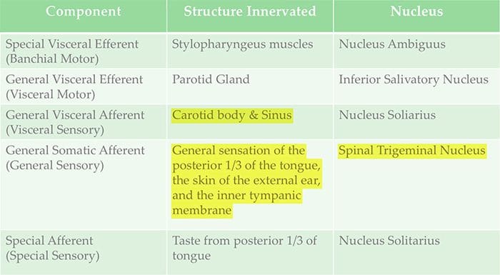
The glossopharyngeal nerve (IX) innervates the stylopharyngeus muscle and supplies sensory innervation to the oropharynx and back of the tongue. The glossopharyngeal nerve additionally supplies parasympathetic innervation to the parotid gland. Unilateral absence of a gag reflex suggests a lesion of the glossopharyngeal nerve (IX), and perhaps the vagus nerve (X).
Vagus Nerve (X)
Reduction of function of the vagus nerve (X) can lead to a reduction of parasympathetic innervation to quite a high number of structures. Important consequences of damage to the vagus nerve could include an increase in blood pressure and heart rate. Isolated dysfunction of just the vagus nerve is rare, but can be diagnosed with a hoarse voice, because of dysfunction of one of its branches, the recurrent laryngeal nerve. Damage to this nerve may result in difficulties swallowing.
Shoulder Elevation and Head-Turning (XI)
Damage to the accessory nerve (XI) can lead to ipsilateral weakness in the trapezius muscle. This can be tested by asking the patient to elevate their shoulders or shrug, where the shoulder blade, or scapula, will protrude to a winged position. Additionally, if the nerve is damaged, weakness or an inability to elevate the scapula may be present because the levator scapulae muscle is only able to provide this function. Based on the location of the lesion, there may also be weakness within the sternocleidomastoid muscle, which then acts to reverse the head so that the face points to the other side.
Tongue Movement (XII)
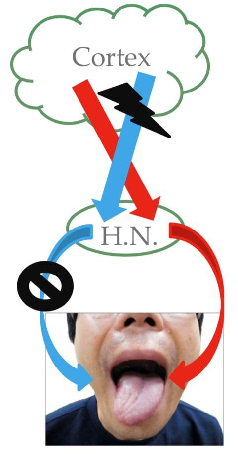
The hypoglossal nerve (XII) is unique in that it is innervated in the motor cortices of both hemispheres of the brain. Damage to the nerve at lower motor neuron level may cause fasciculations or atrophy of the muscles of the tongue. The fasciculations of the tongue are sometimes said to look like a”bag of worms”. Upper motor neuron damage won’t cause atrophy or fasciculations, but only weakness of the innervated muscles. Once the nerve is damaged, it will lead to weakness of tongue movement on one side. When damaged and extended, the tongue will move towards the weaker or damaged side, as shown in the image.
Dr. Alex Jimenez’s Insights
The cranial nerves are a set of 12 nerves which emerge directly from the brain. The first two nerves, known as the olfactory nerve and the optic nerve, come out from the cerebellum, where the remaining ten cranial nerves emerge from the brain stem. The names of the cranial nerves relate directly to their function and they are also numerically identified in roman numerals I-XII by their specific location of the brain and by the order in which they exit the cranium. Damage to any of the above mentioned cranial nerves can cause health issues associated to the specific structure and function of each nerve. Common signs and symptoms in these regions can help healthcare professionals identify the affected cranial nerves.
The scope of our information is limited to chiropractic as well as to spinal injuries and conditions. To discuss the subject matter, please feel free to ask Dr. Jimenez or contact us at 915-850-0900 .
Curated by Dr. Alex Jimenez
Additional Topics: Sciatica
Sciatica is medically referred to as a collection of symptoms, rather than a single injury and/or condition. Symptoms of sciatic nerve pain, or sciatica, can vary in frequency and intensity, however, it is most commonly described as a sudden, sharp (knife-like) or electrical pain that radiates from the low back down the buttocks, hips, thighs and legs into the foot. Other symptoms of sciatica may include, tingling or burning sensations, numbness and weakness along the length of the sciatic nerve. Sciatica most frequently affects individuals between the ages of 30 and 50 years. It may often develop as a result of the degeneration of the spine due to age, however, the compression and irritation of the sciatic nerve caused by a bulging or herniated disc, among other spinal health issues, may also cause sciatic nerve pain.
Human Cranial nerves are a set of 12 paired nerves that come directly from the brain. The first two (olfactory and optic) come from the cerebrum, with the remaining ten come from the brain stem. The names of the these nerves relate to what function they perform and are also numerically identified in roman numerals (I-XII).�The�nerves serve in functions of smell, sight, eye movement, and feeling in the face. These�nerves also control balance, hearing, and swallowing.
As with all nerves, symptoms describe the location of the lesion
Lesion in the lingual nerve will result in loss of taste, general sensation in tongue & salivary secretion
Lesion proximal to the branching of the chorda tympani such as in the facial canal will result in the same symptoms without the loss of general sensation of the tongue (because V3 has not yet joined the CN VII)
Corticobulbar innervation is asymmetric to the upper and lower parts of the Facial Motor Nucleus
If there is an UMN lesion (lesion to the corticobulbar fibers) the patient will have paralysis of the muscles of facial expression in the contralateral lower quadrant
If there is a LMN lesion (lesion to the facial nerve itself) the patient will have paralysis of the muscles of facial expression in the ipsilateral half of the face
Bell�s Palsy
Testing Cranial Nerve CN VII
Ask the patient to mimic you or follow instructions to make certain facial expressions
Be sure to assess all four quadrants of the face
Raise eyebrows
Puff cheeks
Smile
Close eyes tightly
Check for strength of the buccinator muscle against resistance
Ask patient to hold air in their cheeks as you press gently from the outside
Patient should be able to hold air in against resistance
Cranial Nerve VIII – Vestibulocochlear
Cranial Nerve VIII Clinically
Changes in hearing alone are most often due to
Infections (otitis media)
Skull fracture
The most common lesion to this nerve is caused by an acoustic neuroma
This affects CN VII and CNVIII (cochlear AND vestibular divisions) due to proximity in the internal auditory meatus
Symptoms include nausea, vomiting, dizziness, hearing loss, tinnitus, and bell�s palsy etc.
Testing Cranial Nerve CN VIII
Otoscopic Exam
Scratch Test
Can the patient hear equally on both sides?
Weber Test
Tests for lateralization
256 Hz tuning fork placed on top of the patient�s head in the center, is it louder on one side than the other?
Rinne Test
Compares air conduction to bone conduction
Normally, air conduction should last 1.5-2 as long as bone conduction
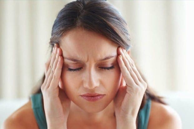
Almost everyone can say that they’ve experienced a feeling of unsteadiness or a spinning/whirling sensation in their heads at one point in their lifetimes. Usually it’s narrowed down to dizziness, however, dizziness is a broad term that can mean different things to different individuals. It is a prevalent complaint which can also be serious. Dizziness has no specific medical definition, but there are four common conditions which can be considered types of dizziness:
Vertigo. The feeling of motion where there is no movement, as if you were spinning or your environment is whirling. Spinning/whirling yourself around and around, then abruptly stopping, can produce temporary vertigo. However, when it occurs throughout an individual’s regular course of living, it could mean that there’s an underlying health issue in the vestibular system of the inner ear, the body’s equilibrium system which tells you which way is up or down and senses the position of your head. About half of all dizziness complaints are diagnosed as vertigo.
Lightheadedness. Also referred to as near syncope or pre-syncope, lightheadedness is the feeling that you’re about to faint. It is commonly believed to occur from standing up too fast or by breathing deeply enough times to generate the sensation.
Disequilibrium. A problem with walking. People with disequilibrium feel unsteady on their feet or feel as if they will fall.
Anxiety. Individuals who are scared, worried, depressed, or fearful of open spaces can use the term “dizzy” to imply feeling frightened, depressed, or anxious.
Individuals who frequently suffer from dizziness may also ultimately complain of more than one type of dizziness. For instance, people with vertigo may also feel anxious. Dizziness may be a one-time event, or it can be a chronic, long-lasting issue. Nearly everyone who experiences some form of dizziness will recover over time. This is because an individual’s sense of balance is an intricate interaction between the brain, each ear’s different vestibular system, sensors in the muscles, and sense of vision. When one component experiences dysfunction, others can generally learn how to compensate. Below, we will be narrowing down the four common types of dizziness.
Vertigo, the sensation of spinning or whirling, can be divided into two different categories: peripheral vertigo and central vertigo. Peripheral vertigo is more common than central vertigo and it typically develops due to damage to the inner ear or CN VIII. This type of vertigo produces abnormal eye movements, referred to as nystagmus, which may be horizontal or rotary.
Nystagmus is usually jerky in nature with a fast and slow phase, however it is often named for the direction of the fast phase. Peripheral vertigo may worsen when the patient looks to the side of the fast phase of nystagmus. Furthermore, the severity of nystagmus can correlate with the severity of the patient’s vertigo. Peripheral vertigo is also characterized as having no other signs and/or symptoms of CNS dysfunction. Patient may describe having symptoms of nausea or may present difficulty when walking, but only due to vestibular dysfunction. The patient may also have hearing loss or tinnitus if the CN VIII or auditory mechanism function is damaged.
The causes of peripheral vertigo are typically benign, including: benign paroxysmal positional vertigo, or BPPV, cervicogenic vertigo, acute labyrinthitis/vestibular neuronitis, Meniere’s disease, perilymph fistula, and acoustic neuroma. Identifying a patient’s cause of vertigo can be determined by narrowing down the symptoms through proper diagnosis from a healthcare professional. If movements, especially of the neck and head, aggravate vertigo, it may be attributed to BPPV, vertebrobasilar artery insufficiency or cervicogenic vertigo. If noise manifests episodes of vertigo, it may be attributed to Meniere’s disease or perilymph fistula.
Common Causes of Dizziness
Vertigo can be Brought on by many things:
Infections, such as the ones which cause the frequent cold or diarrhea, can lead to temporary vertigo through an ear infection. This inner ear disease is generally viral, benign, and usually goes away in one to six weeks, however, drugs and/or medications are readily available if these become too severe.
Benign paroxysmal positional vertigo, or BPPV, is caused by the motion of a misplaced otolith, a tiny calcium particle the size of a grain of sand, from the component of the inner ear which senses gravity into the part that senses head position. The individual feels as if their head is turning when it isn’t. After diagnosis of BPPV using a special methods known as the Dix-Hallpike test, treatment done right in the doctor’s office can help move the otolith back where it belongs and fix the health issue. This therapy, known as the Epley maneuver, has been accounted to cure vertigo 80 percent of the time.
Meniere’s disease is a disorder characterized by long-lasting episodes of severe vertigo. Other symptoms of Meniere’s disease are tinnitus, or ringing in the ears, hearing loss, and fullness or pressure in the ear.
Dandy’s syndrome is a feeling of everything bouncing up and down. It may occur to individuals who take an antibiotic that is toxic to the ear. However, it usually improves over time.
Less frequent, deadly diseases may also result in vertigo, like tumors or stroke.
Below, we will be narrowing down some of the common causes of vertigo, described above, in further detail.
Benign Paroxysmal Positional Vertigo (BPPV)
Benign paroxysmal positional vertigo, or BPPV, may develop spontaneously, particularly in the elderly. It may also commonly develop as a result of head trauma or head injury, such as that resulting from an automobile accident. Vertiginous episodes associated with BPPV may manifest through specific movements, including, looking at a high shelf, referred to as top-shelf vertigo, bending over, and rolling over in bed at night. The onset of vertigo with BPPV can begin a few seconds after movement and often resolves within a minute. As mentioned above, the diagnostic test commonly utilized to diagnose BPPV is the Dix-Hallpike maneuver. Treatment procedures to treat BPPV include the Epley maneuver and Brandt-Daroff Exercises. Furthermore, benign paroxysmal positional vertigo may also resolve on its own as the loose crystals in the inner ear dissolve, however, it may take months and new otoliths can also become displaced.
Cervicogenic vertigo occurs after a neck or head injury, however, it is not very common. It’s generally accompanied by pain and/or joint restriction where vertigo and nystagmus are less severe than that in BPPV. Cervicogenic vertigo manifests with changes in head position but does not subside as quickly as it does with benign paroxysmal positional vertigo.
Vertebrobasilar Artery Insufficiency
Vertebrobasilar artery insufficiency occurs if the vertebral artery is compressed during head rotation or extension. In this instance, the onset of vertigo is delayed more than in BPPV or cervicogenic vertigo due to the fact that ischemia often takes up to 15 seconds to occur. Orthopedic tests for vertebrobasilar artery insufficiency may help in its diagnosis. Diagnostic tests include the�Barre?-Lie?ou sign, DeKlyn Test or Dix-Hallpike Maneuver, Hautant test, Underberg test and the vertebrobasilar after functional maneuver.
Acute Labyrinthitis and Vestibular Neuronitis
Acute labyrinthitis and vestibular neuronitis are not well understood, however, they’re believed to develop as a result of inflammation. These conditions generally follow after a viral infection or may occur seemingly without a cause.�Acute labyrinthitis and vestibular neuronitis are characterized by a single, monophasic attack of vertigo which typically resolves in days to a few weeks and generally does not reoccur.
Meniere’s Disease
Meniere’s disease is characterized by increased pressure in the endolymph which causes membrane ruptures and a sudden mixture of endolymph and perilymph. With Meniere’s disease, episodes of vertigo can last from 30 minutes to several hours, or until equilibrium between the fluids in the inner ears to be reached. Over time, these episodes can damage vestibular and cochlear hair cells, resulting in low-pitch buzzing tinnitus and the loss of hearing of low tones. In comparison to Meniere’s disease, Meniere’s syndrome is when the symptoms of Meniere’s disease are found to be secondary to another condition, such as: hypothyroidism, acoustic neuroma, superior semicircular canal dehiscence or SCDS, or perilymph fistula. True Meniere’s disease is idiopathic.
Perilymph Fistula
Perilymph fistula is an abnormal connection, or tear, which causes a small leak within the inner ear due to trauma or injury, especially barotrauma. Perilymph fistula can look very similar symptomatically to Meniere’s disease/syndrome and it’s often aggravated by changes in pressure causes by airplane rides or driving uphill. Another symptom of perilymph fistula includes Hennebert’s sign, where a vertigo or nystagmus episode is brought on by sealing pressure of the ear, such as by inserting an otoscope.
Central vertigo, another category of vertigo, is less common than peripheral vertigo, as described above. It is caused by damage to the processing center of vestibular information in the brain stem and the cerebral cortex. However, episodes of dizziness are considered to be less severe than with peripheral vertigo while episodes of nystagmus are more severe than the patient’s complaint or description. This specific nystagmus associated with central vertigo may go in multiple directions, including vertical. Central vertigo may or may not have other CNS findings upon diagnosis or examination and no changes in hearing can be expected with this form of vertigo. The most common causes of central vertigo include: cerebrovascular disease, such as transient ischemic attacks, multiple sclerosis, Arnold-Chiari malformation, damage to caudal brainstem or vestibulocerebellum and/or migraine condition.
Lightheadedness, or pre-syncope dizziness, is generally caused by some surrounding circumstance impairing blood flow into the brain when an individual is standing up. Blame this problem on our ancestors who learned to walk upright, placing our brain above our heart. It is a challenge for your heart to keep the brain supplied with blood and it is easy for this system to break down. When blood vessels in the brain become dilated, or enlarged, as a result of elevated fever, excitement or hyperventilation, alcohol ingestion, or prescription drugs and/or medications, such as antidepressants, it’s no wonder someone may commonly get lightheaded. There can also be serious causes, however, such as a stroke and cardiovascular disease.
Pre-syncope dizziness is specifically from cardiac origin, such as output disorders, arrhythmias, Holter monitor testing. It may also be caused by postural/orthostatic hypotension, which may be secondary to other health issues like diabetic neuropathy, adrenal hypofunction, Parkinsons, certain drugs and/or medications, etc. Light-headedness can involve vasovagal episodes accompanied by slow heart rate with low blood pressure often caused by stress, anxiety or hyperventilation. Finally, pre-syncope dizziness can be caused by migraine headaches due to cerebrovascular instability and blood sugar dysregulation.
Disequlibrium, can be caused by:
A type of arthritis in the neck called cervical spondylosis, which puts stress on the spinal cord.
Parkinson’s disease or related disorders that cause an individual to stoop forward.
Disorders involving part of the brain known as the cerebellum. The cerebellum is the part of the brain responsible for coordination and balance.
Diseases like diabetes that can lead to lack of sensation in the legs.
Disequilibrium is most common in the elderly and it generally occurs due to sensory deficits. In addition, disequilibrium has a gradual onset which worsens with reduced vision, darkness, eyes closed and visual acuity losses. However, it is improved by touching a stationary object which is often subjective as dizziness improves with a gait assistive device like a cane, walker, etc.
Dr. Alex Jimenez’s Insights
If you’ve ever experienced a sudden spinning or whirling sensation or even felt faint, woozy or unsteady, you’re not alone. Dizziness is a term used to describe a range of sensations and it is one of the most common reasons why many adults visit their healthcare professionals. While these false sensations can rarely signal a life-threatening condition, frequent episodes can significantly affect an individual’s quality of life. Diagnosis and treatment of dizziness can depend largely on the cause of the symptoms. Fortunately, many treatment methods used to treat dizziness are considered safe and effective.
Other causes of dizziness can be attributed to psychological stress. In this instance, the patient will describe their dizziness as a “floating” sensation.�Dizziness in the kind of anxiety is frequently, but not always, caused by depression. In addition, it can be attributed to an anxiety disorder or anxiety. Various medications can also cause dizziness as a side effect. It’s essential for a healthcare professional to rule out this type of dizziness caused by hyperventilation as well as other types of dizziness. The scope of our information is limited to chiropractic as well as to spinal injuries and conditions. To discuss the subject matter, please feel free to ask Dr. Jimenez or contact us at 915-850-0900 .
Curated by Dr. Alex Jimenez
Additional Topics: Sciatica
Sciatica is medically referred to as a collection of symptoms, rather than a single injury and/or condition. Symptoms of sciatic nerve pain, or sciatica, can vary in frequency and intensity, however, it is most commonly described as a sudden, sharp (knife-like) or electrical pain that radiates from the low back down the buttocks, hips, thighs and legs into the foot. Other symptoms of sciatica may include, tingling or burning sensations, numbness and weakness along the length of the sciatic nerve. Sciatica most frequently affects individuals between the ages of 30 and 50 years. It may often develop as a result of the degeneration of the spine due to age, however, the compression and irritation of the sciatic nerve caused by a bulging or herniated disc, among other spinal health issues, may also cause sciatic nerve pain.
Ataxia is a medical term used to describe a lack of muscle control or coordination of voluntary movements, including everyday physical activities like walking or picking up objects. Often referred to as a symptoms of an underlying health issue, ataxia can affect various movements, causing difficulties with speech patterns and language, eye movement and even swallowing.
Persistent ataxia generally results from damage to the part of the brain which controls muscle coordination, known as the cerebellum. Many causes and conditions can lead to ataxia, such as alcohol abuse, certain drugs and/or medications, stroke, tumors, cerebral palsy, brain degeneration and multiple sclerosis. Inherited faulty genes have also been associated to lead to ataxia.
Diagnosis and treatment for ataxia depends largely on the cause and/or condition. Adaptive devices, including walkers or canes, can help patients with ataxia maintain their independence. Chiropractic care, physical therapy, occupational therapy, speech therapy and regular aerobic stretches and exercises can also help improve the symptoms associated with this health issue.
Symptoms of Ataxia
Ataxia is a health issue which can develop gradually over time or it can come on unexpectedly. As a symptom of a number of neurological disorders, ataxia may ultimately lead to:
Poor coordination
Unsteady walk along with a tendency to stumble
Difficulty with fine motor tasks, such as eating, writing or buttoning a shirt
Changes in speech
Involuntary back-and-forth eye movements, known as nystagmus
Difficulty swallowing
When to Visit a Doctor
In the instance that a patient is not aware of whether they may have an underlying health issue that causes ataxia, such as multiple sclerosis, it’s essential to visit a doctor immediately if the patient:
Loses equilibrium
Loses muscle coordination at a hand, leg or arm
Has difficulty walking
Slurs their speech
Has trouble swallowing
Causes of Ataxia
Damage, degeneration or loss of neural cells in the section of the brain which controls muscle coordination, or the cerebellum, often results in ataxia. The cerebellum is made up of two pingpong-ball-sized parts of folded tissue located at the base of the brain close to the brainstem. The right side of the cerebellum controls coordination over the right side of the body; the left side of the cerebellum controls coordination on the left side of the body. Diseases that damage the spinal cord and peripheral nerves which connect the cerebellum to the muscles can also lead to ataxia. Ataxia causes include:
Head trauma. Damage to the brain or spinal cord due to a blow to the head, such as in the case of an automobile accident, can cause acute cerebellar ataxia, which comes on unexpectedly.
Stroke. After the blood supply to part of the brain is interrupted or severely reduced, depriving brain tissue of nutrients and oxygen, brain cells die.
Cerebral palsy. This can be a general term for a group of disorders brought on by damage to a child’s brain during early development, before, during or shortly after birth, which affects the child’s ability to coordinate body movements.
Autoimmune diseases. Multiple sclerosis, sarcoidosis, celiac disease and other autoimmune conditions can cause ataxia.
Infections. Ataxia may be an uncommon complication of chickenpox and other viral ailments. It may manifest in the healing phases of the infection and can last for days or weeks. Generally, the ataxia resolves over time.
Paraneoplastic syndromes. These are rare, degenerative health issues triggered by the body’s own immune system’s reaction to a cancerous tumor, referred to as neoplasm, most frequently from lung, ovarian, breast or lymphatic cancer. Ataxia can appear months or years before the cancer is even diagnosed.
Tumors. A growth on the brain, cancerous, or malignant, or noncancerous, or benign, can also harm the cerebellum, leading to ataxia.
Toxic reaction. Ataxia is a possible side effect of certain drugs and/or medications, particularly barbiturates, like phenobarbital; sedatives, like benzodiazepines; as well as some kinds of chemotherapy. These are important to diagnose because the effects are usually reversible. Also, some drugs and/or medications can cause problems with age, which means a person may need to reduce their dose or discontinue its use. Alcohol and drug intoxication; heavy metal poisoning, such as from mercury or lead; and solvent poisoning, like from paint thinner, can also cause ataxia.
Vitamin E, vitamin B-12 or thiamine deficiency. Not getting enough of these nutrients, due to the inability to absorb them enough, alcohol misuse or other reasons, may also ultimately lead to ataxia.
For a number of adults that develop sporadic ataxia, no particular cause is found. Sporadic ataxia can take lots of forms, including multiple system atrophy, a progressive and degenerative disease.
Dr. Alex Jimenez’s Insights
The cerebellum is the region of the brain which is in charge of controlling movement in the body. Electrical signals are transmitted from the brain through the spinal cord and into the peripheral nerves to stimulate a muscle to contract and initiate movement. Sensory nerves also gather data from the environment regarding position and proprioception. When one or more of these pathway components experiences a problem, it can subsequently lead to ataxia. Ataxia is a medical term utilized to describe the lack of muscle coordination when a voluntary movement is attempted. It can make any motion which requires muscles to function a challenge, from walking to picking up an object, even swallowing. Diagnosis and treatment can help manage and improve the symptoms associated with ataxia.
Diagnosis of Ataxia
If an individual has developed symptoms of ataxia, a healthcare professional may perform a diagnosis in order to look for a treatable cause. Besides running a physical examination and a neurological examination, including assessing a patient’s memory and concentration, vision, hearing, balance, coordination, and reflexes, your doctor might request lab tests, including:
Imaging studies. A CT scan or MRI of a patient’s brain might help determine possible causes of ataxia. An MRI can sometimes reveal shrinkage of the cerebellum and other brain structures in people with ataxia. It might also demonstrate other findings that are treatable, such as a blood clot or benign tumor, which may be pressing on the cerebellum.
Lumbar puncture (spinal tap). A needle is inserted into the lower spine, or the lumbar spine, between two lumbar bones, or vertebrae, to remove a sample of cerebrospinal fluid. The fluid, which surrounds and protects the brain and spinal cord, is transported to a laboratory for testing.
Genetic testing. A healthcare professional might recommend genetic testing to determine whether a child has the gene mutation which causes hereditary ataxia. Gene tests are available for many but not all of the hereditary ataxias.
Furthermore, diagnosing ataxia may depend on which system is affected. For instance,�if the health issue lies in the vestibular system, the patient will experience dizziness, possibly having vertigo or nystagmus. They may also be unable to walk in a straight line and when walking, they will tend to veer to one side. If the health issue lies in the cerebellar system, cerebellar gaits present with a wide-base and generally involves staggering and titubation. Patient will also have difficulty doing the Rhomberg�s test with their eyes open or closed, because they cannot stand with their feet together, as described below.
Testing the Vestibular System
Testing the vestibular system to determine the diagnosis of ataxia can include the Fakuda Stepping Test and the Rhomberg Test. The�Fakuda Stepping Test is performed by having the patient march in place with their eyes closed and their arms raised to 90 degrees in front of them. If they rotate more than 30 degrees, the test is considered to be positive. It’s important to note that the patient will rotate toward the side of the vestibular dysfunction. The Rhomberg Test will confirm a diagnosis of ataxia if the patient sways a different direction every time their eyes are closed, as this may indicate vestibular dysfunction.
Testing the Cerebellar System
Testing the cerebellar system to determine the diagnosis of ataxia can include the piano-playing test and the hand-patting test as well as the finger-to-nose test. The piano-playing test and hand-patting test both assess for dysdiadochokinesia. Also in both tests, the patient will have more difficulty moving the limb on the side of cerebellar dysfunction. With the finger-to-nose test, the patient may be hyper/hypo metric in movement and intention tremor may be reveled.
Joint Position Sense
In patients with changes to their joint position sense, conscious proprioception may be diminished, especially in elderly patients and patients with neuropathy. Patients with joint position sense losses often rely on visual information to help compensate. When visual input is removed or diminished, these patient�s have exaggerated ataxia.
Motor Strength and Coordination
If the patient has reduced frontal lobe control, they may end up with an apraxia of gait, where they have difficult with the volitional control of movement. Extrapyramidal disorders, such as Parkinson disease, result in the inability to control motor coordination. Pelvic girdle muscle weakness due to a myopathy in this instance will produce an abnormal gait pattern.
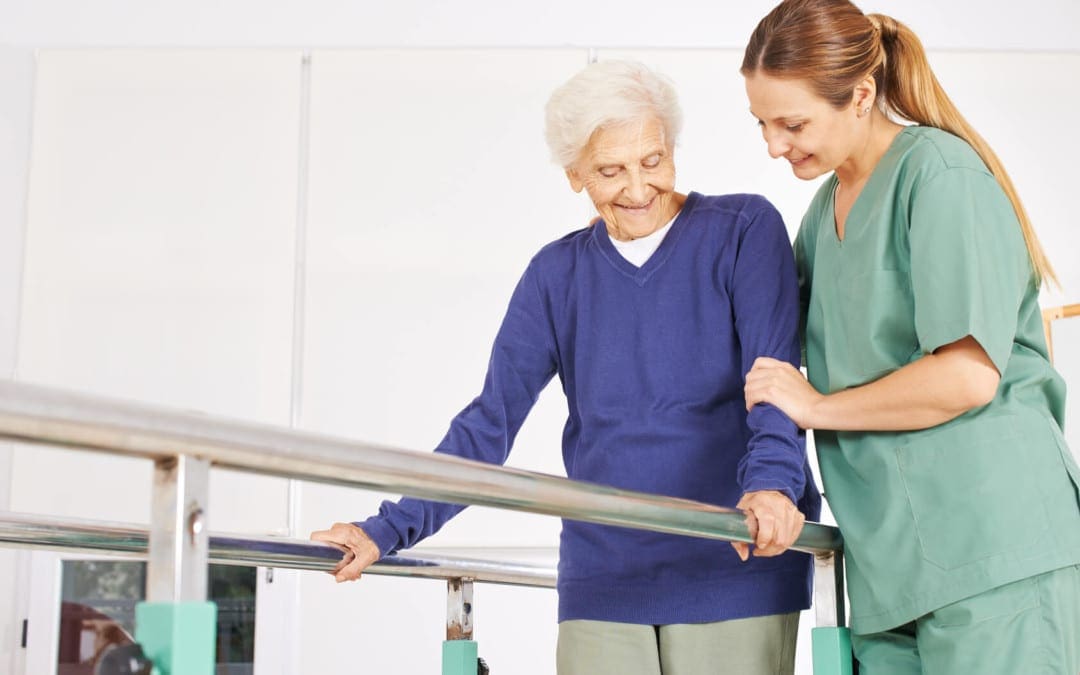
There’s no specific treatment for ataxia. In some cases, treating the underlying health issue often resolves the ataxia, such as quitting the use of drugs and/or medications that cause it. In other cases, such as ataxia that results from chickenpox or other viral infection, it’s likely to resolve on its own. A healthcare professional might recommend treatment to manage symptoms, such as pain, fatigue or nausea, or they may recommend the use of adaptive devices or therapies to help with ataxia. Chiropractic care is a safe and effective, alternative treatment option which focuses on the treatment of a variety of injuries and/or conditions associated with the musculoskeletal and nervous system. A chiropractor commonly uses spinal adjustments and manual manipulations to correct any spinal misalignment, or subluxation, which may be causing a patient’s symptoms. In addition, a doctor of chiropractic, or chiropractor, may also recommend a series of appropriate lifestyle modifications, including nutritional advice and exercise plans, in order to restore a patient’s strength, mobility and flexibility. Chiropractic care together with the proper fitness routine can help speed up the patient’s recovery process.
Adaptive Devices
Ataxia brought on by conditions like multiple sclerosis or cerebral palsy might not be curable. In that circumstance, a healthcare professional might have the ability to recommend adaptive devices. These can include:
Hiking sticks or walkers for walking
Modified utensils for eating
Communication aids for speaking
Other therapies
A patient with ataxia might benefit from particular therapies, including: physical therapy to help improve coordination and enhance mobility; occupational treatment to help with daily living activities, such as eating on their own; and speech therapy to improve speech as well as aid with swallowing.
Coping and Support
The challenges a person face when living with ataxia or with a child with the condition might make the patient feel lonely or it may contribute to depression and anxiety. Talking to a counselor or therapist may help. Or perhaps the patient may find encouragement and understanding in a support group, possibly for ataxia or for their specific underlying condition, such as cancer or multiple sclerosis.
Although support groups aren’t for everyone, they may be good sources of advice. Group members often know about the newest treatments and tend to share their own experiences. If you’re interested, your healthcare professional may be able to recommend a group in your area. The scope of our information is limited to chiropractic as well as to spinal injuries and conditions. To discuss the subject matter, please feel free to ask Dr. Jimenez or contact us at 915-850-0900 .
Curated by Dr. Alex Jimenez
Additional Topics: Sciatica
Sciatica is medically referred to as a collection of symptoms, rather than a single injury and/or condition. Symptoms of sciatic nerve pain, or sciatica, can vary in frequency and intensity, however, it is most commonly described as a sudden, sharp (knife-like) or electrical pain that radiates from the low back down the buttocks, hips, thighs and legs into the foot. Other symptoms of sciatica may include, tingling or burning sensations, numbness and weakness along the length of the sciatic nerve. Sciatica most frequently affects individuals between the ages of 30 and 50 years. It may often develop as a result of the degeneration of the spine due to age, however, the compression and irritation of the sciatic nerve caused by a bulging or herniated disc, among other spinal health issues, may also cause sciatic nerve pain.
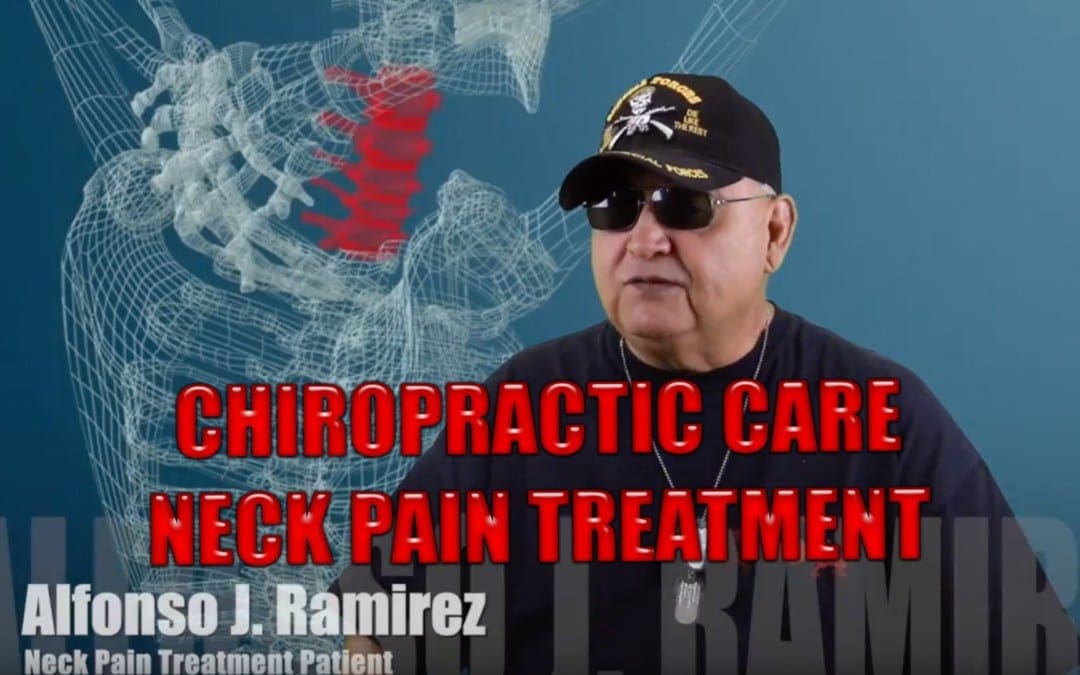
Alfonso J. Ramirez, now retired, found follow-up treatment with Dr. Alex Jimenez for his neck pain. Mr. Ramirez experienced chronic pain and headaches, but after receiving chiropractic care, he found relief from his symptoms. Since then, Alfonso Ramirez has continued to maintain the alignment of his spine with Dr. Jimenez. Mr. Ramirez is grateful for the chiropractic care he’s received for his neck pain and for his shoulder and knee pain. Alfonso J. Ramirez recommends Dr. Alex Jimenez as the non-surgical choice for neck pain.
Chiropractic Care Neck Pain Treatment
Neck pain (or cervical Gia) is a frequent problem, together with two-thirds of the population experience neck pain at any time in their lives. Neck pain, although felt in the neck, can be brought on by many other spinal issues. Neck pain may arise because of muscular tightness in both the neck and upper back, or pinching of the nerves emanating from the cervical vertebrae. Joint disruption in the neck also creates pain, as does joint disruption in the top back. Neck pain affects about 5 percent of the global population as of 2010.
We are blessed to present to you�El Paso�s Premier Wellness & Injury Care Clinic.
As El Paso�s Chiropractic Rehabilitation Clinic & Integrated Medicine Center,�we passionately are focused treating patients after frustrating injuries and chronic pain syndromes. We focus on improving your ability through flexibility, mobility and agility programs tailored for all age groups and disabilities.
If you have enjoyed this video and/or we have helped you in any way please feel free to subscribe and share us.
Benign paroxysmal positional vertigo, or BPPV, is a mechanical issue in the inner ear. It occurs when some of the calcium carbonate crystals (otoconia) that are normally embedded in gel at the utricle become dislodged and migrate to at least one of those 3 fluid-filled semicircular canals, where they are not supposed to be. When enough of these particles accumulate in one of the canals they interfere with the normal fluid motion that these canals utilize to sense head motion, causing the inner ear to send false signals to the mind.
Fluid in the semi-circular canals doesn’t normally react to gravity. However, the crystals do proceed with gravity, thereby shifting the fluid when it normally would be still. When the fluid moves, nerve endings in the canal are eager and send a message to the brain the mind is moving, even though it is not. This false information doesn’t match what another ear is sensing, together with what the eyes are seeing, or with what the joints and muscles are doing, and also this mismatched information is perceived by the brain as a turning sensation, or vertigo, which generally lasts less than one minute. Between vertigo spells some people today feel symptom-free, while some feel a mild sense of imbalance or disequilibrium.
A healthcare professional will execute a collection of tests and evaluations in order to properly diagnose the individual’s BPPV. Regular medical imaging (e.g. an MRI) is not helpful in diagnosing BPPV, because it doesn’t show the crystals which have moved to the semi-circular canals. But when someone with BPPV has their own head moved into a position that produces the dislodged crystals move within a tube, the error signals cause the eyes to move in a very specific pattern, called”nystagmus”.
The nystagmus will possess distinct characteristics that let a trained practitioner to identify which ear the crystals that are displaced are in, and then canal(s) they have moved into. Tests such as the Dix-Hallpike or Roll Tests involve moving the head into specific orientations, allowing gravity to move the dislodged crystals and activate the vertigo while the professional watches for the tell-tale eye movements, or nystagmus.�To execute the Dix-Hallpike test, a healthcare professional will ask the patient to sit on the test table with their legs stretched out. They will then turn the head 45 degrees to one side, which contrasts the right posterior semicircular canal with the sagittal plane of the body, then they are going to allow the patient to lie back quickly, while the eyes are open, so that their head hangs slightly over the edge of the desk.
When the health care provider has finished the diagnosis, then they can perform the appropriate treatment maneuver. The maneuvers make use of gravity to guide the crystals back to the room where they are supposed to be via a very specific series of head movements, commonly referred to as Repositioning Maneuvers. Repositioning maneuvers are highly effective in treating BPPV, inexpensive, and easy to apply.
Dr. Alex Jimenez’s Insights
While the use of surgical interventions as well as that of drugs and/or medications are occasionally recommended to relieve the symptoms associated with benign paroxysmal positional vertigo, or BPPV, they do not treat the underlying health issue. Repositioning maneuvers, like the ones demonstrated below, are considered to be safe yet effective treatment options for BPPV. There is good evidence to support the treatment of BPPV with the Epley maneuver. Although less amounts of research studies have been conducted on other repositioning maneuvers, outcome measures of a variety of patients with BPPV have benefitted from the other treatment options for benign paroxysmal positional vertigo.
Considering that the therapeutic efficacy among maneuvers for every canal is comparable, the option of treatment is generally predicated on clinician preference, complexity of their maneuvers themselves, therapy response to certain maneuvers, as well as musculoskeletal considerations, such as arthritic changes and range of motion of the cervical spine. Below, many repositioning maneuvers are demonstrated, for instance, deep mind hanging maneuver, the Lempert (BBQ) maneuver and the Epley maneuver.
The deep head hanging maneuver is a repositioning maneuver which is used for one of the least common places where BPPV occurs, the superior semi-circular canal, amounting to only about 2 percent of most benign paroxysmal positional vertigo instances. However, the advantage of deep head hanging maneuvers is that they may be effectively performed without knowledge of the side involved. It consists of three steps with four position changes at intervals of approximately 30 seconds.
The deep head hanging maneuver is performed with the patient at the long-sitting position, while the head is brought to a minimum of 30� below the horizontal with the head straight up. When the nystagmus induced by this measure is finished, the head is brought up rapidly to touch the chest while the patient remains supine, and after 30 seconds, the individual has been brought back to a seated position with head flexion maintained. Finally, the patient will be brought back to a neutral head position.
The Lempert maneuver, also referred to as the Barbeque maneuver or the Roll maneuver, is a repositioning maneuver commonly utilized to help treat canilithiasis of the horizontal and lateral canal. It might occur as a complication of posterior canal BPPV treatment repositioning maneuvers. The side with the most notable horizontal nystagmus is assumed to be the affected side.
To perform the Lempert maneuver, the patient should lie supine on the exam table, using the affected ear facing down. Afterward, the healthcare professional will quickly turn the head 90� towards the unaffected side, facing up, waiting 15-20 minutes between each head turn. The medical professional will subsequently turn the head 90� so the affected ear is currently facing up. The next step includes having the individual tuck their arms to their torso, in order to allow the doctor to roll the patient to a more moderate position with their head down. The individual must be turned on their side since the physician rolls their head 90� (returning them to their original position, with the affected ear facing down ). At length, the medical professional should place the patient so that they are face up and bring them into a sitting posture.
Treatment with the Lempert maneuver is efficient approximately 75% of the moment, however, the effectiveness can vary from individual to individual. It is important to keep in mind that longer periods of time between head turns may provoke nausea. This sort of repositioning maneuver shouldn’t be done on patients in which it isn’t safe to move their mind, including in the case of cervical spine injuries.
Epley Maneuver for BPPV
The most common repositioning maneuver for the treatment of benign paroxysmal positional vertigo, or BPPV, is known as the Epley maneuver. The Epley maneuver, occasionally referred to as the canalith repositioning maneuver, is a process which involves a series of head movements, normally performed by a healthcare professional who’s experienced and qualified in the treatment of vestibular disorders, so as to relieve the symptoms associated with BPPV, including dizziness.
The Epley maneuver is performed by placing the patient’s mind at an angle in where gravity can help alleviate the symptoms. Tilting the mind can move the crystals out of the semicircular canals of the inner ear. This means that they will quit displacing the fluid, relieving the dizziness and nausea they may have been causing. In this manner, the Epley maneuver alleviates the symptoms of BPPV. But, it may have to be repeated more than once, as occasionally, some head movements can once again displace the small crystals of the internal ear, once they had been repositions after the first treatment.
Research studies have shown that the Epley maneuver is a safe and effective treatment for the specific vertigo disorder, offering both long-term and immediate relief. The Epley maneuver, named after Dr. John Epley, has been named the canalith repositioning maneuver because it helps to reposition the small crystals at a person’s inner ear, which might be causing the sensation of dizziness.
Repositioning these tiny crystals called otoconia helps to ease BPPV symptoms.�There are two types of BPPV: one where the loose crystals can move freely in the fluid of the canal (canalithiasis), and, more rarely, one where the crystals are thought to be �hung up� on the bundle of nerves that sense the fluid movement (cupulolithiasis).�It is important to make this distinction, as each repositioning maneuver may affect each variant differently. The scope of our information is limited to chiropractic as well as to spinal injuries and conditions. To discuss the subject matter, please feel free to ask Dr. Jimenez or contact us at 915-850-0900 .
Curated by Dr. Alex Jimenez
Additional Topics: Sciatica
Sciatica is medically referred to as a collection of symptoms, rather than a single injury and/or condition. Symptoms of sciatic nerve pain, or sciatica, can vary in frequency and intensity, however, it is most commonly described as a sudden, sharp (knife-like) or electrical pain that radiates from the low back down the buttocks, hips, thighs and legs into the foot. Other symptoms of sciatica may include, tingling or burning sensations, numbness and weakness along the length of the sciatic nerve. Sciatica most frequently affects individuals between the ages of 30 and 50 years. It may often develop as a result of the degeneration of the spine due to age, however, the compression and irritation of the sciatic nerve caused by a bulging or herniated disc, among other spinal health issues, may also cause sciatic nerve pain.
IFM's Find A Practitioner tool is the largest referral network in Functional Medicine, created to help patients locate Functional Medicine practitioners anywhere in the world. IFM Certified Practitioners are listed first in the search results, given their extensive education in Functional Medicine


 CN I Clinically
 Cranial Nerve II Clinically
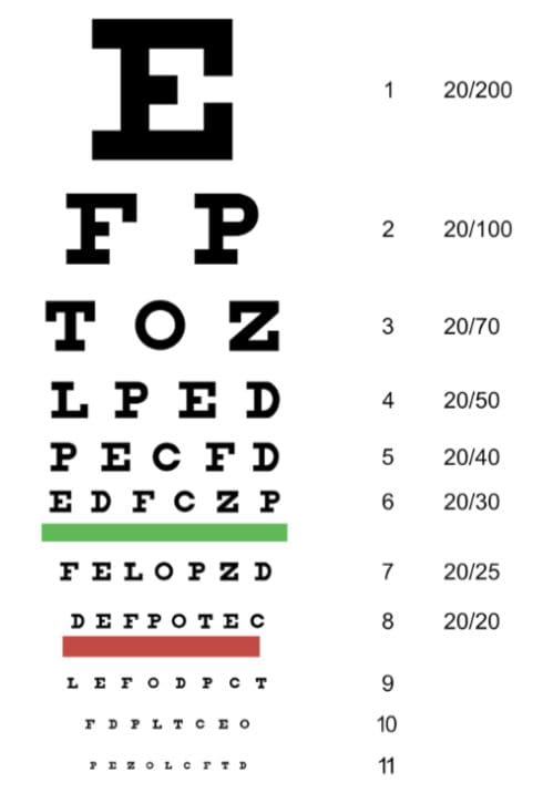
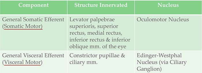 Cranial Nerve III Clinically
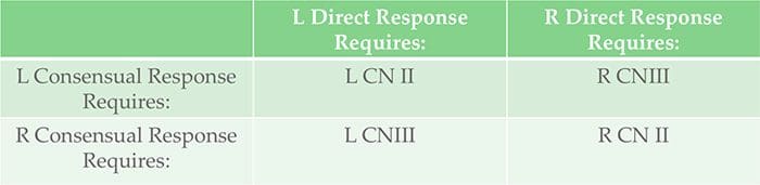 Testing Cranial Nerve CN II & III
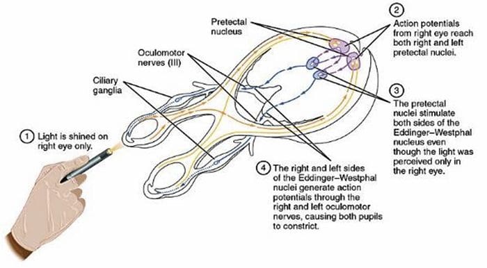
 Cranial Nerve IV Clinically
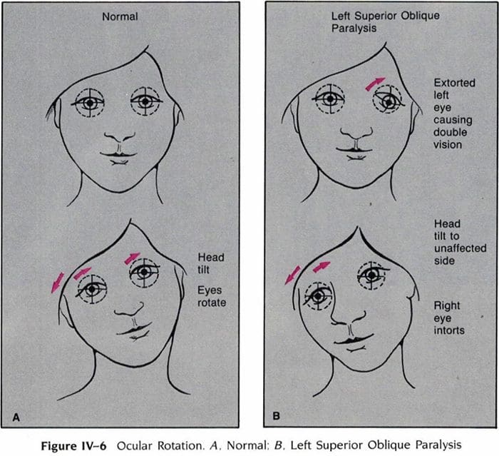
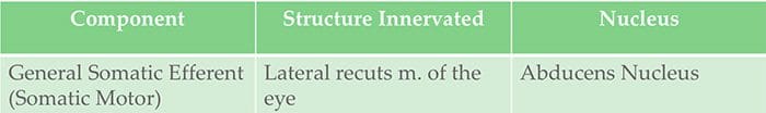 Cranial Nerve VI Clinically
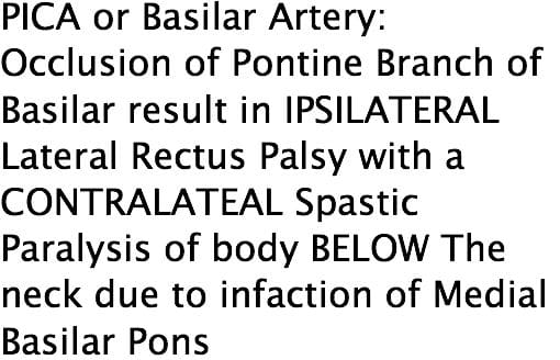
 Cranial Nerve V Clinically
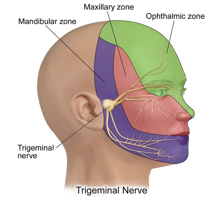
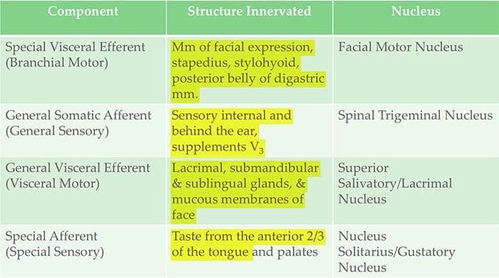 Cranial Nerve VII Clinically
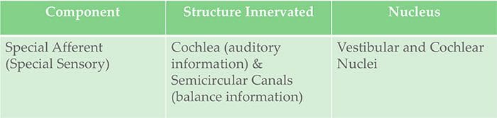 Cranial Nerve VIII Clinically
 Cranial Nerve IX Clinically
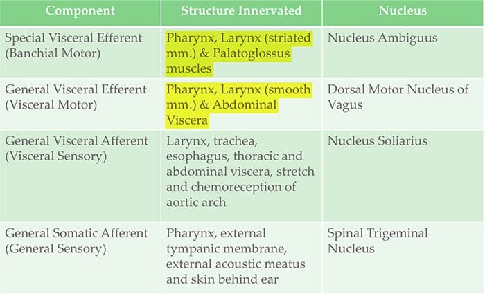 Cranial Nerve X Clinically
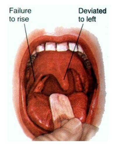
 Cranial Nerve XI Clinically
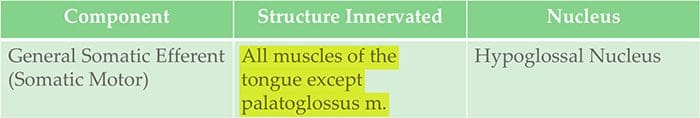 Cranial Nerve XII Clinically