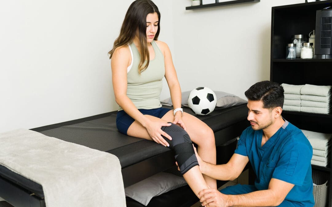
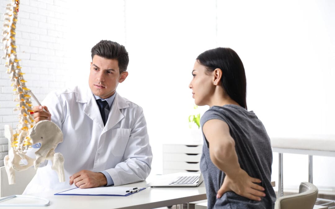
How Integrative Chiropractic Care Prevents Future Injuries in Athletes Using Functional Movement Assessments
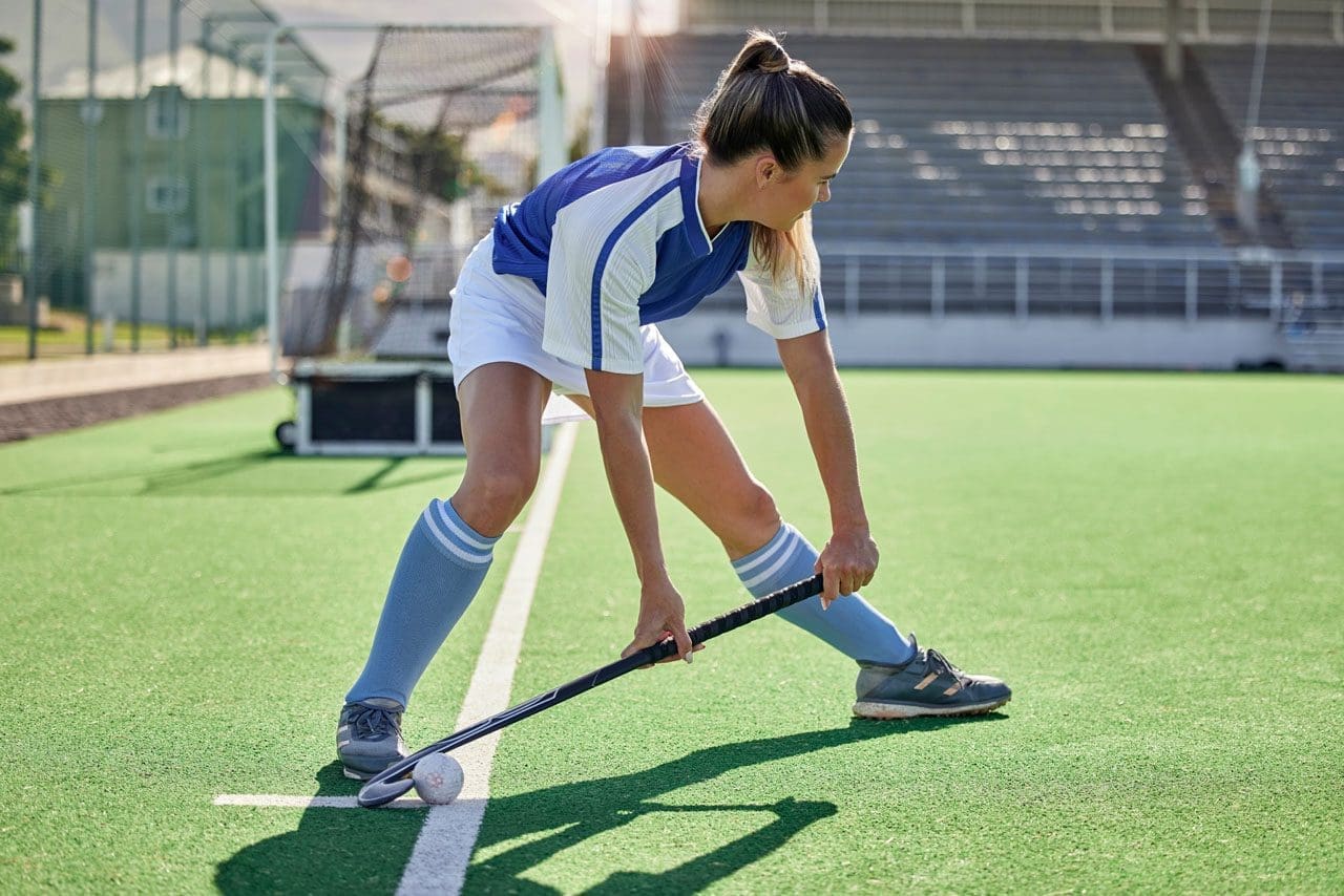
Sports: an athlete is in action on the field, ready to hit the ball during the game.
Athletes often push their bodies hard during training and competition. Small problems can build up over time and turn into painful injuries that force time off from sports. To catch these issues early, many athletes now ask for functional movement assessments as part of integrative chiropractic care. This method spots hidden imbalances like muscle tightness, weak spots, or stiff joints before pain starts. By addressing these problems with adjustments, soft-tissue work, and targeted exercises, practitioners help athletes stay healthy, move better, and avoid overuse injuries.
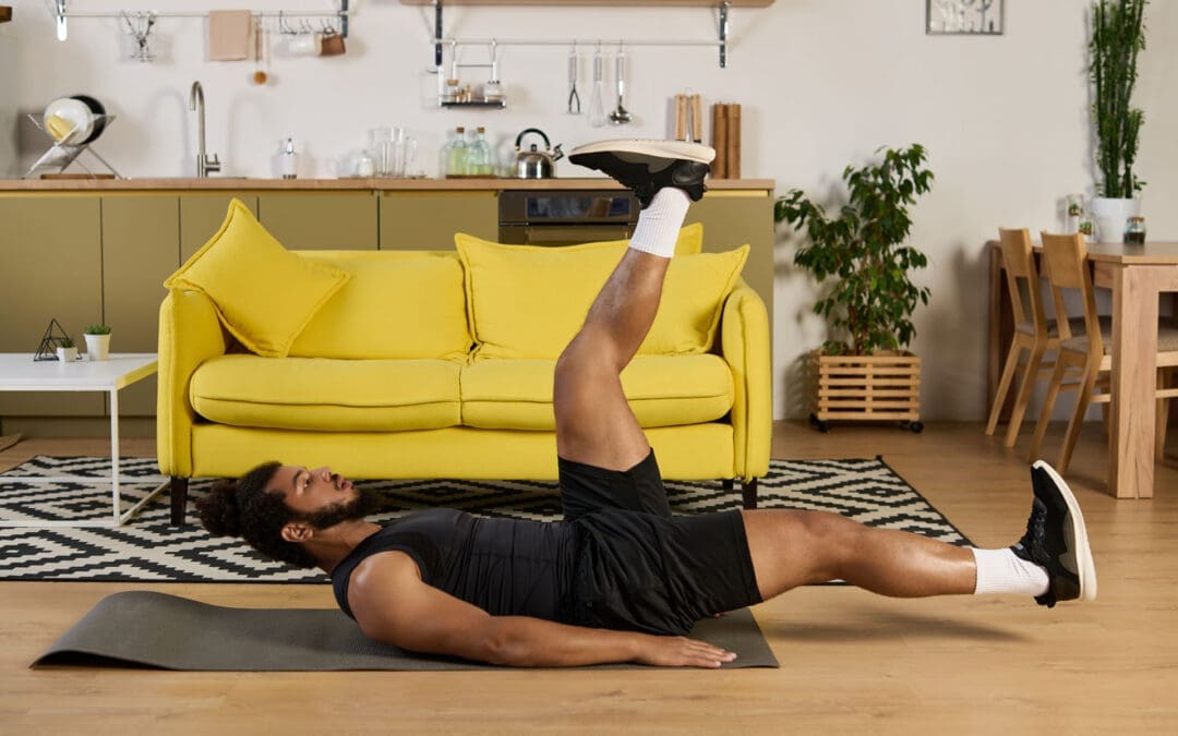
Functional movement assessments check how the body moves during everyday and sport-specific actions. These tests look at mobility, stability, balance, and coordination. Common movements include squats, lunges, reaching overhead, or stepping in different directions. The goal is to find areas where the body does not move smoothly or evenly. Even if nothing hurts yet, these assessments reveal subclinical imbalances—small issues that do not cause pain right away but can lead to bigger problems later.
Early detection of poor posture or uneven weight distribution
Spotting a limited range of motion in the hips, shoulders, or ankles
Identifying weak core or glute muscles that affect overall stability
Noting tight muscles that pull joints out of proper alignment
Integrative chiropractic care
Integrative chiropractic care combines spinal adjustments, soft-tissue therapies, and corrective exercises to effectively address these findings. Gentle adjustments move joints back into better positions, improving nerve signals and reducing pressure on surrounding tissues. Soft tissue work, such as massage or instrument-assisted techniques, loosens tight muscles and breaks up scar tissue. Corrective exercises then build strength and teach proper movement patterns. Together, these steps enhance nervous system function, optimize biomechanics, and stop the body from developing harmful compensation patterns.
The nervous system controls every muscle movement. When the spine or joints are misaligned, nerve messages can get disrupted. This leads to weaker muscle coordination or slower reaction times. Chiropractic adjustments help restore clear nerve pathways, so muscles fire at the right time and with the right force. Better biomechanics means joints move through their full, natural range without extra stress. This reduces wear and tear on knees, hips, shoulders, and the lower back.
Compensation patterns occur when one part of the body works harder to compensate for a weakness elsewhere. For example, tight hip flexors or a tilted pelvis in runners can cause the knees to track incorrectly, leading to pain or stress fractures over time. Faulty shoulder mechanics in swimmers or weightlifters can overload the rotator cuff. Integrative care addresses these root causes rather than just treating symptoms later.
Common subclinical imbalances identified through functional movement assessments include:
Muscle tension in the lower back or hamstrings that limits forward bending
Weak glute muscles that fail to stabilize the pelvis during running or jumping
Joint restrictions in the ankles that change walking or landing mechanics
Uneven shoulder mobility that affects throwing or overhead lifting
Poor core stability causes excessive arching in the lower back during lifts
By addressing these early, athletes lower their injury risk and maintain consistent training. Regular care also speeds recovery if minor issues arise, resulting in less downtime overall.
Practitioners often start with a thorough history and physical exam. They watch the athlete perform key movements and note any asymmetries or compensations. Based on the results, they create a personalized plan. Spinal adjustments realign the vertebrae to take pressure off nerves. Soft tissue therapies release tight fascia and muscles. Then, corrective exercises strengthen weak areas and retrain proper form. Over time, these steps improve balance, coordination, flexibility, and power output.
Key benefits of combining functional movement assessments with integrative chiropractic care:
Reduced chance of sprains, strains, tendonitis, and stress fractures
Improved joint mobility and muscle flexibility for better performance
Faster reaction times and coordination through better nerve function
Less inflammation and quicker recovery between workouts
Longer sports careers by preventing chronic overuse problems
Runners frequently show pelvic imbalances that tilt the hips and strain the iliotibial band or shins. Chiropractic adjustments and exercises that strengthen the glutes and core help keep the pelvis level, improving stride efficiency and cutting injury risk. Weightlifters with restricted shoulder mobility may compensate by excessively arching their backs, which can lead to low-back strain. Targeted soft tissue work and mobility drills correct this pattern before pain develops.
Football players and other contact-sport athletes benefit from regular checks of spinal alignment to better handle impacts. Swimmers gain from improved shoulder mechanics that prevent rotator cuff irritation. Weekend warriors who lift weights or cycle also see gains in endurance and reduced soreness. The approach works for athletes of all levels because it focuses on the root causes rather than waiting for symptoms.
Dr. Alexander Jimenez, DC, APRN, FNP-BC, brings valuable clinical observations to this field. As a chiropractor and board-certified family nurse practitioner with certifications in functional medicine, he emphasizes non-invasive, root-cause approaches. His work highlights how chiropractic adjustments, combined with functional assessments of mobility and biomechanics, help treat sports injuries, sciatica, and musculoskeletal imbalances. Dr. Jimenez observes that addressing nerve compression, inflammation, and movement dysfunction early—through adjustments, nutrition support, and tailored rehabilitation—enhances recovery and prevents recurrence in athletes and active individuals. His integrative practice in El Paso integrates chiropractic care with functional medicine to optimize performance, reduce chronic pain, and support long-term wellness.
This holistic view aligns with broader chiropractic principles that view the body as interconnected. When one area is restricted, it affects the whole kinetic chain. Integrative care breaks that cycle by restoring proper alignment and teaching sustainable movement habits.
Additional advantages athletes notice include:
Better posture during daily activities and sports
Enhanced proprioception (body awareness) for safer landings and cuts
Decreased muscle fatigue during long training sessions
Greater overall strength and power from efficient mechanics
Support for mental focus through reduced nagging discomfort
Preventing injuries this way also saves time and money by avoiding expensive treatments or missed competitions later. Many athletes report feeling stronger, more balanced, and more confident in their movements after consistent care.
To maintain results, athletes typically schedule regular visits. Frequency depends on training intensity, sport demands, and individual findings. Some come weekly during heavy training periods, while others maintain monthly check-ins. Between visits, they perform prescribed exercises at home or in the gym to reinforce new patterns.
Education plays a big role, too. Chiropractors teach proper warm-up routines, cool-down stretches, and body mechanics for specific sports. Nutritional guidance can sometimes complement care to support tissue repair and reduce inflammation. Collaboration with coaches, physical therapists, or trainers creates a complete support team.
In summary, functional movement assessments allow integrative chiropractic care to identify subclinical imbalances long before pain appears. Adjustments restore joint function, soft tissue therapies release restrictions, and corrective exercises build resilience. This combination enhances nervous system communication, optimizes biomechanics, and prevents compensation patterns that cause overuse injuries. Athletes—from runners dealing with pelvic tilts to lifters correcting shoulder mechanics—benefit by training more consistently, performing at higher levels, and enjoying longer, healthier careers. By addressing small issues proactively, this approach helps athletes stay in the game without painful interruptions.
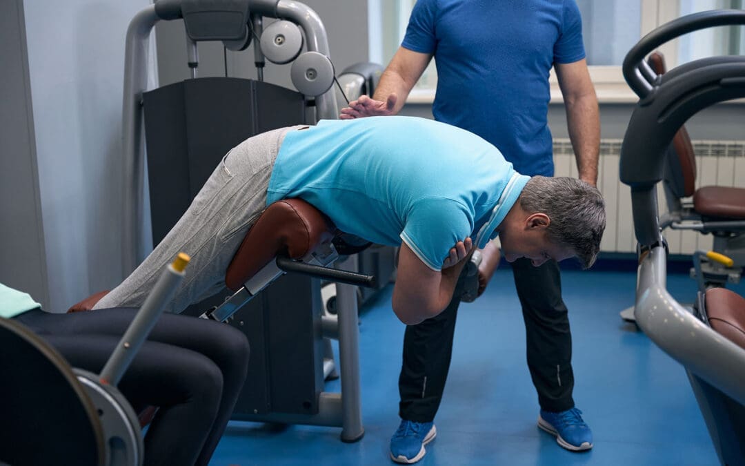
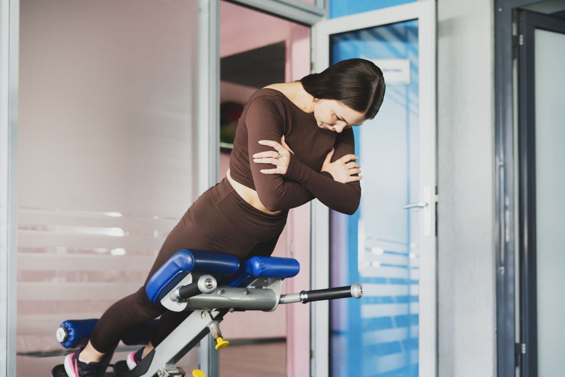
Back Extension Machine (Roman Chair) Training for a Stronger Back
A woman engages in back extension exercises to strengthen back muscles, improve core stability, and relieve chronic back pain.
A practical, El Paso Back Clinic–style guide to core stability, safer form, and pain prevention
If you’ve ever used a back extension machine—also called a hyperextension bench or Roman chair—you already know it looks simple. You lock your feet, rest your hips on the pad, and hinge forward and back up.
But the best results come from how you do it.
At El Paso Back Clinic, the goal is not just “stronger muscles.” It’s a smarter plan that supports spine stability, hip power, and better movement habits—especially for people who deal with recurring low back tightness, desk-related stiffness, or training-related flare-ups. Dr. Alexander Jimenez, DC, APRN, FNP-BC, often emphasizes that many back problems improve when you combine movement quality, targeted strengthening, and a whole-person plan (Jimenez, n.d.-a; Jimenez, n.d.-b).
This article explains:
what the back extension machine actually trains,
how to set it up correctly,
how to avoid the common mistakes that irritate backs,
and how integrative care (chiropractic + NP-style whole-body support) fits into a complete plan.
What the Back Extension Machine Trains (and Why It Matters)
Back extensions are a posterior chain exercise. That means they train the muscles on the back side of your body, including:
Erector spinae (spinal extensor muscles that help you stay upright) (MasterClass, 2021).
Glutes (hip extension power and pelvic support) (MasterClass, 2021).
Hamstrings (help control the lowering phase and assist hip extension) (MasterClass, 2021).
Deep core stabilizers (the “bracing” muscles that keep the spine steady while the hips move) (WebMD, 2024).
This is important because many people think “core” means only the abs. In real life, core stability is about the ability to resist unwanted motion and control the spine while the hips move.
A back extension machine helps train that pattern if you do it as a hip hinge, not as a “low back bend.” (More on that below.)
Roman Chair vs. Back Extension Machine: Same Goal, Different Feel
You’ll see a few styles:
45-degree hyperextension bench (most common “Roman chair” style)
90-degree Roman chair (more upright)
Seated back extension machine (you sit and extend backward against resistance)
Verywell Fit notes that these machines are often grouped together because they train similar movement patterns and posterior chain muscles, even though the setup and feel can differ (Verywell Fit, 2025).
If you’re choosing equipment for home or clinic use, adjustability matters. Many benches are built to adjust pad position and angle so different body types can hinge correctly (Valor Fitness, n.d.).
Step 1: Set Up the Machine Correctly (This Is Where Most People Go Wrong)
Before you do a single rep, take 30 seconds to set it up.
The best setup checkpoints
Hip pad position: The pad should sit around your hip crease (where your hips fold). If it’s too high, you can’t hinge well. If it’s too low, you may feel unstable (WebMD, 2024).
Feet locked in: Your heels and feet should feel secure in the restraints (WebMD, 2024).
Top position posture: At the top, you want a straight line from head to hips—not a “lean back” pose (MasterClass, 2021).
Quick self-test
If you feel the movement mostly in your low back joints (pinchy or compressed) rather than in your glutes/hamstrings, your setup or technique needs adjustment.
Step 2: Use the Right Form (Neutral Spine + Hip Hinge)
A safer back extension is controlled and clean. The spine stays neutral, and the movement comes mostly from the hips.
How to do it (simple steps)
Brace first: Take a breath and tighten your midsection like you’re preparing to be lightly bumped.
Hinge down: Push your hips back and lower your chest slowly. Keep your neck neutral.
Drive up: Squeeze glutes and hamstrings to lift your torso back up.
Stop at neutral: Finish tall and braced. Do not crank into hyperextension (MasterClass, 2021; WebMD, 2024).
Good cues that help
“Hips back, not ribs up.”
“Move like a hinge, not a bendy straw.”
“Glutes finish the rep.”
Chuze Fitness also describes back extensions as a way to work against gravity and build strength in a simple, repeatable pattern, with the option to progress by adding load later (Chuze Fitness, n.d.-a).
The #1 Mistake: Hyperextending at the Top
One of the biggest errors is leaning back too far at the top. People do it to “feel” the lower back more, but it often adds compression where you don’t want it.
What you want instead: a neutral, stacked finish.
Ribs down
Glutes tight
Spine tall
No “backward bend” finish (MasterClass, 2021).
If you can’t stop at neutral, reduce the range of motion and slow the tempo.
Another Common Mistake: Turning It Into a Low-Back Exercise Only
Back extensions are often taught as if they only train the lower back. In reality, they work best when the hips do the job and the trunk stays braced.
A helpful way to think:
The hips create motion
The spine controls motion
That is a big reason back extensions can be useful for stability—when done correctly (WebMD, 2024).
Reps and Sets: Simple Programming That Works
The “right” plan depends on your goal and your history.
Beginner (control first)
2–3 sets of 8–12 reps
Bodyweight only
Slow lowering (2–3 seconds down)
General strength and pain prevention
3 sets of 10–15 reps
Add light load only if form stays clean (Chuze Fitness, n.d.-a).
Stronger posterior chain (experienced lifters)
3–5 sets of 6–10 reps
More rest
Still stop at neutral (no hyperextension)
Rule: load is earned by control.
Verywell Fit’s equipment review also highlights that comfort, stability, and fit matter for consistent training—especially for people using these tools as part of a back-strengthening routine (Verywell Fit, 2025).
Safer Progressions (If Your Back Is Sensitive)
If your back flares easily, you can still train the posterior chain—you just need smarter progressions.
Options that tend to be more back-friendly:
Shorter-range back extensions (only move where you can stay neutral)
Isometric holds at neutral (hold 10–20 seconds)
Lower load, slower tempo
Add glute-focused assistance work (like bridges) alongside back extensions
At El Paso Back Clinic, Dr. Jimenez often frames strengthening as part of a bigger plan: improve mechanics, build tolerance, and progress gradually based on the person’s symptoms and daily demands (Jimenez, n.d.-a; Jimenez, n.d.-c).
When to Pause and Get Checked (Red Flags)
Back extension training should feel like muscular effort, not nerve pain.
Stop and seek professional guidance if you have:
Pain shooting down the leg
Numbness or tingling
Weakness in the foot/leg
Pain that worsens over time with extension-based movements
WebMD also encourages careful form and smart choices when using back extensions, especially when they’re used for “back health” rather than just bodybuilding (WebMD, 2024).
How This Fits the El Paso Back Clinic Approach: Strength + Mobility + Whole-Person Support
Many people try one thing:
“I’ll just strengthen my back.”
Or:
“I’ll just stretch more.”
Or:
“I’ll just get adjusted.”
But most lasting results come from combining the right tools in the right order.
Chiropractic care to improve mechanics
Chiropractic-focused care often aims to:
improve joint motion where stiffness limits your hinge,
reduce irritation that changes how you move,
and help you restore better spinal and pelvic mechanics.
El Paso Back Clinic content emphasizes a whole-body view of pain and function, including movement habits and multi-step plans (Jimenez, n.d.-c).
Exercise to build stability and strength
Once movement is cleaner, exercises like the Roman chair can help you:
reinforce a strong hinge,
strengthen posterior chain muscles,
and build stability that carries into work, lifting, and sports (MasterClass, 2021).
Nurse practitioner support to address barriers to recovery
NP-style integrative support often helps by addressing factors that keep people “stuck,” such as:
sleep quality,
stress load,
inflammation drivers,
safe pain management planning (when appropriate),
and screening for problems that need further testing or referral.
In short: your back isn’t separate from the rest of you.
A Simple 3-Phase Plan You Can Follow
Here is a practical approach that matches how many integrative clinics structure back-pain recovery and performance.
Phase 1: Calm things down and restore motion (1–2 weeks)
Gentle mobility (hips + mid-back)
Light back extensions with short range
Walk daily if tolerated
Focus on bracing and hinge control
Phase 2: Build capacity (3–6 weeks)
Back extensions: 2–3 days/week
Add glute and hamstring work
Add core stability work
Slowly add reps before adding load
Phase 3: Build real-world resilience (ongoing)
Add load gradually (only if neutral form is automatic)
Transfer strength into squats, hinges, and carries
Keep a weekly routine of mobility + stability work
This kind of integrated plan—adjustments plus exercise and habit change—is also described in chiropractic-focused integration articles discussing the value of combining care approaches to improve outcomes (OPTMZ State, 2026).
Key Takeaways
The back extension machine is best used as a hip-hinge strength tool, not a “bend your spine” tool (MasterClass, 2021).
Proper setup (hip pad alignment + stable feet) helps you move safely (WebMD, 2024).
Avoid the big mistake: hyperextending at the top. Stop at neutral.
Strong results often come from a full plan: chiropractic mechanics + targeted exercise + whole-person support, a theme repeated across El Paso Back Clinic education from Dr. Jimenez (Jimenez, n.d.-a; Jimenez, n.d.-c).
Common Motor Vehicle Accidents in El Paso: Recovery and Healing at El Paso Back Clinic®
An injured woman in a stretcher after a car accident, covered by a thermal blanket.
Motor vehicle accidents, or MVAs, are a big issue in El Paso. This city sits on the border, with lots of trucks and cars zooming on roads like I-10 and Loop 375. Accidents often result from drivers not paying attention, drinking, or speeding. They can lead to injuries like neck pain or broken bones. At El Paso Back Clinic®, we help people heal from these injuries. Our team, led by Dr. Alexander Jimenez, uses integrative chiropractic care. This mixes spine fixes with massage, exercise, and healthy eating tips. It treats the whole body and mind. In this article, we discuss common crashes in El Paso, the harm they cause, and how our clinic supports recovery. We draw on Dr. Jimenez’s expertise at our locations in El Paso, TX.
El Paso has many crashes each year. Recent data shows thousands of wrecks, with injuries and even deaths. The border sees heavy truck traffic, upping the risks. Dust storms or rain-slick roads. Work zones add hazards. Knowing this helps folks drive safely. At El Paso Back Clinic®, we see many patients from these events. Our care focuses on pain relief and full health.
Common Types of Motor Vehicle Accidents in El Paso
El Paso’s roads mix locals, visitors, and cross-border traffic. This leads to jam-ups and crashes. Here are the key types:
Distracted Driving Accidents: Phones or snacks pull drivers’ eyes from the road. In El Paso, this sparks many wrecks. Texting hits hard at spots like Mesa and Stanton streets. Texas-wide, it caused over 84,000 crashes in one year.
Drunk or Impaired Driving: Booze or drugs slow folks down. Crashes spike nights and weekends. It’s a top cause in Texas spots like El Paso. They pop up near fun zones like Cincinnati Avenue.
Speeding-Related Crashes: Too fast means tough stops. It makes up 30% of Texas wrecks. On I-10 and Loop 375, speed leads to bad hits. Winds make it worse.
Rear-End Collisions: Cars bump backs from close follows or late brakes. Common on Loop 375 in traffic or near shops like Cielo Vista. Distractions or weather help cause them.
Intersection Crashes: Red-light runs or no yields cause side smacks. Over half happen at crossings like Montana or Zaragoza. The Spaghetti Bowl adds mess. Stop sign skips are big faults.
Pedestrian Incidents: Walkers get struck when drivers miss spots or speed. Downtown, schools, or UTEP see many. Poor walks led to many deaths lately.
Truck Accidents: Border hauls mean big trucks everywhere. Thousands cross yearly. Recent counts show many truck wrecks with injuries. Tired drivers, heavy loads, or blind areas cause them. Spots like I-10, US-54, and Loop 375 are hot.
Pile-ups hit in storms on I-10. Lane changes in builds confuse. Hit-runs occur in town. Stay alert, slow down, and watch out for trucks to avoid.
At El Paso Back Clinic®, we treat folks from all these. Our team knows border traffic woes. We offer care plans for quick heals.
Common Injuries Sustained in Motor Vehicle Accidents
MVAs jolt bodies hard. Sudden moves cause hidden hurts. Here are the usual ones:
Whiplash: Neck snaps cause pain, stiffness, headaches, and dizziness. Top in rear-ends.
Neck and Back Sprains: Pulls or tears cause pain and reduced movement. Low back twists.
Soft Tissue Damage: Bruises, rips in muscles. Swell, stiff. Deep ones last.
Fractures: Breaks from hits. Ribs puncture lungs. Bad ones need ops. Limbs, spine too.
Traumatic Brain Injuries (TBIs): Head knocks cause mix-ups, forgetfulness, and eye issues. Change lives, cost lots.
Shoulders, knees, and inside bleed too. Burns and scars are possible. Trucks crush more. Walkers break bones, heads. Minor ones spark worry or PTSD.
At our clinic, we spot these early. Dr. Jimenez’s team uses checks to plan care.
How These Injuries Occur
Crashes stop or hit fast. Bodies fly in cars. Belts save, but force hurts. Rear-ends jerk heads, stretch necks for whiplash. Sides twist spines for sprains, disc slips. Heads hit for TBIs. Knees dash-bang for sprains. Moves inflame tissues. Trucks smash small cars, break bones. Walkers fly, land hard. Signs may be delayed, so check soon.
We urge quick visits. Our El Paso spots offer fast help.
Integrative Chiropractic Care at El Paso Back Clinic® for MVA Recovery
Our integrative care treats all of you. We fix spines hands-on, easing pain without pills or cuts first. Mixes old ways with massage, PT, and nutrition. Speeds heal, drops swell. Here’s our approach:
Spinal Adjustments: Move bones right, cut nerve pinch, up move. Great for whiplash, back.
Physical Therapy: Builds strong, flexible. Restores after sprains and breaks.
Nutritional Support: Food advice; adds fight-swell, up mood.
Other Therapies: Needle work or disc pull. Ease pain, stress.
We speed recovery, hit the body and feelings. Start in 72 hours, best. Stops long pain. Our functional medicine finds roots.
Insights from Dr. Alexander Jimenez and El Paso Back Clinic®
Dr. Alexander Jimenez, DC, APRN, FNP-BC, has headed El Paso Back Clinic® for 30+ years. He excels in MVA, which includes injuries like whiplash and TBIs. We use functional medicine, nutrition, and rehab. Holistic care heals body and mind from trauma. Cases show fast recovery from car and truck hits. Border traffic brings many to us. Our spots at 11860 Vista Del Sol and 6440 Gateway East offer full care. Call 915-850-0900 for help.
Conclusion
El Paso MVAs from busy roads hurt many. From whiplash to TBIs, harms vary. El Paso Back Clinic® gives natural healing. We cut pain, restore movement. See us after crashes. Safe drives prevent woes. Visit elpasobackclinic.com or call for wellness.
Mobility Challenges in Mexican and Mexican American Communities: Insights from El Paso Back Clinic®
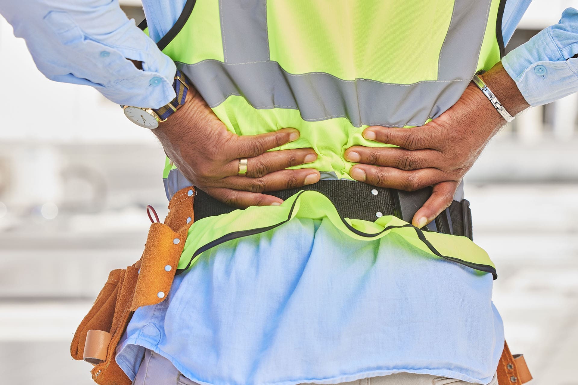
Mexican-American with back pain at a construction site.
At El Paso Back Clinic® in El Paso, TX, we see many patients from Mexican and Mexican American backgrounds facing mobility issues. These problems often stem from tough jobs, health factors like obesity, and aging. Our wellness chiropractic care focuses on pain relief and improved movement. This article discusses common issues such as arthritis and back pain, supported by studies. We’ll explain how our team, including Dr. Alexander Jimenez, DC, APRN, FNP-BC, uses integrative approaches to help. If you’re in El Paso dealing with these, our clinic is here for you.
Common Musculoskeletal Mobility Issues We Treat
Musculoskeletal problems affect your bones, muscles, and joints, making it difficult to move freely. At our clinic, we see these issues often in our community, where many work in demanding fields like farming or construction.
Arthritis, especially in the knees, is a top concern. It causes joint wear-related swelling and pain. In Mexico, about 20-25% of adults aged 40+ have it, with higher rates among women (Villarreal Rizzo et al., 2025). Mexican Americans in the U.S. also face risks, like osteoporosis weakening bones in 16% of women (Wright et al., n.d.). At El Paso Back Clinic®, we help ease this with gentle adjustments and exercises.
Chronic low back pain hits hard, too. It comes from prolonged lifting or standing. In Mexico, it’s the leading cause of disability, with 840.6 cases per 100,000 in 2021 (Clark et al., 2023). Among farmworkers here in Texas, 46.9% report back issues affecting daily life (Weigel et al., 2013). Our chiropractic care targets this to get you moving again.
Work injuries often involve the shoulders, wrists, and legs. Repetitive tasks in jobs cause rotator cuff problems in 19.1% and elbow pain in 20.2% of Latino workers (Mora et al., 2014). Older adults in our area are at risk of frailty due to ongoing pain, leading to reduced mobility (National Institutes of Health, n.d.). Women face more disability in tasks like walking, with arthritis raising risks by 35% over time (Rodriguez et al., 2021).
Here are key facts we see in our patients:
Arthritis rates: 19.6% for knee issues in Mexicans over 40, up to 24.2% in women (Ciampi de Andrade et al., 2022).
Back pain: Affects 16.9% of farmworkers from repetitive strain (Mora et al., 2014).
Craft-related injuries: Neck and knee pain from activities like weaving (Jeanson et al., 2025).
Disability trends: Physical function declines by 0.18 points per year with arthritis (Rodriguez et al., 2021).
Jobs in agriculture and construction drive these, plus obesity adds joint stress. In our Mexican American patients, higher BMI initially slows strength loss but worsens it later (Davis & Al Snih, 2025). About 83% of Hispanic men are overweight, linked to less activity (Valdez et al., 2019). At El Paso Back Clinic®, we address this with personalized plans.
Neuromusculoskeletal Issues Addressed at Our Clinic
These issues combine nerve problems with muscle and bone pain, leading to numbness or weakness. Our wellness approach helps restore nerve function and reduce discomfort.
Chronic low back pain is common, often due to nerve compression. It’s the main cause of disability in Mexico (Alva Staufert et al., 2021). Knee and foot arthritis affects movement, with 25.5% showing joint changes (Ciampi de Andrade et al., 2022). We treat foot pain from standing jobs, seen in 4.8% of workers (Mora et al., 2014).
Shoulder injuries, such as rotator cuff tears, are associated with overhead work and affect 19.1% (Mora et al., 2014). Elbow issues, or epicondylitis, affected 20.2% due to tool use (Mora et al., 2014). MSDs in Mexico rose 57.3% over 30 years (Clark et al., 2023). Obesity plays a role, with 40% of Hispanic men affected (Valdez et al., 2019).
In border areas like El Paso, women report 29.8% low back and 38.3% upper back pain from factory jobs (Harlow et al., 1999). Older patients walk more slowly due to leg pain (Quiben & Hazuda, 2015).
Common issues we handle:
Low back pain: Top disability driver, tied to work and weight (Alva Staufert et al., 2021).
Knee/foot arthritis: More in women, causing stiffness (Ciampi de Andrade et al., 2022).
Rotator cuff: From arm overuse in construction (Mora et al., 2014).
Epicondylitis: Elbow strain, common in 20% (Mora et al., 2014).
How El Paso Back Clinic® Helps with Integrative Care
Our clinic combines nurse practitioners (NPs) and chiropractic methods for culturally sensitive help. We focus on pain management and rehab to fit our community’s needs.
NPs at our clinic offer full check-ups that consider culture and history. They suggest diets rich in veggies and yoga for detox and pain relief (Jimenez, 2026a). We team up for whole-body care (Jimenez, 2026b).
Chiropractic adjustments realign the spine to ease nerve compression. For sitting-related back pain, we restore curves and strengthen the core (El Paso Back Pain Clinic, n.d.). Access to this care is key, though Hispanics use it less (Roseen, 2023).
Dr. Alexander Jimenez shares from his experience: Chronic back pain worsens with poor posture, but adjustments and exercises help (Jimenez, n.d.). For sciatica, decompression relieves pressure on nerves, which is common in laborers. Neuropathy gets therapy for tingling (Jimenez, n.d.). He uses functional medicine to tackle stress, diet, and job factors in our Mexican American patients.
We include mindfulness and natural remedies. Cultural factors, such as family support, help recovery, but delays worsen pain (Arthritis Foundation, n.d.). Our NPs create home plans (Pérez-Stable et al., 2003).
Rehab strengthens areas such as the legs and shoulders (Mora et al., 2014). It cuts frailty risks (National Institutes of Health, n.d.). For farmworkers, it reduces disability (Weigel et al., 2013).
Our care benefits:
Cultural match: Understanding barriers like work migration (Harlow et al., 1999).
Strength building: Targeted exercises (Mora et al., 2014).
Prevention: Nutrition against obesity (Valdez et al., 2019).
Why Choose El Paso Back Clinic® for Your Mobility Needs
In El Paso, with our diverse community, these issues are common but treatable. Our clinic specializes in wellness chiropractic to help you stay active. Contact us for a consultation with Dr. Jimenez and our team.
Fun Ways to Stay Active: Alternatives to Boring Workouts for Better Health
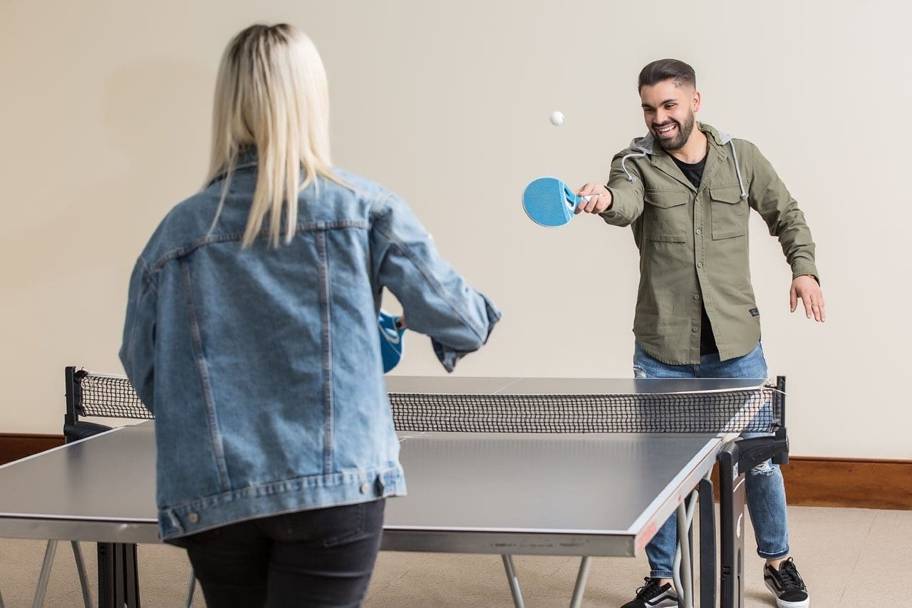
Friends play table tennis as a way to start making fitness fun and as a doable health goal.
Many people start the new year with big fitness goals. They promise to hit the gym every day or run miles each week. But often, these plans fall apart quickly. Life gets busy, motivation fades, and suddenly, exercise feels like a chore. If this sounds like you, don’t worry. Giving up on strict resolutions doesn’t mean giving up on health. Instead, shift to activities that feel more like play than work. Fun sports and easy movements can keep you moving without the dread of traditional workouts. This approach makes staying active sustainable and enjoyable, leading to better long-term habits (Bayou Bend Health System, n.d.).
Research shows that making physical activity fun boosts your chances of sticking with it. For example, choosing things you enjoy turns exercise into a hobby. This can improve your mood, reduce stress, and even help with weight management. Health experts recommend at least 150 minutes of moderate activity per week for adults, but it doesn’t have to be in a gym (NHS, n.d.). Simple swaps like walking in nature or dancing to music can meet these goals while feeling effortless.
In this article, we’ll explore ways to restart your fitness journey with joy. We’ll cover fun sports, social options, and relaxing practices. We’ll also discuss low-impact choices for those who struggle with standard routines. Plus, learn how professionals like integrative chiropractors and nurse practitioners can guide you. Drawing from expert insights, including clinical observations from Dr. Alexander Jimenez, DC, APRN, FNP-BC, this guide offers practical tips to get back on track.
Restarting After a Failed New Year’s Resolution
If your resolution crashed early, it’s time for a fresh start. The key is picking activities that excite you. Fun, easy sports like hiking, dancing, swimming, or biking can make movement feel rewarding. These options build fitness without the pressure of sets and reps.
Hiking: Head to a trail for fresh air and views. It’s a great way to explore while getting your heart rate up. Start with short paths and build up. Hiking strengthens legs and improves balance, all while enjoying nature (MultiCare Clinic, n.d.).
Dancing: Put on your favorite tunes and move freely. Whether alone or in a class, dancing boosts cardio and coordination. It’s low-pressure and can burn calories without feeling like exercise (Whispering Oaks Senior Living, n.d.).
Swimming: Water supports your body, making it gentle on joints. Swim laps or just splash around for fun. It’s ideal for all ages and helps with endurance (Vista Springs Living, n.d.).
Biking: Ride a bike around your neighborhood or on paths. It’s easy to adjust speed and distance. Biking tones muscles and can be a social outing (Blue Cross NC, n.d.).
These activities trick your brain into thinking you’re playing, not working out. Studies support this: enjoyable exercise leads to better adherence and health outcomes (Exercise is Medicine, n.d.).
Beyond solo sports, join social activities to add fun. Pickleball, tennis, or team sports bring people together, making commitment easier.
Pickleball: A mix of tennis and ping-pong, it’s easy to learn and play. Courts are popping up everywhere, and it’s great for quick games with friends (Nerd Fitness, n.d.).
Tennis: Hit the court for rallies that improve agility. Doubles makes it less intense and more chatty (Athlean-X, n.d.).
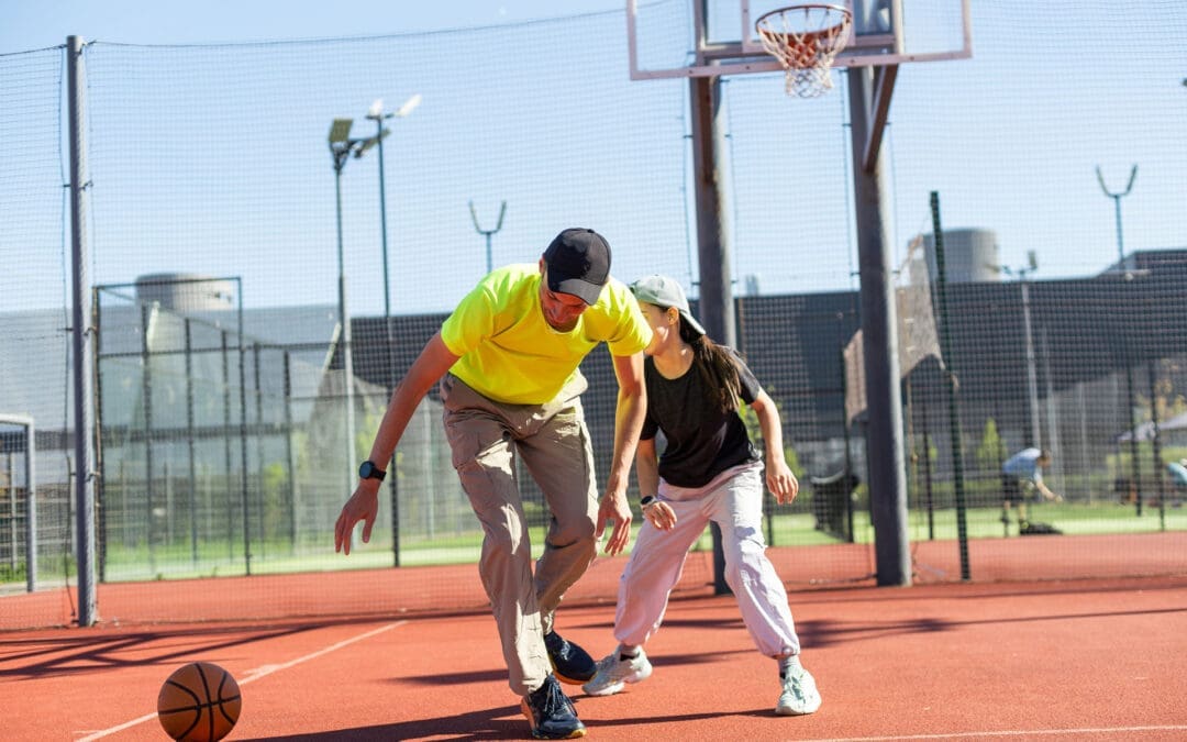
Team Sports: Join a recreational league for soccer, basketball, or volleyball. The group vibe keeps you motivated, and games feel like events, not drills (Quora, n.d.).
Social exercise can reduce feelings of isolation while building strength. One study notes that group activities enhance mental health alongside physical benefits (Reddit, n.d.).
For a calmer approach, try mind-body practices like yoga or Tai Chi. These are low-impact and focus on relaxation.
Yoga: Gentle poses improve flexibility and reduce stress. Start with beginner videos at home. It helps with breathing and mindfulness (Piedmont Wellness Center, n.d.).
Tai Chi: Slow, flowing movements build balance and calm the mind. It’s perfect for easing into activity without strain (Care Insurance, n.d.).
These practices are adaptable for any fitness level. They promote relaxation, which can lower blood pressure and improve sleep (NHLBI, n.d.).
To build habits, start small. Aim for 10–15 minute sessions a few times a week. Gradually increase as you gain confidence. This prevents burnout and lets your body adjust (Bayou Bend Health System, n.d.). Track progress in a journal to see improvements, like feeling more energetic.
Options for Those Who Dislike Traditional Workouts
Not everyone loves the gym or running. If weights and treadmills bore you, low-impact or sociable sports offer alternatives. These keep you active without the monotony, focusing on enjoyment and variety.
Swimming and biking stand out as low-impact favorites. Swimming provides a full-body workout in a supportive environment, reducing joint stress (Seniors Helping Seniors, n.d.). Biking lets you control the pace, making it accessible for beginners (MultiCare Clinic, n.d.).
Hiking and dancing add adventure. Hiking varies with terrain, keeping things interesting, while dancing lets you express yourself creatively (Blue Cross NC, n.d.; Whispering Oaks Senior Living, n.d.).
For a challenge, try rock climbing. It’s low-impact but builds strength and problem-solving skills. You can start indoors at a gym with easy walls (The Telegraph, n.d.).
Joining a recreational sports league brings community. Options like softball or ultimate frisbee emphasize fun over competition (Nerd Fitness, n.d.).
Benefits of These Activities:
More engaging than repetitive workouts.
Build social connections.
Adaptable to your energy level.
Improve mood through endorphins (Sanguina, n.d.).
These choices make the activity feel natural. For instance, walking briskly counts as exercise and can be done anywhere (Quora, n.d.). Or jump rope for short bursts—it’s simple and effective for cardio (MCU, n.d.).
If mobility is an issue, modify exercises. Chair-based routines or water aerobics allow movement without strain (ParentGiving, n.d.; Care.com, n.d.). The goal is consistency over intensity.
Experts agree: low-impact options like these support heart health and flexibility, especially for those with limits (Gaddis Premier, n.d.; Prairie Hills at Independence, n.d.).
How Integrative Professionals Can Help
When starting or restarting activity, professional guidance ensures safety. Integrative chiropractors and nurse practitioners offer tailored care, especially if you have physical limits.
Integrative chiropractors focus on the whole body. They use adjustments to align the spine, reducing pain and improving movement. This holistic approach addresses root causes rather than just symptoms (Integral Chiropractic, n.d.; Impastato Chiropractic, n.d.).
For example, if joint pain stops you from hiking, a chiropractor can ease stiffness through manipulations and exercises (Elysian Wellness Centre, n.d.; De Integrative Healthcare, n.d.). They often include nutrition and lifestyle advice for better results (AFP Fitness, n.d.; Together4Health Wellness, n.d.).
Nurse practitioners add medical expertise. They assess your health and create plans that address limits, such as suggesting low-impact swimming for arthritis (Buckner Parkway Place, n.d.; Cor Health Ontario, n.d.).
Together, these pros provide personalized care. They work with your abilities to help you enjoy activities again (Wellness Center FW, n.d.; Fortitude Health, n.d.).
Dr. Alexander Jimenez, DC, APRN, FNP-BC, embodies this integrated approach. With over 30 years in practice, he combines chiropractic and nursing for comprehensive care. His clinical observations highlight non-invasive methods for pain management and mobility.
In his work, Dr. Jimenez notes that tailored programs, like resistance band exercises, strengthen muscles without high impact. This helps people with injuries return to fun activities like biking or dancing. He emphasizes flexibility for joint health, noting that restricted movement can lead to pain, but gentle practices like yoga can restore it.
On LinkedIn, Dr. Jimenez shares insights on sciatica and back pain, recommending core exercises like modified squats for those with limitations. He advocates stretching to prevent stiffness, noting, “If you don’t stretch, your body ‘pays interest'” in reduced mobility.
His practice includes functional medicine, addressing nutrition and the environment for wellness. For example, he uses assessments to create plans that fit patients’ lifestyles, helping them stay active despite chronic conditions (All Injury Rehab, n.d.; Motus Integrative Health, n.d.).
How They Help:
Assess limits and set realistic goals.
Provide exercises like water aerobics for joint relief (Activ Therapy, n.d.).
Offer advice on enjoyable activities to build habits (Nepute Wellness Center, n.d.).
Monitor progress to adjust plans.
This support makes returning to movement less daunting. Integrative care focuses on harmony in physical, mental, and emotional health (Wellness Center FW, n.d.).
Wrapping Up: Make Movement Joyful
Staying active doesn’t require grueling workouts. By choosing fun options like hiking or yoga and seeking professional help when needed, you can rebuild habits. Remember Dr. Jimenez’s observation: personalized, holistic care unlocks better mobility. Start small, stay consistent, and enjoy the process. Your health will thank you.
Dr. Alex Jimenez at El Paso Back Clinic®: Beating Back Pain from Long Desk Hours
Businesswoman experiences worsening back pain while sitting at her desk.
If your back pain gets worse the longer you sit at your desk, you are not alone. Many people in El Paso face this issue due to long hours spent in sedentary jobs. Sitting for extended periods can put pressure on the spine, tighten muscles, and reduce blood flow, leading to stiffness, aches, and, in some cases, chronic problems (Colorado Pain Care, n.d.). The positive news is that you can take simple steps to reduce the pain and prevent it from worsening. At El Paso Back Clinic® in El Paso, TX, the wellness chiropractic care team, led by Dr. Alex Jimenez, DC, APRN, FNP-BC, focuses on helping people just like you find natural, long-term relief through personalized plans.
Prolonged sitting stresses the lower back by increasing disc pressure by up to 90% compared to standing. It flattens the spine’s natural curve, strains muscles, and creates imbalances (Colorado Pain Care, n.d.). Slouching or leaning forward adds extra load to the neck and upper back. Over time, this can lead to tight hips, weak core muscles, and ongoing discomfort that affects daily life.
At El Paso Back Clinic®, our experts understand these issues caused by sedentary work. They use a holistic approach that combines chiropractic adjustments, functional medicine, and rehab to address root causes like poor posture and muscle imbalances from desk jobs (Jimenez, n.d.-a).
Here are practical changes to start today:
Move often: Get up every 30 minutes to stand, walk, or shift positions. Short 1-2 minute breaks improve circulation and ease tension (Huntsville Hospital Health System, n.d.; Sydney West Physio, n.d.).
Use regular breaks: Set a timer for quick walks to get water or to stretch. This habit prevents stiffness from building up throughout the day.
Add dynamic movement: While sitting, shift weight, uncross legs periodically, or use a footrest to change angles. These small actions keep the spine mobile (Colorado Pain Care, n.d.).
A proper ergonomic setup supports optimal posture and reduces strain.
Follow these key tips:
Set your chair so that your feet are flat on the floor, your knees are at 90 degrees, and your hips are level with or above your knees.
Add lumbar support (a small pillow or rolled towel works) to maintain the lower back’s curve.
Place your screen at eye level to avoid looking down or up too much.
Keep the keyboard and mouse close so elbows bend at 90 degrees and shoulders stay relaxed.
Avoid crossing legs for long, as it can tilt the pelvis (Senara Chiropractic & Med Spa, n.d.; Huntsville Hospital Health System, n.d.).
Consider alternating between sitting and standing with a standing desk. Even partial standing reduces spinal pressure.
Stretches help loosen tight spots from sitting, such as the hips, shoulders, and neck.
Try these simple ones:
Hip flexor stretch: Kneel on one knee, gently push hips forward, and hold 20-30 seconds per side.
Chest and shoulder opener: Clasp hands behind your back or use a wall to stretch forward.
Neck tilts: Slowly tilt the head side to side or forward/back; hold for 10-15 seconds.
Upper back extension: Hands behind head, gently arch upper back (Sydney West Physio, n.d.).
Do them hourly or during breaks for better flexibility.
Strengthening the core supports the spine and improves posture long-term.
Include these:
Planks: Hold forearm plank 20-30 seconds.
Cat-camel: On hands and knees, arch and round back slowly.
Bridges: Lie back, lift hips while squeezing glutes.
Walking or gentle yoga: Build overall strength (Huntsville Hospital Health System, n.d.; Sydney West Physio, n.d.).
Aim for 20-30 minutes of activity most days.
For lasting relief, professional care targets alignment, mobility, and personalized fixes. At El Paso Back Clinic®, Dr. Alex Jimenez leads a team offering integrated chiropractic care. This includes spinal adjustments to correct misalignments, non-surgical spinal decompression for disc relief, acupuncture, functional medicine for nutrition and stress, and rehab exercises tailored to desk-related issues.
Dr. Jimenez, with dual expertise as a chiropractor and nurse practitioner, emphasizes posture correction, mobility training, and the prevention of sedentary pain through evidence-based methods. The clinic helps restore function without drugs or surgery, focusing on root causes like imbalances from prolonged sitting (Jimenez, n.d.-a; Jimenez, n.d.-b).
Other options in El Paso exist, but El Paso Back Clinic® stands out for its comprehensive wellness approach, advanced diagnostics, and patient-centered plans that go beyond basic adjustments.
If pain includes numbness, tingling, or weakness in the legs, or persists despite changes, seek evaluation to rule out serious conditions (University of Maryland Medical System, n.d.).
Start small: improve movement, setup, and stretches. If needed, contact El Paso Back Clinic® for expert help. Many in El Paso regain comfort and stay active with this care.
Spinal Hygiene Explained: Daily Habits to Keep Your Spine Strong and Pain-Free
A woman performs spinal hygiene exercises on a fitness ball at home, strengthening her back muscles.
Spinal hygiene is the practice of caring for your spine every day to keep it strong, flexible, and healthy. Just like brushing your teeth helps prevent cavities, spinal hygiene helps avoid back problems and keeps your body moving well. The spine supports your whole body, protects the nervous system, and lets you bend, twist, and stand tall. Good habits can make a big difference in how you feel now and as you get older.
What Spinal Hygiene Means
Spinal hygiene includes simple daily actions to protect the spine’s natural shape and movement. The spine has gentle curves that help absorb shock and allow smooth motion. When these curves stay balanced, and the spine moves freely, you feel better overall.
Proper posture: Sit and stand with your shoulders back, head aligned over your spine, and pelvis in a neutral position to avoid extra strain.
Regular movement and exercise: Stay active with walking, swimming, or stretching to keep joints loose and muscles strong.
Proper body mechanics: Lift things by bending your knees, keeping objects close to your body, and avoiding twists to prevent injury.
Core strength: Build muscles around your midsection for better support and stability.
Hydration and nutrition: Drink plenty of water to keep spinal discs cushioned, and eat foods rich in calcium, vitamin D, magnesium, and omega-3 fatty acids to support bone and tissue health.
Stress management: Use deep breathing, meditation, or yoga to reduce tension that tightens back muscles.
These steps help maintain the spine’s integrity and prevent issues like stiffness or pain (Spinenpain.org, n.d.; Lifemovesmt.com, n.d.).
Neglecting spinal hygiene can lead to problems over time. Poor habits can cause muscle imbalances, joint wear, reduced motion, and conditions such as herniated discs or chronic back pain. This can affect your nervous system, since the spine houses nerves that control body functions. When the spine is out of alignment, it may press on nerves, leading to pain, weakness, or other issues (Servinglifedallas.com, n.d.; Drmmalone.com, n.d.).
Benefits of Good Spinal Hygiene
Taking care of your spine brings many advantages. It reduces the chance of back pain, improves how easily you move, and supports better posture. A healthy spine also helps the nervous system function smoothly, boosting energy, coordination, and overall well-being. Regular care can slow age-related changes, lower injury risk, and help you stay active longer.
Prevents muscle tightness and joint problems
Improves blood flow and nutrient delivery to spinal tissues
Enhances balance and reduces fall risk
Supports better sleep and less daily discomfort
Studies and experts note that these habits promote long-term health and vitality (Illinoisspinalcare.com, n.d.; Spinehealth.org, n.d.).
How Chiropractic Care Fits In
Chiropractic care plays a key role in spinal hygiene. Chiropractors use gentle adjustments to fix misalignments, called subluxations, that can stress the spine and nerves. These adjustments restore proper movement, reduce pain, and improve function. Regular chiropractic visits act as preventive maintenance, catching small issues before they grow.
Many people combine chiropractic with other habits for the best results. Adjustments improve alignment, while daily posture and exercise help maintain gains. This approach helps address common complaints such as neck or low back pain and supports recovery from injuries (Illinoisspinalcare.com, n.d.; Eastportlandchiropractor.com, n.d.).
The Power of Integrative Care with Chiropractors and Nurse Practitioners
An integrative approach brings together diverse experts to achieve stronger results. Chiropractors focus on the spine’s structure, alignment, and movement through adjustments and rehab. Nurse practitioners (NPs), especially those with advanced training, address broader health needs such as nutrition, stress management, hormone balance, and lifestyle changes.
This team effort addresses both the physical spine and daily habits that affect it. For example, a chiropractor might correct alignment after an injury, while an NP guides on anti-inflammatory foods or stress reduction to aid healing. Together, they create personalized plans that work better than one alone, especially for complex pain, chronic issues, or recovery from accidents.
Dr. Alexander Jimenez, DC, APRN, FNP-BC, stands out in this field. As a Doctor of Chiropractic and board-certified Family Nurse Practitioner, he combines spinal adjustments, decompression therapy, and functional medicine. His practice emphasizes root-cause care, using nutrition, lifestyle tweaks, and advanced diagnostics for musculoskeletal problems, personal injuries, and overall wellness. Clinical observations from his work show that integrative methods restore mobility, reduce pain, and improve quality of life by treating the whole person—not just symptoms (Dralexjimenez.com, n.d.; A4m.com, n.d.).
Simple Ways to Start Spinal Hygiene Today
You can begin with easy changes that fit into daily life.
Posture checks: Set reminders to sit tall and take breaks from sitting every 30 minutes.
Daily stretches: Try cat-cow pose, child’s pose, or seated twists for 5-10 minutes.
Safe lifting: Always bend at the knees and use your legs.
Stay hydrated: Drink water throughout the day to keep your discs healthy.
Eat spine-friendly foods: Include leafy greens, fish, nuts, and dairy or alternatives for key nutrients.
Move often: Walk or do low-impact activity for at least 30 minutes most days.
Manage stress: Practice deep breathing or short meditation sessions.
Adding regular chiropractic check-ups can help monitor and maintain progress (Lifemovesmt.com, n.d.; Newlifefamilychiropractic.net, n.d.).
Final Thoughts
Spinal hygiene is a smart, everyday way to protect one of your body’s most important parts. By focusing on posture, movement, nutrition, and professional care when needed, you support a strong spine, healthy nerves, and a better quality of life. Small habits add up to big benefits, helping you stay active and pain-free for years.
IFM's Find A Practitioner tool is the largest referral network in Functional Medicine, created to help patients locate Functional Medicine practitioners anywhere in the world. IFM Certified Practitioners are listed first in the search results, given their extensive education in Functional Medicine