Understanding Core Overtraining Injuries: Wellness Strategies and Chiropractic Solutions at El Paso Back Clinic
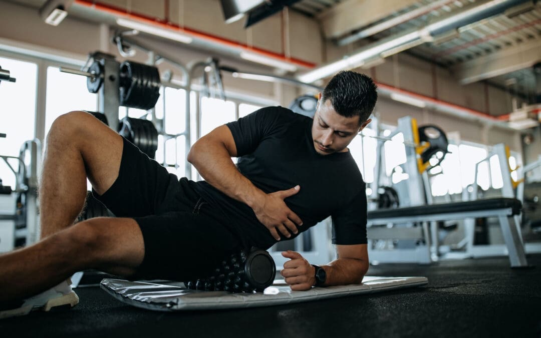
A man is training in a gym to build strong and healthy muscles.
Pushing your core muscles too far without proper rest can create big issues for your overall health, especially your back. At El Paso Back Clinic® in El Paso, TX, we specialize in wellness chiropractic care that helps people recover from these problems and stay strong. Core overtraining affects the muscles around your midsection, which support your spine and daily movements. This article breaks down the injuries that can happen, why they occur, ways to stop them, and how our chiropractic methods provide relief. We focus on natural wellness to keep your back and body in balance.
The Role of Core Muscles in Back Health and Overtraining Basics
Your core is like the foundation of a house—it holds everything together. It includes muscles in your stomach, sides, lower back, and hips. These help with bending, twisting, and standing straight. When you overtrain, you repeat exercises like sit-ups or lifts too much, without breaks. This wears down tissues faster than they can heal. Wellness experts note that this leads to lasting soreness, reduced energy, and risks to your spine.
At our clinic, we treat many cases where core issues cause back pain. Overtraining disrupts the natural alignment, pulling on the back. Science shows repetitive stress changes how muscles and bones work together, leading to problems.
Everyday Muscle Strains from Pushing the Core Too Hard
Strains are pulls or small tears in muscles. They are common when the core gets overworked and can’t support the body well.
Strains Around the Groin
These happen in the inner thigh muscles tied to the core. Quick stops and starts in activities like hiking or playing kickball can trigger them. If the core is weak from too much training, it adds extra pull. You feel a sudden sharp pain, maybe see bruising, and have trouble moving your legs inward. In the back, this strain can tug on the lower spine, causing aches there too.
Strains in the Stomach Area
Abdominal strains come from forceful turns, like swinging a racket or carrying heavy bags. Overdoing core workouts builds up damage over time. Pain hits when you tense up or laugh, and the area feels sore to the touch. This connects to back health because weak abs force the back muscles to overcompensate, leading to stiffness.
Problems with Hip Flexors
These muscles help raise your legs and link directly to the core. Running uphill or doing too many leg raises without rest inflames them. Symptoms include a tight feeling in the front of the hip and pain when stepping up. Poor core balance makes the back arch unnatural, adding pressure.
Our wellness approach at El Paso Back Clinic uses gentle checks to find these strains early and guide healing without harsh methods.
Deeper Issues: Fractures and Bone Stress from Core Overuse
When overtraining goes on, it can harm bones, which support the core and back.
Fractures in the Ribs
Rib stress fractures are tiny breaks from constant tugging by core muscles. This shows up in paddling sports or even heavy coughing fits from overuse. The muscles contract hard, stressing the bone until it cracks. Pain sharpens with deep breaths or twists, and it can feel tender. Since ribs protect the upper back area, this injury often leads to posture problems and back discomfort.
Other Stress Fractures
These small cracks appear in weight-bearing bones like the pelvis or lower spine from ongoing impact. Walkers or dancers who ignore rest will experience issues when bone repair lags behind damage. Early signs are dull aches that worsen with activity. In young active people, it might involve cartilage issues, too. Back clinic patients often report these symptoms linked to core weakness, causing spinal instability.
Healing takes rest, but our chiropractic wellness plans speed it up safely.
Extra Effects Like Ongoing Pain, Loss of Power, and Stiffness
Overtraining doesn’t stop at big injuries—it brings smaller but nagging problems.
Lasting Pain and Rigid Muscles
You might wake up stiff or feel constant soreness in the core. This spreads to the back, making sitting or standing tough. It’s a sign the body is inflamed and needs recovery time.
Weaker Muscles Overall
Tired core muscles can’t hold strong, leading to drops in power. One side might become too tight while the other weakens, throwing off balance. This imbalance pulls on the back, increasing the risk of slips or strains during daily tasks.
Tight Spots in the Legs and Sides
Muscles like the back of the thighs (hamstrings) or the outer thigh band (IT band) tighten to make up for a worn-out core. This causes knee or hip issues that refer pain to the lower back. Wellness care addresses these chains of problems.
Signs also include getting sick more or feeling down, as the body fights overload.
How These Injuries Develop and What Increases the Risk
Injuries build from too much activity without balance. Body mechanics play a part—bad posture during exercises adds uneven stress. Muscles need time to fix small wear, but skipping rest lets damage grow.
In jobs with lifting or sports with jumps, core pulls transfer to bones. Poor shoes or low nutrients weaken things further. At our back clinic, we look at the whole picture, including how back alignment affects core strain.
Smart Ways to Avoid Core Overtraining Problems
Wellness starts with prevention. Ramp up workouts slowly, adding just a bit more each week. Switch activities to give muscles variety. Take full rest days and stretch gently.
Eat foods rich in vitamins for strong bones, and use supportive gear. Pay attention to body signals like unusual tiredness. Our clinic offers wellness checks to catch risks early.
Chiropractic Wellness Care for Healing Core Injuries
At El Paso Back Clinic, we use integrated chiropractic to resolve core issues and boost back health.
Adjustments to the Spine
These hands-on moves realign the back, easing nerve pressure and helping muscles relax. It improves how the core and back communicate, reducing pain fast.
Therapies for Soft Tissues
Massage-like techniques release tight spots, increase blood flow, and calm inflammation. This works well for strains and stiffness.
Our methods treat current pain while building wellness to avoid repeats. They enhance flexibility, strength, and nerve health for long-term back care.
Expertise from Dr. Alexander Jimenez at El Paso Back Clinic
Dr. Alexander Jimenez, DC, APRN, FNP-BC, leads our wellness efforts with deep knowledge in chiropractic science.
Linking Injuries to Patient Histories
He connects core overtraining to back pain through detailed stories, exams, and imaging. For instance, work lifts or sports twists can lead to patterns in muscle and nerve issues.
Diagnosis with Dual Expertise
Using his skills as a nurse practitioner and chiropractor, he does thorough assessments. Advanced scans reveal hidden bone or tissue problems in core areas.
Step-by-Step Treatments
Procedures include spine adjustments, tissue work, and guided exercises. For sports or personal injuries, he adds strength training. In car accidents, focus is on impact-related core and back strains.
Care for Medical and Legal Needs
The clinic manages full treatment plans, including reports for insurance or court in injury cases. This covers work, auto, or everyday accidents with clear documentation.
Dr. Jimenez promotes education on wellness to empower patients.
Bringing It All Together for Better Health
Core overtraining brings strains, fractures, pain, and imbalances that hit the back hard. Prevention through smart habits and chiropractic care keeps you moving well. At El Paso Back Clinic®, our wellness focus helps restore balance naturally.
Chiropractic Solutions for Sudden Movement Injuries
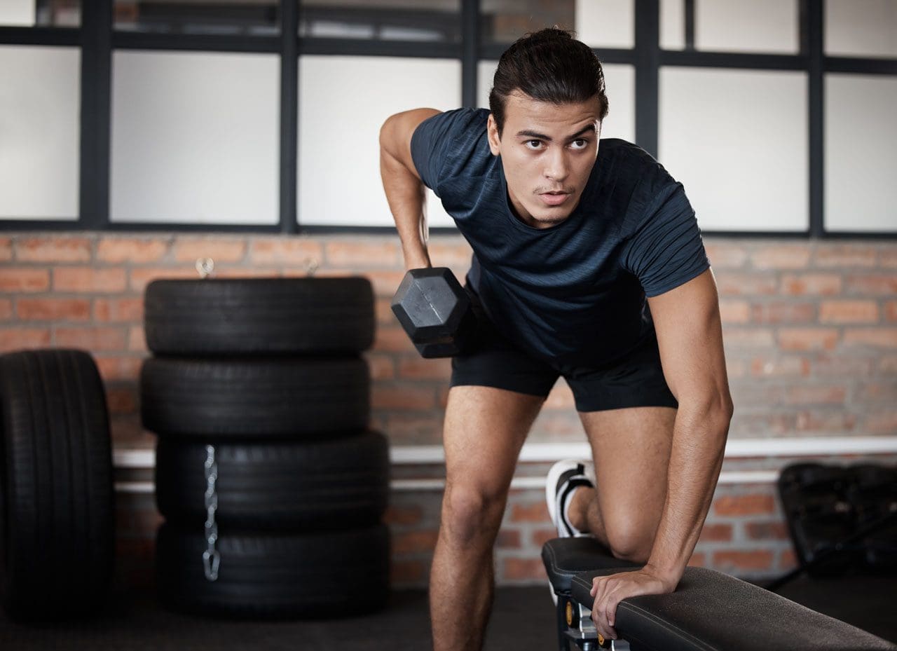
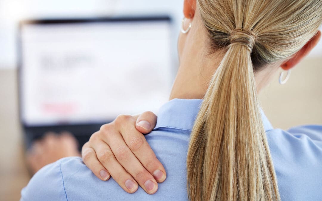
Businesswoman experiencing back pain while working at her desk in a modern office.
Introduction
Picture yourself dashing to catch a ball, only to twist your ankle with a sharp sting. Or imagine your neck jerking in a car crash, leaving you stiff and sore. These are sudden movement injuries—quick, forceful actions that strain muscles, sprain joints, or sometimes result from involuntary jerks due to medical conditions (Hopkins Medicine, n.d.; Verywell Health, 2022). Sudden movement injuries are acute musculoskeletal issues, such as sprains or strains, caused by a single traumatic event, or they can result from neurological conditions that lead to uncontrollable motions (Cleveland Clinic, 2023a; UF Health, n.d.).
Chiropractic integrative care can help by easing pain, reducing swelling, and restoring movement naturally. At El Paso Back Clinic, Dr. Alexander Jimenez, DC, APRN, FNP-BC, uses spinal adjustments, nutrition, and therapies like massage to heal these injuries (Jimenez, n.d.a). This article explores what sudden movement injuries are, how they happen, and how Dr. Jimenez’s holistic approach speeds recovery. You’ll learn practical tips to heal and prevent future issues, all backed by science.
Whether from a sports slip or an unexpected jolt, these injuries can disrupt daily life. With the right care, you can bounce back stronger and stay active (Cleveland Clinic, 2023b).
Understanding Sudden Movement Injuries
Sudden movement injuries come in two forms. Acute soft-tissue injuries, like strains (stretched muscles or tendons) or sprains (stretched ligaments), happen from a single, forceful action, such as twisting a knee or jerking your neck in a crash (Hopkins Medicine, n.d.; Cleveland Clinic, 2023c). These often occur in sports, work accidents, or falls, causing pain, swelling, or limited motion (UPMC, n.d.).
Involuntary movement injuries, like jerks or tremors, stem from neurological conditions such as myoclonus or ataxia (Verywell Health, 2022; Children’s Hospital, n.d.). These can result from brain injuries, seizures, or migraines, leading to uncontrolled motions that may strain muscles or joints (Edward K. Le, 2023; Movement Disorders, n.d.).
Both types limit mobility and can lead to chronic pain if untreated. Acute injuries cause immediate swelling or bruising, while neurological ones may add balance issues or anxiety (Cleveland Clinic, 2023a; UF Health, n.d.). Early treatment prevents long-term problems like arthritis or weakness (Cleveland Clinic, 2023b).
How Sudden Movement Injuries Occur
Acute soft-tissue injuries arise from sudden force. A quick pivot in basketball can sprain an ankle, or lifting a heavy box awkwardly can strain a back muscle (Cleveland Clinic, 2023c). Common triggers include:
Sports Accidents: Sudden twists or tackles in soccer or running (Cleveland Clinic, 2023b).
Car Crashes: Whiplash from neck jerking (Cleveland Clinic, 2023d).
Slips or Falls: Tripping on a curb, straining a knee (Pain Care Florida, n.d.).
No Warm-Up: Jumping into exercise without stretching (Cleveland Clinic, 2023c).
Involuntary movement injuries come from medical issues. Myoclonus, which causes jerky motions, can result from epilepsy or brain trauma, straining muscles during spasms (Movement Disorders, n.d.). Ataxia, causing unsteady movement, might follow a stroke, leading to falls or sprains (Children’s Hospital, n.d.). Risk factors include age, weak muscles, or prior injuries, which make joints less stable (UPMC, n.d.).
Both types disrupt normal movement. A sprained ankle swells, limiting walking, while involuntary jerks can cause falls, leading to additional injuries (Edward K. Le, 2023).
Signs and Effects of Sudden Movement Injuries
Signs vary by injury type. For soft-tissue injuries, you might notice:
Sharp pain or swelling, like a sore ankle after a twist (Hopkins Medicine, n.d.).
Bruising or stiffness, making joint movement tough (Cleveland Clinic, 2023c).
Weakness, like struggling to lift after a shoulder strain (UPMC, n.d.).
Involuntary movement injuries show differently:
Sudden twitches or tremors, like myoclonus spasms (Movement Disorders, n.d.).
Unsteady walking or balance loss from ataxia (Children’s Hospital, n.d.).
Muscle soreness from repetitive jerks (Verywell Health, 2022).
These injuries can make daily tasks hard—walking hurts with a sprained knee, or involuntary jerks cause embarrassment (Cleveland Clinic, 2023a). Untreated, they risk chronic pain, joint damage, or falls, especially in older adults (Cleveland Clinic, 2023b). Acting early stops small issues from growing.
Chiropractic Care for Recovery
Chiropractic care helps sudden movement injuries by fixing spinal misalignments that disrupt nerve signals, easing pain and swelling (New Edge Family Chiropractic, n.d.). Adjustments realign the spine, improving joint function and muscle coordination (Rangeline Chiropractic, n.d.). For a sprained ankle, adjustments reduce nerve pressure, speeding healing (Texas Medical Institute, n.d.).
For involuntary movements, chiropractic care calms nervous system stress, reducing spasms in conditions like myoclonus (Jimenez, n.d.a). Patients often feel less pain and better mobility after a few sessions (Cleveland Clinic, 2023b). It’s like resetting a stuck gear, letting your body move freely.
Dr. Jimenez’s Expertise at El Paso Back Clinic
At El Paso Back Clinic, Dr. Alexander Jimenez, DC, APRN, FNP-BC, uses his dual expertise as a chiropractor and nurse practitioner to treat sudden movement injuries from work, sports, personal falls, or motor vehicle accidents (MVAs). “Trauma misaligns the spine, blocking healing,” he explains (Jimenez, n.d.b).
His clinic uses advanced diagnostics: X-rays for neuromusculoskeletal imaging and blood tests to check inflammation. A sports injury, like a twisted knee, might show nerve pinches affecting mobility (Jimenez, n.d.a). Treatments are non-surgical: adjustments restore alignment, ultrasound reduces swelling, and exercises strengthen muscles. For MVAs, Dr. Jimenez provides detailed medical-legal documentation, working with specialists for smooth claims.
Integrative therapies boost recovery. Massage improves blood flow, speeding tissue repair; acupuncture reduces pain for easier motion; and nutrition plans with anti-inflammatory foods support healing (Jimenez, n.d.b). A worker with a strained back from lifting moved freely after adjustments and massage. Dr. Jimenez targets root causes, like poor form, to prevent chronic issues.
Integrative Therapies for Healing
El Paso Back Clinic’s integrative approach enhances recovery. Massage therapy relaxes tight muscles, boosting circulation to heal sprains faster (Texas Medical Institute, n.d.). Acupuncture targets points to ease pain and calm spasms, helping with involuntary movements (Jimenez, n.d.b). Exercises like leg lifts rebuild strength and stabilize joints (Sport and Spinal Physio, n.d.).
The RICE method (rest, ice, compression, elevation) helps reduce swelling in soft-tissue injuries early on (Cleveland Clinic, 2023e). These therapies, paired with chiropractic, speed recovery and prevent issues like arthritis (Cleveland Clinic, 2023b).
Nutrition to Support Recovery
Nutrition aids healing from sudden movement injuries. Omega-3-rich foods like salmon reduce inflammation, easing joint pain (Best Grand Rapids Chiropractor, n.d.). Leafy greens like spinach provide antioxidants to protect tissues (Spine, n.d., p. 417). Lean proteins like chicken rebuild muscles and ligaments (Human Care NY, n.d.).
Calcium from yogurt strengthens bones, while magnesium in nuts prevents spasms (Foot and Ankle Experts, n.d.). Try salmon salads or berry smoothies to support recovery. These foods work with chiropractic to speed healing (Rangeline Chiropractic, n.d.).
Preventing Future Injuries
Prevent injuries with smart habits. Warm up before sports with stretches to cut strain risks (Cleveland Clinic, 2023c). Strengthen core muscles with planks to stabilize joints (Sport and Spinal Physio, n.d.). Use proper form when lifting—bend knees, keep back straight (UPMC, n.d.).
For neurological issues, manage conditions like epilepsy with doctor guidance to reduce spasms (Verywell Health, 2022). Regular chiropractic check-ups catch misalignments early (New Edge Family Chiropractic, n.d.). These steps keep you moving safely.
Success Stories from El Paso Back Clinic
At El Paso Back Clinic, a runner with a sprained ankle healed with adjustments and protein-rich meals, returning to races. A driver post-MVA eased neck pain with acupuncture and greens. These stories show how integrative care restores mobility.
Conclusion
Sudden movement injuries, from sprains to involuntary jerks, can disrupt life, but chiropractic care at El Paso Back Clinic, led by Dr. Jimenez, heals them naturally. Using adjustments, nutrition, and therapies like massage, the clinic restores movement. Try warm-ups, eat omega-3s, and visit the clinic. Stay active and pain-free.
Weekend Athletes Injury Solutions: A Simple, Evidence-Based Guide for Safer Play and Faster Recovery
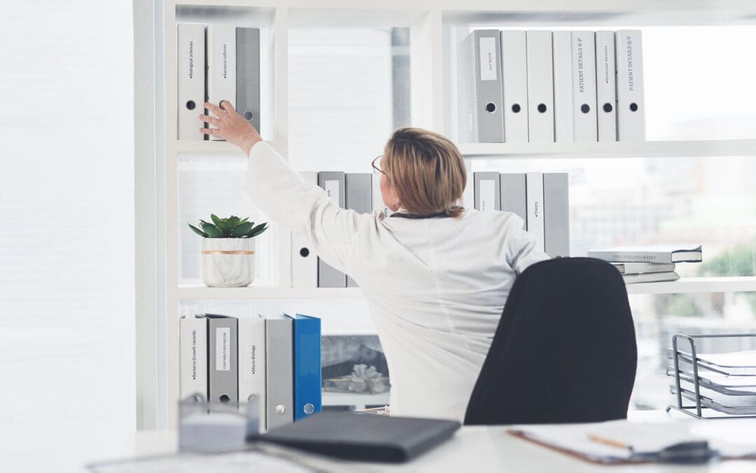
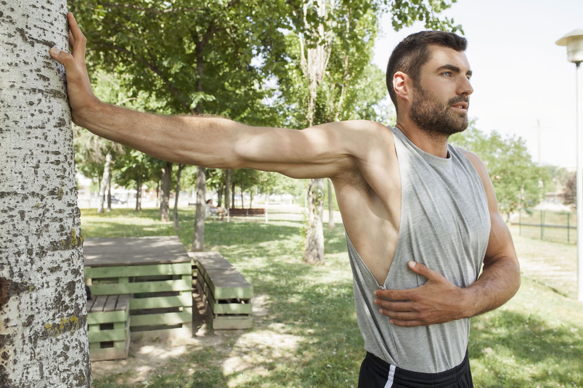
A handsome, muscular man in sportswear is stretching his muscles in a sunny park.
Who this is for: adults who sit most of the week and then go hard on the weekend (a.k.a. “weekend warriors”). What you’ll get: clear reasons these injuries happen, what to do first, how to prevent them, and how integrative chiropractic care—like the approach used in El Paso—helps you recover and return to activity safely.
Weekend warriors 101
A weekend warrior is someone who does most of their intense activity on one or two days after a mostly sedentary week. That pattern can still deliver strong health benefits if you meet weekly exercise targets, but the sudden spike in effort raises the risk of sprains, strains, and overuse problems—especially when you skip warm-ups or jump in too fast (Riverside Health System, 2025; Weill Cornell Medicine, 2024). (riversideonline.com)
Large studies show that “condensed” exercisers can gain health benefits similar to those who spread workouts throughout the week—as long as the total weekly minutes reach the recommended amounts. The catch: your muscles, tendons, and joints still need gradual loading to stay injury-resistant (American Heart Association News, 2024; Shiroma et al., 2019). (www.heart.org)
Why weekend athletes get hurt
Most weekend injuries come down to three drivers:
Overuse: repeating motions your tissues aren’t ready for (long runs, repetitive swings).
Sudden movement: fast cuts, awkward landings, or twisting under load.
Poor preparation: no warm-up, weak stabilizers, and worn-out shoes.
These factors underlie many musculoskeletal problems seen by orthopedic and emergency clinicians (Aligned Orthopedic Partners, 2024; Weill Cornell Medicine, 2024). (Aligned Orthopedic Partners)
What typically gets injured (and what it feels like)
Emergency physicians most often treat injuries to the knees, shoulders, and ankles, with sprains and strains outnumbering fractures (Weill Cornell Medicine, 2024). (weillcornell.org)
Ankle sprain (ligament): twist/roll, swelling, tenderness, sometimes bruising.
Knee sprain/overuse pain: instability, joint-line pain, and pain after cutting or pivoting.
Achilles tendinopathy: stiff, sore area above the heel (often worse in the morning).
Rotator cuff irritation: pain with overhead reach or lying on the shoulder.
Shin splints: aching along the shin after running on hard surfaces (Riverside Health System, 2025). (riversideonline.com)
Sprain vs. strain (plain words): Sprain = ligament (joint stabilizer). Strain = muscle or tendon (mover). Sprains can feel unstable and bruise; strains feel like a pull with spasm or weakness (Aligned Orthopedic Partners, 2024). (Aligned Orthopedic Partners)
Your job habits shape your weekend risk
Repetitive tasks and long sitting can irritate tissues before you ever play. Those weekday loads stack with Saturday’s game and can tip you into pain. Tendinitis, for example, often develops from repeated motions (MyShortlister, 2023). Short micro-breaks, posture changes, and light mid-week movement help. (Shortlister)
First aid: what to do in the first 24–72 hours
For many fresh soft-tissue injuries, start with the PRICE method: Protect, Rest, Ice (20 minutes on), Compress, Elevate. Don’t push through sharp pain. Seek urgent care for a “pop,” severe swelling, numbness/weakness, deformity, or inability to bear weight (Weill Cornell Medicine, 2024). (weillcornell.org)
When imaging is useful (and what usually comes first)
You don’t need an MRI for every sprain. Clinicians begin with a history and examination; an X-ray is often the first test if a fracture is suspected. Musculoskeletal ultrasound or MRI follows when soft-tissue damage is suspected, symptoms persist, or nerve signs appear (Weill Cornell Medicine, 2024). (weillcornell.org)
In work, sport, or motor-vehicle accident (MVA) cases, advanced imaging also supports clear medical-legal documentation—a key part of comprehensive injury care (El Paso Back Clinic; Dr. Jimenez). (elpasobackclinic.com)
Practical prevention that actually works
Warm up and cool down. Do 5–10 minutes of light cardio and dynamic moves (leg swings, lunges, and arm circles). Ease into slow stretches after play (Riverside Health System, 2025; Appleton Chiropractic Center, n.d.). (riversideonline.com)
Build up gradually. Increase time or intensity by ~10% per week. Rotate high- and low-impact days (Center for Orthopedic Surgery & Sports Medicine, n.d.). (COSM)
Use the right gear. Replace worn shoes; match footwear to your sport (Riverside Health System, 2025). (riversideonline.com)
Hydrate, fuel, and sleep. Under-fueling and short sleep increase the risk of cramps and strains (Riverside Health System, 2025). (riversideonline.com)
Add two short mid-week sessions. Even 20–30 minutes of exercise twice a week improves tissue tolerance and reduces the risk of weekend injuries (Mayo Clinic Sports Medicine, n.d.). (sportsmedicine.mayoclinic.org)
Simple self-care roadmaps
Ankle sprain
Days 0–2: PRICE, gentle ankle pumps, compression sleeve.
Days 3–7: pain-free range of motion; start weight bearing as tolerated.
Weeks 2–4: add balance drills and band work.
See a clinician if you can’t bear weight or feel instability (Weill Cornell Medicine, 2024). (weillcornell.org)
Achilles tendinopathy
Reduce jumping/sprinting while painful.
Begin slow calf raises (progress to eccentrics); increase load gradually (Aligned Orthopedic Partners, 2024). (Aligned Orthopedic Partners)
Shoulder soreness (rotator cuff pattern)
Short rest (not total rest), then scapular control and light external-rotation drills; limit overhead volume and improve thoracic mobility (Aligned Orthopedic Partners, 2024). (Aligned Orthopedic Partners)
Low-back strain
After 24–48 hours, try gentle mobility exercises (such as pelvic tilts and cat-camel), followed by core endurance exercises (like planks) and hip-hinge practice. If pain persists or travels below the knee or you notice weakness, seek evaluation (Weill Cornell Medicine, 2024). (weillcornell.org)
2 rounds: push-ups 8–12; band rows 12–15; band “T” raises 10–12
Dead bug 6/side; bird-dog 6/side
3–5 min pec stretch + thoracic rotations
Short “bridge” sessions like these raise tissue tolerance and make weekend play safer (Center for Orthopedic Surgery & Sports Medicine, n.d.). (COSM)
How integrative chiropractic care supports weekend athletes
Integrative chiropractic care blends joint-specific manual therapy with targeted exercise, soft-tissue work, and—when indicated—acupuncture, bracing/taping, and coordinated medical evaluation. The goal is to improve mechanics (how you move) and capacity (what your tissues can handle), so you heal and resist re-injury (Radiant Life Chiropractic, 2024; Aligned Orthopedic Partners, 2024). (Radiant Life Chiropractic)
At El Paso Back Clinic, this approach is paired with a dual-scope model (chiropractic plus nurse practitioner care) for sports, work, personal, and MVA injuries. The team can:
Perform focused orthopedic and neurological exams.
Order X-ray, MRI, CT, or musculoskeletal ultrasound when the exam suggests more than a simple sprain.
Coordinate medical-legal documentation (mechanism, findings, imaging, functional limits, and response to care) for injury cases.
Guide progressive rehab and return-to-play plans based on pain-free motion, strength, and sport-specific tasks (El Paso Back Clinic; Jimenez, 2025). (elpasobackclinic.com)
Local context: Recent clinic articles from El Paso highlight dual-scope evaluation, the role of advanced imaging, and clear documentation for personal-injury cases—useful if your injury involves work or an auto crash (El Paso Back Clinic). (elpasobackclinic.com)
A smarter return-to-play checklist (advance only when all are true)
Daily tasks are pain-free, and you’re sleeping normally.
Full, pain-free range of motion for the injured area.
Strength feels symmetrical from side to side in simple tests.
You can do basic sport drills (jog-cut-jog; easy swings/serves) without symptoms.
If a step hurts, back up, adjust the load, and rebuild capacity (Weill Cornell Medicine, 2024). (weillcornell.org)
Key takeaways
Weekend-only training can be beneficial—the total weekly activity level matters most—but spikes in workload increase the risk of injury (AHA News, 2024; Riverside Health System, 2025). (www.heart.org)
Most common issues include sprains, strains, and overuse injuries in the ankle, knee, and shoulder (Weill Cornell Medicine, 2024). (weillcornell.org)
Warm up, build gradually, and add two short mid-week sessions to cut risk (Riverside Health System, 2025; Center for Orthopedic Surgery & Sports Medicine, n.d.). (riversideonline.com)
Integrative chiropractic care—with exam, imaging when needed, progressive exercise, and thorough documentation—helps you recover and return to play stronger and safer (El Paso Back Clinic; Radiant Life Chiropractic, 2024). (elpasobackclinic.com)
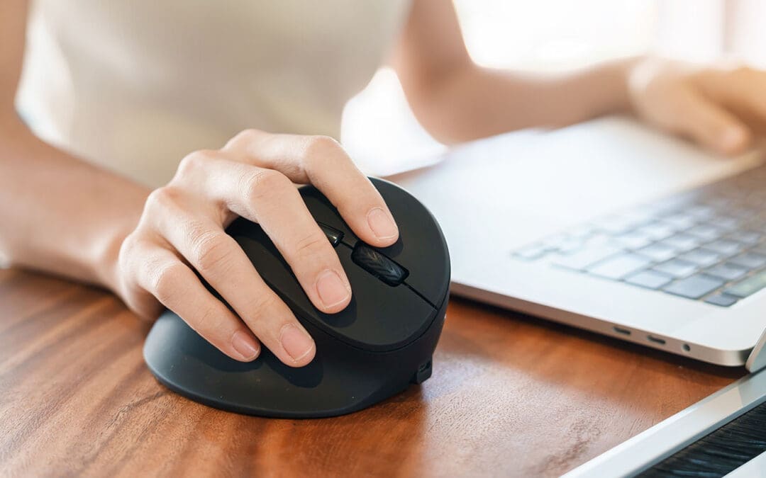
El Paso Back Clinic’s Guide to Ergonomic Mice for Pain-Free Hands
Spending hours at a computer can strain your hands, wrists, and arms, especially after injuries from accidents or repetitive tasks. At El Paso Back Clinic in El Paso, TX, led by Dr. Alex Jimenez, DC, APRN, FNP-BC, we specialize in providing holistic solutions to help patients overcome pain. An ergonomic mouse, designed to fit your hand’s natural shape, reduces strain and helps prevent conditions like carpal tunnel syndrome and tendonitis. Paired with our chiropractic care, advanced diagnostics, and integrative therapies, it supports recovery and long-term wellness. This article explains how El Paso Back Clinic uses ergonomic tools and expert care to restore health and prevent future issues.
Why Choose an Ergonomic Mouse?
Unlike standard flat mice, an ergonomic mouse curves to match your hand, often tilting upright in a manner similar to a handshake grip. This keeps your wrist straight, easing muscle and nerve strain (Goldtouch, 2023a). At El Paso Back Clinic, we recommend these for patients with desk jobs or those recovering from accidents.
Traditional mice twist your forearm, pinching nerves. Ergonomic designs hold your arm neutrally, reducing fatigue (Logitech, n.d.). For example, Logitech’s MX Vertical tilts at 57 degrees, cutting wrist tension (Logitech, n.d.). Our patients report less pain after switching, helping them work or recover comfortably.
Pick a mouse with thumb rests or adjustable angles to suit your hand. Our clinic guides you to the best choice for your needs (ProtoArc, 2023).
Supporting Natural Posture for Comfort
Your hand’s position affects your entire arm. Regular mice force your wrist to bend inward, stressing bones and nerves (ZDNet, 2023). An ergonomic mouse reduces this twist, called pronation, keeping your hand in a relaxed position (Goldtouch, 2023a).
Studies show these mice cut muscle effort by up to four times (Logitech, n.d.). They also help ease shoulder and neck tension, which is crucial for those recovering from injuries (Kosak Chiropractic, n.d.). At El Paso Back Clinic, we have seen patients benefit from this switch, especially those who have experienced motor vehicle accidents (MVAs) or repetitive strain injuries.
Reducing Repetitive Strain Injuries
Repetitive strain injuries (RSI) from constant clicking cause tingling, numbness, or pain (EffyDesk, 2023). Ergonomic mice minimize hand movements, featuring curves that allow fingers to rest naturally (Goldtouch, 2023b).
Thumb rests stop over-gripping, and lightweight designs make moving easier (ProtoArc, 2023). Our patients, from office workers to MVA survivors, use these to avoid worsening injuries. This supports healing during rehabilitation.
Preventing Carpal Tunnel and Tendonitis
Carpal tunnel syndrome squeezes the wrist’s median nerve, causing tingling or a weak grip. Tendonitis inflames tendons from overuse (FlexiSpot, n.d.). Both are common in desk workers and individuals who have been in accidents. Ergonomic mice open the wrist’s tunnel, reducing pressure by up to 30% (Goldtouch, 2023a).
They also limit bends that inflame tissues (ZDNet, 2023). For tendonitis, less forearm twist eases elbow strain, preventing long-term damage (Lowery Chiropractic, n.d.). El Paso Back Clinic patients who use these mice often stop the progression of injury early.
Setting Up Your Workstation for Health
An ergonomic mouse works best with a properly set-up desk. At El Paso Back Clinic, we recommend adjusting your chair to a 90-degree elbow angle with your feet flat. Keep your mouse at elbow height to avoid reaching (Kosak Chiropractic, n.d.).
Use a keyboard tray to maintain a straight wrist position and set your monitor at eye level to prevent neck strain (Kosak Chiropractic, n.d.). Take hourly breaks—stretch wrists, roll shoulders—to boost blood flow (EffyDesk, 2023). Our team offers personalized tips to make your workspace support recovery.
El Paso Back Clinic’s Holistic Healing Approach
Our clinic blends chiropractic adjustments, acupuncture, and rehabilitation to treat pain holistically. Adjustments realign joints, easing nerve pressure and swelling (Rozenhart Chiropractic, n.d.). For wrist pain, we target hand-to-elbow alignment to relieve carpal tunnel (Lowery Chiropractic, n.d.).
We utilize integrative therapies, such as ultrasound to warm tissues and electrical stimulation to calm nerves (Lowery Chiropractic, n.d.). Nutrition counseling helps reduce inflammation, thereby aiding recovery (Evolve Chiropractic, n.d.). Dr. Jimenez creates custom plans to address the causes of injuries, not just their symptoms.
Dr. Alex Jimenez’s Expertise in Injury Care
Dr. Alex Jimenez, a chiropractor and nurse practitioner, leads El Paso Back Clinic with dual expertise. He treats work, sports, personal, and MVA injuries using advanced neuromusculoskeletal imaging and dual-scope diagnosis to pinpoint issues like nerve compression (Jimenez, n.d.a).
For MVAs, he links whiplash to arm pain, using scans to guide treatment (Jimenez, n.d.b). Care includes adjustments, exercises, and massage to restore function. Acupuncture boosts natural healing (Evolve Chiropractic, n.d.). We also manage legal documentation for injury claims, easing patient stress (Jimenez, n.d.a).
A recent patient, following a motor vehicle accident (MVA), utilized an ergonomic mouse and our care plan. Pain dropped 70% in weeks, avoiding surgery (Jimenez, n.d.b). Dr. Jimenez focuses on natural healing over medication.
Targeted Therapies for Lasting Relief
We pair ergonomic tools with rehab. Grip exercises strengthen the hands, while wrist stretches build flexibility (EffyDesk, 2023). Acupuncture targets specific pain points, and massage helps loosen muscles (Rozenhart Chiropractic, n.d.).
Dr. Jimenez utilizes electro-acupuncture for nerve recovery, which has been shown to be effective for chronic pain (Jimenez, n.d.a). Patients track their progress with pain logs to achieve steady improvement. Our El Paso clinic provides these therapies for seamless care.
Success Stories at El Paso Back Clinic
Anna, a receptionist, switched to an ergonomic mouse and received our adjustments. Her wrist pain faded in weeks, improving her work (Goldtouch, 2023a). Carlos, an MVA survivor, worked with Dr. Jimenez. Adjustments and exercises restored his arm strength (Jimenez, n.d.b).
These stories show our approach delivers. Small changes, combined with expert care, transform lives.
Building a Pain-Free Future
Start with an ergonomic mouse and a tuned workspace. Experience the benefits of our chiropractic care, acupuncture, and nutrition for lasting health. Short walks and breathing exercises boost recovery (Evolve Chiropractic, n.d.).
Visit El Paso Back Clinic for a custom plan. Dr. Jimenez’s team treats all injuries naturally, from desk strain to MVAs (Jimenez, n.d.a). Act early to stay pain-free.
Conclusion: Heal with El Paso Back Clinic
An ergonomic mouse supports natural hand posture, cutting strain. Paired with our chiropractic and integrative care, it helps prevent and manage issues such as carpal tunnel syndrome. Dr. Jimenez’s expertise ensures effective recovery. Call +1 (915) 850-0900 to start your pain-free journey today.
Strumming Without Pain: Chiropractic Solutions for Guitarists and Bassists at El Paso Back Clinic
Playing guitar or bass fills life with rhythm and joy. The thrill of strumming chords or plucking deep notes creates unforgettable moments. But for many string players in El Paso, Texas, this passion can lead to pain. Hours of practice can strain hands, wrists, forearms, elbows, and shoulders, leading to repetitive strain injuries (RSIs) such as tendonitis. These injuries bring swelling, stiffness, and aches that make playing tough. At El Paso Back Clinic, led by Dr. Alexander Jimenez, DC, APRN, FNP-BC, we offer integrative chiropractic care to tackle these issues, helping musicians heal naturally and keep the music alive.
This article explains why guitarists and bassists are prone to RSIs, how tendonitis affects key areas, and how our clinic’s holistic approach—combining chiropractic adjustments, massage, acupuncture, and nutrition—restores health. With insights from Dr. Jimenez’s 30+ years of expertise, we’ll show how El Paso Back Clinic helps local musicians recover from injuries and prevent future pain, so they can strum and pluck without worry.
Why String Players Face Repetitive Strain Injuries
Guitarists and bassists repeat the same motions for hours: fretting chords, strumming strings, or plucking heavy bass lines. These actions stress tendons—the tough bands connecting muscles to bones. Over time, small tears form, which can lead to inflammation or tendonitis. Unlike a one-time injury, RSIs develop gradually from overuse, making them common among musicians (Pianucci et al., 2021).
The fretting hand curls tightly to press strings, while the strumming or plucking arm moves fast. Bassists face extra strain from thicker strings that need more force. Poor posture, like slouching over a guitar, adds pressure to the shoulders and neck. Heavy instruments—guitars at 7-10 pounds and basses up to 12—strain the body more during gigs (Pain Free NY, n.d.). Cold El Paso nights or long jam sessions at local venues like Lowbrow Palace can worsen symptoms by stiffening muscles.
Other factors increase risks. Older players over 40 have less flexible tendons (Bend Total Body Chiropractic, n.d.). Poor habits, such as gripping picks too hard or skipping warm-ups, can speed up strain. Diet matters too—sugary or fatty foods fuel inflammation, slowing recovery (Healthline, 2022). El Paso’s active music scene, with frequent gigs and rehearsals, means local players often push their limits, increasing the risk of RSI.
Where It Hurts: Tendonitis in Musicians’ Bodies
Tendonitis hits specific spots based on how guitarists and bassists play. Here’s where pain strikes:
Hands and Fingers: Fretting chords strains finger tendons, especially at the thumb base. Thumb tendonitis (De Quervain’s) causes sharp pain when gripping the neck. Swelling or a gritty feel signals trouble (Guitar Strength Project, n.d.).
Wrists: Strumming and plucking bend wrists repeatedly, inflaming tendons on top (extensor) or below (flexor). Stiffness after waking or a weak grip are signs. Carpal tunnel syndrome may add tingling or numbness (Rawlogy, n.d.).
Forearms: Constant flexing causes the forearm muscles to burn. Redness, warmth, or lumps show tendonitis. Bassists feel it more from forceful plucks (Healthline, 2022).
Elbows: “Guitar elbow” mimics tennis elbow, with pain on the outer elbow from strained tendons. Inner elbow pain (golfer’s elbow) also hits. Both weaken grip, making it hard to hold picks or instruments (Tennis Elbow Classroom, n.d.).
Shoulders: Holding arms out for chords strains the rotator cuff tendons, causing aches that spread down the arm. Slouching worsens it (Smithsonian Folkways, n.d.).
These areas link up. Hand pain can trigger elbow issues, and shoulder misalignment can strain wrists. Catching early signs—such as soreness or fatigue—prevents more severe problems.
Symptoms That Stop the Show
Tendonitis symptoms creep in but hit hard. Pain starts as a dull ache during play, then sharpens at rest. Swelling puffs up joints, and stiffness locks fingers, especially in the morning. Numbness or tingling buzzes in cold venues, sometimes with fingers turning blue from poor blood flow (Pain Free NY, n.d.). Weakness, drops, and fatigue, as well as burning or throbbing sensations, often linger after gigs. A grating sensation hints at the presence of scar tissue.
For El Paso musicians, long practices for gigs at Tricky Falls or house shows can exacerbate symptoms. Stress from late-night sets or cold weather can cause muscles to tighten, exacerbating pain. If symptoms last for weeks, it’s time to visit El Paso Back Clinic for expert care.
Quick Relief at Home
Before professional help, try these steps to ease tendonitis:
RICE Method: Rest by avoiding play and using splints. Ice for 15 minutes, wrapped, several times daily. Compress with elastic wraps, not too tight. Elevate arms on pillows (Mayo Clinic, 2023).
Meds: Ibuprofen reduces swelling, but ask a doctor first.
Stretches: Gentle wrist circles, finger spreads, or forearm pulls, held 15-30 seconds (Healthline, 2022).
Massage: Use massage balls to roll out knots gently (Rawlogy, n.d.).
Diet: Eat berries, fish, and leafy greens to help combat inflammation. Avoid sugary snacks common at El Paso food trucks.
These help, but don’t address the root cause. For lasting relief, see the experts at El Paso Back Clinic.
Chiropractic Care at El Paso Back Clinic
At El Paso Back Clinic, Dr. Alexander Jimenez and his team utilize chiropractic care to effectively treat RSIs. Adjustments realign joints in the wrist, elbow, or shoulder, freeing nerves and boosting blood flow. For elbow tendonitis, specific adjustments reduce pain and swelling, with patients often regaining full motion in weeks (Stamford Spine, n.d.).
Our clinic checks the whole body. A misaligned shoulder can strain wrists, so we adjust the entire chain. Soft tissue work, such as Graston therapy, breaks down scar tissue in the wrists. Laser therapy reduces inflammation, and taping supports joints during physical activity (Pinnacle Hill Chiropractic, 2024). Regular visits help keep the body aligned, reducing the risk of re-injury by up to 50% (Chiro One, n.d.).
Dr. Jimenez’s dual training as a chiropractor and nurse practitioner ensures precise diagnosis and treatment. Using advanced imaging like MRIs, we pinpoint tendon tears or nerve issues. Treatments are safe, with only mild soreness possible, and tailored to each musician’s needs (Bend Total Body Chiropractic, n.d.).
Integrative Healing for El Paso Musicians
Our integrative approach goes beyond adjustments. We combine:
Massage Therapy: Kneads forearm knots, easing tension from long sets (Beech Street Health, n.d.).
Acupuncture: Calms nerves, reducing wrist tingling for smoother playing.
Exercises: Wrist curls with light weights or finger bands build strength (Chiro One, n.d.).
Nutrition: Collagen supplements and omega-3 fatty acids accelerate tendon repair. We guide patients to local El Paso markets for healthy foods.
Ergonomics: Adjust guitar straps or use lighter picks. Take breaks every 20 minutes during practice (Smithsonian Folkways, n.d.).
This mix helps heal faster and prevents future pain, allowing musicians to stay on stage.
Dr. Jimenez’s Expertise at El Paso Back Clinic
Dr. Alexander Jimenez brings over 30 years of experience to El Paso Back Clinic. His dual-scope approach—combining chiropractic and functional medicine—targets the root causes of injuries. We provide personalized plans for musicians, workers, athletes, and individuals who have experienced accidents. Advanced tools, such as neuromusculoskeletal imaging, can reveal hidden damage, while assessments also consider lifestyle and genetics (Jimenez, n.d.a).
For a local guitarist who was injured in a car accident, we utilized adjustments, massage, and nutrition to restore their fretting ability. Our clinic also handles legal documentation for injury claims, ensuring smooth insurance processes (Jimenez, n.d.b). From whiplash to wrist strain, we help El Paso’s music community heal naturally.
Preventing Pain for Lifelong Playing
Prevention keeps musicians playing. Try these:
Exercises: Wrist stretches, towel twists, or 1-pound weight curls, 10 reps, three times weekly (Healthline, 2022).
Warm-Ups: 10-minute finger flexes and arm circles before gigs.
Technique: Use loose grips and neutral wrists. Alternate hands for songs (No Treble, 2011).
Gear: Ergonomic straps and lighter instruments ease shoulder strain.
Breaks: Rest every 20 minutes. Relax with meditation to cut stress.
El Paso Back Clinic offers tailored plans to keep your body gig-ready.
Keep the Music Playing
Tendonitis doesn’t have to silence your strings. At El Paso Back Clinic, Dr. Jimenez and our team use chiropractic and integrative care to heal RSIs and prevent pain. From hands to shoulders, we address the root causes so you can play without fear. Visit us in El Paso to get back to strumming and plucking with ease.
Contact El Paso Back Clinic at 915-850-0900 or dralexjimenez.com to schedule your consultation today.
Understanding Nerve Conditions of the Spine: Causes, Symptoms, and Treatments
The spine is a critical part of the body, serving as a highway for nerves that transmit signals between the brain and the rest of the body. When something goes wrong with these nerves—whether they’re compressed, irritated, or damaged—it can lead to a range of uncomfortable symptoms like pain, numbness, tingling, or weakness. These issues, known as nerve-related spine conditions, can affect the back, arms, or legs and stem from various causes, including injuries, degenerative conditions, or infections. In this article, we’ll explore these conditions, their symptoms, causes, and how they’re diagnosed and treated, with a special focus on integrative approaches like those used by Dr. Alexander Jimenez, a chiropractor and nurse practitioner in El Paso, Texas. We’ll also look at how chiropractic care, targeted exercises, massage therapy, acupuncture, and integrative medicine can promote healing and prevent long-term problems.
What Are Nerve-Related Spine Conditions?
Nerve-related spine conditions happen when the spinal nerves or spinal cord are compressed, irritated, or damaged. The spine is made up of bones called vertebrae, which protect the spinal cord—a bundle of nerves that carries messages to and from the brain. Between the vertebrae are intervertebral discs, which act as cushions, and small openings called foramina, where nerve roots exit the spinal cord to connect to other parts of the body. When these nerves or the spinal cord itself are affected, it can disrupt the signals, leading to symptoms like pain, numbness, tingling, or weakness (Mayo Clinic Health System, n.d.).
Some of the most common nerve-related spine conditions include:
Radiculopathy: Often referred to as a “pinched nerve,” this condition occurs when a nerve root is compressed or irritated as it exits the spine. It can cause pain, numbness, or weakness that radiates along the nerve’s path. For example, lumbar radiculopathy can lead to sciatica, a condition characterized by pain that shoots from the lower back down the leg (Cleveland Clinic, n.d.).
Spinal stenosis refers to the narrowing of the spinal canal, which puts pressure on the spinal cord or nerve roots. It’s often caused by aging or degenerative changes and can lead to symptoms like back pain, numbness, or difficulty walking (HSS Education, n.d.).
Herniated or Bulging Discs: Discs can bulge or herniate (when the inner gel-like material pushes out), pressing on nearby nerves. This can cause pain, tingling, or weakness in the arms or legs, depending on where the disc is located (Penn Medicine, n.d.).
Degenerative Conditions: Conditions like arthritis or bone spurs can narrow the spaces where nerves travel, causing compression and symptoms like pain or stiffness (Health Central, n.d.).
Trauma or Injury: Accidents, such as car crashes or falls, can damage the spine and compress nerves, leading to immediate or delayed symptoms (Verywell Health, n.d.).
Infections or Structural Abnormalities: Infections, tumors, or abnormal spine alignment (like scoliosis) can also press on nerves, causing similar symptoms (MSD Manuals, n.d.).
These conditions can range from mild annoyances to serious issues requiring immediate medical attention, especially if they cause severe symptoms like loss of bladder or bowel control, which may indicate cauda equina syndrome, a medical emergency (Verywell Health, n.d.).
Symptoms of Nerve-Related Spine Conditions
The symptoms of nerve-related spine conditions depend on where the nerve compression or damage occurs and the severity of the condition. Common symptoms include:
Pain: This can be sharp, burning, or aching and may stay in one spot (like the neck or lower back) or radiate to other areas, such as the arms, buttocks, or legs. For example, sciatica often causes burning pain that travels from the lower back to the legs (Penn Medicine, n.d.).
Numbness or Tingling: These sensations, often described as “pins and needles,” can occur in the hands, arms, feet, or legs, depending on the affected nerve (Cleveland Clinic, n.d.).
Weakness: Muscle weakness in the arms, hands, or legs can make it hard to lift objects, walk, or maintain balance. In severe cases, it can cause issues like foot drop, where a person struggles to lift their foot while walking (Johns Hopkins Medicine, n.d.).
Loss of Coordination: Compression of the spinal cord (myelopathy) can affect fine motor skills, making tasks like buttoning a shirt or writing difficult (Verywell Health, n.d.).
Balance Issues: Spinal stenosis or myelopathy can cause trouble walking or maintaining balance, sometimes described as feeling like “walking through mud” (Spine-health, n.d.).
Loss of Bladder or Bowel Control: This is a rare but serious symptom that requires immediate medical attention, as it may signal cauda equina syndrome (HSS Education, n.d.).
Symptoms can develop suddenly, like after an injury, or gradually, as with degenerative conditions like arthritis. If you experience severe or worsening symptoms, especially loss of bladder or bowel control, seek medical care right away.
Causes of Nerve-Related Spine Conditions
Nerve-related spine conditions can have many causes, ranging from natural aging to sudden injuries. Here are some of the main culprits:
Degenerative Changes: As people age, the spine can undergo wear and tear. Osteoarthritis can cause bone spurs, and degenerative disc disease can lead to bulging or herniated discs, both of which can press on nerves (Mayo Clinic Health System, n.d.).
Herniated or Bulging Discs: When a disc’s inner material bulges or herniates, it can push against nearby nerves, causing pain or numbness. This is a common cause of radiculopathy, including sciatica (Penn Medicine, n.d.).
Spinal Stenosis: The spinal canal can narrow due to thickened ligaments, bone spurs, or other changes, putting pressure on the spinal cord or nerve roots (Cleveland Clinic, n.d.).
Trauma: Car accidents, sports injuries, or falls can fracture vertebrae, dislocate joints, or cause swelling that compresses nerves, leading to severe consequences. For example, a car crash can lead to whiplash, which may cause nerve damage in the neck (Solomon Law, n.d.).
Infections: Spinal infections, like abscesses, can press on the spinal cord or nerves, causing pain and neurological symptoms (MSD Manuals, n.d.).
Structural Abnormalities: Conditions like scoliosis (abnormal spine curvature) or tumors can compress nerves, leading to symptoms like pain or weakness (Johns Hopkins Medicine, n.d.).
Inflammatory or Autoimmune Conditions: Diseases like rheumatoid arthritis can cause inflammation that compresses nerves, contributing to symptoms (OrthoTOC, n.d.).
Each cause can lead to different symptoms and requires specific diagnostic and treatment approaches to address the root issue.
Diagnosing Nerve-Related Spine Conditions
Diagnosing nerve-related spine conditions starts with a doctor asking about your symptoms and medical history, followed by a physical exam to check for numbness, weakness, reflexes, and posture. Depending on the findings, additional tests may be needed to pinpoint the cause (Penn Medicine, n.d.). Common diagnostic tools include:
Imaging tests, such as X-rays, CT scans, or MRIs, can reveal the spine’s structure, including bones, discs, and nerves, to identify compression or damage (Spine Info, n.d.).
Nerve Conduction Studies (NCS) and Electromyography (EMG): These tests assess the function of nerves and muscles, and can help confirm nerve damage (Spine Info, n.d.).
Myelogram: A special X-ray or CT scan with contrast dye can highlight pressure on the spinal cord or nerves (Spine Info, n.d.).
Dr. Alexander Jimenez, a chiropractor and nurse practitioner in El Paso, Texas, uses a dual-scope approach to diagnosis, combining his expertise in chiropractic care and advanced nursing. His clinic utilizes advanced neuromusculoskeletal imaging techniques, such as MRIs and CT scans, to obtain a clear picture of the spine’s condition. Dr. Jimenez correlates patient injuries—whether from work, sports, car accidents, or personal incidents—with clinical findings to create a precise diagnosis. This approach ensures that the treatment plan targets the specific cause of the nerve issue, whether it’s a herniated disc, spinal stenosis, or trauma-related damage (Jimenez, n.d.).
Treatment Options for Nerve-Related Spine Conditions
Treatment for nerve-related spine conditions depends on the cause, severity, and symptoms. Most doctors start with conservative (non-surgical) treatments, moving to surgery only if needed. Here’s an overview of common treatments:
Non-Surgical Treatments
Medications: Over-the-counter pain relievers, such as ibuprofen, or prescription medications, like gabapentin, can help manage pain and inflammation (Spine Info, n.d.).
Physical Therapy: Targeted exercises can strengthen muscles, improve posture, and reduce pressure on nerves. Physical therapy is often effective for radiculopathy and spinal stenosis (Cleveland Clinic, n.d.).
Epidural Steroid Injections: These deliver anti-inflammatory medication directly to the affected nerve root, reducing pain and swelling (Penn Medicine, n.d.).
Chiropractic Care: Adjustments and manipulations can realign the spine, relieving pressure on nerves. Dr. Jimenez’s clinic utilizes chiropractic techniques to treat conditions such as sciatica and herniated discs, with a focus on restoring spinal alignment (Jimenez, n.d.).
Massage Therapy: This can relax tight muscles, improve blood flow, and reduce nerve irritation, especially for conditions caused by muscle tension or spasms (Inova, n.d.).
Acupuncture: By stimulating specific points, acupuncture can reduce pain and promote natural healing, often used alongside other treatments (Total Spine Ortho, n.d.).
Activity Modification: Avoiding activities that worsen symptoms, like heavy lifting, can help the spine heal (Penn Medicine, n.d.).
Surgical Treatments
If conservative treatments are not effective, surgery may be necessary. Common procedures include:
Laminectomy: Removes part of a vertebra to create more space for nerves, often used for spinal stenosis (Spine Info, n.d.).
Microdiscectomy: Removes part of a herniated disc that’s pressing on a nerve, commonly used for radiculopathy (Spine Info, n.d.).
Spinal Fusion: Fuses vertebrae together to stabilize the spine, used for severe degenerative conditions or trauma (Inova, n.d.).
Dr. Jimenez’s clinic takes an integrative approach, combining chiropractic care with targeted exercises, massage therapy, and acupuncture to treat nerve-related spine conditions. For example, a patient with sciatica resulting from a herniated disc may receive spinal adjustments to realign the spine, exercises to strengthen core muscles, and acupuncture to alleviate pain. This holistic approach addresses the root cause while promoting long-term healing and preventing future problems (Jimenez, n.d.).
Dr. Alexander Jimenez’s Integrative Approach in El Paso
Dr. Alexander Jimenez, a chiropractor and nurse practitioner in El Paso, Texas, has extensive experience treating nerve-related spine conditions caused by work, sports, personal, or motor vehicle accident injuries. His clinic uses a dual-scope approach, blending chiropractic expertise with advanced medical knowledge to provide comprehensive care. Here’s how his clinic handles these cases:
Treating Different Types of Injuries
Work Injuries: Repetitive motions or heavy lifting at work can lead to conditions like herniated discs or radiculopathy. Dr. Jimenez uses spinal adjustments, targeted exercises, and ergonomic advice to relieve nerve compression and prevent recurrence (Jimenez, n.d.).
Sports Injuries: Athletes may suffer nerve compression from trauma or overuse. The clinic employs chiropractic care, physical therapy, and massage to restore function and reduce pain, helping athletes return to their activities (Jimenez, n.d.).
Personal Injuries: Falls or other accidents can cause nerve damage. Dr. Jimenez’s team uses advanced imaging to assess the injury and creates personalized treatment plans, often including acupuncture and exercise (Jimenez, n.d.).
Motor Vehicle Accident (MVA) Injuries: Car crashes can cause whiplash or other trauma that compresses nerves. The clinic provides detailed diagnostic assessments, including MRIs, to identify nerve damage and offers treatments like spinal adjustments and massage to promote healing (Solomon Law, n.d.; Jimenez, n.d.).
Medical Care and Legal Documentation
Dr. Jimenez’s clinic is skilled in handling the medical and legal aspects of injury cases, especially for MVAs. They provide thorough documentation of injuries, diagnoses, and treatments, which is critical for insurance claims or legal cases. For example, if a patient has radiculopathy from a car accident, the clinic documents the injury’s impact on their daily life, the diagnostic findings (like MRI results), and the treatment plan. This detailed paperwork supports patients in legal proceedings while ensuring they receive proper medical care (Jimenez, n.d.).
Integrative Medicine for Healing and Prevention
Dr. Jimenez’s approach emphasizes integrative medicine, combining chiropractic care with other therapies to address the cause of nerve issues and enhance overall health. For instance:
Chiropractic Adjustments: Realign the spine to relieve nerve pressure, effective for conditions like sciatica or herniated discs.
Targeted Exercises: Strengthen muscles around the spine to improve stability and prevent future injuries.
Massage Therapy: Reduces muscle tension and improves circulation, aiding in nerve healing.
Acupuncture: Stimulates natural pain relief and promotes recovery, especially for chronic pain.
Lifestyle Changes: Advice on posture, ergonomics, and nutrition helps prevent long-term problems (Jimenez, n.d.).
This integrative approach not only treats the immediate symptoms but also focuses on long-term health, reducing the risk of chronic pain or recurring issues.
How Integrative Medicine Promotes Healing
Integrative medicine, as practiced by Dr. Jimenez, combines conventional medical treatments with complementary therapies to address the whole person, not just the symptoms. For nerve-related spine conditions, this approach offers several benefits:
Natural Healing: Chiropractic care and acupuncture stimulate the body’s natural healing processes, reducing reliance on medications (Total Spine Ortho, n.d.).
Pain Reduction: Therapies such as massage and acupuncture can help reduce pain levels, thereby improving quality of life (Inova, n.d.).
Improved Function: Exercises and adjustments restore mobility and strength, helping patients return to normal activities (Cleveland Clinic, n.d.).
Prevention: By addressing underlying causes, like poor posture or weak muscles, integrative medicine reduces the risk of future nerve problems (Jimenez, n.d.).
For example, a patient with spinal stenosis might receive adjustments to improve spinal alignment, exercises to strengthen their core, and massage to relax tight muscles. Over time, these treatments can reduce nerve compression, improve mobility, and prevent the condition from worsening.
Preventing Long-Term Problems
Preventing long-term nerve-related spine issues involves addressing the root causes and maintaining spinal health. Here are some strategies:
Maintain Good Posture: Proper posture reduces strain on the spine and nerves (Mayo Clinic Health System, n.d.).
Stay Active: Regular exercise, especially core-strengthening workouts, supports the spine and prevents injuries (Cleveland Clinic, n.d.).
Utilize ergonomics: Adjust workstations or lifting techniques to prevent repetitive strain (Jimenez, n.d.).
Manage Weight: Excess weight can put pressure on the spine, worsening nerve conditions (Health Central, n.d.).
Seek Early Treatment: Addressing symptoms early with chiropractic care or physical therapy can prevent conditions like radiculopathy from becoming chronic (Spine Info, n.d.).
Dr. Jimenez’s clinic emphasizes these preventive measures, educating patients on lifestyle changes to keep their spines healthy and reduce the risk of future nerve issues.
Conclusion
Nerve-related spine conditions, like radiculopathy, spinal stenosis, and herniated discs, can cause significant discomfort and disrupt daily life. These conditions stem from various causes, including degenerative changes, trauma, infections, or structural issues, and lead to symptoms like pain, numbness, tingling, and weakness. Through proper diagnosis using imaging and clinical assessments, doctors can pinpoint the cause and recommend treatments, ranging from medications and physical therapy to surgery in severe cases. Integrative approaches, like those used by Dr. Alexander Jimenez in El Paso, combine chiropractic care, targeted exercises, massage therapy, and acupuncture to treat injuries from work, sports, or accidents while promoting natural healing. By addressing the root cause and focusing on prevention, these methods can help patients recover and avoid long-term problems. If you’re experiencing symptoms of a nerve-related spine condition, consult a healthcare provider to explore your treatment options and start your journey to recovery.
MVA and Workplace Injuries: How El Paso Back Clinic Helps You Heal Better
Introduction: Healing That Goes Beyond Pain Relief
At El Paso Back Clinic, Dr. Alexander Jimenez, DC, FNP‑C, leads a caring, multidisciplinary team focused on more than just pain—for over 25 years, they’ve helped patients regain mobility, lift heavy objects, or just live comfortably again (El Paso Back Clinic About Us, n.d. (El Paso Back Clinic® • 915-850-0900, El Paso Back Clinic® • 915-850-0900)). This clinic combines chiropractic care, functional medicine, fitness training, and rehabilitation with the goal of improving your function—not just masking symptoms.
Hidden Injuries After a Car Crash: More Than You Think
Motor vehicle accidents can cause “invisible” damage. Soft tissues, spinal joints, and nerves may be injured without obvious signs. Research shows that a neck injury during an MVC significantly raises the chance of future neck pain—by more than twice, in fact ([Nolet et al., 2019] meta‑analysis (PMC)). That’s why treating car accident injuries early and thoroughly matters.
Why Work Tasks Can Trigger Pain After an MVA
Even if you’re back to work after a car wreck, lingering misalignments or muscle weakness may make your body more sensitive to strains. Whether you’re sitting at a desk, lifting boxes, or driving long hours, those lingering dysfunctions can worsen work-related pain—unless the underlying injury is addressed.
Dr. Jimenez’s All-in-One Approach
What sets El Paso Back Clinic apart is the combination of:
Chiropractic adjustments for alignment and nerve relief
Functional Medicine assessments to tackle inflammation, nutrition, and hormonal imbalances
Advanced rehabilitation training for strength and flexibility
Chiropractic Care: Resetting Your Body’s Alignment
Chiropractic adjustments at El Paso Back Clinic help restore proper spine alignment and reduce pressure on nerves and joints. Especially after auto accidents, this can rapidly improve pain levels and mobility ([Dr. Hudak, 2025] (El Paso Back Clinic® • 915-850-0900)).
Functional Medicine and Nutrition: Treating the Whole You
El Paso Back Clinic doesn’t just treat symptoms—they identify root causes. Using tools like Living Matrix assessments and nutritional profiles, Dr. Jimenez addresses systemic contributors like inflammation, metabolism, or hormonal imbalances, empowering you to recover not just physically but holistically (El Paso Back Clinic Blog, n.d. (El Paso Back Clinic® • 915-850-0900)).
Fitness, Strength Training, and Rehab Workouts
Recovery doesn’t happen sitting still. Your rehabilitation may include:
El Paso Back Clinic shares patient stories and research-backed protocols that reflect their commitment to proven, safe care (El Paso Back Clinic Videos, n.d. (El Paso Back Clinic® • 915-850-0900)). From acute auto injuries to chronic pain syndromes, the clinic offers tailored pathways back to health.
Conclusion: Return to Life, Stronger and Smarter
Auto and workplace injuries often overlap—one can increase the risk or severity of the other. At El Paso Back Clinic, Dr. Jimenez delivers integrated, functional recovery through chiropractic, fitness, functional medicine, and rehab care. It’s about easing pain—while helping you regain mobility, strength, and confidence to live fully.
IFM's Find A Practitioner tool is the largest referral network in Functional Medicine, created to help patients locate Functional Medicine practitioners anywhere in the world. IFM Certified Practitioners are listed first in the search results, given their extensive education in Functional Medicine