Accident/Collisions are seeing a rise and along with those crashes are spinal injuries. What you need to know about:
Accident-related injuries
Treatment
Recovery
Legal issues
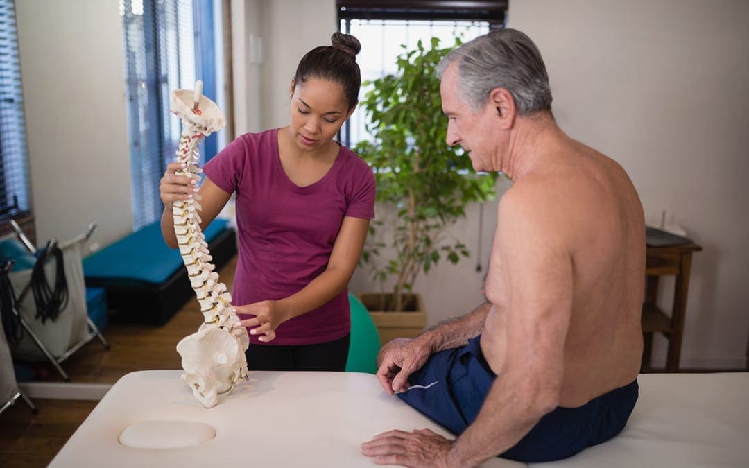
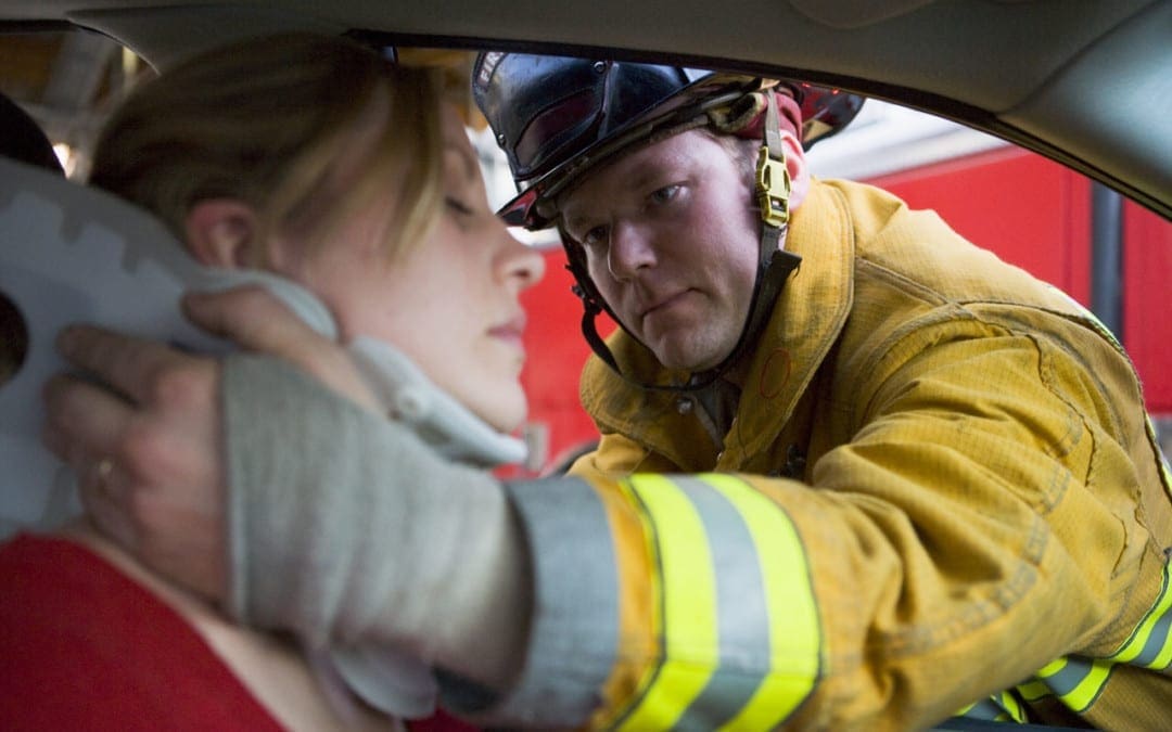
We take a risk, however small, every time we get in the car/truck, of being involved in a car wreck that results in injuries and specifically a spinal injury/s. The National Spinal Cord Injury Statistical Center has seen that motor vehicle accidents/collisions with the majority being car/truck accidents. �
�
However, with all the construction taking place nowadays, excavators, bulldozers, steamrollers, forklifts, and other vehicles are also seeing a rise in accidents. This is also generating a rise in spinal cord injury/s. Auto accidents are now ranked as the number one cause of spinal injury. The risk of an accident is small and the risk of a resulting serious spine injury is smaller still, it�s not anything. What you need to know about a motor vehicle accident/collision includes:
Injuries
Treatment
Recovery
Insurance
Legal issues
Common Motor Vehicle Accidents/Collisions
�
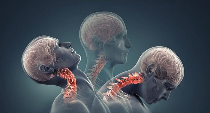
Whiplash
Whiplash is one of the most typical injuries following an accident, especially when rear-ended or a rear-end collision. Its a neck injury that happens when the neck snaps suddenly back and forth causing trauma to the tissues in the neck. Symptoms often develop a few days after the accident including:
Limited neck movement
Stiffness and pain in the neck
Pain or tenderness in the upper back, shoulders, and arms
Numbness or tingling in the arms
Dizzyness
Headaches starting at the base of the skull
Problems with concentration or memory
Ringing in the ears
Sleep issues
Depression
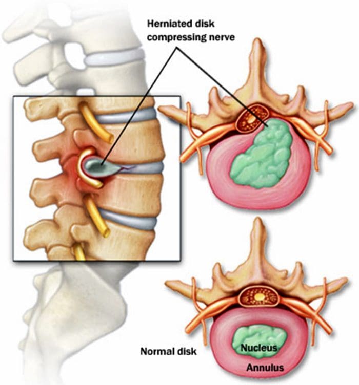
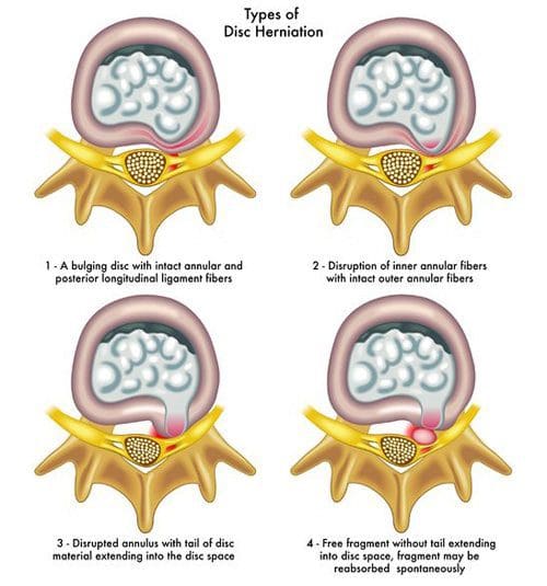
Herniated Disc
�
The discs in the spine cushion the vertebrae by absorbing the weight, force, and overall impact of a regular day. They are made of a soft, gel-like substance in the center that behaves like the gel in foot orthotics, shoes, mattresses, etc made to feel soft and comfortable. It has a tough outer membrane.
A herniated disc happens when that soft gel springs a leak/s out from a tear, meaning the shock-absorbing cushion has been compromised and is not delivering the absorption it’s supposed to and places added pressure on the surrounding nerve/s and roots. Herniations can happen naturally from age and from jobs that involve consistent and constant repetitive:
Pushing
Pulling
Bending
Twisting
Herniations also happen after going through some type of physical trauma like a motor vehicle accident/collision. Symptoms depend on where the herniation occurs and include:
Muscle weakness around the affected nerve/s
Sharp shooting pain that can spread out from the shoulders to the arms, legs and low back
Tingling in arms or legs
Numbness
There could also be no symptoms and no discovery of a herniated disc until tested for something else.
�
Vertebral Fractures
�
The vertebrae are highly susceptible to fractures of all types and can appear at any spot along the spine. For many, the injuries are mild and heal with non-surgical treatment and time. Major trauma to the spine can cause severe injuries/conditions which include:
Burst fractures
�
This is where the vertebra fractures in multiple places into bony fragments that fall into the spinal cord getting lodged inside with the jagged edges of the bones creating tears, cuts, etc that can result in paralysis and even death.
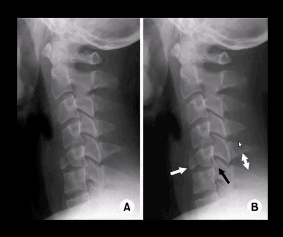
Flexion fracture
�
This is an injury seen in head-on collisions where the upper part of the body gets thrown forward and the bottom part stays in place likely from the seatbelt. This tears the vertebra apart resulting in a flexion teardrop fracture.
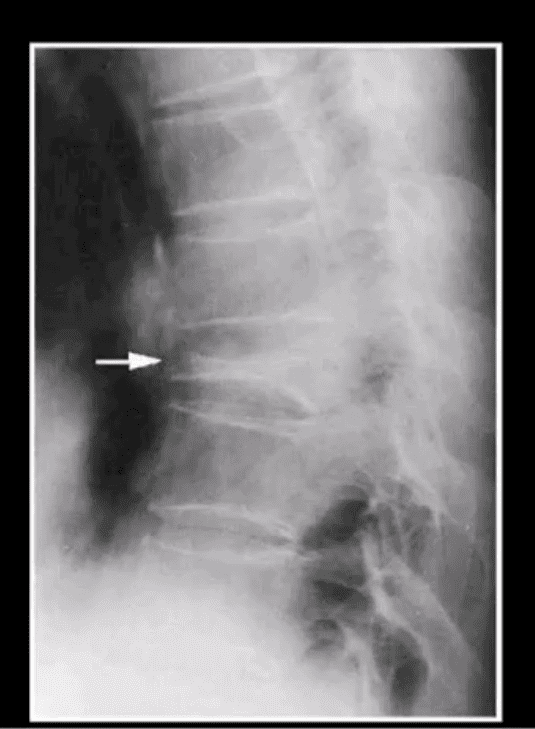
Vertebral compression
�
These types cause the front of the vertebra to collapse while the back keeps its position that forms a wedge-like shape. However, more often it is associated with osteoporosis, healthy individuals can experience a vertebral compression fracture from a serious traumatic event like an auto accident. Fractures can cause mild to severe pain that is exacerbated with movement. If the spinal cord is injured the individual could experience:
Tingling
Numbness
Weakness in the limbs
Loss of bladder/bowel function
Because of the increased safety features in today’s vehicles, fractures of the spine are rare except for severe motor vehicle accidents/collisions.
�
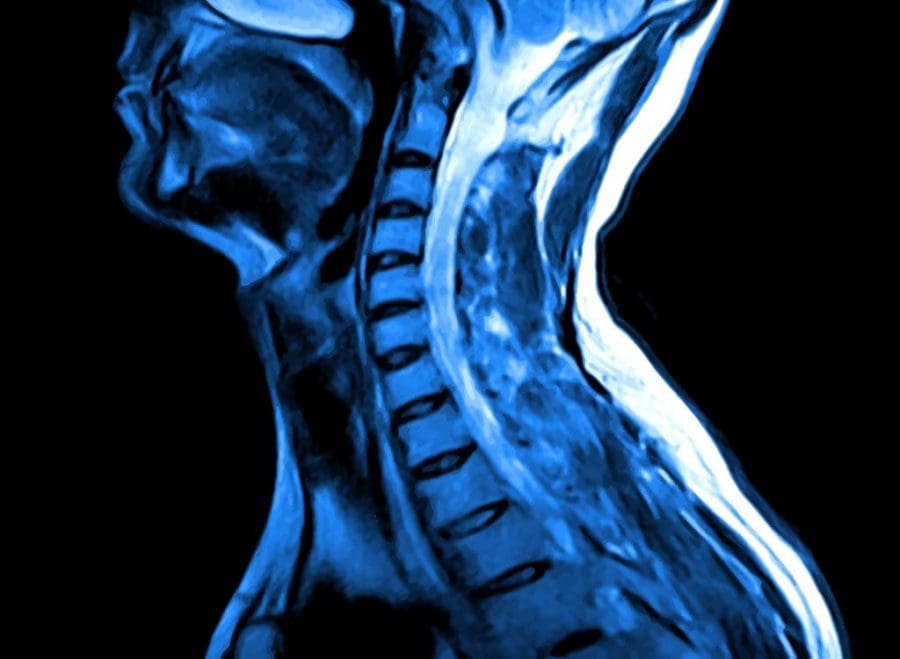
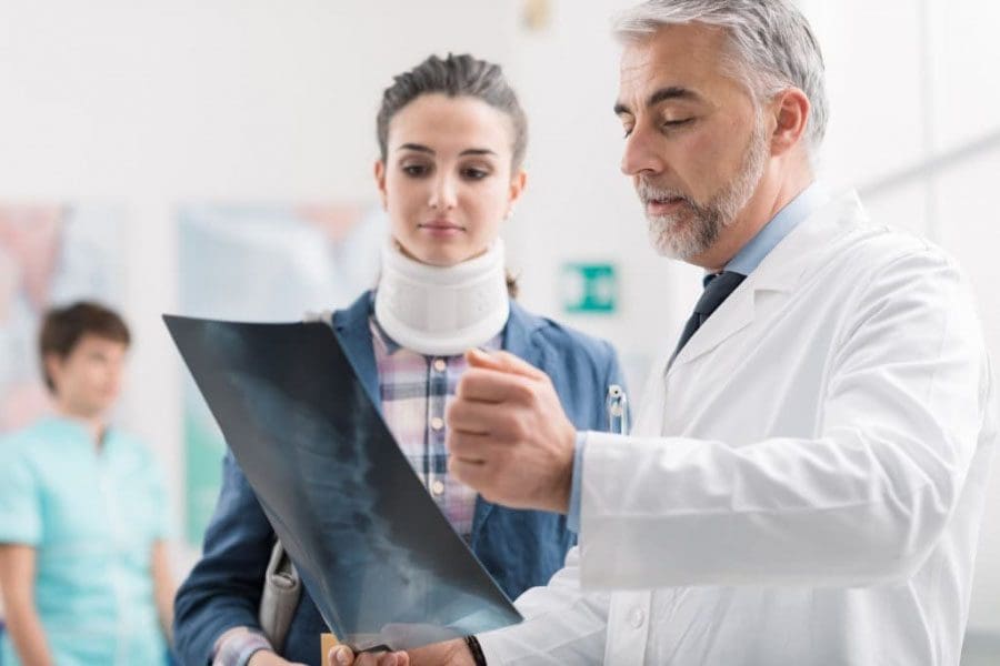
Diagnosis and treatment
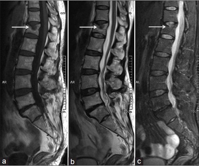
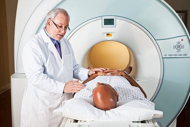
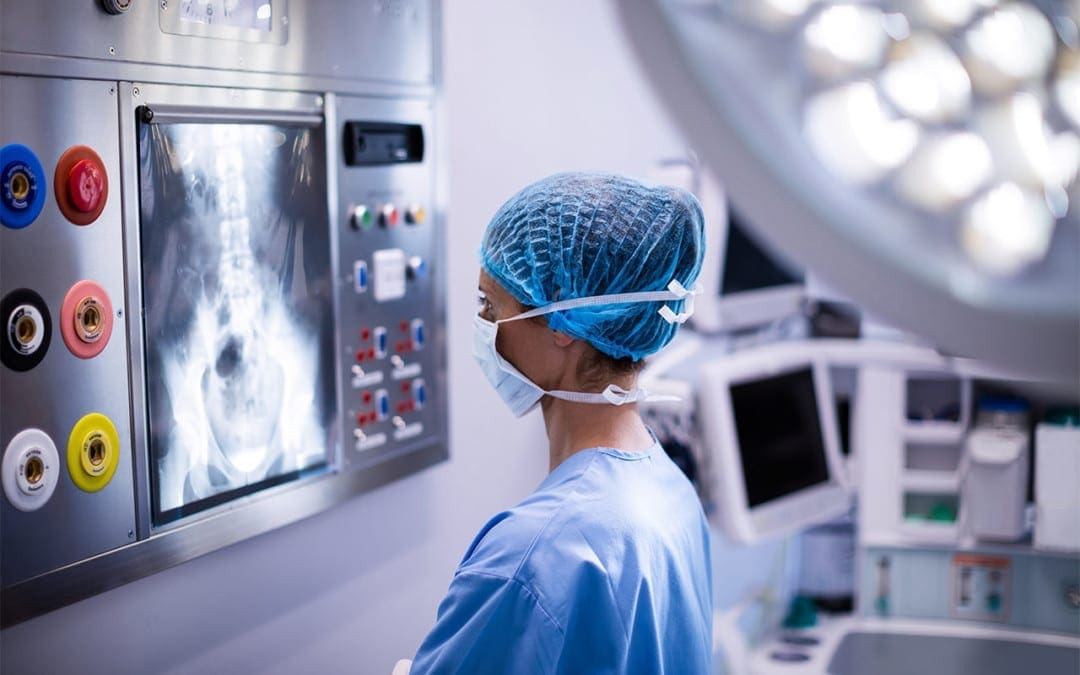
A doctor will review medical history along with the accident information. Imaging tests will follow like:
X-ray
CT or computed tomography scan
MRI or magnetic resonance imaging
�
The way these techniques of imaging are done depends on the accident and the state of the spine. Being brought into the hospital from a motor vehicle accident/collision with a suspected spinal injury means the imaging will be done first to rule out or not potentially life-threatening injury/s to the spine. Treatment for spinal injuries can range from:
Soft collar
Chiropractic
Over-the-counter anti-inflammatory medications
Corticosteroid injection/s
Nerve blocks
Physical therapy
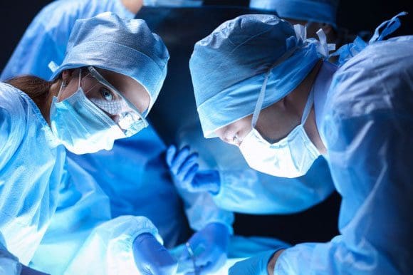
Surgery to correct certain injuries when all other forms of treatment are not working
�
Recovery
Every case, accident, and injury is different�and depends on several factors, like age, health, and how severe the accident/collision was. Severe and extreme injures like a burst fracture can take a long time to heal. �
�
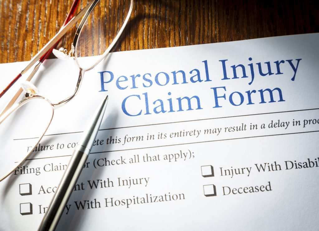
Legal Issues
Individuals with a spinal injury can face thousands in medical bills. If there is medical equipment involved along with therapeutic services for long-term care, like physical therapy then bills will accumulate.
Also, a personal injury claim could be necessary to pay for everything, especially, if the accident/collision is no fault of your own. Compensation could help with:
Loss of employment
Employment benefits
Wages lost
Ability to work/earn income
Medical expenses
Pain
Suffering
Consult with a specialist when considering filing a personal injury claim
�
Work Compensation
If a spinal injury accident happens at work there could be worker’s compensation. Workers� compensation is insurance that replaces wages and medical benefits to workers that have been injured while doing their job. These are injuries that happened during the operation of a motor vehicle, like a truck, or forklift. The worker must file an injury report immediately so there is documentation supporting the injury claim. Waiting to file can make the employer question if there even was an injury. A workers� compensation claim works differently than a personal injury claim, based primarily on what is covered under the job’s insurance policies.
An example is the legal term pain and suffering. This is not covered by workers� compensation. However, a work training accident would be covered by work comp in the event that the individual cannot return to their job/occupation after the injury. However, any injury/s after a motor vehicle accident/collision should never be taken lightly or ignored. Individuals must be proactive in their treatment after an accident/collision. This is to prevent and avoid further injury.
�
Auto Accident Doctors & Chiropractor Treatments
Dr. Alex Jimenez�s Blog Post Disclaimer
The scope of our information is limited to chiropractic, musculoskeletal, physical medicines, wellness, and sensitive health issues and/or functional medicine articles, topics, and discussions. We use functional health & wellness protocols to treat and support care for injuries or disorders of the musculoskeletal system. Our posts, topics, subjects, and insights cover clinical matters, issues, and topics that relate and support directly or indirectly our clinical scope of practice.*
Our office has made a reasonable attempt to provide supportive citations and has identified the relevant research study or studies supporting our posts. We also make copies of supporting research studies available to the board and or the public upon request. We understand that we cover matters that require an additional explanation as to how it may assist in a particular care plan or treatment protocol; therefore, to further discuss the subject matter above, please feel free to ask Dr. Alex Jimenez or contact us at 915-850-0900. The provider(s) Licensed in Texas& New Mexico*
Discitis affects around 1 out of every 100,000 people. This means that it is not a common spinal disease. Discitis can occur in adults and children, however, it is more common in children. �
�
Discitis mostly occurs in the low back region of the spine
Followed by the neck region
Finally the middle-back region
It accompanies vertebral osteomyelitis. Both types of infections share many of the same symptoms/characteristics. Although these are uncommon conditions, they can produce severe symptoms affecting an individual’s quality of life. This is why early diagnosis and treatment are essential.
�
Discitis Causes
There are two recognized causes of discitis. The rarest form comes from a prior surgical or diagnostic procedure. This usually happens when a needle or other tool/device transfers the infection. The other is the more common, and it is known as spontaneous discitis. Here the infection develops from a bacterial or viral organism that travels to the disc/s via the blood supply from another part of the body.
When an infection starts somewhere else and then travels to the disc, it is called transient bacteremia, which is bacteria in the bloodstream that has a short life. Ear infections along with skin infections are perfect examples of infections that can lead to transient bacteremia and discitis. �
After a disc becomes infected, it can be quite difficult for the body to fight the infection. The disc/s are the largest avascular organs in the body, which means they do not have their own blood supply. The discs get their nutrition and blood supply, which includes the white blood cells for fighting infections, from the vertebral endplates. Because the discs lack the resources to fight infections on their own, there is a struggle when trying to protect against infection.
Because discitis is usually caused by an infection that developed in another area of the body, individuals with medical conditions are at a higher risk for developing discitis. These conditions include:
Diabetes
A.I.D.S
Cancer
Chronic kidney disease
�
Symptoms
Intense back pain that starts gradually is the distinctive characteristic symptom of discitis. The pain is usually localized to the area where the infection is located. This means that the pain doesn’t radiate or spread out like other types of back pain conditions. �
�
Diagnosis
A doctor, spine specialist, or chiropractor will review medical history and symptoms with the individual. A fever is normally not present once the infection is inside the disc, along with the white blood cell count being normal.
However, the erythrocyte sedimentation rate increases. This is a blood test that examines how fast red blood cells fall to the bottom of a tube. The faster that they fall to the bottom, the more likely there is inflammation somewhere in the body.
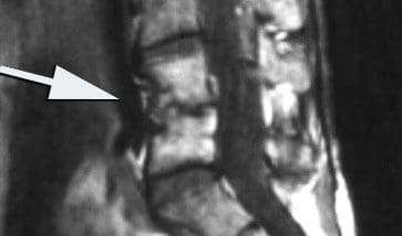
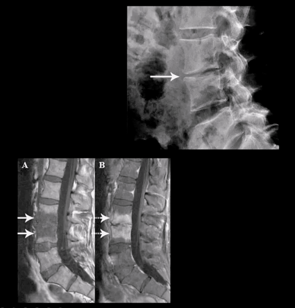
Blood tests can be utilized during diagnosis, however, the most accurate diagnostic tool to confirm discitis is magnetic resonance imaging or MRI that shows if an infection is present. �
�
Treatment
Treatment can be challenging. This is because of the fact that the discs do not have a blood supply, and medications/antibiotics travel through the blood. It is treatable and is usually done within a six to eight-week course of antibiotics intravenously or through an IV.
IV administered antibiotics could require treatment on an outpatient basis. The entire course of antibiotics must be completed in its entirety in order to manage the discitis. A doctor could also prescribe a spinal brace to help stabilize the spine and reduce pain. A brace can limit movement, however, it will help ensure proper healing.
�
Spinal Infections
Spinal infections can present spontaneously or as secondary conditions, e.g. after a surgical procedure. Spinal infections can affect different structures, like the:
Vertebral column or the bones of the spine
Intervertebral disc space, which is the cushion-gel structures between the vertebrae
� Here are some facts about the occurrence and prevalence of different infections of the spine:
Vertebral osteomyelitis is the most common type of infection. It affects an estimated 27,000 to 66,000 people a year.
Epidural abscess is an infection inside the spinal canal that affects up to two cases per 10,000 in hospital admissions around the U.S. It is pretty common in individuals with vertebral osteomyelitis or discitis. Eighteen percent of those individuals can develop this infection. However, it is more common in people fifty and older.
Discitis, as aforementioned is a pretty uncommon condition. Although, treatment has advanced, around twenty percent of individuals with this infection do not survive.
�
Infection Risk Factors
There are certain factors that increase the risk of developing an infection. These factors include:
Symptoms from a spinal infection can vary. However, continuous back pain with no history of trauma or injury. Usually, there is a delay in the diagnosis for an infection of the spine because of the:
Subtle nature of the symptoms
Individual’s belief that the pain is not serious
Absence of body-wide symptoms like a fever
Lab results can also complicate the diagnostic process, as they can be misleading. There could be normal white blood cell counts, x-rays that show no abnormalities, and a sensitive diagnostic test like a bone scan might not show that an individual is positive until a week later.
An erythrocyte sedimentation rate is a valuable screening test when it comes to spinal infections. The test can measure inflammation and infection in the body. If a spinal infection is suspected, an MRI could be the most reliable tool to confirm early diagnosis.
Health & Immunity Series
�
Dr. Alex Jimenez�s Blog Post Disclaimer
The scope of our information is limited to chiropractic, musculoskeletal, physical medicines, wellness, and sensitive health issues and/or functional medicine articles, topics, and discussions. We use functional health & wellness protocols to treat and support care for injuries or disorders of the musculoskeletal system. Our posts, topics, subjects, and insights cover clinical matters, issues, and topics that relate and support directly or indirectly our clinical scope of practice.*
Our office has made a reasonable attempt to provide supportive citations and has identified the relevant research study or studies supporting our posts. We also make copies of supporting research studies available to the board and or the public upon request. We understand that we cover matters that require an additional explanation as to how it may assist in a particular care plan or treatment protocol; therefore, to further discuss the subject matter above, please feel free to ask Dr. Alex Jimenez or contact us at�915-850-0900. The provider(s) Licensed in Texas& New Mexico
A bone graft is defined as using bone-in spine fusion surgery. Spinal fusion’s purpose is to link or weld bones together, in this case, the spinal bones. There are a variety of spinal conditions cause instability and pain:
Degenerative disc disease
Scoliosis
Trauma from an auto accident, sports injury, slip, and fall accident
Spine surgeons use a bone graft to:
Stop motion between two or more vertebrae
Stabilize a spinal deformity
Repair fractures of the spine
�
�
Spinal Fusion Stimulates New Bone Growth
A bone graft does not heal or fuse the spine instantly. Rather a bone graft sets up a foundational frame for the individual’s body to generate and grow new bone. A bone graft stimulates new bone production. It is when this new bone begins to grow and solidify, that fusion takes place.
With these types of surgeries, instrumentation like screws, and rods are typically used for the beginning stabilization. But it is the actual healing of the bone that welds the vertebrae together creating long-term stability.
A bone graft can be used for structural purposes for supporting the spine, usually this is done in place of a disc or bone that was removed. Or it can be an onlay, this means that a mass of bone fragments will grow together to stabilize the spine bridging the joint.
There are two generalized bone graft types:
Real bone
Substituted bone graft
Real bone can come from the patient, which is called an auto-graft or from a donor’s bone, called an allograft.
�
The Individual’s Bone or Auto-graft
An auto-graft is bone taken or harvested from the individual’s body and transplanted to a specific area, in this case, the spine. An auto-graft is considered the gold standard because it is the individual’s own bone, which contains:
These all help to stimulate the healing of the fusion. There are advantages for an auto-graft, which include a higher probability for fusion success and a lower risk for disease transmission. The only real setback for individuals of an auto-graft is the post-operative pain that usually comes with the procedure when harvesting an individuals’ bone. Bone can be harvested from one of the individual’s:
Iliac crests
Pelvic bones
Ribs
Spine
� Bone graft harvesting creates a new set of risks. These include:
Because of these risks and the possibility that the bone could be poor quality, a surgeon could decide to use another type of bone graft. When this happens a surgeon could go with what is known as a local auto-graft. This is bone harvested from the decompression itself.
These are the parts that are removed to decompress the nerves. They usually consist of bone spurs, lamina, and portions of the spinous process. These same bone pieces can be reused to assist with the fusion of the decompressed areas.
�
Donor Bone or Allograft
An allograft is a bone harvested from another person, usually from a tissue bank. Tissue banks harvest bone and other tissues from cadavers for medical purposes. An allograft is prepared by freezing or freeze-drying the bone or tissues. This helps limit the risk of graft rejection. Bone from an allograft does not have living bone cells and is not as effective at fusion stimulation when compared to an autograft. However, it still does work. Tissue banks:
Screen all their donors
Supervise bone recovery
Test donations
Sterilize donations
Store for use
Look for tissue banks that are accredited by the American Association of Tissue Banks. US Food and Drug Administration has strict regulations when it comes to human cell and tissue processing. These include rules about the eligibility of donors. These guidelines/protocols help reduce the risk of tissue contamination and the spread of disease.
�
Bone Graft Substitute
These substitutes are man-made or are made from a manipulated version of a natural product. These alternatives are safe and can provide a solid foundation for the individual’s body to grow bone. Substitutes have similar properties of human bone, which include a porous structure and proteins that stimulate healing.
�
Demineralized Bone Matrix – DBM
A demineralized bone matrix is an allograft that has gone through a process where the mineral content has been removed. This demineralization helps reveal bone-forming proteins like collagen, and growth factors hidden within the bone that can stimulate healing.
This procedure is often considered a bone graft extender. It is not considered a replacement. This is because its ability to fuse the human spine on its own has not been proven. DBM can be combined with the regular bone for more volume and is available in these forms:
Chip
Granule
Gel
Powder
Putty
�
Ceramic-based Extenders
Ceramic-based extenders are mixed in combination with other sources of bone. This is because they consist of calcium matrix for fusion, but there are no cells or proteins to stimulate the healing process. These include:
Ceramic-based extenders do not present a risk for disease transfer but can cause inflammation. They are available in porous and mesh forms.
�
Morphogenetic Protein – BMP
Different types of bone morphogenetic proteins or BMP’s are used to stimulate new bone growth. These proteins are found in human bone, however, they are trace amounts. They are then produced in larger amounts through genetic engineering.
This all depends on the type of spine surgery an individual undergoes. Bone morphogenetic protein could be considered an option in promoting new bone growth along with healing fusion.
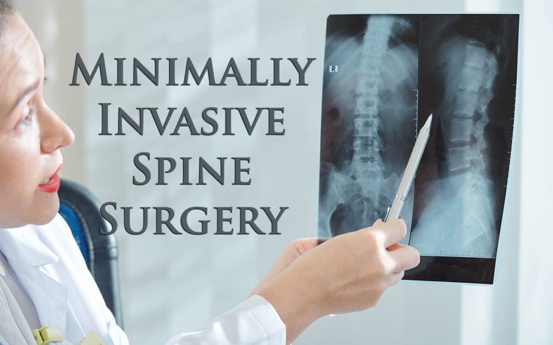
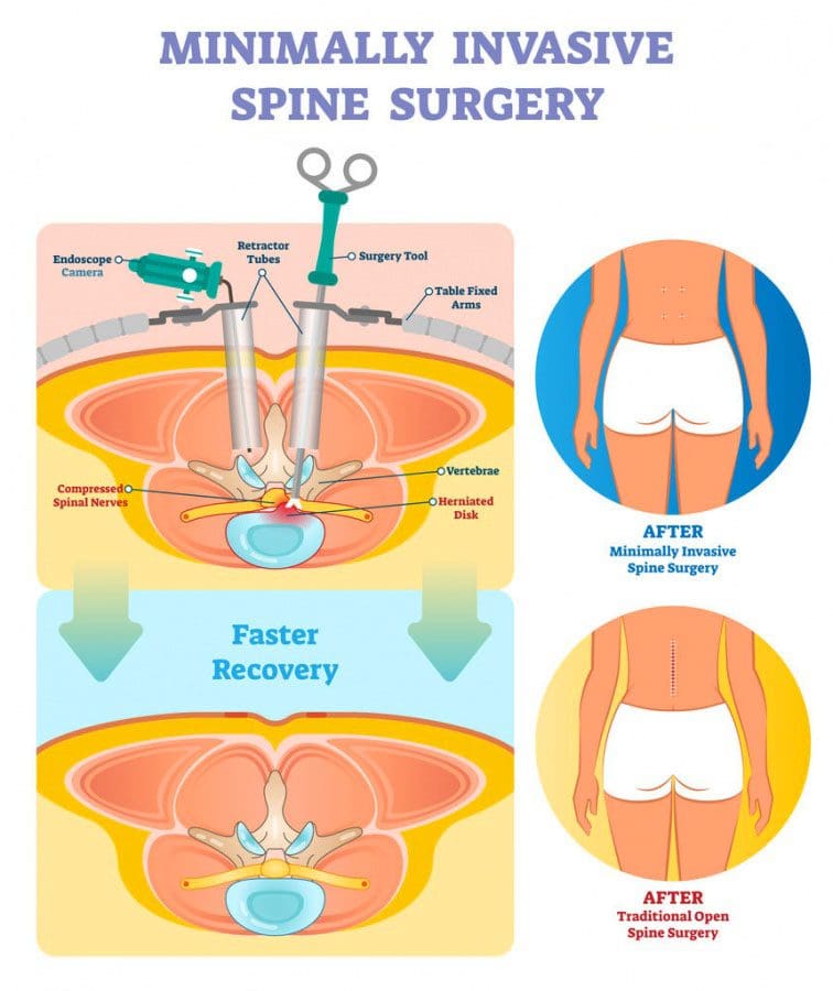
Minimally invasive spine surgery known as M.I.S.S is an option to traditional open surgical procedures, as well as an alternative when non-surgical approaches are working but the pain or condition is becoming worse, regardless. These are performed to treat a variety of spinal disorders like:
Bone spurs
Degenerative disc disease
Herniated disc
Scoliosis
Spinal instability
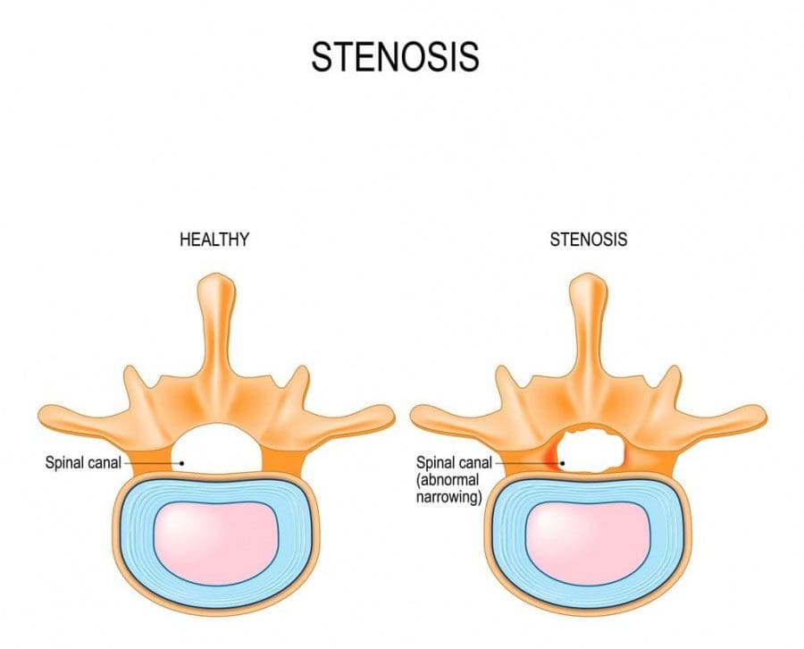
Spinal stenosis
Spinal tumors
Minimally invasive surgery can offer potential benefits. These include
A small/tiny incision/s
Minimal cutting through soft tissues like ligaments, and muscles
Outpatient option/s
Reduced post-operative pain
Quicker recovery
�
The Focus of Spine Surgery
There are two main goals when it comes to spine surgery or rather the goal/focus of the surgery. These are decompressing and stabilizing the spine.
�
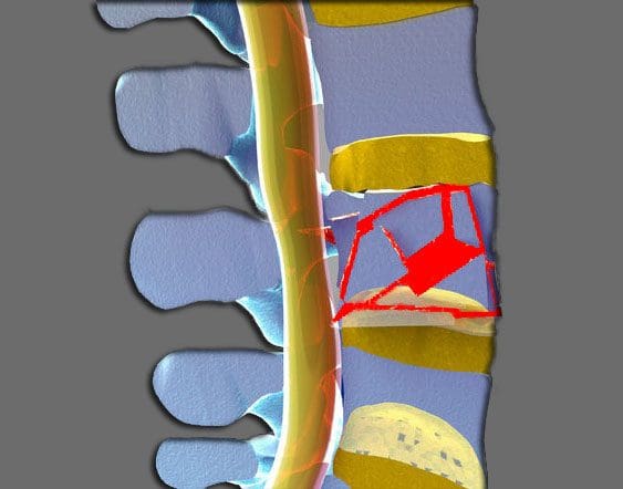
Decompression of the spine
Spinal decompression involves removing any tissue/s that are compressing/pinching the nerve structures like a spinal nerve root or the spinal cord itself. Bone spurs and fragments from a herniated disc are the types of tissue/s that can cause neural compression.
�
Stabilization of the spine
An abnormal movement of one or more levels/segments of the spinal cord can cause back pain, neck pain, or both. Surgeries that are meant to stabilize and stop these abnormal movements utilize spine instrumentation combined with fusion.
�
Spine Surgery Techniques
Minimally invasive spine surgery techniques include:
Rather than cutting through soft tissues, a tubular retraction instrument generates a tunnel that expands and passes between the muscle/s to access the spine’s column. Then an endoscope or a tiny video camera goes in and around the area, projecting a visualization of what’s happening on a monitor during the procedure.
�
This is the surgeon’s/team’s eyes as they work to repair the damage. The surgery is run through the tubular retraction system along with any specially designed instruments that are needed. Types of surgical procedures performed with minimally invasive surgery include:
Discectomy
Microdiscectomy
Foraminotomy
Microforaminotomy
Microlaminectomy
Microlaminotomy
The micro means that the surgery is done using a special microscopic camera to view the disc/s and nerve/s. Imaging scans, systems, and image-guidance technologies, like fluoroscopy, which is a real-time x-ray are utilized during the surgery pinpointing the key aspects of the patient�s spinal anatomy. The surgical imaging shows 2D and 3D views, which guides the placement of any instrumentation, like pedicle screws.
�
Disorders Treated with Minimally Invasive Surgery
�
Degenerative disc disease
Degenerative disc disease is known as DDD often develops progressively in older adults and affects the intervertebral discs. The normal wear and tear of cellular age-related changes in the body can cause the spine’s discs to:
Stiffen
Lose Flexibility
Loss of Strength
Loss of Height
Lose shape, along with the ability to absorb/distribute the forces associated with moving
These structural changes increase the risk of disc herniation and subluxations.
�
Herniated discs
A herniated disc also called a slipped, bulging, and ruptured disc. This happens when the soft gel cushion of a disc breaks through the protective outer layer. Other than the damaged disc, the loose interior gel can also irritate and inflame the nerves causing back pain. �
�
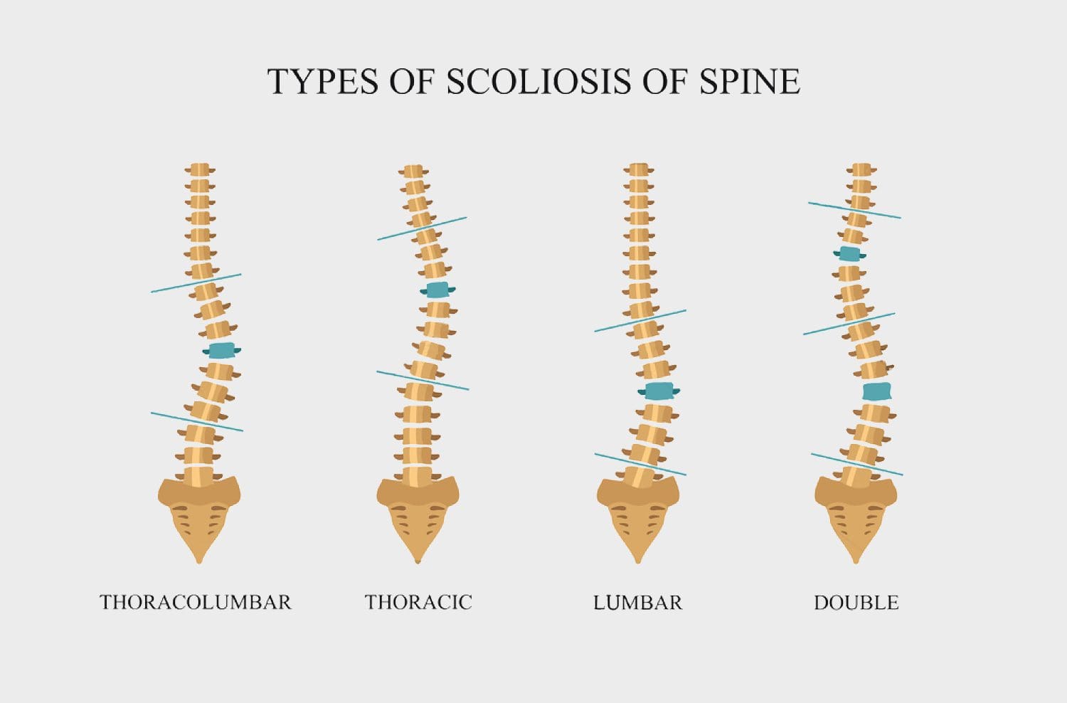
Scoliosis
Scoliosis is an abnormal sideways curve of the spine that can cause progressive spinal deformity. A scoliotic curve can look like an �S� or �C.� Most cases have no known cause, and while the condition is more commonly associated with children, adults can develop scoliosis, as well.
�
Spinal stenosis
Spinal stenosis happens when the spinal nerve roots and the spinal cord become compressed/pinched. These nerves branch off the spinal cord and exit the spinal canal through passageways called neuroforamen. Nerve and spinal cord compression can cause symptoms like:
Pain
Weakness
Tingling sensations
Numbness
Sometimes, pain can travel into the arms or legs
�
Spine Surgery Risks
With any spine surgery there are potential risks and complications that can occur. Here are some possible complications that can happen during and after surgery, with both open and minimally invasive procedures.
Minimally invasive spine surgery does offer many benefits:
Tiny incision
Less pain
Reduced risk
Faster recovery
Let’s not forget that M.I.S.S is still surgery. Less than 5% of people with back or neck pain need spine surgery and, surgery is the last resort for treating pain and symptoms caused by a spinal condition/disorder.
It is only when non-surgical treatments like chiropractic, acupuncture, physical therapy, medication, or spinal injections do not reduce symptoms in 3 to 6 months. This is when you qualify to be a candidate for spine surgery. There are certain types of spinal disorders that require urgent or immediate surgical intervention.
Talk with your doctor, chiropractor, or spine specialist about the pain, the symptoms, and compare the results of the different therapies/treatments and go from there. With any type of surgery there are many considerations to discuss before making a decision to treat back or neck pain and if minimally invasive surgery could be an option.
Staying at home means it can be tough to see a doctor, chiropractor, spine specialist, or neurosurgeon to handle back pain, especially when it tends to flare up at the most inconvenient times. There are still options, here�s what to do. What options are available when you want to see a doctor about back pain, but getting to the clinic can be a challenge.
Fortunately, there are a variety of tools to handle back pain that can provide some relief.
Over-the-counter pain medications like Motrin are one of the best medicines for non-traumatic back pain inflammation.
�
Heat Packs/Heat Therapy
Heat therapy promotes vasodilation and draws nutrient-rich blood into the targeted tissues. Increased blood flow delivers oxygen and nutrients and cell waste is removed. The warmth decreases muscle spasms, relaxes tense muscles, relieves pain, and increases range of motion.
Superficial heat is available in different forms, which include:
Hot and moist compresses
Dry or moist heating pads
Hydrotherapy
Commercial chemical/gel packs
Remember heat packs in any form should be wrapped in a towel to prevent burns, as a punctured heat pack should be discarded, as the chemical agent/gel can burn skin. �
�
Cold Packs/Cold Therapy
Cold therapy produces vasoconstriction. This slows blood circulation, which reduces inflammation, muscle spasms, and pain. Superficial cold is also available in different forms, which include:
Commercial cold packs
Ice cubes
Iced towels/compresses
Hydrotherapy.
The application of cold therapy is usually less than 15 minutes, as the effects of cold are known to last longer than heat. Cold packs or ice should never be applied directly to the skin.
A towel, should be placed between the cold object and the skin surface to prevent any skin and nerve damage. A punctured cold pack should be discarded, as the chemical agent/gel will also burn the skin.
�
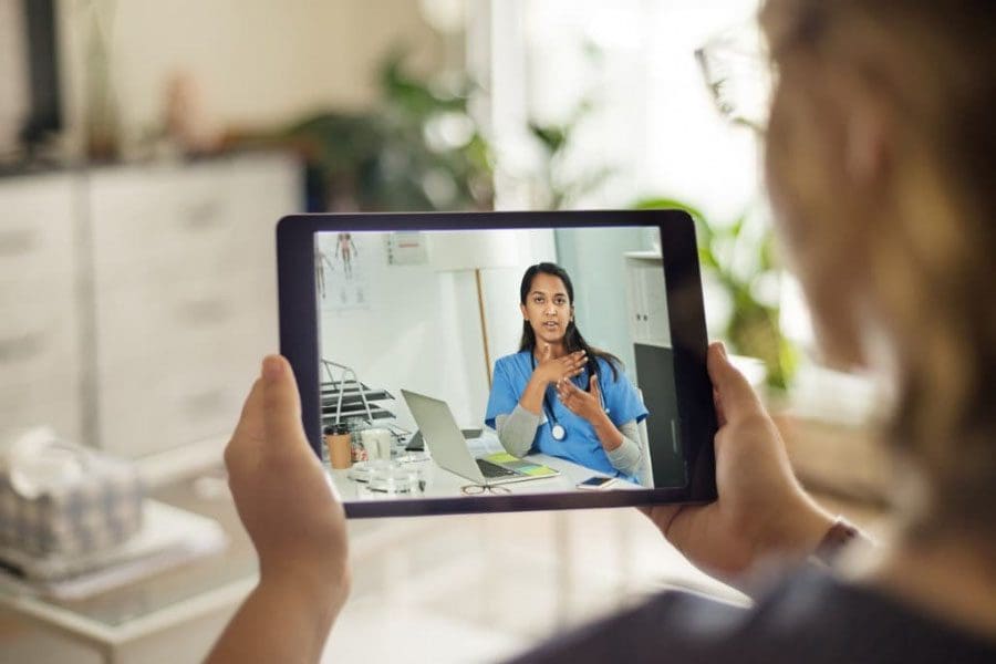
Telemedicine
It might be hard to believe that a virtual video visit can work to handle back pain. On a video call, a chiropractor is unable to physically palpate the sore areas and measure the range of motion and strength. However, this should not discourage you from scheduling a virtual appointment.
Telemedicine, without a physical examination, can be highly beneficial. A chiropractor can start the process of ordering tests, like MRI, X-ray, etc. Even if the pain is tolerable, meaning the kind that doesn�t need medicine or imaging tests, this should not be an excuse to skip an orthopedic visit.
With telemedicine, a chiropractor can still give advice, show back stretches, exercises, order back pain supplements, and talk about the risks and benefits of treatments available to try on your own. �
�
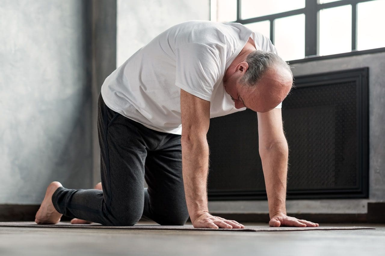
Physical Therapy
With chronic low back pain, chances are your chiropractor suggested physical therapy or PT. Now is the time to bring back those PT exercises, especially with a back-pain flare-up.
Low-back pain or any back pain for that matter with no neurological issues, could mean that a stretching and exercise program is all that is needed. Find out if your chiropractor or a physical therapist offers other options:
Patient portal communication or e-visits.
Uploads of illustrated handouts describing how to do various stretches and exercises.
Remote evaluation. The individual submits pictures or a video of their movements for personalized feedback, which the chiropractor or physical therapist evaluates and provides.
�
Get Active
Evidence shows that being active is better than resting. Moving increases the blood flow to the muscles, which helps with muscle spasms, trigger points, tense muscles/ligaments, and other issues.
�
Pilates
Pilates focuses on controlled movement, breathing, and stretching. A review found Pilates can be a highly effective and beneficial approach to handle back pain and related discomfort. Check out beginner Pilates videos. Be sure to avoid any move/s that cause pain, worsens the existing pain, or generate new pain.
�
Yoga
A review found that yoga can help improve mobility and decrease pain. If this is a new practice, start with gentle yoga or restorative yoga.
�
Walking
Going for a walk is easy, accessible, and is beneficial for the spine. Walking can be as effective as non-drug interventions in decreasing pain and discomfort in chronic low-back pain. Simple movements along with rollers and massagers can handle back pain as well. These include:
Self-massage with a tennis ball
Foam rolling
Hand-held massager
Stretching
McKenzie Method, comprised of gentle stretching exercises
These strategies and approaches can become the methods and techniques for the relief of existing back-pain in the absence of a doctor, chiropractor, or physical therapist.
PODCAST: Ryan Welage and Alexander Jimenez, both medical students at the National University of Health Sciences, discuss the several new approaches that they developed in order to help people continue to engage and participate in exercise from the comfort of their own homes. Using their advanced understanding of functional medicine, biomechanics, and nutrition, they undertake explaining simple methods and techniques for complex movement protocols. Moreover, Alexander Jimenez and Ryan Welage discuss how diet can be an essential element in overall health and wellness. Dr. Alex Jimenez offers additional guidelines with the Functional Fitness Fellas, among further advice. – Podcast Insight
If you have enjoyed this video and/or we have helped you in any way
please feel free to subscribe and share us.
Thank You & God Bless.
Dr. Alex Jimenez RN, DC, MSACP, CCST
IFM's Find A Practitioner tool is the largest referral network in Functional Medicine, created to help patients locate Functional Medicine practitioners anywhere in the world. IFM Certified Practitioners are listed first in the search results, given their extensive education in Functional Medicine
 �
�