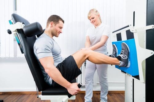
Blood Flow Restriction training (BFR) is a style of resistance training that utilizes the custom of wrapping a kind of tourniquet around a limb and training with a relatively light load. It is a practice that has gained quite a bit of popularity in the resistance coaching realm over the last few decades and is something which can benefit training protocols.
If used properly, practical blood flow restriction training (BFR) could help you through hypertrophy plateaus, pack on additional mass and even aid in growth or maintenance of muscle mass during times in which lifting heavy weight is either laborious or impossible. Let’s understand what’s actually going on in the body when it is used by you.
As mentioned prior, BFR demands using some form of tourniquet around a limb so as to inhibit blood flow. However, not all of blood flow is restricted. The purpose of the tourniquet is to prevent what’s known as ‘venous return’ . When you contract a muscle, more blood than ordinary is shuttled to provide the muscle with a myriad of different nutrients, such as oxygen. Typically, if un-wrapped, the blood then returns to the heart through veins so as to rid the muscle of metabolic bi-products like carbon dioxide, lactate, and hydrogen ions (the acidity that makes your muscle “burn off”).
The role of using some form of tourniquet is to inhibit the ‘venous return’ of blood to the heart while still allowing arterial blood circulation to the muscle. By doing this, the blood continues to be shuttled to the muscle and pools without having the ability to escape. It’s believed that the accumulation of blood and bi-products contributes to activation of fast-twitch muscle fibers, which is typically thought to only happen after these are fatigues or due to using fairly heavy loads. By doing this, you increase the potential for the muscle t.
In fact, a recent analysis by suggested that when participants used the same load (40% of 1 RM) and either used a tourniquet or didn’t, the team using BFR observed the same gains in strength and muscle volume as the group that did not. The catch: the BFR team had finished significantly less repeats, and thus less quantity, in addition to less time under pressure. This implies the exact same advantage was observed by them, but achieved in time.
The research appears to indicate that you could complete less work in order to achieve the very same results. Utilizing blood flow restriction training is ideal for times that you are fatigued or simply too sore to execute resistance training that is significant or are just at a time crunch. Additionally, using BFR is a candidate for instances when usage of significant weight is apparently impossible or ill advised, for example post-injury or operation, or being elderly.
Gear
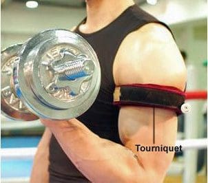
Considering the nature of this kind of training, BFR requires using some form of tourniquet. The easiest and most convenient way to achieve this would be to use some form of strap like an ace bandage or weightlifting knee wraps. If you are able to discover a strap using a comparable elasticity diameter that is smaller, this would be more optimal. When wrapping your limbs, you want to prevent wrap ‘over’ the limb. Otherwise you can risk limiting the muscle’s capacity to contract and your range of movement.
Placement
You will want to put the wrap around the proximal portion of the muscle you’re working. This implies over the muscle and close to the torso. If you are thinking about training forearms and your biceps, you should set the wrap beneath the deltoid. Using this technique for the body requires some careful instructions. Some experts say that when practicing BFR for the body, your leg ought to be wrapped close to the groin area, over the quadriceps. If you’re training calves this would be included. When training BFR for calves, its wise to wrap over the calf and beneath the knee. This is because the common wraps are not really large enough to effectively wrap over the quadriceps.
Wrapping Pressure
When wrapping your muscle, remember to keep in mind that you aren’t attempting to completely restrict blood flow. You still require blood circulation to the muscle. As such, when you wrap, you should try to shoot for wrapping the arm at about a 7 out of 10, with ten being very painful and a complete loss of blood flow. If your arm is totally asleep before you even begin training, the wrap is too tight.�If you complete a set of exercises and your arm is not pumped or fatigued, then you’ve probably not wrapped the bands tight enough.
First and foremost, a majority of experts concur that this kind of training is in fact a safe practice provided that it’s executed properly. To be able to maintain proper safety, ensure that you have not completely restricted blood circulation. Further, as soon as you’ve finished your sets, be certain that you remove the wrap in order to give the muscle blood supply and permit the used blood to be recycled. Should you have them too tight or keep the wraps on too long, you run the danger of inducing tissue and cell death. This isn’t advised. Further, if you have higher blood pressure or heart problems BFR, or blood flow restriction, training is not suggested.
There’s also some evidence to indicate that musculature which isn’t directly occluded, for example chest and shoulders, can experience some benefit from BFR. That is interesting because there was a long belief that advantage would be seen by muscle below the tourniquet. A current meta-analysis indicated that despite evidence, the indirect muscle (chest and shoulders) may see increased benefit in comparison to the same training without a tourniquet. If you are feeling tired, yet still want to get a chest and shoulder pump, then it may help you to wrap your arms.
Finally, BFR shouldn’t be used only in place of different sorts of training. Outcomes like power, power output, hypertrophy and force production rely on coaching specificity and varying immunity (i.e to be able to maximize strength, you need to train with heavier loads to get lower repetitions). The study suggests that blood flow restriction training could be as good as other types of instruction, not exceptional. Therefore, blood flow restriction, or BFR, training may be a useful tool within a resistance-training schedule that is well-rounded.
The scope of our information is limited to chiropractic and spinal injuries and conditions. To discuss options on the subject matter, please feel free to ask Dr. Jimenez or contact us at 915-850-0900 .�
By Dr. Alex Jimenez
Additional Topics: Sports Care
Athletes engage in a series of stretches and exercises on a daily basis in order to prevent damage or injury from their specific sports or physical activities as well as to promote and maintain strength, mobility and flexibility. However, when injuries or conditions occur as a result of an accident or due to repetitive degeneration, getting the proper care and treatment can change an athlete’s ability to return to play as soon as possible and restore their original health.
In order to comprehend how BFR, or blood flow restriction, functions, it is important to perform a quick debriefing on how your circulatory system, also called vascular or cardiovascular system, works. Your arteries are blood vessels that carry oxygenated blood away from your heart to your body. Your veins are blood vessels that carry blood from the body back to the heart.
The objective of blood flow restriction training would be to restrict venous return while still allowing arterial flow by strategically wrapping the lightest portion of your own limbs. Blood can keep pooling to a muscle by restricting the veins rather than the arteries and it remains trapped there. It is like filling a water balloon to max capacity (with no popping up, of course).
By gathering all of the blood to the working muscles without letting it leave, a couple key things happen:�One, you receive a crazy pump and your muscles become supersized. The concept is that this contributes to cellular swelling that shocks the muscles into growth. Second, it’s gonna burn tremendously. Your muscles become deprived of oxygen and can not eliminate accumulating waste materials and this creates a great deal of acidosis or strain. Metabolic stress is just one of the three major mechanisms of muscle development and shouldn’t be dismissed.
The Science of BFR
Dr. Brad Schoenfeld is a regular contributor on hypertrophy (the scientific term for muscle growth). In his book Science and maturation of Muscle Hypertrophy, ” he states: “The prevailing body of literature shows that BFR training stimulates anabolic signaling and muscle protein synthesis and markedly increases muscle development despite using loads frequently considered too low to encourage substantial hypertrophy.” Brad goes on further, saying that “it has been speculated that metabolic stress would be the driving force behind BFR-induced muscle hypertrophy.”
Another interesting matter that occurs with blood flow restriction training is since your oxygen-dependent slow-twitch fibers fatigue way quicker than normal, you have to quickly begin tapping into the fast-twitch muscle fibers, which have the biggest potential for growth.
Interestingly enough, your fast-twitch fibers typically don’t get hit unless you’re using heavy loads or pretty hefty loads performed explosively. But BFR lets you really go fast-twitch with loads less than 50 percent of your own one-rep max. Actually, one study from the Journal of Applied Physiology revealed increased muscle cross-sectional area with BFR training using loads as light as 20 percent of one-rep maximum.
What this means for you is that with BFR training you can utilize lighter loads to construct muscle while sparing your muscles from heavy loading and without fatiguing your central nervous system. Additionally, it is important to note that research has proven the gains are not just for legs and the arms but also for muscle groups over the wraps.
How to Wrap For BFR Training
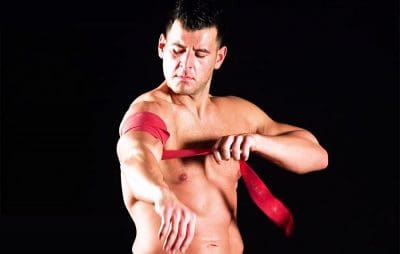
There are some high-end pressure cuffs which may be used to wrap your limbs for BFR, however any wraps will get the job done. Some people utilize knee/elbow or ace bandages wraps. Others use hospital tourniquets that are run-of-the-mill.
For your upper body, wrap it only beneath the shoulder at the top of upper arm so that the wrapping is nestling into your armpit.
For the lower body, wrap only below the gluteal fold from the back and just below the hip flexor in the front.
For both the upper and lower body, you want to wrap at about a 7 out of 10 on the tightness scale (10 being as tight as you can).
You shouldn’t feel any numbness or tingling sensations. That usually means you wrapped it tight, if you do. Wrapping it too tight will limit flow and prevent blood from pooling in the gut, so it defeats the purpose. When in doubt, wrap at first, particularly around the back side of the spectrum.
How can you know whether you wrapped it right? In the event you get your life’s muscular pump. Recall, if it feels sketchy just take off the wraps and re-wrap a tiny bit looser. There is a bit of a learning curve and thus don’t place too much stress to nail it on the first try.
How BFR Training Builds Muscle
The secret to effective BFR training is using light loads (40 to 50 percent of your one-rep maxor less), high repetitions (10 to 15 repetitions or longer), and short rest periods (30 minutes or less). In addition, it is important to note that BFR does not replace your regular training–it just enhances it. Here are my three favorite ways to execute BFR training:
BFR Finishers
After performing your main work out, hit a BFR finisher. If you completed an upper-body workout, hit an upper-body BFR finisher. If you finished a lower-body workout, hit on a BFR finisher. Hit on one for the upper and lower body if you do total-body sessions.
Extra Training Volume and Frequency
BFR is a excellent way to increase training volume (how much work you do) and coaching frequency (how often you train) without impairing your recovery. As an example, to bump up your training volume, if you did 3 routine sets of an exercise with heavier loads, try adding in an additional couple sets of BFR training to the same movement pattern or muscle group using a lighter load for higher reps and shorter rest periods between sets.
Active Recovery and Deloading
Since BFR training requires having lighter loads, it is considerably easier to recover from deeper training. This makes it a process to employ but still want to train. It’s also great to use if you integrate regular deloads–or intervals of decreased loading or training volume–into your training schedule.
It is worth mentioning that BFR is being used with remarkable success in rehabilitation settings, especially with wounded athletes. Being able to operate and develop muscles after an injury or operation with loads is a joint-sparing feature unique to BFR training. As always, consult your physician or physical therapist to find out if BFR training is right for you in such circumstances.
The scope of our information is limited to chiropractic and spinal injuries and conditions. To discuss options on the subject matter, please feel free to ask Dr. Jimenez or contact us at 915-850-0900 .�
By Dr. Alex Jimenez
Additional Topics: Sports Care
Athletes engage in a series of stretches and exercises on a daily basis in order to prevent damage or injury from their specific sports or physical activities as well as to promote and maintain strength, mobility and flexibility. However, when injuries or conditions occur as a result of an accident or due to repetitive degeneration, getting the proper care and treatment can change an athlete’s ability to return to play as soon as possible and restore their original health.
Athletes face extreme pressure to return to play when they are hurt however, the true challenge for physicians is to get them back in the game safely. Athletes should be tough and maintain a positive attitude whilst regularly going through pain. When they’re made to sit out due to an accident, they should be focused and motivated to return to play as quickly as possible. They rehabilitate and rest as they trust that their bodies will ready after a full treatment plan.
This is the idealistic perspective of injury associated with athletes in their specific sport or physical activity. However, the reality is that accidents are an unavoidable by product of being an athlete and the transition from “active athlete” to “injured athlete” and back to “active athlete” does not always happen without complications.
Injured athletes fight with anxiety, frustration, anger and sometimes depression during their time away from play, which might also keep them from following their rehabilitation program effectively. Additionally, the return to the sport itself yields a fresh pair of adversities as athletes should browse through personal fears and a desire to come back to their pre-injury condition with the support of their family and healthcare physician.
Importance of Support for Injured Athletes
Social support can come from various forms, ranging from emotional support to task challenge assistance. Some wounded athletes want a caring individual simply to listen to their anxieties while others might prefer a challenging drive to work harder during rehab. Studies looking at the supply of social support have found that athletes feel most satisfied with the support provided by professionals in comparison to support supplied by teammates or coaches.
It would appear obvious that athletes would need support to assist with the injury recovery process. Because teams have access to trainers in a school setting, this additional support is possible. However, injuries are not unique to the collegiate population, which makes it important to address that �and provide this service.
Researchers who immediately addressed athletes’ tastes from healthcare professionals found that the desire to learn more concerning the injury resulted in a clearer timeline for return to play along with an open environment where athletes felt comfortable asking questions. In respect to athletes not fully understanding their injuries, they noticed that they would have appreciated the use of models and more sophisticated explanations from their physicians. It’s essential for healthcare professionals to take the time to help these athletes that are injured throughout the rehabilitation and recovery process and return to play with expertise.
Even though a complete return to play could be potential in time, it won’t happen immediately and teammates, parents, the athletes and coaches need to understand this. Trainers who have missed those who have been inactive for any period of time or numerous practices will require a slow progression back to their previous degree. This is bothersome for coaches who may “need” that athlete and also for the athlete who wants to return so as not to let the team down, trainer or themselves. Additionally, while appeasing the team and coach, the athlete may want to listen to doctors to ensure a safe recovery.
Goal-Setting to Facilitate Confidence And Motivation
Throughout the rehabilitation process, athletes should set modest goals, adjust their mindset, surround themselves with supportive people and develop their patience. It is important for others such as doctors, parents and trainers to understand the process, and provide athletes with resources and support to help them construct in these areas. Like setting rehabilitation targets that are daily followed by exercise goals, simple strategies can help athletes experience modest successes and build their own confidence.
Every injured athlete would like to return to 100 percent but it is going to take some time to reach that degree. They’re very likely to eliminate the drive and motivation to continue, if they don’t see improvements over time. The athlete has to set goals based on their current status. The athlete will see little daily improvements leading them in the path of better performances in the future.
Building and/or maintaining confidence is vital, and it cannot be connected to results. Athletes need to realize that confidence keeps them trying even if scenarios aren’t going their way, and helps them push through failures. Confidence is a way of behaving and thinking that should be evident in everything one does regardless of the outcome.
In Conclusion
Given potential effects related to harm and the emotions, it’s clear that more education is essential to guarantee positive consequences for athletes who’ve experienced sports injuries. Injuries are unavoidable but they do not need to be devastating to well-being and one’s life if handled effectively. It’s apparent that athletes encounter adversity due to the injury and due to the change for their own lives and daily routines. The recovery is sometimes more easy than the yield to perform since the bone may heal and the tear could be mended, but the brain doesn’t change as easily.
It is necessary for everyone involved to understand that helping an athlete recovery in order to return to play as soon as possible demands attention to both the body and the brain. This can be accomplished by one with awareness, education and effort of coaches, doctors, athletes and parents alike.
The scope of our information is limited to chiropractic and spinal injuries and conditions. To discuss options on the subject matter, please feel free to ask Dr. Jimenez or contact us at 915-850-0900 .�
By Dr. Alex Jimenez
Additional Topics: Sports Care
Athletes engage in a series of stretches and exercises on a daily basis in order to prevent damage or injury from their specific sports or physical activities as well as to promote and maintain strength, mobility and flexibility. However, when injuries or conditions occur as a result of an accident or due to repetitive degeneration, getting the proper care and treatment can change an athlete’s ability to return to play as soon as possible and restore their original health.
Injury is a common occurrence in sport participation. Ask any athlete and they’ll tell you that one of the drawbacks they can experience in their specific physical activity is injury.
Being hurt can mean a number of things to an athlete out of the pain they experience. Firstly, injury can bring a stop to training (i.e., coaching) and may indicate that what they’ve devoted lots of their time and energy and can too be removed quite suddenly (Crossman, 1997). Sport participation is a part of the identity of an athlete and so sports are a tremendous portion of their lives. When that is removed, albeit for a short time period, this can have a possible psychological effect on how an athlete views themselves.
Additionally, injury can take away the positive reinforcements sport provides where athletes undergo a feeling of mastery, autonomy and sense of control (Deutsch, 1985). Injury might be thought of as a setback because sport is used by athletes as a means of managing anxiety, stress and depression, among other things.
Psychological Effects on Injured Athletes
Understandably then, it may be anticipated that athletes can undergo a number of psychological reactions and stress upon becoming injured. Athletes’ psychological experiences differ as no one person experiences injury precisely in the same manner. Yet some emotions are more commonly reported than others and include stress, fear, anger, tension, fatigue, doubt, lack of motivation, and aggravation (Ahern & Lohr, 1997; American College of Sports Medicine, 2001; Klenk, 2006).
Of course it is normal for athletes to experience these emotions in reaction to trauma or injury and it is therefore necessary to be aware that not all athletes encounter an observable psychological disturbance to being hurt. They are athletes who seem to take being injured in their stride and their emotional reactions appear to resolve. On the flip side, other athletes appear to fight emotionally and their responses become problematic when symptoms do not resolve.
Though there’s no predictable sequence of an athlete’s psychological responses to injury, athletes often exhibit three classes of reaction to their injury. To help come to terms with their injury, athletes often attempt to get and interpret as much injury-relevant information they can (i.e., “How bad is it?” , “How long?” , “What can/can’t I do”, “Just how can I fix it?”) . As previously discussed, athletes may experience reactive behavior and psychological upheaval . Often athletes may ask questions or have thoughts that are like the following: “I can’t believe this has happened today”, “I’ll never return to 100%”, and “I’m no good to the group today”. Athletes with apparent psychological effects can frequently display a range of signs suggesting poor adjustment to the injuries, including:
Feelings of anger & confusion
Obsession with �when can I return to play?�
Trying to do too much too soon in terms of rehabilitation program (pushing the limits)
Denial (e.g., �The injury is no big deal�)
Repeatedly returning to play too soon & experiencing re-injury
Exaggerated bragging about accomplishments
Dwelling on minor physical complaints
Sleep disturbances
Alterations in diet
Guilt about letting the team down
Withdrawal from significant others
Rapid mood swings
Statements like �no matter what is done, it will never get better�
The final category indicates that athletes come to terms with the injury and engage in successful coping. If there is anything they could do at home or may help out in training athletes voice that the injury is starting to appear good or often think so, and ask their service network if their responses resolves than becomes debatable. But if an athlete is exhibiting problematic signs of adverse effect as a consequence of their injury, it is very important for them to find help from a sport psychologist who can assist them manage and cope more effectively with their injury thus assisting their injury recovery procedure.
Research has shown that negative emotions experienced by injured athletes may affect athletes’ attitudes toward and subsequent recovery from trauma (Ahern & Lohr, 1997; Crossman, 1997). Using psychological strategies have been found to improve injury recovery, mood through healing, coping, confidence restoration, pain control, and adherence to treatment protocols (Brewer et al., 2000).
Improving Athlete’s Psychological Skills
Psychological skills like goal setting, imagery and relaxation helps athletes cope better with stress, reducing likelihood of harm and stress of harm should it occur. In addition, even athletes that deal with injury can benefit from studying these strategies as they are sometimes utilized to boost performance on a basis that is constant.
Other psychological skills utilized to cope effectively with trauma but can also be used to enhance operation after experiencing injury include self-talk to help athletes have a positive attitude to rehabilitation and build confidence as well as problem solving to help deal with setbacks and search for opportunities. In addition to abilities, it is essential for athletes to be more educated in the recovery procedure and their injury to help reduce uncertainty and provide them with clear expectations and also to keep them informed.
The scope of our information is limited to chiropractic and spinal injuries and conditions. To discuss options on the subject matter, please feel free to ask Dr. Jimenez or contact us at 915-850-0900 .�
By Dr. Alex Jimenez
Additional Topics: Sports Care
Athletes engage in a series of stretches and exercises on a daily basis in order to prevent damage or injury from their specific sports or physical activities as well as to promote and maintain strength, mobility and flexibility. However, when injuries or conditions occur as a result of an accident or due to repetitive degeneration, getting the proper care and treatment can change an athlete’s ability to return to play as soon as possible and restore their original health.
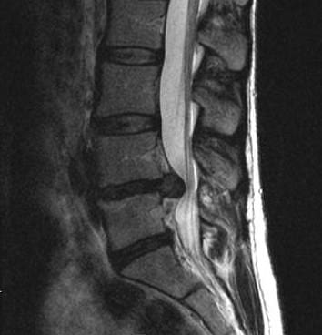
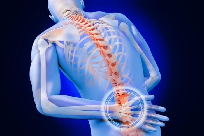
A herniated disc can lead to pain as well as disrupt your daily activities, as you likely know. That is probably what brings you to the office of the doctor: You have back pain or neck pain, and you’d love to understand why.
Your doctor will ask you questions and execute a few exams. This is to try to find the origin of your pain and also to find out which intervertebral disks are herniated. An accurate diagnosis will help your doctor develop a treatment plan method to help you recover and to handle your herniated disc pain and other spine symptoms.
Physical Exam: Herniated Disc Diagnosis
As part of the physical exam, your doctor will ask about your current symptoms and remedies you have already tried for your pain. Some average herniated disc diagnostic questions include:
When did the pain begin? Where’s the pain (cervical, thoracic or mid-back, or lumbar or lower back)?
What activities did you lately do?
What do you do for your herniated disc pain?
Can the disc herniation pain radiate or travel to other parts of your body?
Does anything reduce the disk pain or make it even worse?
Your doctor may also observe your position, range of movement, and physical condition both lying down and standing up. Movement that causes pain will be noticed. A Las�gue evaluation, also referred to as the Straight-Leg Raising evaluation, may be accomplished. You’ll be asked to lie down and extend your knee with your hip bent. If it produces pain or makes your pain worse, this may indicate a herniated disc.
With a herniated disc (or a bulging or ruptured disc), you might feel stiff and may have lost your normal spinal curvature because of muscle strain. Your physician may also feel for tightness and note the spine’s curvature and alignment.
Neurological Exam: Herniated Disc Diagnosis
Your spine specialist will also run a neurological exam, which tests your reflexes, muscle strength, other nerve changes, and pain disperse. Radicular pain (pain that travels away from the source of the pain) can increase when stress is applied directly to the affected area. You might, for instance, have sciatica; this is radicular pain that might be caused by the herniated disk. Since the disc is compressing a nerve, you might experience pain and symptoms in other areas of the body, although the origin of the pain is on your spine.
Imaging Tests for Herniated Discs
Your spine specialist may order imaging tests to help diagnose your injury or condition; you might have to see an imaging facility for those evaluations.
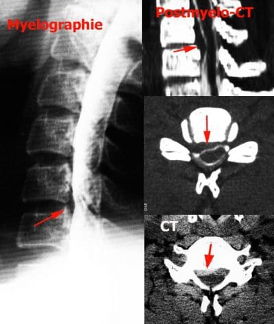
An X-ray may demonstrate a secondhand disk space, fracture, bone spur, or arthritis, which might rule out disk herniation. A computerized axial tomography scan (a CT or CAT scan) or a magnetic resonance imaging test (an MRI) equally can show soft tissue of a bulging disk or herniateddisc. So that you may get treatment these tests will demonstrate location and the stage of the herniated discs.
Other Tests to Diagnose�a Herniated Disc
To obtain the most accurate identification, your spine specialist may order additional tests, for example:
Electromyography (EMG): He or she may order an examination known as an electromyography to measure your nerves respond, if your spine pro suspects you’ve got nerve damage.
Discogram or discography: A sterile procedure where dye is injected into one of your vertebral disc and seen under special conditions (fluoroscopy). The goal is to pinpoint which disk(s) might be causing your pain.
Bone scan: This technique generates film or computer images of bones. A very small number of radioactive substance is injected into a blood vessel throughout the blood flow. It collects on your bones and can be detected by a scanner. This procedure helps doctors detect spinal problems such as disease, a fracture, tumor, or arthritis.
Laboratory evaluations: Typically blood is attracted (venipuncture) and tested to determine if the blood cells are normal or abnormal. A metabolic disease which might be contributing to a back pain may be indicated by Chemical changes in the blood.
The scope of our information is limited to chiropractic and spinal injuries and conditions. To discuss options on the subject matter, please feel free to ask Dr. Jimenez or contact us at 915-850-0900 .�
By Dr. Alex Jimenez
Additional Topics: Sciatica
Lower back pain is one of the most commonly reported symptoms among the general population. Sciatica, is well-known group of symptoms, including lower back pain, numbness and tingling sensations, which often describe the source of an individual’s lumbar spine issues. Sciatica can be due to a variety of injuries and/or conditions, such as spinal misalignment, or subluxation, disc herniation and even spinal degeneration.
There are a number of important factors to take into consideration, such as the timing of when an MRI scan must be performed and limitations with interpretation of findings, to get an MRI scan for herniated discs.
To begin with, the difficulty with the results of an MRI scan, as with a number of other diagnostic studies, is that the abnormality may not always be the source of an individual’s back pain or other symptoms. Numerous studies have shown that approximately 30 percent of people in their twenties and forties have a lumbar disc herniation in their MRI scan, even though they don’t have any pain.
An MRI scan cannot be interpreted on its own. Everything Has to Be well-correlated into the individual patient’s condition, for example:
Symptoms (such as the duration, location, and severity of pain)
Any deficits in their examination
Another concern with MRI scans is the time of when the scan is done. When a patient has experienced the following symptoms would be the only time that an MRI scan is needed immediately:
Bowel or bladder incontinence
Progressive weakness due to nerve damage in the legs.
Herniated Disc Analysis with MRI
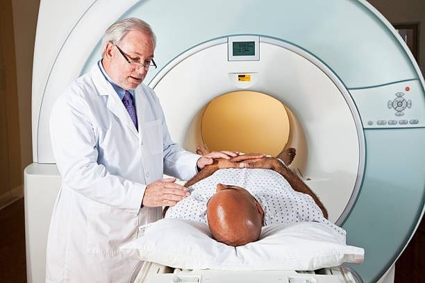
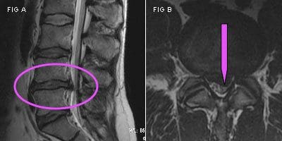
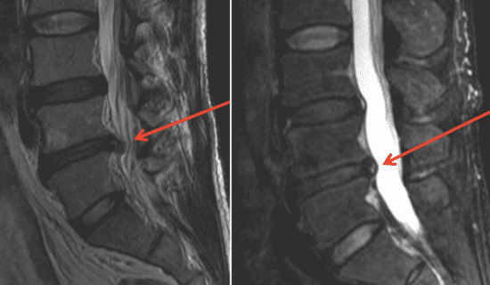
Obtaining an MRI (magnetic resonance imaging) can be an important step in correctly assessing a herniated disc in the spine. Unlike an X-ray, MRI uses a magnetic field and a computer to create and record detailed pictures of the internal workings of your entire body. This technology can also be capable of producing cross-sectional views in identifying a disc of the body, which greatly help doctors. MRI scans are based on new technology, but they have become essential in diagnosing a number of back and neck issues, such as spinal stenosis, herniated discs and bone spurs.
An MRI scan has a number of benefits that greatly help a herniated disc patient. The advantages of an MRI can be:
Unobtrusive
Painless and free of radiation
Can focus on a particular part of the entire body
Extremely accurate
Diagnosing Disc Herniation
Should you believe you have a herniated disc in the neck or back, the very first step would be to visit a physician. Your physician will have the ability to supply you with a complete evaluation and inspection of your medical history to create a identification. Following that, you may be referred to execute an MRI stabilize and to confirm the herniated disc.
At the imaging center you’ll be put to the tubular MRI machine to get a body scan. You may remain enclosed in the MRI device for up to an hour while the comprehensive scan of place where the herniated disc along the spine is completed. The MRI can reveal the exact condition of the herniated disc and surrounding arrangements. This allows your doctor to produce the treatment plan that is right for you and to understand the origin of the disc damage and pain.
Herniated Disc Follow-Up Treatment
Most patients are able to successfully treat herniated disc pain using nonsurgical standard treatments prescribed by their physician. These include relaxation, compression treatment and mild exercise. Surgery can then be explored when months or weeks of treatment do not bring a return to previous action.
If you’re researching surgical options and have become concerned by a number of the risks and unsuccessful results of traditional open back operation, contact a specialist. Spine surgery specialists perform minimally invasive spine surgery, including invasive stabilization surgeries and minimally invasive decompression, which can treat a number of the very acute herniated discs. They may review your MRI to determine if you are a candidate for minimally invasive spine surgery, which may help you get your life back.
The scope of our information is limited to chiropractic and spinal injuries and conditions. To discuss options on the subject matter, please feel free to ask Dr. Jimenez or contact us at 915-850-0900 .�
By Dr. Alex Jimenez
Additional Topics: Sciatica
Lower back pain is one of the most commonly reported symptoms among the general population. Sciatica, is well-known group of symptoms, including lower back pain, numbness and tingling sensations, which often describe the source of an individual’s lumbar spine issues. Sciatica can be due to a variety of injuries and/or conditions, such as spinal misalignment, or subluxation, disc herniation and even spinal degeneration.
A healthcare professional’s clinical diagnosis focuses on finding out the source of a patient’s pain. For this reason, the clinical identification of pain in the herniated disc relies on more than only the findings from a diagnostic evaluation, like CT scan or an MRI scan.
The spine care professional arrives at a clinical diagnosis of the cause of the patient’s pain by means of a combination of findings by a comprehensive medical history, conducting a complete physical exam, and, if appropriate, running one or more diagnostic tests:
Medical history: The physician will choose the patient’s medical history, such as a description of if sciatica, the back pain or other symptoms occur, a description of how the pain feels, what remedies, positions or activities make the pain feel better and more.
Physical examination: The physicians will conduct a physical exam of the individual, such as muscle power and analyzing neural function in parts of the leg or arm, analyzing for pain in positions and much more. Ordinarily, this series of physical tests will give a good idea of the type of back issue the individual has to the spine professional.
Diagnostic tests: After the physician has a fantastic idea of the origin of the patient’s pain, a diagnostic evaluation, such as a CT scan or a MRI scan, is often ordered to confirm the presence of an anatomical lesion at the backbone. The evaluations can give a picture of the location of nerve roots and the disc.
It’s important to emphasize that MRI scans and other diagnostic tests aren’t utilized to diagnose the patient’s pain; rather, they are only utilized to confirm the existence of an anatomical problem that was suspected or identified throughout the medical history and physical examination. Because of this, while the radiographic findings on an MRI scan or other tests are significant, they aren’t as important in diagnosing the reason for the patient’s pain (that the clinical investigation demonstrated) as are the findings from the medical history and physical examination. Many times, an MRI scan or other kind of evaluation will be used for the purpose of treatment, so the healthcare specialist can determine the way it’s currently impinging on the nerve root and precisely where the herniated disc is.
When MRI is Used to Diagnose Herniated Discs
When patients have predominantly experienced leg pain along with a lumbar disc herniation, MRI scans are usually recommended early in a patient’s path of pain.
Therefore, physicians often recommend waiting 3 to 6 months (following the onset of lower back pain) prior to having an MRI scan done as a way to see whether the pain will get better with conservative (nonsurgical) remedies. As a very general guideline, if the results of the MRI scan aren’t likely to affect a patient’s further back pain therapy, and �the patient will continue with non-surgical treatments such as chiropractic treatments, physical therapy and drugs, waiting to acquire an MRI scan, as well as other imaging scans, in most situations is a fair option.
What Happens When a Disc Herniates
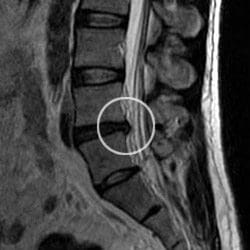
Though the spinal discs are made to withstand significant amounts of force, injury and other issues with the disc can happen. After the disc ages or is injured, the outer portion (annulus fibrosus) of a disk may be torn as well as the disc’s inner substance (nucleus pulposus) can herniate or extrude out of the disk. Nerves, and the inner portion of the disc surround each spinal disc that leaks out comprises proteins, therefore when this material comes in contact with a nerve wracking pain that may travel down the length of the nerve can be caused by it. Even a tiny disk herniation which enables a small quantity of the inner disc material to touch the nerve may cause pain.
Pain from a Herniated Disc vs. Degenerative Disc Disease
A herniated disc will generally create another type of pain than degenerative disk disease (another common disc problem).
When a patient has a symptomatic degenerated disc (one which causes pain or other symptoms), it’s the disc space itself which is debilitating and is the origin of pain. This type of pain is called axial pain.
When a patient has a symptomatic herniated disc, it is not the disk space itself that hurts, but rather the disc difficulty is causing pain in a nerve in the spine. This kind of pain is typically called radicular pain (nerve root pain, or tingling from a lumbar herniated disk).
In conclusion, when an individual begins to experience painful symptoms along their lower back, or lumbar spine, although they may sometimes not experience any symptoms, it a herniated disc is suspected, its recommended to seek immediate medical attention and to consider having an MRI, CT scan or other imaging tests to properly diagnose the presence of a herniated disc or other injury and/or condition before following with treatment.
The scope of our information is limited to chiropractic and spinal injuries and conditions. To discuss options on the subject matter, please feel free to ask Dr. Jimenez or contact us at 915-850-0900 .�
By Dr. Alex Jimenez
Additional Topics: Sciatica
Lower back pain is one of the most commonly reported symptoms among the general population. Sciatica, is well-known group of symptoms, including lower back pain, numbness and tingling sensations, which often describe the source of an individual’s lumbar spine issues. Sciatica can be due to a variety of injuries and/or conditions, such as spinal misalignment, or subluxation, disc herniation and even spinal degeneration.
IFM's Find A Practitioner tool is the largest referral network in Functional Medicine, created to help patients locate Functional Medicine practitioners anywhere in the world. IFM Certified Practitioners are listed first in the search results, given their extensive education in Functional Medicine