The tendons are powerful soft tissues which connect the muscles to the bones. One of these tendons, the quadriceps tendon, works together with the muscles found at the front of the thigh in order to straighten the leg. A quadriceps tendon rupture can affect an individual’s quality of life.
A quadriceps tendon rupture can be a debilitating injury and it usually requires rehabilitation and surgical interventions to restore knee function. These type of injuries are rare. Quadriceps tendon ruptures commonly occur among athletes who perform jumping or running sports.
Quadriceps Tendon Rupture Description
The four quadriceps muscles come together above the kneecap, or patella, to form the quadriceps tendon. The quadriceps tendon joins the quadriceps muscles into the patella. The patella is connected to the shinbone, or tibia, by the patellar tendon. Working collectively, the quadriceps muscles, the quadriceps tendon, and the patellar tendon, straighten the knee.
A quadriceps tendon rupture can be partial or complete. Many partial tears don’t completely disrupt the soft tissues. However, a full tear will divide the soft tissues�into two parts. If the quadriceps tendon ruptures entirely, the muscle is no longer attached to the kneecap or patella. As a result, the knee is unable to straighten�out when the quadriceps muscles contract.
Quadriceps Tendon Rupture Causes
A quadriceps tendon rupture frequently occurs due to an increased load on the leg where the foot is planted and the knee is somewhat flexed. By way of instance, when landing from an awkward jump, the power is too much for the soft tissues to bear, causing a partial or complete tear. Tears may also be due to falls, direct impacts to the knee, and lacerations or cuts.
A weakened quadriceps tendon is also more likely to rupture. Several factors may result in tendon weakness, including quadriceps tendinitis, the inflammation of the quadriceps tendon, called quadriceps tendinitis. Quadriceps tendinitis is one of the most common sports injuries in athletes who participate in sports or physicial�activities which involve jumping.
Weakened soft tissues may also be brought on by diseases that interrupt blood flow to the knee or patella. Utilizing corticosteroids and some antibiotics have also been connected to weakness associated with quadriceps tendon ruptures. Immobilization for an extended period of time can also decrease strength in the quadriceps tendons. Finally, quadriceps tendon ruptures can occur due to dislocations and/or surgery.
Quadriceps Tendon Rupture Symptoms
A popping or tearing feeling is one of the most common symptoms associated with a quadriceps tendon rupture. Pain followed by swelling and inflammation of the knee�might make the individual unable to straighten out their knee. Other symptoms of a quadriceps tendon rupture include:
An indentation at the top of the kneecap or patella of the affected site
Bruising
Tenderness
Cramping
Sagging or drooping of the kneecap or patella where the tendon tore
Difficulty walking because the knee is buckling or giving away
Quadriceps Tendon Rupture Evaluation
The healthcare professional will perform an evaluation to diagnose a quadriceps tendon rupture by first discussing the patient’s symptoms�and medical history.�After talking about the patient’s symptoms and medical history, the doctor will conduct a comprehensive evaluation of the knee.
To ascertain the precise cause of the patient’s symptoms, the healthcare professional will examine how well it is possible to stretch, or straighten,�the knee. Although this area of the evaluation can be debilitating, it’s essential to diagnose a quadriceps tendon rupture.
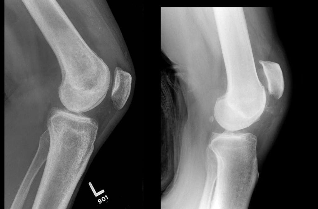
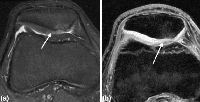
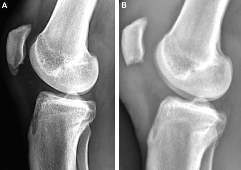
To verify a quadriceps tendon rupture diagnosis, the doctor may order some imaging tests, like an x-ray or magnetic resonance imaging, or MRI, scan. The kneecap moves from place once the quadriceps tendon ruptures. This can be quite evident on a sideways x-ray perspective of the knee.
Complete tears may frequently be identified with x-rays alone. The MRI can reveal the quantity of tendon torn along with the positioning of the tear. From time to time, an MRI will also rule out another injury with similar symptoms. Diagnostic imaging is helpful in the evaluation of sports injuries.
The quadriceps tendon is the large tendon found just above the kneecap, or patella, which allows us to straighten out our knee. While the quadriceps tendon is a strong, fibrous cord which can withstand tremendous amounts of force, sports injuries or other health issues may lead to a quadriceps tendon rupture. Quadriceps tendon ruptures are debilitating problems which can affect a patient’s quality of life.
Dr. Alex Jimenez D.C., C.C.S.T. Insight
Quadriceps Tendon Rupture Treatment
Non-Surgical Treatment
A majority of partial tears react well to non-surgical treatment approaches. The doctor may advise the patient to utilize a knee immobilizer or brace to allow the quadriceps tendon to heal. Crutches will help avoid placing weight onto the leg. A knee immobilizer or brace is used�for 3 to 6 months.
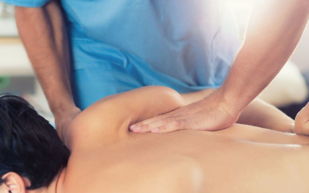
Once the initial pain, swelling, and inflammation have�decreased, alternative treatment options, such as chiropractic care and physical therapy, can be utilized. A doctor of chiropractic, or chiropractor, utilizes spinal adjustments and manual manipulations to carefully correct any spinal misalignments, or subluxations, which may be causing problems.
Furthermore, chiropractic care and physical therapy can provide lifestyle modifications, including physical activity and exercise programs to help speed up the recovery process. The patient may be recommended a variety of stretches and exercises to improve strength, flexibility and mobility. The healthcare professional will determine when it’s safe to return-to-play.
Surgical Treatment
Many individuals with complete tears require surgery to repair a quadriceps tendon rupture. Surgical interventions depend on the patient’s age, actions, and prior level of function. Surgery for quadriceps tendon ruptures involves re-attaching the tendon to the kneecap or patella. Surgery is carried out with regional spinal anesthetic or general anesthetic.
To reattach the tendon, sutures are put in the tendon and then threaded through drill holes at the kneecap. The stitches are attached in the base of the kneecap. The�physician will tie the sutures to find the ideal tension in the kneecap or patella. This will also make sure that the place of the kneecap closely matches that of the uninjured patella or kneecap.
A knee immobilizer, brace or a long leg cast may be utilized following the surgery. The patient may be allowed to set weight on their leg by means of crutches. Stretches and exercises are added into a rehabilitation program by a chiropractor or physical therapist after a surgical intervention.
The precise timeline for chiropractic care and physical therapy following a surgery for those patients that require it will be individualized personally. The patient’s rehabilitation program will be contingent upon the kind of tear, their surgery, medical condition, along with other requirements.
Conclusion
The majority of patients can return to their original routines after recovering from a quadriceps tendon rupture. The individual’s return will be addressed very carefully by the healthcare professional.�The scope of our information is limited to chiropractic and spinal health issues. To discuss the subject matter, please feel free to ask Dr. Jimenez or contact us at�915-850-0900�.
Curated by Dr. Alex Jimenez
Additional Topic Discussion: Relieving Knee Pain without Surgery
Knee pain is a well-known symptom which can occur due to a variety of knee injuries and/or conditions, including�sports injuries. The knee is one of the most complex joints in the human body as it is made-up of the intersection of four bones, four ligaments, various tendons, two menisci, and cartilage. According to the American Academy of Family Physicians, the most common causes of knee pain include patellar subluxation, patellar tendinitis or jumper’s knee, and Osgood-Schlatter disease. Although knee pain is most likely to occur in people over 60 years old, knee pain can also occur in children and adolescents. Knee pain can be treated at home following the RICE methods, however, severe knee injuries may require immediate medical attention, including chiropractic care.
The knee is a made up of a variety of complex soft tissues. Enclosing the knee joint is a fold at its membrane known as the plica. The knee is encapsulated�by a fluid-filled structure called the synovial membrane. Three of these capsules, known as the synovial plicae, develop around the knee joint throughout the fetal stage and are absorbed before birth.
However, during one research study in 2006, researchers found that 95 percent of patients undergoing arthroscopic surgery had remnants of their synovial plicae. Knee plica syndrome occurs when the plica becomes inflamed, generally due to sports injuries.�This often takes place in the center of the kneecap, known as medial patellar plica syndrome.
What are the Symptoms of Knee Plica Syndrome?
The most common symptom of knee plica syndrome is knee pain, although a variety of health issues can also cause these symptoms. Knee pain associated with knee plica syndrome is generally: achy, instead of sharp or shooting; and worse when using stairs, squatting, or bending. Other symptoms of knee plica syndrome can also include the following:�
a catching or locking sensation on the�knee while getting up from a chair after sitting for an extended period of time,
difficulty sitting for extended intervals,
a cracking or clicking noise when bending or stretching the knee,
a feeling that the knee is slowly giving out,
a sense of instability on slopes and stairs,
and may feel swollen plica when pushing on the knee cap.
What are the Causes of Knee Plica Syndrome?
Knee plica syndrome is commonly caused as�a result of an excess of stress or pressure being placed on the knee or due to overuse. This can be brought on by physical activities and exercises which require the individual to bend and extend the knee like running, biking, or utilizing a stair-climbing machine. An automobile accident injury or�a�slip-and-fall accident can also cause knee plica syndrome.
�
Knee plica syndrome, commonly referred to as medial patellar plica syndrome, is a health issue which occurs when the plica, a structure which surrounds the synovial capsule of the knee, becomes irritated and inflamed. Knee plica syndrome can occur due to sports injuries, automobile accident injuries, and slip-and-fall accidents, among other types of health issues. The symptoms of knee plica syndrome may commonly be mistaken for chondromalacia patella. Diagnostic imaging can help diagnose the problem to continue with treatment.
Dr. Alex Jimenez D.C., C.C.S.T. Insight
How is Knee Plica Syndrome Diagnosed?
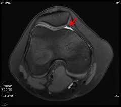
In order to diagnose medial patellar plica syndrome, the healthcare professional will first perform a physical examination. They will use the evaluation to rule out any other potential causes of knee pain, such as a torn meniscus, tendonitis, and broken bones or fractures. Be sure to talk to your doctor about any physical activities you participate in along with any recent health issues. The healthcare professional might also utilize an X-ray or MRI to have a better look at your knee.
What is the Treatment for Knee Plica Syndrome?�
Most instances of medial patellar plica syndrome respond well to alternative treatment options, such as chiropractic care, physical therapy or even a physical activity or exercise plan at home. Chiropractic care uses spinal adjustments and manual manipulations to safely and effectively correct a variety of health issues associated with the musculoskeletal and nervous system. Moreover, chiropractic care and physical therapy can include a series of stretches and exercises to help restore strength, mobility, and flexibility to the hamstrings and quadriceps. These stretches and exercises are described below.
Quadriceps Strengthening
The medial plica is attached to the quadriceps, a major muscle on the thighs. An individual with weakened quadriceps has a higher chance of developing knee plica syndrome. You can strengthen your quadriceps by performing the stretches and exercises as follow:
quadriceps sets or muscle tightening
straight leg raises
leg presses
mini-squats
biking, swimming, walking, or use an elliptical machine.
Hamstring Stretching
The hamstrings are the muscles which extend down the back of the thighs, from the pelvis to the shin bone. These help flex the knee. Tight hamstrings place more stress and pressure on the front of the knee, or the plica. A chiropractor or physical therapist will guide the patient through numerous stretches and exercises which may help unwind the nerves. As soon as the patient learns these moves, they may perform them a few times each day to keep the muscles relaxed.
Corticosteroid Injections
Some healthcare professionals may provide corticosteroid injections for the knee if the pain and inflammation causes a restriction in function. Corticosteroid injections can help temporarily reduce painful symptoms, however, it’s essential for the patient to continue with treatment to heal knee plica syndrome. The painful symptoms may return when the corticosteroid burns off if not treated.
Surgery
If chiropractic care, physical therapy, or the treatment described above does not help heal knee plica syndrome, a procedure known as arthroscopic resection may be needed. To perform this process, the doctor will insert a small camera, called an arthroscope, via a tiny cut at the side of the knee. Small surgical instruments are then inserted through a second small cut to take out the plica or correct its position.
After surgery, your doctor will consult with a chiropractor or physical therapist for a rehabilitation program.�Recovering from surgery for knee plica syndrome is dependent upon many factors, including the patient’s overall health and wellness. The patient may recover within a few days in case the knee has been changed. Remember to wair a few weeks before returning to a routine levels of exercise and physical activity.
Living with Knee Plica Syndrome
Plica syndrome is generally easy to treat with chiropractic care, physical therapy,�and other treatment approaches, as described above. Should you need surgery, the approach is minimally invasive and requires less recovery compared to a number of different types of knee surgery.
Talk to your healthcare professional to determine the best treatment choice for your knee plica syndrome. The scope of our information is limited to chiropractic and spinal health issues. To discuss the subject matter, please feel free to ask Dr. Jimenez or contact us at�915-850-0900�.
Curated by Dr. Alex Jimenez
Additional Topic Discussion: Relieving Knee Pain without Surgery
Knee pain is a well-known symptom which can occur due to a variety of knee injuries and/or conditions, including�sports injuries. The knee is one of the most complex joints in the human body as it is made-up of the intersection of four bones, four ligaments, various tendons, two menisci, and cartilage. According to the American Academy of Family Physicians, the most common causes of knee pain include patellar subluxation, patellar tendinitis or jumper’s knee, and Osgood-Schlatter disease. Although knee pain is most likely to occur in people over 60 years old, knee pain can also occur in children and adolescents. Knee pain can be treated at home following the RICE methods, however, severe knee injuries may require immediate medical attention, including chiropractic care.
EXTRA EXTRA | IMPORTANT TOPIC: El Paso, TX Chiropractor Recommended
Many people think of joints, bones, and the, skeletal system when they think of chiropractic, but in fact, the muscles also play an integral part in supporting the body. The muscles are layers and interwoven work to move and stabilize the spine, facilitate the movement of the body�s joints, and aid in respiration. When there is pain within this system, chiropractic can be a very effective treatment. More patients are turning to chiropractic care to treat a variety of painful conditions because it does not use addictive pharmaceuticals with unpleasant side effects; it is completely natural. Chiropractic can also keep patients from requiring surgery in many cases. So when it comes to myofascial pain and trigger points, this form of treatment is often considered optimal.
What is Myofascial Pain?
In simple terms, myofascial pain is simply pain in the muscles. When you break down the word, �myo� means muscle and �fascia� refers to the connective tissue that are interwoven throughout the body.
The pain originates in specific trigger points that are located in the muscles and fascia at various areas of the body. The pain can range in intensity from mild and annoying to severe and debilitating.
What are Trigger Points?
Trigger points are tightened, hypersensitive spots that can be located in any muscle. Different people may have different trigger points. It isn�t like specific lower back pain or neck pain which occur in particular areas of the body. Trigger points can vary from person to person.
When trigger points form, they become nodules or spots that exist in one of the muscle�s taut bands. The patient may experience a variety of symptoms including pain, weakness, burning, tingling, and other symptoms.
What often makes trigger points challenging to locate is that they cause what is known as referred pain. In other words, the person may experience the pain at the exact location of the trigger point, or the pain can be referred to other areas in the body. Referred pain usually has fairly consistent pain patterns so it can be traced to the origin � eventually.
Around 85% of the pain that individuals experience is attributed to myofascial pain. The trigger points determine whether the pain is chronic or acute. It is a condition that is very common.
How do Trigger Points Form?
Trigger points form when the muscle undergoes trauma of some type. The trauma can come from disease, accidents, related work conditions (from persistent, repetitive motion), and sports injuries.
Activities or habits that place a repetitive, long-term strain on the muscles can also cause trigger points. Poor posture, improper ergonomics, and repetitive movements are the most common of these types of activities. Emotional and physical stress are often identified as causes of irritating trigger points.
Benefits of Chiropractic for Myofascial Pain and Trigger Points
Chiropractic care is often a preferred treatment for myofascial pain due to its effectiveness and drug-free approach. Patients who undergo treatment will usually experience a dramatic decrease in their pain level, or it will be eliminated.
They also enjoy increased strength, flexibility, and range of motion. With continued chiropractic care, they will find that they have more endurance for work and recreational activities and even sleep better. It should be noted that sleep disruptions are a common complaint associated with myofascial pain.
Overall, chiropractic can give patients with myofascial pain a better quality of life with decreased incidence of injury. They are often able to lower their pain medication or eliminate it.
Because chiropractic is a whole-body approach, patients learn healthy habits including diet, exercise, and mental wellness. Most of all, they can live with less pain or no pain at all.
Chondromalacia patellae, also referred to as runner’s knee, is a health issue in which the cartilage beneath the patella,�or kneecap, becomes soft�and ultimately degenerates. This problem is prevalent among young athletes,�however, it may also develop in older adults who suffer from arthritis of the knee.
Sports injuries like chondromalacia patellae are frequently regarded as an overuse injury. Taking some time off from participating in physical activities and exercise may produce superior outcomes. In the instance that the individual’s health issues are due to improper knee alignment, rest may not offer pain relief. Symptoms of runner’s knee include knee pain and grinding sensations.
What Causes Chondromalacia Patellae?
The kneecap,�or the patella, is generally found through the front of the knee joint. If you bend your knee, the rear end of your kneecap slips over the cartilage of your femur, or thigh bone, at the knee. Complex soft tissues, such as tendons and ligaments, connect the kneecap to the shinbone and thigh muscle. Chondromalacia patellae�can commonly occur when any of these structures fail to move accordingly, causing the kneecap to rub against the�thigh bone. Poor kneecap motion may result from:
Misalignment due to a congenital health issue
Weakened hamstrings and quadriceps, or the muscles of the thighs
Muscle imbalance between the adductors and abductors, the muscles on the inside and outside of the thighs
Continuous pressure to the knee joints from certain physical activities and exercise like running, skiing, or jumping
a direct blow or injury for a kneecap
Who is at Risk for Chondromalacia Patellae?
Below is an assortment of factors which may increase an individual’s chance for developing chondromalacia patellae.
Age
Adolescents and young adults have the highest risk for this health issue. During growth spurts, bones and muscles can often grow too rapidly, causing short-term muscle and bone imbalances in the human body.
Gender
Females are more likely than males to develop runner’s knee, because women generally possess less muscle mass than men. This may result in abnormal knee placement, and more lateral pressure on the kneecap.
Flat Feet
Individuals who have flat feet can add more strain to the knee joints as compared to individuals who have higher arches.
Past Injury
Previous injuries to the kneecap, including a dislocation, can raise the chance of developing chondromalacia patellae.
Increased Physical Activity
Increased levels of physical activities and exercise can place pressure on the knee joints, which may raise the risk for knee issues.
Arthritis
Runner’s knee may also be an indication of arthritis, a well-known problem causing pain and inflammation to the tissue and joint. Swelling can prevent the proper function of the knee and its complex structures.
What are the Symptoms of Chondromalacia Patellae?
Chondromalacia patellae will generally present as pain in the knee, called patellofemoral pain, accompanied by sensations of cracking or grinding when extending or bending the knee. Pain may worsen after sitting for an extended period of time or through physical activities and exercises that apply intense pressure for your knees, like standing. It’s essential for the individual to seek immediate medical attention if the symptoms of chondromalacia patellae, or runner’s knee, do not resolve on their own.
Diagnosis and Chondromalacia Patellae Grading
A healthcare professional will search for areas of pain and inflammation on the knee. They might also look at the way the kneecap aligns with the thigh bone. A misalignment may indicate the presence of chondromalacia patellae. The doctor may also perform a series of evaluations to ascertain the presence of this health issue.
The healthcare professional may also ask for any of the following tests to help diagnose chondromalacia patellae, including:�x-rays to show bone damage or misalignments or arthritis; magnetic resonance imaging, or MRI, to see cartilage wear and tear; and�arthroscopic examination, a minimally invasive procedure which involves inserting an endoscope and camera inside the knee joint.
Grading
There are four levels of chondromalacia patellae, ranging from grade 1 to 4, which characterize the level of the patient’s runner’s knee. Grade 1 is considered mild while grade�4 is considered severe.
Grade 1 indicates the softening of the cartilage in the knee region.
Grade 2 suggests a softening of the cartilage followed by abnormal surface features, the start of degeneration.
Grade 3 reveals the thinning of the cartilage together with active degeneration of the complex soft tissues of the knee.
Grade 4, or the most severe grade, demonstrates exposure of the bone through a substantial part of the cartilage Bone exposure means that bone-to-bone rubbing is most likely happening in the knee.
What is the Treatment for Chondromalacia Patellae?
The goal of treatment for chondromalacia patellae is to first decrease the strain being placed on the kneecap, or patella, and the femur, or thigh bone. Rest and the use of ice and heat agains the affected knee joint is generally the first line of treatment. The cartilage damage associated with runner’s knee may often repair itself with these remedies along.
Moreover, the healthcare professional may prescribe anti-inflammatory drugs and/or medications, such as ibuprofen, to decrease pain and inflammation around the knee joint. When tenderness, swelling, and pain persist, the following treatment options could be explored. As mentioned above, individuals should seek immediate medical attention if symptoms persist.�
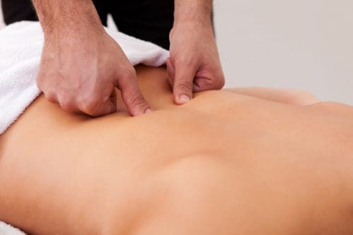
Chiropractic Care
Chiropractic care is a safe and effective, alternative treatment option which focuses on the diagnosis, treatment, and prevention of a variety of injuries and/or conditions associated with the musculoskeletal and nervous system, including chondromalacia patellae. Occasionally,�knee pain may originate due to spinal misalignments or subluxations. A doctor of chiropractic, or chiropractor, will use spinal adjustments and manual manipulations to carefully restore the natural integrity of the spine.�
Furthermore, a chiropractor may also recommend a series of lifestyle modifications, including nutritional advice and a physical activity or exercise guide to help ease symptoms associated with chondromalacia patellae. Rehabilitation may also focus on�strengthening the quadriceps, hamstrings, adductors, and abductors to improve muscular strength, flexibility, and mobility. The purpos of muscle balance is also to assist in preventing knee misalignment, among other complications.
Surgery
Arthroscopic surgery might be required to inspect the joint and ascertain whether there is a misalignment of the knee. This operation involves inserting a camera in the knee joint through a very small incision. A surgical procedure can repair the issue. One�common process is a lateral release. This surgery involves cutting a number of the ligaments to release tension and permit for more movement. Additional surgery may entail implanting the back of the kneecap, inserting a cartilage graft, or transferring the thigh muscle.
�
Chondromalacia patellae is characterized as the inflammation of the underside of the patella, or kneecap, caused by the softening of the cartilage surrounding the soft tissues of the knee joint. This well-known health issue is generally caused due to sports injuries in young athletes, although chondromalacia patellae may also occur in older adults with arthritis in the knee. Chiropractic care can help restore strength and balance to the knee joint and its surrounding soft tissues.
Dr. Alex Jimenez D.C., C.C.S.T. Insight
How to Prevent Chondromalacia Patellae
A patient can ultimately lower their chance of developing runner’s knee, or chondromalacia patellae, by:�
Avoiding repeated stress on the knees. In case the individual needs to spend time on their knees, they could wear kneepads.
Produce muscle balance by strengthening the quadriceps, hamstrings, abductors, and adductors.
Wear shoe inserts that correct flat feet. This may reduce the amount of pressure being placed on the knees to realign the kneecap, or patella.
Keeping a healthy body weight can also help prevent chondromalacia patellae. Following the nutritional advice and guidance from a healthcare profesional can help promote a healthy body weight. The scope of our information is limited to chiropractic and spinal health issues. To discuss the subject matter, please feel free to ask Dr. Jimenez or contact us at�915-850-0900�.
Curated by Dr. Alex Jimenez
Additional Topic Discussion: Relieving Knee Pain without Surgery
Knee pain is a well-known symptom which can occur due to a variety of knee injuries and/or conditions, including�sports injuries. The knee is one of the most complex joints in the human body as it is made-up of the intersection of four bones, four ligaments, various tendons, two menisci, and cartilage. According to the American Academy of Family Physicians, the most common causes of knee pain include patellar subluxation, patellar tendinitis or jumper’s knee, and Osgood-Schlatter disease. Although knee pain is most likely to occur in people over 60 years old, knee pain can also occur in children and adolescents. Knee pain can be treated at home following the RICE methods, however, severe knee injuries may require immediate medical attention, including chiropractic care.
Have you ever had a pain in the neck? And your kids or significant other don’t count. If you’ve ever had a stiff, sore neck, then you’ve more than likely experienced cervicalgia. You’re not alone. The American Osteopathic Association estimates that more than 25% of Americans have experienced or chronically experience neck pain. Neck pain is one of the primary causes of chronic pain, ranking number three behind knee pain (number two) and back pain (number one). Chronic pain affects around 65% of people in the United States, ranging in age 18 to 34. They either have experienced it firsthand or care for someone who has recently experienced it. That number increases as the population ages.
It is also worth noting that most doctors prescribe pain medications, but more than 33% of patients with chronic pain won’t take them because they are afraid of becoming addicted.
What is Cervicalgia?
Cervicalgia is a blanket term used to describe neck pain. It can range from a simple crick in the neck to severe pain that prevents you from turning your head.
Knowing the term for the pain, though, does not help when it comes to treatment because treatment lies in the cause of the pain. It can become quite complex because there are so many causes for the pain. Sometimes the cause itself must be eliminated before the treatments for the pain can be effective.
What are the Causes of Cervicalgia?
The causes of cervicalgia are vast and varied. A patient who sits at their desk for too long or sleeps in a poor position can develop neck pain.
Injuries such as sports injuries and whiplash fall at the more severe end of the spectrum. Even simple gravity can be a culprit.
The human head can weigh as much as 10 pounds, sometimes even more, and the neck is tasked with keeping it upright. Just the action of fighting gravity and keeping the head erect for long periods of time (like all day) can cause the neck muscles to become strained and fatigued. This can also cause neck injuries to heal slower because the neck is almost always in use and under consistent stress.
How is Cervicalgia Treated?
Treatment for cervicalgia depends on both the symptoms and the cause. If you have been injured, you should immediately seek medical attention to assess the injury’s severity.
You can apply ice to help reduce inflammation and swelling, but do not delay a medical evaluation. Some neck injuries can be severe, causing severe conditions, including paralysis.
After an assessment, your doctor may prescribe medication such as anti-inflammatories and stronger painkillers. A cervical collar may also be recommended since it allows the neck to rest, which will promote healing.
If the pain is caused by other reasons such as stress, poor posture, or sleeping on the wrong pillow (in other words, you have a crick in your neck), you can use an over-the-counter anti-inflammatory medication, and using a heating pad will help. Massage is also effective.
However, prevention is the best cure. When you know what is causing your cervicalgia, you can take steps to prevent it. Chiropractic can help both in prevent cervicalgia and in treating it.
Chiropractic for Cervicalgia
Chiropractic treatment can help relieve cervicalgia pain for many of the causes, including injury, stress, and misalignment. Depending on the cause, the chiropractor will use specific techniques to treat the root of the problem.
They will bring the body back into alignment, which also helps to prevent the pain of cervicalgia. The most attractive aspect is that it allows for pain management without the use of any medications.
When you get regular chiropractic care, you can reduce your chances of experiencing pain in your neck and back. That is why so many people are choosing chiropractic care for their neck and back pain instead of turning to traditional medicine because it works.
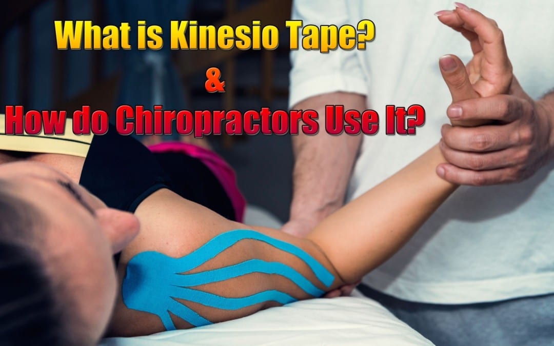
You may have seen professional athletes, dancers, gymnasts, and others who engage in extremely physical activities using a type of tape on various points of their bodies. It is sometimes colored and does not seem to inhibit range of motion. While it looks like tape or maybe a very fancy bandage, it is a highly technical, specialized tape that is used to treat patients of all ages and activity levels. It is called Kinesio tape, and it is often used by chiropractors to help address specific injuries.
What is Kinesio Tape?
Also called Kinesio Tex Tape, Kinesio Tape is a special adhesive tape that has elastic properties. It was developed by Dr. Kenzo Kase, a chiropractor, and acupuncturist, in 1979. It is safe for all ages including pediatric and geriatric patients. The tape is comprised of a 100% cotton fiber strip with medical grade acrylic adhesive. It is soft and gentle, but it works.
The tape is hypoallergenic and latex free, so it is appropriate for a vast audience. It is also water resistant, making it wearable for many activities. When worn, the tape does not limit or inhibit range of motion, and the same tape can be worn for several days without losing its effectiveness.
How does Kinesio Tape Work?
The tape can stimulate or relax muscles, depending on the tension that it puts on the body when applied. When worn, it lifts the skin by microscopic increments which aids in lymphatic drainage.
It also helps to decrease inflammation and swelling which reduces pressure in the area. This allows the blood and lymphatic fluid to flow freer and more effectively in and out of the affected area.
When used correctly, Kinesio Tape can reduce inflammation, promote better circulation, prevent injury, facilitate healing, the re-educate the neuromuscular system. This helps the body return to homeostasis. It can be applied in many different configurations, but often the applications are a single �I,� �Y,� or �X.�
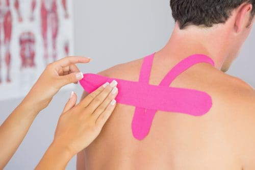
The Kinesio Taping Method
The Kinesio taping method is a systematic, therapeutic technique that offers a two-prong approach of supporting the patient and rehabilitating the condition or affected area. It can alleviate pain reduce swelling, providing relief to the patient. There are specific taping shapes that are specialized to address certain areas of the body as well as certain conditions. While it provides stability and support to the body�s joints and muscles, it does not restrict the range of motion. The technique is designed to address soft tissue injury by manipulating that area and facilitating healing.
What Conditions is Kinesio Taping used to Treat?
Many conditions throughout the body can benefit from Kinesio Taping. Chiropractors use it for:
Lower back strain
Plantar fasciitis
Back strain
Carpal tunnel syndrome
Rotator cuff injury
Ankle Sprains
Whiplash
Herniated disc
Post-surgery edema
Tennis elbow
Pre-surgery edema
Patella tracking
Athletes may also use it for additional support or to prevent injury. Because it relies on the body�s natural healing process, many people find it to be preferred treatment for many painful conditions. When combined with chiropractic care, Kinesio Tape is very effective.
When treating a condition with Kinesio Tape, the chiropractor may use a variety of techniques, depending on the illness or injury. They may use spinal manipulation, massage, and other treatments, combining them with recommendations for lifestyle changes and diet modifications.
The draw for this treatment is that it encourages the body to heal itself, eliminating the need for drugs with their undesired and unpleasant side effects, or more invasive procedures like surgery. Kinesio Taping is safe, natural, and a perfect complement to chiropractic care.
Sinding-Larsen-Johansson, or SLJ, syndrome is a debilitating knee condition that most commonly affects teens during periods of rapid growth. The kneecap, or patella, is attached to the shinbone, or tibia, from the patellar tendon. The tendon connects to an expansion plate at the bottom of the kneecap throughout growth.
Repetitive stress on the patellar tendon can make the growth plate within the knee become inflamed and irritated. SLJ mainly develops in children and adolescents between the ages of 10 and 15 because that is when most people experience growth spurts. SLJ is most common in young athletes due to excess or repetitive strain in the knee.
Causes of SLJ Syndrome
The large muscle group at the front of the upper leg is known as the quadriceps. When straightening the leg, the quadriceps pull to deliver the leg forward. This puts pressure on the growth plate at the bottom of the kneecap. During rapid growth, the bones and muscles don’t always grow at precisely the same rate.
Since the bones grow, tendons and muscles can get tight and stretched. This increases the strain around the patellar tendon and also on the growth plate it’s attached to. Repetitive or extra stress and pressure in this area can cause the growth plate to become irritated and painful. Matters that can contribute to growing SLJ syndrome are comprised of:
Sports that involve a lot of running and jumping, such as field and track or other sports such as football, gymnastics, basketball, lacrosse, and field hockey, can place stress on the knees.
Increased or incorrect physical activity can add strain on the knees. Improper form while training, shoes that don’t support the toes or an unusual way of jogging can increase chances of SLJ syndrome.
Tight or stiff quadriceps muscles can also lead to SLJ syndrome. Muscles that are more powerful and more elastic will work better, reducing the strain on the patellar and kneecap tendon.
Activities that place more pressure on the knees or demanding tasks for the knees, such as lifting heavy items, walking up and down stairs, and squatting can cause SLJ syndrome. If there’s already pain on the knee, then these movements may make it worse.
Symptoms of SLJ Syndrome
Symptoms demonstrating the presence of�Sinding-Larsen-Johansson, or SLJ, syndrome include: pain at the front of the knee or near the bottom of the kneecap, as this is the main symptom of SLJ; swelling and tenderness around the kneecap; pain that increases with physical activities like jogging, climbing stairs, or leaping; pain that becomes more acute when kneeling or squatting; and a swollen or bony bump at the bottom of the kneecap.
Sinding-Larsen-Johansson, or SLJ, syndrome is medically referred to as a juvenile osteochondrosis which affects the patella tendon in the kneecap which attaches to the inferior pole of the patella in the shinbone. Commonly characterized by knee pain and inflammation, SLJ is considered an overuse knee injury rather than a traumatic injury. Sinding-Larsen-Johansson syndrome is similar to Osgood-Schlatter syndrome.
Dr. Alex Jimenez D.C., C.C.S.T. Insight
Diagnosis of SLJ
Should you see a healthcare professional for knee problems, they will generally ask questions about how much pain the patient is experiencing and if they do any sports or other physical activities and exercises. Whether or not the patient has also had a recent growth spurt, the doctor will examine the patient’s knee for swelling and tenderness.
In very rare instances, the healthcare professional may also ask patients to acquire an X-ray or other imaging diagnostics, such as magnetic resonance imaging, or MRI, to rule out other health issues like fracture or disease.
Prevention of SLJ
The most significant way that patients can prevent getting SLJ is to stop doing physical activities which cause pain in the knee. The patient should limit themselves before the pain goes off.
It is crucial to warm up well and stretch before exercising, playing sports or engaging in any other physical activities. A jog around the track for a couple of minutes and some dynamic stretching is enough to warm up the body.
If the quadriceps muscles are tight, then you might want to do some specialized exercise and physical activity routines. Talk to your healthcare professional, such as a chiropractor or physical therapist, to discuss what’s best for you. Doing a few stretches and warm up exercises after sports or physical activities can help prevent SLJ syndrome from developing.
Treatment of SLJ
The first and most important way to treat SLJ is to stop any action that causes irritation in the knee. It’s essential for a patient to not resume any physical activities without first being cleared by a healthcare professional.
SLJ can be challenging to treat since it may not completely resolve before the bones have completely matured and the growth plates are completely shut. During physical activities, knee pain may come and go in the meantime. Other treatments to help ease SLJ syndrome include:
Use the RICE formula.
Rest. Limit physical activities as much as possible and keep weight off the knee. Walking must be kept to a minimum.
Ice. Apply ice or a cold compress to the affected area for 15 to 20 minutes every few hours. Repeat this for 2 to 3 days or until the painful symptoms have decreased.
Compress. Give the knee additional support with a strap, a band, or a ribbon. This will also�help manage symptoms.
Elevate. Keep the knee higher than the heart to reduce swelling.
Take anti-inflammatory or painkilling drugs. Painkillers like acetaminophen and ibuprofen can help relieve pain and decrease swelling.
Begin a stretching and strengthening program. After the pain and tenderness on your knee have been gone, speak with your physician or sports injury professional about a physical rehabilitation program to strengthen the muscles of your leg and increase their flexibility and range of movement.
It’s easy to become impatient when sidelined by an injury, but the proper treatment can help build the strength needed for future physical activities.�The scope of our information is limited to chiropractic as well as to spinal injuries and conditions. To discuss the subject matter, please feel free to ask Dr. Jimenez or contact us at�915-850-0900�.
Curated by Dr. Alex Jimenez
Additional Topic Discussion: Relieving Knee Pain without Surgery
Knee pain is a well-known symptom which can occur due to a variety of knee injuries and/or conditions, including sports injuries. The knee is one of the most complex joints in the human body as it is made-up of the intersection of four bones, four ligaments, various tendons, two menisci, and cartilage. According to the American Academy of Family Physicians, the most common causes of knee pain include patellar subluxation, patellar tendinitis or jumper’s knee, and Osgood-Schlatter disease. Although knee pain is most likely to occur in people over 60 years old, knee pain can also occur in children and adolescents. Knee pain can be treated at home following the RICE methods, however, severe knee injuries may require immediate medical attention, including chiropractic care.
IFM's Find A Practitioner tool is the largest referral network in Functional Medicine, created to help patients locate Functional Medicine practitioners anywhere in the world. IFM Certified Practitioners are listed first in the search results, given their extensive education in Functional Medicine