Back pain can affect every part of the body, including the:
Head
Neck
Legs
Feet
Dental complications are common for people using medications for acute and chronic back pain. Therefore, regular dental care is necessary to spot these problems before they become serious.
Pain medications can at times be the root cause of some dental decay issues.
Myofascial pain syndrome (MPS) requires the highest level of specialized dental care because of the highly sensitive nature of the condition.
I asked a local dentist that specializes in treating patients with chronic pain conditions why dental care is important?
Dental
Regular dental visits are as important as general check-ups with a primary doctor.
Any issues will have time to grow and become more complicated to treat once diagnosed.
Locate a dentist who treats patients with chronic pain issues, specifically in the upper body, and understands the special needs.
Ask about specific tools to help minimize and control pain:
Before
During
After treatment
Call your primary or pain management physician to ask if there are any specific treatment that the dentist needs to know about.
Dry Mouth
People with chronic pain usually have to use various medications.
These medications can cause dry mouth that can lead to tooth decay and gum disease.
Dry mouth condition is called xerostomia and can cause major issues like:
Normal swallowing
Taste problems
Speech problems
Oral tissue integrity
Chronic mouth irritation
Inflammation
Dental decay
Erosion
Stay hydrated by keeping water around throughout the day, and chew sugarless gum or keep sugarless hard candy around to keep saliva flow.
There is also moisturizing mouth spray, that a dentist can provide.
Periodontal Disease
Periodontal disease is when over time, gums pull away from the teeth and form pockets that can get infected.
The body�s immune system fights the bacteria as the plaque�grows and begins to spread out.
Bacterial toxins and the body�s natural fight response to the infection begin to break down bone and the connective tissue that holds the teeth in place.
If left untreated the:
Bones
Gums
Tissue
That support the teeth are destroyed,� which means that the infected tooth has to be pulled.
Tooth Extraction
People with chronic pain try there best to avoid potential pain triggers, which include dental exams.
Tooth or gum pain/sensitivity usually presents in the later stages of decay when the tooth cannot be saved.
Unfortunately, the result is tooth removal.
There are medications that are known to contribute or cause tooth decay:
Antidepressants
Anti-inflammatories
Aspirin
Methadone
Best Defense Strong Offense
So brush your teeth at least twice a day and floss. Although we’ve all heard this throughout our lives, it is true. Just remember brushing and flossing can go a long way in dealing and managing chronic pain issues.
Give the tongue a good brushing to remove bacteria that can lead to plaque and chronic bad breath.
Tooth cleanings and exams are necessary to prevent any issues before they become serious and require major surgery.
Talk with your primary caregiver before visiting the dentist to figure out any special protocol or medication requirements needed.
After dental treatment, allow plenty of time to rest and recover.
Eat soft food during recovery and avoid:
Meats
Popcorn
Hard candy
These can become lodged in the teeth.
Prevention of dental disease will definitely pay off in the end and allow your immune system to perform at its optimal level.
It is worth the effort, I want all my patients to be in top health and proper oral hygiene can prevent so many diseases and is an essential part of a healthy lifestyle.
*CHRONIC* pain Chiropractic Relief | El Paso, Tx
Living with chronic pain symptoms can tremendously affect an individual’s quality of life. Neck and back pain caused by a variety of health issues, such as herniated discs and/or automobile accident injuries, can cause persistent symptoms which may last weeks, months, even years if left untreated.
Dr. Alex Jimenez is a chiropractor in El Paso, TX, who has helped his patients with chronic neck and back pain find the treatment they deserve. Patients describe how Dr. Alex Jimenez has helped them find pain relief and achieve overall health and wellness.
NCBI Resources
Dental visits help you maintain healthy teeth and gums. Gum disease and poor dental health have been linked to a variety of health issues, including heart disease. Researchers have drawn a direct line between tooth loss and heart disease. What�s more, regular oral exams and teeth cleaning can lead to the detection of early-stage medical conditions, some of which can be life-threatening which include:
Do you have low brain endurance when it comes to focus and concentration? Do you often feel like you must drink coffee or exercise to improve brain function? Have you been experiencing noticeable variation in your mental speed? How often do you pick up your smartphone and forget why? Many women commonly struggle to remember everyday tasks throughout their 40’s and 50’s. Research studies have determined that menopause is a prevalent cause of brain fog in women. �
What is Menopause Brain Fog?
Many women between the ages of 40 and 50 may be going through menopause or the end of their menstrual cycles.� Symptoms may vary for every woman and these can range from thinning hair to weight gain to night sweats. Many other women may also have general forgetfulness or �brain fog� which can ultimately make it hard for them to concentrate. �
During one research study, healthcare professionals found that about 60 percent of middle-aged women had trouble focusing or concentrating and other health issues associated with cognitive problems. These health issues increased in women going through perimenopause. Perimenopause is the stage before the menstrual cycle stops entirely. �
The women in the research study also reported experiencing subtle changes in memory but researchers believe that a �negative effect� may have worsened these symptoms. The researchers also found that women going through menopause generally experience negative changes in mood and other memory problems. Moreover, the research study found that brain fog may also be associated with sleep issues and other vascular symptoms associated with menopause like hot flashes. �
Another research study also found that women in the early stages of menopause may experience more noticeable cognition problems. Women during the first year of their last menstrual period scored the lowest on tests evaluating: �
attention
memory
verbal learning
working memory tasks
motor function
Memory for the women improved over time, which is the opposite of what the researchers had initially hypothesized. � Furthermore, healthcare professionals believe that midlife brain fog in women may be associated with hormonal changes. �
Estrogen, progesterone, follicle-stimulating hormone, and luteinizing hormone, are all responsible for different processes in the human body, including brain function. Perimenopause lasts an average of 4 years, during which time the hormone levels may ultimately fluctuate wildly and cause a variety of symptoms as the mind and the body adjust to these hormonal changes. �
Brain Fog and Alzheimer’s Disease in Women
Memory problems during menopause can be completely normal. You may forget where you placed your smartphone or you may have trouble remembering an old coworker’s name. However, if your cognitive problems begin to negatively affect your everyday life, it may be best for you to see your healthcare professional immediately to receive a proper diagnosis. �
Dementia is another well-known health issue that may also cause brain fog. Alzheimer�s disease is the most prevalent cause of dementia in older women. It generally starts with trouble remembering things as well as difficulty organizing thoughts. Unlike the brain fog associated with menopause, Alzheimer�s disease is a health issue that progressively worsens over time. �
Other common symptoms associated with Alzheimer�s disease and dementia include: �
trouble finding the right words to identify different objects
repeating questions or statements over and over
difficulty making decisions
difficulty performing daily tasks
changes in mood, personality, or behavior
getting lost, even in familiar places
Menopause Brain Fog Treatment
Menopause brain fog may be moderate and may go away on its own over time. Severe memory health issues may cause you to neglect your personal hygiene, forget the name of familiar objects, or even have difficulty following directions. �
Once your healthcare professional has ruled out other health issues like dementia and Alzheimer’s disease, you may explore menopausal hormone therapy (MHT). This treatment involves taking low-dose estrogen or a combination of estrogen and progestin. These hormones may help with other symptoms you may experience during menopause, not just memory loss. �
According to healthcare professionals, however, long-term use of estrogen may increase the risk of breast cancer, cardiovascular disease, and other health issues. Speak with your doctor to see if this type of treatment is right for you. �
Menopause Brain Fog Prevention
While you may not be able to prevent the brain fog associated with menopause, there are several lifestyle modifications you can do to help you ease into your symptoms as well as to help improve your memory and overall health and wellness. �
Eat a Balanced Diet
A balanced diet that�s rich in low-density lipoprotein (LDL) cholesterol and fat may be bad for both your brain and your heart. Instead, try a balanced diet that’s rich in whole foods and healthy fats. The Mediterranean diet, by way of instance, may help with brain health because it�s rich in omega-3 fatty acids and other unsaturated fats. Good food choices include: �
fresh fruits and vegetables
whole grains
beans and nuts
olive oil
fish
Exercise the Body
Getting regular exercise and/or physical activity is recommended for all people, including women going through menopause. Researchers and healthcare professionals believe that exercise may even help with brain fog and other memory problems. �
Get Enough Sleep
Your quality of sleep may affect brain fog. With sleep problems high on the list of symptoms associated with menopause, getting enough sleep can be a tall order. As a matter of fact, 61 percent of postmenopausal women report having insomnia. �
According to research studies, hormonal changes in women going through menopause can cause brain fog and other memory health issues. However, these memory as well as cognition problems associated with hormonal changes and menopause, may ultimately improve on their own over time. Several treatment and prevention options can help ease menopause brain fog. If brain fog symptoms become worse, a doctor can help rule out other health issues like Alzheimer’s disease and dementia, among others. – Dr. Alex Jimenez D.C., C.C.S.T. Insight
Neurotransmitter Assessment Form
The following Neurotransmitter Assessment Form can be filled out and presented to Dr. Alex Jimenez. Symptoms listed on this form are not intended to be utilized as a diagnosis of any type of disease, condition, or any other type of health issue. �
In honor of Governor Abbott’s proclamation, October is Chiropractic Health Month. Learn more about the proposal. �
Do you have low brain endurance when it comes to focus and concentration? Do you often feel like you must drink coffee or exercise to improve brain function? Have you been experiencing noticeable variation in your mental speed? How often do you pick up your smartphone and forget why? Many women commonly struggle to remember everyday tasks throughout their 40’s and 50’s. Research studies have determined that menopause is a prevalent cause of brain fog in women. �
The scope of our information is limited to chiropractic, musculoskeletal and nervous health issues or functional medicine articles, topics, and discussions. We use functional health protocols to treat injuries or disorders of the musculoskeletal system. To further discuss the subject matter above, please feel free to ask Dr. Alex Jimenez or contact us at 915-850-0900 . �
Curated by Dr. Alex Jimenez �
Additional Topic Discussion: Chronic Pain
Sudden pain is a natural response of the nervous system which helps to demonstrate possible injury. By way of instance, pain signals travel from an injured region through the nerves and spinal cord to the brain. Pain is generally less severe as the injury heals, however, chronic pain is different than the average type of pain. With chronic pain, the human body will continue sending pain signals to the brain, regardless if the injury has healed. Chronic pain can last for several weeks to even several years. Chronic pain can tremendously affect a patient’s mobility and it can reduce flexibility, strength, and endurance.
Neural Zoomer Plus for Neurological Disease
Dr. Alex Jimenez utilizes a series of tests to help evaluate neurological diseases. The Neural ZoomerTM Plus is an array of neurological autoantibodies which offers specific antibody-to-antigen recognition. The Vibrant Neural ZoomerTM Plus is designed to assess an individual�s reactivity to 48 neurological antigens with connections to a variety of neurologically related diseases. The Vibrant Neural ZoomerTM Plus aims to reduce neurological conditions by empowering patients and physicians with a vital resource for early risk detection and an enhanced focus on personalized primary prevention. �
Formulas for Methylation Support
XYMOGEN�s Exclusive Professional Formulas are available through select licensed health care professionals. The internet sale and discounting of XYMOGEN formulas are strictly prohibited.
Proudly,�Dr. Alexander Jimenez makes XYMOGEN formulas available only to patients under our care.
Please call our office in order for us to assign a doctor consultation for immediate access.
If you are a patient of Injury Medical & Chiropractic�Clinic, you may inquire about XYMOGEN by calling 915-850-0900.
�
For your convenience and review of the XYMOGEN products please review the following link. *XYMOGEN-Catalog-Download �
* All of the above XYMOGEN policies remain strictly in force.
Stomach pains, burning, or aching 1-4 hours after eating?
Excessive belching, burping, or bloating?
Do digestive problems subside with relaxation?
Abdominal distention after certain probiotics?
Frequent use of medication?
If you are experiencing any of these situations, then you might be experiencing some bacterial problems in the immune system that can be lowered with the antimicrobial agent: silver.
Silver the Antimicrobial Agent
It is known for being an antimicrobial agent in the form of cream ointment for second and third-degree burns on individuals. Silver has been longed used as a cationic polymer to produce bactericidal materials and has been applied to biosensors, drug delivery systems, and medical devices that require antimicrobial properties.
The full antimicrobial potential of oral silver has yet to be capitalized upon despite the research that has a large amount of promising of the usage of silver. The research stated that silver had been known for addressing contemporary problems in infection control like multi-drug resistance, which is causing a rising number of dangerous hospital and community-acquired infections in individuals.
fTo maximize the antimicrobial efficacy of silver in the body, it must be enhanced through the absorption and has to interact with cell membranes in the body. Silver has to be reduced to the size between 1 and 100 nanometers (nm), which is classifies the therapeutical effect of the “silver nanoparticle.”
Silver Nanoparticles
The silver nanoparticles are active antibacterial agents in the body. The crucial factors of silver nanoparticles are that they can affect the bactericidal activity due to their size, shape, surface function, and stability. The antibacterial properties of silver nanoparticles are found to increase with a decrease in their diameter, and the direct interaction of silver nanoparticles with bacteria mainly occurs when the diameters are around 1-10 nm.
Once the silver nanoparticles are absorbed into the bloodstream, it then adheres and accumulates on the bacterial cell walls. Once they attach to the bacterial cell walls, then they can elicit irreversible damage by causing structural changes and deforms in the walls of the bacterial cell by creating gaps and increasing permeability. The smaller the size of silver nanoparticles is, the higher the ability it has to penetrate and increase the types of bacteria, which silver nanoparticles can kill.
Several studies have been shown that silver nanoparticles activity is strongly dependent on size. For example, silver nanoparticles that are less than 30 nm are effective against Staphylococcus aureus and Klebsiella pneumoniae. The silver bactericidal not only is weakening the cell walls of the bacteria (like the mechanisms of action in common antibiotics in the penicillin and cephalosporin categories), but once they are inside, they can exert the bactericidal activities by damaging protein and DNA through denaturation and blocking the transcription/translation. Finally, the silver nanoparticles can release ROS (reactive oxygen species), which enhances the former bactericidal activities.
Silver Nanoparticles as Antibiotics
In many ways, silver nanoparticles can function like traditional antibiotics; however, unlike pharmaceuticals, silver nanoparticles are effective against the broad-spectrum of Gram-positive and Gram-negative bacteria. Silver nanoparticles are more effective against Gram-negative bacteria, and unlike traditional antibiotics, they are effective when they destroy viruses and fungus. A 2019 study stated that silver nanoparticles between 10 and 20nm were shown to possess a significant amount of antibacterial action against bacteria like Bacillus cereus, Listeria monocytogenes, Staphylococcus aureus, Staphylococcus saprophyticus, Escherichia coli, Pseudomonas putida when it is analyzed by well diffusion assay.
Another 2019 study showed that silver nanoparticles are effective in eradicating Gram-negative Helicobacter pylori, which colonizes the gastric epithelium and can be a causative agent in various gastrointestinal diseases like peptic ulcers, gastritis, mucosa-associated lymphoid lymphoma, and adenocarcinoma.
Since multi-drug resistance is a growing problem in the treatment of infectious diseases and the widespread use of broad-spectrum antibiotics has been produced as antibiotic resistance to protect the bacteria species from antibiotic penetration. Luckily though, silver nanoparticles have been shown to prevent glycocalyx formation that is necessary for biofilm production, and silver nanoparticles have been able to breakthrough he biofilms at concentrations lower than 50 �g/mL.
A highlight of silver nanoparticles as antibacterial agents is their effectiveness at minimal concentrations. One study stated that since silver is widely used in industrial applications because of its metallic properties and its antibacterial activities are against various organisms. Like the growth of Escherichia coli, Staphylococcus, Providencia, Serratia, and Pseudomonas aeruginosa are inhibited by the presence of silver at doses of approximately 1 �g/mL.
Since the safety of silver nanoparticles is favorable according to the present research since cytotoxicity is noted in some cell lines and this action is dose-dependent, and the toxicity is low as well as safe for concentrations that are less than 10 �g/mL. The only possible way that toxicity is in the body is the overproduction of ROS. An excessive quantity of ROS can cause oxidative stress in DNA, lipids, and proteins in the body.
Conclusion
In an era where bacterial infections are running rampant and growing to resist antibiotics. Alternative antimicrobial agents like silver nanoparticles must be vital for the body to heal itself. Even though silver nanoparticles are not new agents, they deserve to be given the credit they deserve for addressing this contemporary issue that is infecting the body. Along with silver nanoparticles, some products are known to help the immune system and offer hypoallergic nutrients, metabolic support for enzymes in the body, phytonutrients, target amino acids, and offer gastrointestinal support to make the body function properly.
October is Chiropractic Health Month. To learn more about it, check out Governor Abbott�s declaration on our website to get full details on this historic moment.
The scope of our information is limited to chiropractic, musculoskeletal and nervous health issues as well as functional medicine articles, topics, and discussions. We use functional health protocols to treat injuries or chronic disorders of the musculoskeletal system. To further discuss the subject matter above, please feel free to ask Dr. Alex Jimenez or contact us at 915-850-0900 .
References:
Franci, Gianluigi, et al. �Silver Nanoparticles as Potential Antibacterial Agents.� Molecules (Basel, Switzerland), MDPI, 18 May 2015, www.ncbi.nlm.nih.gov/pmc/articles/PMC6272636/.
Jose, Manu, et al. �Influence of Preparation Procedure on Physicochemical and Antibacterial Properties of Titanate Nanotubes Modified with Silver.� Nanomaterials (Basel, Switzerland), MDPI, 23 May 2019, www.ncbi.nlm.nih.gov/pmc/articles/PMC6566197/.
Nakamura, Shingo, et al. �Synthesis and Application of Silver Nanoparticles (Ag NPs) for the Prevention of Infection in Healthcare Workers.� International Journal of Molecular Sciences, MDPI, 24 July 2019, www.ncbi.nlm.nih.gov/pmc/articles/PMC6695748/.
Patil, Maheshkumar Prakash, et al. �Morphological Changes of Bacterial Cells upon Exposure of Silver-Silver Chloride Nanoparticles Synthesized Using Agrimonia Pilosa.� Microbial Pathogenesis, U.S. National Library of Medicine, Mar. 2018, www.ncbi.nlm.nih.gov/pubmed/29339306.
Saravanakumar, Kandasamy, et al. �Unveiling the Potentials of Biocompatible Silver Nanoparticles on Human Lung Carcinoma A549 Cells and Helicobacter Pylori.� Scientific Reports, Nature Publishing Group UK, 8 Apr. 2019, www.ncbi.nlm.nih.gov/pmc/articles/PMC6453883/.
Team, DFH. �Silver as a Novel Antibacterial Agent.� Designs for Health, 22 Oct. 2019, blog.designsforhealth.com/node/1132.
Ergonomics involves the study and engineering of improving work tools/products to help employees and improve the physical demands of their jobs.
How does sciatica fit into this?
People that have to sit or stand for long periods in their jobs can develop back/hip conditions that can lead to:
Back
Buttock
Leg
Foot pain
And this can make working painfully difficult if not impossible contributing to lost workdays.
Check out these tips and apply ergonomic principles to everyday activities like:
Using a sit-to-stand up desk
Adjust sitting posture
Adjusting standing posture
Proper movement
These tips can help get you through the workday and the workweek with less pain.
Have a Seat
Sitting for long periods is not good for the spine or sciatic pain.
Try to stand up every 20 minutes and walk around the workspace.
Choose a well-designed ergonomic chair.
Add low back support with a lumbar pillow or even a rolled-up towel at the base of the chair.
Added tips to reduce sciatica while sitting:
Do not cross your legs
Keep feet flat on the floor
Keep hips and knees bent at a 45-degree angle
If the chair has wheels, roll around and make it an exercise, instead of twisting/turning the body in an awkward position that can exacerbate the pain.� Use the chair to move as a single unit.
Stand Up
Changing up posture is a wise way to exercise the spine on the job.
Mix it up when it comes to sitting and standing.
Sitting all day is connected to a variety of health problems that go beyond back pain. These include:
Obesity
Type 2 diabetes
There are great benefits when you can sit and stand at work. This can help the work routine immensely.
Standing lowers the risk of type 2 diabetes: Studies have shown people that sit for long periods at work had higher levels of fasting blood sugar.
Standing lowers the risk of cardiovascular problems: Research has linked people who spend even just two hours a day sitting have an increased risk for cardiovascular health problems by one-hundred percent.
Standing helps burn extra calories: A study found regular use of sit-stand desks at work can help burn calories and prevent weight gain when combined with proper diet and exercise.
An easy way is to use a sit-stand desk or sit-to-stand desk.
A sit-stand desk allows adjusting desk height to work seamlessly from sitting to standing.
Before purchasing this type of equipment, research the different styles and types to find the right model for you.
Although standing is important, don’t stand in one place or position for an extended time. Move around and stretch out.
If the job requires standing, rest one foot on a box or stool. and alternate every 10 to 15 minutes.
When standing with sciatica:
Take care when getting up from sitting to a standing position.
When getting up try not to bend at the waist, as this can stretch and aggravate the nerve.
Slide to the front of the seat and stand up by straightening the legs.
Keep Work Within Arms Reach
Keep your work close to avoid bending forward, as this also aggravates the nerve.
Keep your shoulders relaxed, and rest the elbows and arms on the desk or chair arms.
Computer Ergonomics
Create a sciatica-friendly workstation.
Position the monitor at eye level
Keep the keyboard and mouse close
Avoid reaching too far
Choose a proper ergonomic chair
Avoid leaning or slumping forward
Muscle Smarts
Don’t move or lift objects that require great muscular force, like pushing a sofa or picking up a table.
Carrying a:
Purse
Briefcase
Groceries
Luggage
It can be challenging, so carry an equal amount of weight to keep the body balanced.
Anything you don’t need, leave at home. You don’t need the extra weight.
Sleeping On The Proper Mattress Matters
After a long day, get off your feet and rest.
However, if the mattress you sleep on does not support your spine, then sciatica can become a permanent condition.
A soft and lumpy mattress does not properly support the spine, which leads to muscle fatigue and restless sleep.
If these measures:
Time
Ice
Heat
Over-the-counter medications
Don’t help reduce the back and leg pain, definitely call a doctor or chiropractor.
They can determine what is causing sciatica and will create a customtreatment plan to get you working at your best in the shortest amount of time possible.
El Paso, TX Best Sciatica Chiropractor Treatment
Sandra Rubio discusses how Dr. Alex Jimenez and his staff can help relieve your sciatica symptoms. Chiropractic care can improve pain and discomfort as well as reduce irritation and inflammation caused by sciatica. In addition, a chiropractor like Dr. Jimenez can also provide nutritional and fitness advice for sciatic nerve pain. Other treatment methods, like deep-tissue massage, can help relieve sciatica symptoms. Dr. Jimenez is the homeopathic, non-surgical choice for sciatic nerve pain and its associated symptoms.
NCBI Resources
You may be suffering from sciatica if you have ever experienced a shooting, nerve-like pain down one of your legs. The sciatic nerve can be impacted by a number of different things, including injury and degenerative diseases, that can lead to sciatica. Fortunately, chiropractic can be extremely effective for the treatment of sciatica.
A car accident can easily damage the spine and soft tissues. An auto accident may cause a misalignment of the spine, a herniated disc, or other injuries that cause symptoms of sciatica.
Many sufferers of sciatica do not realize that their workplace activities � including repetitive motions and sitting in one position for long periods of time � can lead to sciatica.
Do you have low brain endurance when it comes to focus and concentration? Do you often feel like you must drink coffee or exercise to improve brain function? Have you been experiencing noticeable variation in your mental speed? How often do you pick up your smartphone and forget why? Many women commonly struggle to remember everyday tasks throughout their 40’s and 50’s. Research studies have determined one prevalent cause for midlife brain fog in women: hormones. �
What is Brain Fog?
Brain fog is a health issue associated with feelings of confusion and disorientation. It can make a person feel as if they’re not thinking, understanding, and remembering things as they should. Other terms utilized to describe brain fog include memory fog, forgetfulness, fogginess, and cognitive or cognition issues. People with brain fog frequently forget names they used to know, lack the ability to remember things without writing them down, and they may not feel as sharp as they used to be. �
One research study that began analyzing women at age 35 demonstrated that many women report forgetfulness and concentration problems throughout their menopausal transition. Healthcare professionals can help regulate this process for women who are transitioning into menopause as well as allow them to know how women�s brains can be sensitive to fluctuating levels of estrogen for mood and cognitive ability. Hormones also play an important role in midlife brain fog. �
Brain Fog and Hormones
During an interview with Dr. Marcie Richardson, Women Living Better (WLB) advisor, she states, �there’s a lot of research studies which haven’t proven a definitive connection between brain fog and changing hormones, however, we do know that there are estrogen receptors found in the human brain”. She adds, “but, there is very clear scientific evidence for sleep affecting cognitive function, so if you are dealing with disrupted sleep, it is likely contributing to your cognitive problems�. �
The December 2018 New York Times article by Jane Brody cited a 2010 research study that followed women for 6-years to see whether her anxiety, depression, sleeping problems, and vasomotor symptoms, were the cause of a decline in her cognitive function right before menopause or in late perimenopause. However, it concluded these were not the causes. �
Brain Fog Remedies to Consider
It�s essential to keep in mind that research studies demonstrate that women can begin to experience brain fog early in the menopausal transition which will then resolve on its own. Many women worry that their brain fog may be a sign or symptom of dementia and Alzheimer’s disease, but these have not been associated with each other in research studies to date. While there aren�t any definitive treatments for brain fog, there are several natural remedies or coping techniques available, such as recognizing the symptom, utilizing tools like notes to self, setting alarms for reminders as well as acknowledging and accepting when a name doesn�t come back to you right away, generally when you are no longer feeling stressed out about it. Keep in mind that disrupted sleep, which comes with hormonal changes for many women, can also contribute to brain fog. �
According to Research Studies
A sample of several recent research studies associated with midlife brain fog and cognition problems in adult women has been demonstrated below. As you can see, it�s inconclusive. The research study on midlife brain fog in women suggests that: �
Many types of problems with memory are associated with lower ratings of health and depressed mood. Problems with current memory and remembering past events are associated with higher levels of reported stress, which many women commonly attribute to the increased burden of having to meet multiple role demands, among other demands.
Women in the earliest and middle stages of perimenopause, as well as those who utilize hormones, have more problems associated with memory than the previously mentioned women in late-stage menopause or perimenopause.
About 72 percent of women reported problems remembering names at least some of the time. About 50 percent have a problem remembering where they put things, recent phone numbers, things others told them or they told others, keeping up a correspondence and forgetting what they were doing. None of these are considered a serious problem.
Another research study, from 2009, concluded that �consistent with transitioning women�s perceived memory difficulties, perimenopause was associated with a decrement in cognitive performance, characterized by women not being able to learn as well as they had throughout premenopause. Improvement rebounded to premenopausal levels in postmenopause, suggesting that menopause transition-related cognitive difficulties may be time-limited, according to the research study�. �
A recent meta-analysis set out to �synthesize the existing research studies of the relationship between menopausal stage and neuropsychological performance and depression�. However, it concluded that although �the menopausal transition is a time of increased vulnerability for cognitive decline and increased risk of depressive symptoms and depressive disorders, these results from the research studies looked at can’t necessarily be generalized�. Although it�s reassuring that there was no documentable cognition decline across research studies, the weakness in these research studies is that cognition decline is generally measured as the ability to complete a test in an experimental setting, a setting with one focus and no interruptions. �
The research study doesn�t accurately emulate real-life settings or measure the type of forgetfulness midlife women report. � A fourth research study also found that cognitive function does not change linearly across perimenopause. Decreases in attention/working memory, verbal learning, verbal memory, and fine motor speed may be most evident in the first year after the final menstrual period, according to the research study. Further research studies are required to determine the association between midlife brain fog and hormone changes in perimenopause and menopause in adult women. �
Symptoms for menopause for women in their 40’s or 50’s are different for each woman, and these can commonly include anything from night sweats to weight gain to thinning hair. However, many women have also reported feeling symptoms of brain fog. According to research studies, hormonal changes in women going through menopause or perimenopause can cause brain fog. However, further research studies are required to demonstrate how hormones can ultimately cause brain fog and other mental health issues. – Dr. Alex Jimenez D.C., C.C.S.T. Insight
Neurotransmitter Assessment Form
The following Neurotransmitter Assessment Form can be filled out and presented to Dr. Alex Jimenez. Symptoms listed on this form are not intended to be utilized as a diagnosis of any type of disease, condition, or any other type of health issue. �
In honor of Governor Abbott’s proclamation, October is Chiropractic Health Month. Learn more about the proposal. �
Do you have low brain endurance when it comes to focus and concentration? Do you often feel like you must drink coffee or exercise to improve brain function? Have you been experiencing noticeable variation in your mental speed? How often do you pick up your smartphone and forget why? Many women commonly struggle to remember everyday tasks throughout their 40’s and 50’s. Research studies have determined one prevalent cause for midlife brain fog in women: hormones. �
The scope of our information is limited to chiropractic, musculoskeletal and nervous health issues or functional medicine articles, topics, and discussions. We use functional health protocols to treat injuries or disorders of the musculoskeletal system. To further discuss the subject matter above, please feel free to ask Dr. Alex Jimenez or contact us at 915-850-0900 . �
Curated by Dr. Alex Jimenez �
Additional Topic Discussion: Chronic Pain
Sudden pain is a natural response of the nervous system which helps to demonstrate possible injury. By way of instance, pain signals travel from an injured region through the nerves and spinal cord to the brain. Pain is generally less severe as the injury heals, however, chronic pain is different than the average type of pain. With chronic pain, the human body will continue sending pain signals to the brain, regardless if the injury has healed. Chronic pain can last for several weeks to even several years. Chronic pain can tremendously affect a patient’s mobility and it can reduce flexibility, strength, and endurance.
Neural Zoomer Plus for Neurological Disease
�
Dr. Alex Jimenez utilizes a series of tests to help evaluate neurological diseases. The Neural ZoomerTM Plus is an array of neurological autoantibodies which offers specific antibody-to-antigen recognition. The Vibrant Neural ZoomerTM Plus is designed to assess an individual�s reactivity to 48 neurological antigens with connections to a variety of neurologically related diseases. The Vibrant Neural ZoomerTM Plus aims to reduce neurological conditions by empowering patients and physicians with a vital resource for early risk detection and an enhanced focus on personalized primary prevention. �
Formulas for Methylation Support
XYMOGEN�s Exclusive Professional Formulas are available through select licensed health care professionals. The internet sale and discounting of XYMOGEN formulas are strictly prohibited.
Proudly,�Dr. Alexander Jimenez makes XYMOGEN formulas available only to patients under our care.
Please call our office in order for us to assign a doctor consultation for immediate access.
If you are a patient of Injury Medical & Chiropractic�Clinic, you may inquire about XYMOGEN by calling 915-850-0900.
�
For your convenience and review of the XYMOGEN products please review the following link.*XYMOGEN-Catalog-Download �
* All of the above XYMOGEN policies remain strictly in force.
Q: My primary healthcare provider recently diagnosed me with a herniated disc in the lumbar spine. They referred me to get chiropractic treatment, but I�m nervous because it’s new to me and I’m afraid of being adjusted wrong, paralyzed, etc. Can I trust chiropractic treatment to work?
A: It�s normal to be nervous about going to a chiropractic clinic.
If you’re not sure whether chiropractic is for you, there is scientific evidence that shows how chiropractic techniques like spinal manipulation/spinal adjustment and forms of manual/mechanical therapy are safe and effective for relieving pain and other musculoskeletal pain, conditions, and symptoms.
I encourage everyone to try chiropractic treatment as a non-surgical treatment option for a herniated disc.
It Is Your Decision
At the first appointment, a chiropractor will take a medical history and perform a thorough exam to determine the nature of the symptoms and their possible causes, which include a herniated disc.
Sometimes with a herniated disc, there may be no symptoms at all.
But usually a herniated disc causes:
Back pain
Referred pain or pain that is felt in other parts of the body like the legs, feet, etc.
An irritated spinal nerve can cause symptoms in the legs
This can lead to neurological symptoms like:
Tingling
Numbness
Weakness in the legs
Once the chiropractor determines your symptoms, they may use one or several techniques to relieve the back pain and other symptoms.
Techniques used by chiropractors for disc-related problems include:
Specific self-treatment exercises to improve motion & decrease back pain
Cox technique like spinal traction using special tables
Spinal manipulation
Hands-on techniques that relieve pain and restore movement to the spine and body
These techniques have been proven to be very safe. There are other techniques a chiropractor can recommend for various conditions, as each has their own style and method.
A chiropractic treatment plan will also include:
Education
Self-management instructions
This is to teach you how to control/eliminate pain with proper posture and proper body mechanics.
Whichever treatment the chiropractor recommends, he or she will discuss it with you, including the benefits and risks.
Although the treatments listed above will most likely be a part of your treatment plan, your chiropractor will answer your questions and work with you to select a treatment that meets your specific goals and preferences.
Don’t Be Nervous A Chiropractor Monitors Treatment Progress
If symptoms do not improve within a reasonable time frame, then a chiropractor may refer the patient to other treatments to manage disc-related pain, including:
Physical therapy
Acupuncture
Spinal injections
Surgery
Fortunately, self-management and time can be the best treatment. Allowing the body to heal itself is the way to go. But if rest is not enough then chiropractic treatment may be just what is needed to kick in the body’s self-healing function.
If you decide to give chiropractic treatment a try, don’t be nervous, as a chiropractor will monitor progress throughout the treatment.
In any case, chiropractors are qualified to discuss the benefits and risks of other treatments, depending on the condition.
Hopefully, this article has given you the basics of chiropractic medicine and how it works so you can make the best choice for your herniated disc/s.
Low Back Pain Management El Paso, TX Chiropractor
Denise suffered an auto accident injury which resulted in back pain. When she realized she could not sit, walk or sleep for lengthy periods of time without having painful symptoms, Denise found chiropractic care with Dr. Alex Jimenez at El Paso, TX. Once she received therapy for her automobile accident injuries, Denise experienced relief from her symptoms and she was able to execute her regular tasks once again. Thanks to the education and maintenance Dr. Alex Jimenez supplied, Denise regained her initial health and health.
Back pain is more most common, with roughly nine out of ten adults undergoing it at some time in their lifetime, and five functioning adults developing it annually. Some quote around 95 percent of Americans will experience back pain at some time in their lifetime. It is undoubtedly the typical cause of chronic pain since it’s also a substantial contributor to missed work and handicap. In the United States alone, acute cases of lower back pain are the fifth most frequent reason for doctor visits and cause 40% of missed days off work. What’s more, it is the leading cause of disability worldwide.
NCBI Resources
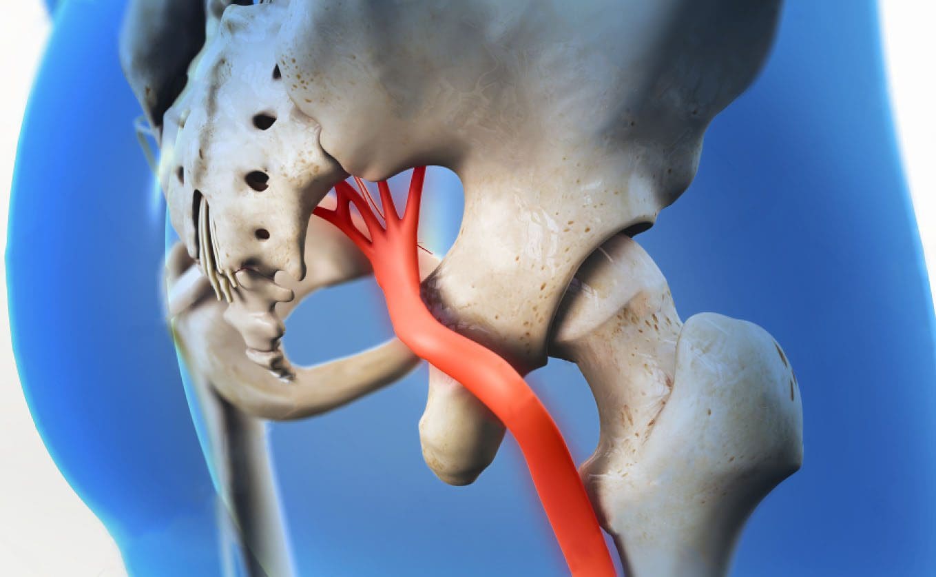
A herniated disc is a common spinal condition that typically affects the cervical spine (neck region) or the lumbar spine (lower back), although it can occur in any part of the spine. Most often, a herniated disc happens at the L4 � L5 and the L5 � S1.� This is because this portion of the spine, the lumbar region, bears the bulk of the body�s weight.
FDA recognizes and approves spinal decompression and its ability to eliminate herniated discs.
On the verge of back surgery, a mason discovered the non-surgical solution to work-related chronic back pain.
A new male patient who works in construction came to see me as a last resort to lessen his back pain brought on from damaged/herniated discs.
His primary caregiver recommended back surgery, but that would have put him on disability for months.
Fortunately, before saying yes to the surgery, a co-worker recommended chiropractic care.
Bricklayers/masons have the highest rate of back injuries with non-paid sick leave.
Constant bending over, even with a back brace, takes its toll on the spine, which in this case resulted in two herniated discs.
Pain medications helped in the beginning but with constant use, put him in a constant brain fog state, along with the expense, which took its toll on the family budget.
Disc Injury & Back Surgery
The doctor did not discuss spinal decompression therapy
�A non-surgical back treatment that slowly and gently stretches the spine.
This stretching lessens the pressure on the compressed nerve root (herniated disc) and results in less and even complete alleviation.
The patient came twice a week with myself and the team working on him over the course of a month, however, every case is different so treatments vary depending on the condition.
With each treatment, the two herniated discs were slowly reverted back to their natural position. This is able to be achieved with less pressure between the discs.
Towards the end of treatment, the patient’s pain was gone by about 90%.
With two weeks of rest, the patient was able to return to work.
The best part was that there was no surgery, pain medications, disability, and hospital bills.
Spine treatment alternative
Chiropractic/Decompression therapy is way less expensive than medication and surgery. It is:
Non-surgical
Recovery time is faster
Completely drug-free
People suffering every day with herniated/injured discs should consider the chiropractic decompression option. You do not have to learn to live with chronic back pain.
If you suffer from:
Herniated discs
Bulging discs
Degenerative disc disease
I encourage you to discuss the condition with an experienced chiropractor. There are many proven alternatives to back surgery and pain meds. People need to be aware of these alternatives for chronic back pain. The right-back pain treatment can definitely improve the quality of life.
Herniated Disc El Paso, TX
Sandra Rubio developed two herniated discs and a bulging disc after suffering from an accident at a young age, which caused her intense pain throughout her youth.
When she became a mother, her symptoms became severe.
After visiting doctors without results, Sandra found chiropractor Dr. Alex Jimenez and found relief from her sciatica and migraines.
The herniated disc treatment she received from Dr. Alex Jimenez was non-surgical.
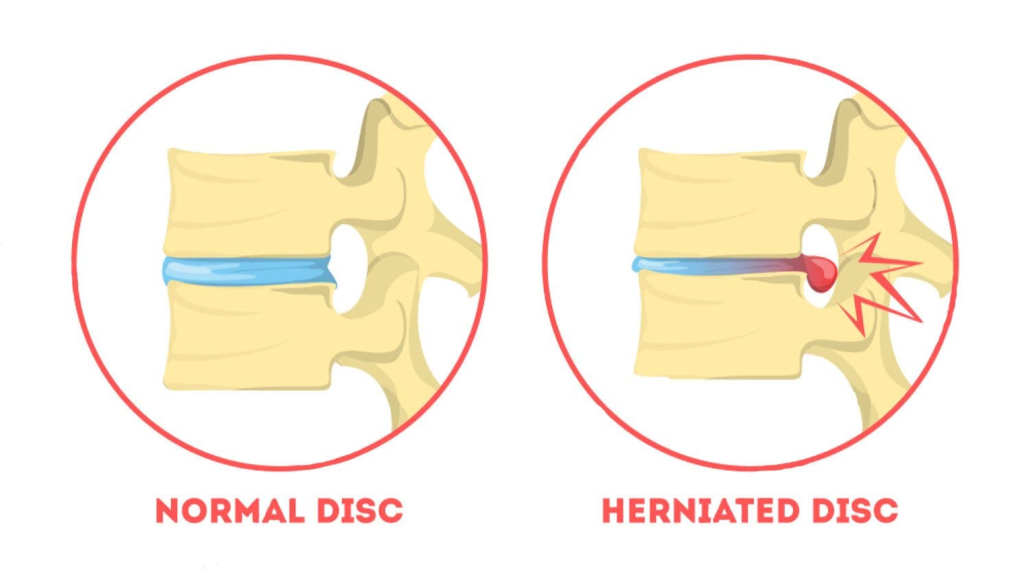
A herniated disc, also known as a slipped disc, is a medical condition in which:
Atear from the outer intervertebral disc allows the soft, central area to bulge out beyond the outer rings.
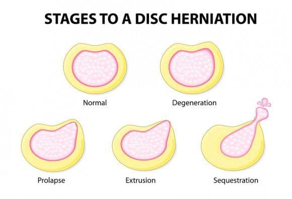
Disc herniation is usually a result of:
Degeneration (wear/tear)
Trauma (auto accident/sports injury)
Lifting injuries
Straining movement
The tear can release the compounds, which cause inflammation and can cause severe pain even if the nerve root not compressed.
A physical examination is usually the first step in diagnosing a herniated disc. The chiropractor will examine the spine while the patient is standing, and while they’re lying down. Depending on the severity and location of the herniation, they may note a decrease in spine curvature.
Radicular pain will be assessed, when the spine is:
Unmoving
In motion
With pressure applied
Other tests may be administered.
X-rays may also be taken, but an MRI is usually more accurate and shows more detail.
Chiropractic has been very effective in helping patients manage their pain and regain their mobility so they can return to their normal life. Therefore, it should be your first option for treatment before you go down the road with drugs or surgery.
NCBI Resources
It is often referred to as a ruptured disc or slipped disc and occurs when the disc moves or slips out of place. It can also be the result of a disc that has a small tear and is leaking the jelly-like substance that is inside. This can put pressure on the surrounding nerves, causing pain and discomfort.
IFM's Find A Practitioner tool is the largest referral network in Functional Medicine, created to help patients locate Functional Medicine practitioners anywhere in the world. IFM Certified Practitioners are listed first in the search results, given their extensive education in Functional Medicine