Discover the clinical approach for substance use disorder, a vital method in addressing challenges related to addiction treatment.
Integrative Management of Substance Use Disorder (SUD) and Musculoskeletal Health: A Collaborative Model for Chiropractors and Nurse Practitioners
Substance use disorder (SUD) is a chronic, treatable medical condition that affects the brain, behavior, and the entire body, including the musculoskeletal system. For many patients, SUD overlaps with chronic pain, injury, emotional distress, and functional limitations. An integrative care model that combines evidence‑based SUD screening and treatment with chiropractic care and nurse practitioner (NP)–led primary care can reduce risk, improve function, and support long‑term recovery (American Medical Association [AMA], n.d.; National Institute on Drug Abuse [NIDA], n.d.; National Institute of Mental Health [NIMH], 2025).
This article explains what SUD is, how it is identified and categorized, how clinicians can manage it using practical workflows, and how integrative chiropractic and NP care can address overlapping risk profiles and musculoskeletal consequences.
What Is Substance Use Disorder (SUD)?
SUD is a medical condition in which the use of alcohol, medications, or other substances leads to significant impairment or distress in daily life. It is not a moral failing or a lack of willpower; it is a chronic, brain‑ and body‑based disease that is treatable (NIDA, n.d.; NIMH, 2025).
SUD exists on a spectrum from mild to severe. People with SUD may:
Use more of the substance than they planned
Try and fail to cut down or stop
Spend a lot of time obtaining, using, or recovering from the substance
Continue to use even though it harms health, work, relationships, or safety (American Psychiatric Association, 2022; NIMH, 2025)
Person‑first, non‑stigmatizing language
Stigma can keep people from seeking care. Using respectful, person‑first language reduces shame and supports engagement. NIDA and the AMA recommend (NIDA, n.d.; AMA, n.d.):
Say “person with a substance use disorder,” not “addict” or “drug abuser.”
Say “substance use” or “misuse,” not “abuse.”
Focus on SUD as a chronic, treatable condition.
Categories and Diagnostic Features of SUD
DSM‑5‑TR framework: Mild, moderate, severe
Diagnostic criteria for SUD come from the Diagnostic and Statistical Manual of Mental Disorders, Fifth Edition, Text Revision (DSM‑5‑TR) (American Psychiatric Association, 2022; NIAAA, 2025). A diagnosis is based on the number of symptoms present over 12 months.
Typical criteria include (paraphrased):
Using more or for longer than intended
Unsuccessful efforts to cut down
Spending a lot of time obtaining, using, or recovering
Cravings or strong urges
Role failures at work, school, or home
Social or interpersonal problems caused or worsened by use
Giving up important activities
Using in physically hazardous situations
Continued use despite physical or psychological problems
Tolerance
Withdrawal
Severity is determined by symptom count (American Psychiatric Association, 2022; NIAAA, 2025):
Mild: 2–3 symptoms
Moderate: 4–5 symptoms
Severe: 6 or more symptoms
Substance‑specific categories
Clinically, SUD is further categorized by substance type (NIDA, n.d.; NIMH, 2025):
Alcohol use disorder (AUD)
Opioid use disorder (e.g., heroin, oxycodone, hydrocodone)
Stimulant use disorder (e.g., cocaine, methamphetamine)
Sedative, hypnotic, or anxiolytic use disorder (e.g., benzodiazepines)
Cannabis, tobacco, hallucinogen, or inhalant use disorders
Each category has similar behavioral criteria but unique medical risks, withdrawal profiles, and treatment options (NIDA, n.d.; NIAAA, 2025).
Risk and severity categories for clinical workflows
For practical care, validated screening tools classify risk that guide next steps (AMA, n.d.; NIDA, n.d.; NIAAA, 2025):
Low/no risk: Negative screen or very low scores
Moderate risk: At‑risk use with potential consequences (e.g., falls, crashes, future disease)
Substantial/severe risk: High scores suggest likely SUD and active harm
For example, adult risk zones using tools like AUDIT and DAST (AMA, n.d.):
Low risk/abstain: AUDIT 0–7; DAST 0–2
Moderate risk: AUDIT 8–15; DAST 3–5
Substantial/severe risk: AUDIT ≥16; DAST ≥6
These categories help teams decide when to give brief interventions, when to intensify care, and when to refer to specialty treatment.
Epidemiology and Public Health Impact
National surveys show that millions of people in the United States live with SUD, yet only a fraction receive treatment (Substance Abuse and Mental Health Services Administration [SAMHSA], 2023). The 2022 National Survey on Drug Use and Health reported high rates of both substance use and serious mental illness, often co‑occurring (SAMHSA, 2023).
Key points from recent federal data (SAMHSA, 2023; NIMH, 2025):
SUD commonly co‑occurs with depression, anxiety, and other mental disorders.
Co‑occurring conditions worsen medical outcomes and increase healthcare use.
Early identification and integrated treatment can improve function, reduce complications, and lower long‑term costs.
Identifying Patients With SUD: Screening and Assessment
Early, routine identification is critical. Primary care teams, NPs, and chiropractic clinics that integrate behavioral health can all play a role (AMA, n.d.; NIDA, n.d.; NIAAA, 2025).
Building a safe, trauma‑informed environment
Before asking about substance use, the team should (AMA, n.d.; NIDA, n.d.):
Explain that “we screen everyone” as part of whole‑person care.
Emphasize confidentiality within legal limits.
Use a calm, nonjudgmental tone and body language.
Offer patients the option not to answer any question.
Acknowledge that stress, trauma, pain, and life pressures often contribute to substance use.
This aligns with trauma‑informed care principles promoted by SAMHSA and helps patients feel safe enough to share (AMA, n.d.).
Validated screening tools
Evidence‑based tools are preferred over informal questioning. Common options include (AMA, n.d.; NIDA, n.d.; NIAAA, 2025):
For adults:
AUDIT or AUDIT‑C (Alcohol Use Disorders Identification Test) – screens for unhealthy alcohol use and risk of AUD.
DAST‑10 (Drug Abuse Screening Test) – screens for non‑alcohol drug use problems.
TAPS Tool (Tobacco, Alcohol, Prescription medication, and other Substances) – combined screen and brief assessment.
For adolescents:
CRAFFT 2.1+N – widely used for youth; captures risk behaviors and problems.
S2BI (Screening to Brief Intervention) and BSTAD – brief tools validated for ages 12–17 (NIDA, n.d.; AMA, n.d.).
For alcohol‑specific quick screens:
AUDIT‑C (3 questions) or full AUDIT
NIAAA Single Alcohol Screening Question (SASQ):
“How many times in the past year have you had 4 (for women) or 5 (for men) or more drinks in a day?” (NIAAA, 2025)
Results guide risk categorization and next steps.
Role of the care team
In integrated practices, roles can be divided (AMA, n.d.):
Medical assistants or nurses
Administer pre‑screens and full questionnaires.
Flag positive or concerning responses.
Nurse practitioners / primary care clinicians
Review screening results.
Deliver brief interventions using motivational interviewing.
Conduct or oversee further assessment.
Prescribe and manage pharmacotherapy for SUD when indicated.
Coordinate referrals and follow‑up.
Behavioral health clinicians (on‑site or virtual)
Perform biopsychosocial in-depth evaluations.
Provide psychotherapy and relapse‑prevention skills.
Support motivational enhancement and family engagement.
Chiropractors and physical‑medicine providers
Screen for substance misuse related to pain, function, and injury patterns.
Observe red flags (frequent lost prescriptions, inconsistent pain reports, sedation, falls).
Communicate concerns to the NP or primary medical provider.
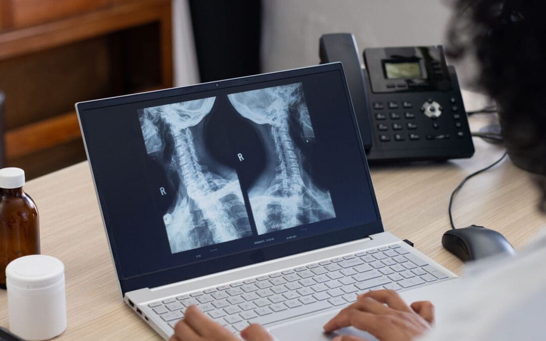
Dr. Alexander Jimenez, DC, APRN, FNP‑BC, exemplifies this dual role. As both a chiropractor and a family practice NP, he combines neuromusculoskeletal assessment with medical screening and functional medicine evaluation to identify root causes of chronic pain and unhealthy substance use patterns (Jimenez, n.d.).
Clinical clues that may suggest SUD
Beyond formal tools, clinicians should stay alert for patterns such as (AMA, n.d.; NIMH, 2025):
Frequent injuries, falls, or motor vehicle accidents
Repeated missed appointments or poor adherence to treatment
Drowsiness, agitation, slurred speech, or odor of alcohol
Unexplained weight loss, infections, or liver abnormalities
Social and financial instability, job loss, or legal problems
In chiropractic and musculoskeletal settings, repeated injuries, delayed healing, inconsistent exam findings, or “pain behaviors” that do not match imaging or biomechanics may prompt gentle, supportive screening and medical referral.
Understanding Long Lasting Injuries- Video
Comprehensive Assessment and Risk Stratification
Once a screen is positive, the next level is a more detailed assessment. This should examine substance type, frequency, amount, impact, withdrawal, mental health, physical comorbidities, and function (AMA, n.d.; NIMH, 2025).
Structured assessment tools
Clinicians may use (AMA, n.d.; NIDA, n.d.; NIAAA, 2025):
Full AUDIT for alcohol
DAST‑10 for general drugs
CRAFFT or GAIN for adolescents
Checklists based directly on DSM‑5‑TR criteria to rate symptom count and severity (NIAAA, 2025).
These tools allow classification into mild, moderate, or severe SUD and support shared decision‑making regarding level of care.
Co‑occurring mental health conditions
SUD frequently co‑occurs with (NIMH, 2025):
Major depressive disorder
Anxiety disorders
Posttraumatic stress disorder (PTSD)
Bipolar disorder
Attention‑deficit/hyperactivity disorder
Co‑occurring disorders can:
Increased risk for self‑medication with substances
Worsen treatment outcomes if not recognized
Require integrated treatment plans (NIMH, 2025)
NPs, behavioral health clinicians, and chiropractors with integrative training should maintain a low threshold for mental health screening and referral.
Managing Patients With SUD: A Practical Clinical Process
Effective SUD care is chronic‑disease care: ongoing, team‑based, and tailored to readiness to change (AMA, n.d.; SAMHSA, 2023).
Core elements of management
Key components include (AMA, n.d.; NIDA, n.d.; NIMH, 2025):
Routine screening and re‑screening
Brief interventions and motivational interviewing
Harm‑reduction strategies
Medications for certain SUDs (when appropriate)
Evidence‑based behavioral therapies
Peer and family support
Long‑term follow‑up and relapse‑prevention planning
Brief intervention and motivational interviewing
For patients with low to moderate risk, brief intervention can be delivered in 5–15 minutes and often by NPs or primary care clinicians (AMA, n.d.; NIAAA, 2025). Using motivational interviewing, clinicians:
Ask open‑ended questions (“What do you enjoy about drinking? What concerns you about it?”)
Reflect and summarize the patient’s own statements
Ask permission before giving advice
Help patients set realistic, patient‑chosen goals (cutting down, abstaining, or seeking treatment)
This approach respects autonomy and builds internal motivation for change.
Determining level of care
The American Society of Addiction Medicine (ASAM) describes a continuum of care (AMA, n.d.; SAMHSA, 2023):
Prevention/early intervention
Brief interventions in primary care
Self‑management support and education
Outpatient services
Office‑based counseling and medications for AUD or opioid use disorder (OUD)
Integrated behavioral health visits
Intensive outpatient / partial hospitalization
Several therapy sessions per week, day or evening programs
Residential/inpatient services
24‑hour structured care for severe or complex cases
Medically managed intensive inpatient services
Medically supervised detoxification and stabilization
NPs and primary care teams decide the appropriate level based on risk severity, co‑occurring medical and psychiatric conditions, social supports, and patient preference (AMA, n.d.; NIMH, 2025).
Medications for SUD
For some patients, medications support recovery by reducing cravings, blocking rewarding effects, or stabilizing brain function (SAMHSA, 2020; AMA, n.d.; NIAAA, 2025). Examples include:
Alcohol use disorder
Acamprosate – supports abstinence after detox
Disulfiram – creates an unpleasant reaction to alcohol, discouraging use
Naltrexone blocks the rewarding effects of alcohol
Opioid use disorder
Buprenorphine – a partial opioid agonist that reduces cravings and overdose risk; often prescribed in primary care with appropriate DEA registration
Methadone – full agonist, dispensed in specialized opioid treatment programs
Naltrexone (extended‑release) – opioid antagonist that prevents relapse after detox
Overdose prevention
Naloxone – rapid opioid‑overdose reversal, recommended for anyone at risk (AMA, n.d.).
NPs managing patients with SUD work within state scope‑of‑practice rules and in collaboration with addiction specialists where needed.
Behavioral therapies and peer support
Evidence‑based therapies include (AMA, n.d.; NIDA, n.d.):
Cognitive behavioral therapy (CBT)
Dialectical behavior therapy (DBT)
Motivational enhancement therapy
The Matrix Model (especially for stimulants)
Family‑based therapy for adolescents
Peer support groups (Alcoholics Anonymous, Narcotics Anonymous, SMART Recovery) can reinforce coping skills, hope, and accountability.
Long‑term follow‑up
SUD is chronic; relapse risk can persist for years. Best practice includes (AMA, n.d.; NIMH, 2025):
Follow‑up within 2 weeks after treatment initiation
Monthly to quarterly visits as patients stabilize
Peer support and care management between visits
Rapid re‑engagement after any relapse or lapse
NASW, NIDA, and NIMH stress that relapse should be treated as a signal to adjust care—not as failure (NIDA, n.d.; NIMH, 2025).
How SUD Affects the Body and the Musculoskeletal System
SUD impacts nearly every organ system. Many effects directly or indirectly worsen neuromusculoskeletal health and pain.
General systemic effects
Common systemic consequences include (NIDA, n.d.; NIMH, 2025; SAMHSA, 2023):
Cardiovascular disease and hypertension
Liver disease and pancreatitis (especially with alcohol)
Respiratory disease (especially with tobacco and some drugs)
Endocrine and hormonal disruption
Immune dysfunction and higher infection risk
Sleep disturbances and fatigue
Worsening of mood, anxiety, and cognitive function
These changes affect healing capacity, resilience, and the way patients perceive pain.
Musculoskeletal and pain‑related effects
Substance use and SUD can influence the musculoskeletal system through several pathways:
Increased injury risk
Impaired judgment, coordination, and reaction time increase the risk of falls, motor vehicle accidents, and sports injuries.
Heavy alcohol use is associated with fractures, soft tissue injuries, and delayed healing (AMA, n.d.; SAMHSA, 2023).
Bone, joint, and muscle changes
Alcohol and some drugs can impair bone density and quality, increasing osteoporosis and fracture risk.
Nutritional deficiencies associated with SUDs weaken connective tissue and muscle function.
Sedentary behavior and deconditioning are common in people with long‑standing SUD.
Chronic pain and central sensitization
Chronic alcohol or opioid use can alter pain pathways in the central nervous system, raising pain sensitivity.
Opioid‑induced hyperalgesia can make pain seem worse even at stable or increasing doses.
Functional and ergonomic stress
Disrupted sleep, poor posture, and prolonged sitting or immobility (for example, in recovery environments or during unemployment) can lead to spinal stress, neck and low back pain, and muscle imbalance.
Clinically, Dr. Jimenez and similar integrative providers often see patients with combined profiles: chronic low back or neck pain, sedentary work, ergonomic strain, poor sleep, high stress, and escalating reliance on medications, including opioids or sedatives. Addressing both the mechanical and behavioral contributors can change the trajectory of pain and SUD risk (Jimenez, n.d.).
Integrative Chiropractic Care in the Context of SUD
Philosophy of integrative chiropractic care
Integrative chiropractic care focuses on restoring alignment, mobility, and neuromuscular control while considering lifestyle, nutrition, sleep, and emotional stress. In the model used by Dr. Jimenez, chiropractic adjustments are combined with functional medicine strategies, targeted exercise, and collaborative medical care (Jimenez, n.d.).
For patients with or at risk of SUD, this approach offers:
Non‑pharmacologic pain management
Improved movement, posture, and ergonomics
Education that empowers patients to self‑manage pain
Reduced reliance on habit‑forming medications
Spinal adjustments and targeted exercises
Spinal and extremity adjustments aim to:
Restore joint mobility
Reduce mechanical irritation of nerves and soft tissues
Improve segmental alignment and overall posture
Targeted exercises are prescribed to:
Strengthen deep stabilizing muscles (core, gluteal, cervical stabilizers)
Correct muscle imbalances and faulty patterns
Increase flexibility and joint range of motion
Enhance proprioception, balance, and movement control
Examples of targeted exercise strategies often used in integrative chiropractic and rehab clinics include (Jimenez, n.d.):
Lumbar stabilization and core‑strengthening sequences
Hip mobility and glute activation drills for low back and sciatica‑like pain
Cervical and scapular stabilization for neck and shoulder pain
Postural retraining, including ergonomic break routines for prolonged sitting
By reducing biomechanical stress and enhancing functional capacity, these interventions may decrease pain intensity, frequency, and flare‑ups, which in turn can lower the drive to self‑medicate with substances.
Reducing overlapping risk profiles
Many risk factors for SUD and for chronic musculoskeletal pain overlap, including (NIMH, 2025; NIDA, n.d.; Jimenez, n.d.):
Chronic stress and trauma
Poor sleep and circadian disruption
Sedentary lifestyle and obesity
Repetitive strain and poor ergonomics
Social isolation and low self‑efficacy
Integrative chiropractic care can help shift these shared risk profiles by:
Encouraging regular physical activity and graded movement
Coaching ergonomic and postural strategies at work and home
Teaching breathing, stretching, and relaxation routines that reduce muscle tension and sympathetic overdrive
Collaborating with NPs and behavioral health clinicians to align interventions with mental health and SUD treatment plans
In Dr. Jimenez’s practice, this often includes structured flexibility, mobility, and agility programs that are adapted to age and functional status, with close monitoring to avoid over‑reliance on medications, including opioids and sedatives (Jimenez, n.d.).
The Nurse Practitioner’s Role in Comprehensive SUD and Musculoskeletal Care
NPs are well-positioned to coordinate SUD care and integrate it with musculoskeletal and chiropractic treatment.
Comprehensive medical management
NP responsibilities typically include (AMA, n.d.; NIMH, 2025; NIAAA, 2025):
Conducting and interpreting SUD screening and risk stratification
Performing physical exams and ordering labs or imaging
Diagnosing SUD and co‑occurring conditions
Prescribing non‑addictive pain strategies and medications where indicated
Managing or co‑managing medications for AUD or OUD (per training and regulations)
Monitoring for drug–drug and drug–disease interactions
Coordinating with behavioral health and community resources
In integrative settings like Dr. Jimenez’s clinic, the NP role is blended with functional medicine principles, looking at nutrition, metabolic health, hormonal balance, and inflammation that influence both pain and SUD risk (Jimenez, n.d.).
Activity pacing and graded return to work or sport
Sleep hygiene and circadian rhythm support
Nutrition strategies that support musculoskeletal healing and brain health
These interventions lower the mechanical load on the spine and joints, reduce fatigue, and increase a patient’s sense of control—all of which help reduce triggers for substance use and relapse.
Care coordination and team communication
NPs often serve as the central coordinator who (AMA, n.d.; NIMH, 2025):
Ensures all team members (chiropractor, physical therapist, behavioral health, addiction medicine, primary care, or specialty providers) share a coherent plan
Tracks progress on pain, function, substance use, mood, and quality of life
Adjusts the plan as conditions change
Supports families and caregivers in understanding both SUD and musculoskeletal needs
In a model like Dr. Jimenez’s, this may involve regular case conferences, shared EHR notes, and integrated treatment plans that align spinal rehabilitation with SUD recovery goals (Jimenez, n.d.).
Practical Clinical Pathway: From First Contact to Long‑Term Recovery
For clinics that combine chiropractic and NP services, a practical, stepwise pathway for patients with possible SUD and musculoskeletal complaints can look like this (AMA, n.d.; NIDA, n.d.; NIAAA, 2025; NIMH, 2025; Jimenez, n.d.):
Step 1: Initial visit and global screening
Intake includes questions on pain, function, injuries, sleep, mood, and substance use.
Staff administer brief tools (for example, AUDIT‑C and DAST‑10 for adults, CRAFFT for adolescents).
The chiropractor documents neuromusculoskeletal findings; the NP reviews medical and behavioral health risks.
Step 2: Identification of SUD risk
Negative or low‑risk screens → brief positive health message and reinforcement of low‑risk behavior.
Moderate risk → NP provides brief intervention, motivational interviewing, and a follow‑up plan.
Substantial or severe risk → NP initiates comprehensive assessment, safety planning, and possible referral to specialized services.
Step 3: Integrated treatment planning
The team crafts a unified plan that may include:
Spinal adjustments and targeted exercises to correct alignment and biomechanics
Gradual increase in physical activity with pain‑sensitive pacing
Behavioral health referral for CBT, trauma‑informed treatment, or other modalities
Consideration of medications for AUD or OUD, if indicated
Harm‑reduction measures (for example, naloxone prescription for those at overdose risk)
Step 4: Ergonomics and lifestyle
NP and chiropractor jointly review workplace and home ergonomics, posture, and activity patterns.
Patients learn micro‑break routines, stretching, and strengthening sequences for high‑risk tasks (for example, lifting or prolonged sitting).
Nutrition, stress‑management, and sleep interventions are introduced or refined.
Step 5: Monitoring and long‑term follow‑up
Regular follow‑up visits evaluate:
Pain levels and functional capacity
Substance use patterns and cravings
Mood, sleep, and quality of life
Adherence to exercise and ergonomic plans
The team updates the treatment plan to respond to progress, setbacks, or new diagnoses.
Patients are coached to view flare-ups or lapses as opportunities to learn and adjust, not as failures.
This kind of coordinated, integrative approach can reduce repeated injuries, unnecessary imaging or surgeries, and long‑term dependence on medications, including opioids.
Clinical Insights from an Integrative Practice Model
Although each practice is unique, Dr. Alexander Jimenez’s clinic illustrates several principles that can guide others (Jimenez, n.d.):
Whole‑person assessment: History taking includes injuries, lifestyle, trauma, nutrition, environment, and psychosocial stressors.
Functional movement focus: Care plans emphasize flexibility, mobility, agility, and strength to restore capacity rather than just relieve symptoms.
Non‑invasive first: Chiropractic adjustments, functional exercise, and lifestyle interventions are prioritized before invasive procedures or long‑term controlled substances.
Integrated roles: As both DC and FNP‑BC, Dr. Jimenez unifies neuromusculoskeletal, primary care, and functional medicine perspectives in a single, coordinated plan.
Patient empowerment: Education, coaching, and accessible care options help patients take a proactive role in maintaining spinal health and reducing SUD risk.
This model aligns with national guidance on behavioral health integration and SUD management in medical settings while adding the musculoskeletal and ergonomic expertise of chiropractic care (AMA, n.d.; NIDA, n.d.; NIMH, 2025).
Key Takeaways
SUD is a chronic, treatable medical condition that often co‑occurs with mental disorders and chronic pain.
Validated screening tools and non‑stigmatizing, trauma‑informed communication are core to early identification.
Risk and severity categories (mild, moderate, severe) guide brief intervention, level of care, and referral decisions.
SUD significantly affects the body, including bone health, soft tissue integrity, injury risk, and chronic pain pathways.
Integrative chiropractic care—with spinal adjustments, targeted exercises, and ergonomic guidance—can reduce pain, improve function, and lower overlapping risk factors for SUD.
Nurse practitioners provide comprehensive SUD management, coordinate care, and deliver ergonomic and lifestyle counseling that complements chiropractic treatment.
A collaborative, long‑term, patient‑centered model—such as the one exemplified by Dr. Alexander Jimenez—offers a promising pathway to healthier spines, healthier brains, and healthier lives.
Conclusion
Substance use disorder is a complex medical condition that requires compassion, evidence‑based screening, and coordinated care across multiple disciplines. For healthcare professionals—whether chiropractors, nurse practitioners, primary care physicians, or behavioral health specialists—the opportunity to identify and support patients with SUD begins with understanding what it is, how to recognize it, and how to respond with respect and proven interventions.
The integration of chiropractic care and nurse practitioner-led primary care offers a distinctive advantage for patients struggling with both chronic pain and substance use. When a patient presents with a work injury, auto accident, or years of poor ergonomics, they may not volunteer that they are also wrestling with alcohol dependence, prescription opioid misuse, or stimulant use. Yet these challenges often coexist. The musculoskeletal system bears the weight of increased fracture risk, muscle wasting, poor healing, and heightened pain sensitivity. The mind and nervous system are equally affected, with sleep disruption, mood changes, and reduced resilience to stress all fueling the cycle of pain and substance use.
Clinics and practices that integrate screening, brief intervention, and coordinated treatment have a powerful tool to interrupt this cycle. Spinal adjustments restore mechanical function. Targeted exercises rebuild strength and proprioception. Ergonomic guidance prevents re‑injury. Nurse practitioners coordinate medications, monitor for drug interactions, and counsel on lifestyle factors that support both spine health and recovery from SUD. Behavioral health clinicians provide therapy, peer support, and relapse prevention. Together, this team addresses root causes, not just symptoms.
The clinical model exemplified by providers like Dr. Alexander Jimenez demonstrates that a single clinician with dual expertise—chiropractic and family practice nurse practitioner credentials—can seamlessly weave these threads into a coherent, patient‑centered plan. Patients benefit from continuity, alignment of goals, and a provider who understands both the biomechanics of a herniated disc and the neurobiology of addiction. Larger practices can achieve similar results through deliberate team communication, shared decision‑making, and a commitment to non‑stigmatizing, trauma‑informed care.
The evidence is clear: early identification saves lives and improves outcomes. Validated screening tools are quick and accurate. Motivational interviewing and brief interventions work. Medications for alcohol and opioid use disorders are safe and effective when used thoughtfully. Non‑pharmacologic approaches—exercise, manual therapy, stress management, social support—are powerful and underutilized. And when musculoskeletal and behavioral health care are woven together, patients heal faster, return to function sooner, and are far less likely to relapse into substance misuse.
For healthcare teams willing to expand their lens beyond isolated complaints—beyond “just” back pain or “just” anxiety—the reward is profound: patients who reclaim their health, their relationships, and their sense of purpose. This is the promise of integrative, collaborative, evidence‑based care for substance use disorder and musculoskeletal health.
References
American Medical Association. (n.d.). Substance use disorder treatment: How‑to guide for primary care integration [PDF]. American Medical Association.
American Psychiatric Association. (2022). Diagnostic and statistical manual of mental disorders (5th ed., text rev.). American Psychiatric Publishing.
Jimenez, A. D. (n.d.). Injury specialists: El Paso family practice nurse practitioner and chiropractor. Dr. Alex Jimenez. https://dralexjimenez.com/
Substance Abuse and Mental Health Services Administration. (2023). 2022 national survey on drug use and health: Annual national report (HHS Publication No. PEP23‑07‑01‑006). U.S. Department of Health and Human Services. https://www.samhsa.gov/data/report/2022-nsduh-annual-national-report
Real-Life Posture Rehab: How El Paso Back Clinic Helps You Move Better Every Day
Move around and change posture positions throughout the day.
Improving posture is one of the fastest ways to feel stronger, breathe easier, and protect your spine—especially if you live with long commutes, heavy work, or hours at a desk, like many people in El Paso. At El Paso Back Clinic, Dr. Alexander Jimenez, DC, APRN, FNP-BC, and his team see every day how targeted physical activity, along with integrative chiropractic and nurse practitioner (NP) care, can turn slouching and stiffness into confident, upright movement. El Paso, TX Doctor Of Chiropractic+1
This article explains, in simple language:
What good posture really is
Recommended physical activities and exercises to enhance posture
How yoga, Pilates, and mind-body practices improve alignment
Easy desk and “tech neck” fixes
How integrative chiropractic care supports posture
How nurse practitioners help with medical, ergonomic, and lifestyle support
How the El Paso Back Clinic combines all of this in real-world care
What “Good Posture” Means (and Why It Matters in Daily Life)
Good posture means your body is stacked in a natural, balanced way:
Ears over shoulders
Shoulders over hips
Hips over knees and ankles
Spine holding its natural curves (neck, mid-back, low back)
When posture is poor—like slouching over a phone or leaning forward at a desk—stress builds up in your neck, shoulders, and back. Over time, this can lead to:
Chronic neck and back pain
Tension headaches
Fatigue and shallow breathing
Tight hip flexors and weak glutes
Early joint wear and tear
Research and clinical guides show that specific exercises and posture-friendly habits can reduce pain and improve alignment by strengthening postural muscles and keeping you moving throughout the day. Healthline+2Harvard Health+2
At El Paso Back Clinic, Dr. Jimenez often reminds patients that posture is not about “standing stiff.” It is about a strong, relaxed, and mobile spine that can handle work, sports, and life in the desert heat. El Paso, TX Doctor Of Chiropractic+1
Core Principles of Posture-Focused Exercise
Most effective posture plans share the same core goals:
Strengthen the core and back—so your spine has solid support
Activate glutes and shoulders—to counter slumping and hip stress
Improve flexibility—especially in chest, hip flexors, and hamstrings
Train body awareness—so you notice and correct slouching
Add low-impact cardio—to boost circulation and recovery
Think of Your Program in Simple Pieces
Try to include each week:
2–3 days of core and back strengthening
2–3 days of mobility and stretching
2–4 days of low-impact cardio like walking or swimming
Daily micro-breaks from sitting or driving
That may sound like a lot, but many of these can be done in 10–20 minute blocks and woven into your normal day.
Foundational Strength Exercises for Better Posture
Many posture programs start with bodyweight moves you can do at home—no machines, no fancy equipment. Sources on physical therapy and spine health support these exercises. Healthline+2Primal Physical Therapy+2
Planks (Front and Side Planks)
Why they help: Planks strengthen your deep core, shoulders, and glutes. A strong core keeps your spine from sagging or arching too much.
Basic front plank:
Start on your forearms and toes
Keep your body in a straight line from head to heels
Gently pull your belly toward your spine
Hold 20–30 seconds, rest, repeat 2–3 times
Side planks add extra stability for your sides and hips, which support upright posture. Woodlands Sports Medicine
Bird-Dog
Why it helps: Bird-dog builds core and back strength while training balance and control.
How to do it:
Start on hands and knees
Extend your right arm forward and left leg back
Keep your hips level; don’t twist
Hold 3–5 seconds, then switch sides
Do 8–10 reps per side
Physical therapists often use this exercise to improve posture and relieve back pain. Primal Physical Therapy+1
Glute Bridges
Why they help: Bridges work the glutes and hamstrings and relieve stress on the lower back.
Lie on your back, knees bent, feet flat
Press through your heels and lift your hips
Squeeze your glutes at the top
Hold 3–5 seconds, then lower
Repeat 10–15 times
Strong glutes help balance tight hip flexors from long periods of sitting, which is very common among drivers and office workers in El Paso. Primal Physical Therapy+1
Superman Exercise
Why it helps: The Superman move targets the “posterior chain,” the muscles along the back of your body that help prevent slouching. Woman & Home
Lie face down
Lift your chest, arms, and legs slightly off the floor
Hold briefly and lower with control
Start with 5–8 reps
This move is especially useful if you sit a lot or look down at screens, as it helps your back muscles stay active.
Rowing Movements (Bands or Dumbbells)
Why they help: Rowing exercises strengthen the upper back and shoulder stabilizers that pull your shoulders back.
Use a resistance band or light dumbbells
Pull your elbows back and squeeze your shoulder blades together
Mobility and Stretching: Releasing the “Posture Brakes”
If strength is the “engine,” tight muscles are the “brakes.” You need both to work well. Stretching and mobility exercises help open areas that tend to tighten up, such as the chest, neck, hips, and upper back. Illinois Back Institute+1
Key Posture Stretches
Chest Opens / Doorway Stretch
Stand in a doorway with your forearms on the frame
Gently lean forward until you feel a stretch across your chest
Gently slide your chin straight back (like a mini “double chin”)
Hold 3–5 seconds
Repeat 10 times
Cat-Cow
On hands and knees
Slowly round your back toward the ceiling, then gently arch it
Move with your breath for 8–10 cycles
Hip Flexor Stretch
In a half-kneeling position, gently shift your hips forward
Keep your torso upright; avoid over-arching your back
Hold 20–30 seconds on each side
These stretches are simple but powerful when done daily—especially if you spend long hours driving I-10 or sitting at a workstation in El Paso. Illinois Back Institute+1
Mind-Body Practices: Yoga, Pilates, and Tai Chi
Mind-body exercises are excellent for posture because they combine strength, flexibility, and body awareness.
Yoga for Alignment and Awareness
Yoga routines often include:
Mountain Pose (Tadasana)—teaches what upright alignment feels like
Child’s Pose and Cat-Cow – gently move and decompress the spine
Bridge Pose – strengthens glutes and back
Chest opener poses—counter phone and computer hunching
Research-based guides show yoga can improve postural muscle endurance and help people become more aware of how they carry themselves. Healthline+1
Pilates for Core Control
Pilates focuses on:
Deep core strength
Controlled breathing
Smooth, precise movements
Many physical therapy and rehab programs use Pilates-style exercises to support spinal alignment and postural stability. Primal Physical Therapy+1
Tai Chi for Balance and Relaxed Upright Posture
Tai chi uses slow, flowing movements with calm breathing. It helps:
Improve balance and coordination
Encourage relaxed, upright posture
Reduce stress and muscle guarding
Chiropractic resources often recommend swimming, walking, yoga, and tai chi as ideal companions to chiropractic care. Muscle and Joint Chiropractic+1
Everyday Physical Activities That Support Posture
You don’t have to become a gym athlete to help your posture. Many everyday activities, done with good form, support a healthier spine.
Helpful posture-friendly options include:
Walking:
Encourages natural spinal motion
Easy to fit into breaks or evenings
Swimming:
Full-body, low-impact workout
Strengthens back and shoulder muscles with less joint stress
Dancing:
Builds coordination and body awareness
Helps you practice an upright chest and an active core
Cycling (with proper bike fit):
Strengthens hips and legs
Supports overall fitness and endurance
Clinics that treat back pain often highlight walking and swimming as key activities for long-term spinal health. Illinois Pain & Spine Institute+1
Desk, Phone, and “Tech Neck”: Quick Fixes You Can Actually Use
Long hours on a computer or phone are a major reason posture has become such a problem. Harvard Health and orthopedic clinics stress the importance of frequent movement breaks and simple desk exercises. Harvard Health+2barringtonortho.com+2
Desk-Friendly Posture Break Routine
Try this mini-routine a few times each day:
Chin tucks – 10 reps
Shoulder blade squeezes – hold 5 seconds × 10 reps
Seated Cat-Cow – 5–10 slow breaths
Forward fold stretch next to your desk—hold 20–30 seconds
Simple Ergonomic Tips
Keep feet flat on the floor
Hips and knees are near 90 degrees
Screen at or just below eye level
Use a small lumbar support or rolled towel behind your low back
Stand and walk at least every 30–60 minutes
Recent expert tips also support using standing desks, wireless headphones for “walking meetings,” and light resistance bands at your station to keep postural muscles awake. Harvard Health+1
How Integrative Chiropractic Care at El Paso Back Clinic Supports Posture
Chiropractic care focuses on the spine, joints, and nervous system. Integrative chiropractic care goes further, combining adjustments with corrective exercises, lifestyle coaching, and medical input from NPs. Advanced Spine & Posture+1
What a Posture-Focused Chiropractic Visit Often Includes
The Nurse Practitioner’s Role in Supporting Posture
At El Paso Back Clinic, Dr. Jimenez works not only as a chiropractor but also as a board-certified family nurse practitioner, which provides a broader, medically informed perspective on posture-related problems. El Paso, TX Doctor Of Chiropractic+1
A nurse practitioner can:
Review your full medical history
Identify arthritis, osteoporosis, nerve issues, or autoimmune conditions that affect posture.
Order and interpret imaging and labs
X-rays, MRIs, and blood work when appropriate
Prescribe or adjust medications
Short-term pain or muscle-relaxant use when necessary
Coordinate referrals
Physical therapy, pain management, and surgical consults if needed
Give lifestyle and ergonomic counseling
Weight management, sleep, stress, and work setup
Use telemedicine for follow-up
To keep you on track with your exercise and pain management plan
This integrative model makes it easier to catch red flags early, adjust plans safely, and provide each patient with a personalized path rather than a one-size-fits-all list of exercises.
How Dr. Alexander Jimenez Combines Physical Activity, Chiropractic Care, and NP Expertise
With decades of experience in personal injury, sports, and functional medicine, Dr. Jimenez has seen the same pattern again and again: posture improves the most when hands-on care, smart exercise, and patient education are combined. El Paso, TX Doctor Of Chiropractic+2El Paso, TX Doctor Of Chiropractic+2
In his clinical observations at El Paso Back Clinic:
Agility and functional training (such as controlled squats, lunges, and balance drills) help patients return to sports, warehouse work, or family life with greater resilience.
Posture work is often integrated with nutrition, sleep, and stress management, because tired, inflamed bodies struggle to maintain good alignment. El Paso, TX Doctor Of Chiropractic+1
This dual license (DC + APRN, FNP-BC) allows Dr. Jimenez to move comfortably between spine mechanics and whole-person health, which is ideal for complex posture and pain cases.
Sample Weekly Posture-Boosting Plan (General Example)
This is a general example for educational purposes, not a personal prescription. Always consult your provider—especially if you have pain, injuries, or medical conditions.
Posture check around your home and car: adjust chairs, pillows, and monitor height
Patients at El Paso Back Clinic often have a plan customized to their injury type (auto accident, work injury, or sports strain) and their job or sport. El Paso Back Clinic® • 915-850-0900+1
Safety Tips: When to Get Help
Stop and get professional care if posture exercises cause:
Sharp or stabbing pain
Numbness or tingling in arms or legs
New weakness or loss of coordination
Trouble walking or standing
Loss of bladder or bowel control (emergency—seek urgent care)
A chiropractor can evaluate your spine and joints; a nurse practitioner can check for underlying medical causes. At El Paso Back Clinic, the team works together to decide whether you need imaging, medication, rehab, or a referral to another specialist. El Paso, TX Doctor Of Chiropractic+1
Bringing It All Together
To enhance posture and protect your spine:
Strengthen your core, back, and glutes with planks, bridges, bird-dogs, rows, and Supermans
Stretch your chest, neck, and hips to release tight, “slouching” muscles
Use mind-body practices like yoga, Pilates, and tai chi to build body awareness
Add low-impact activities like walking and swimming to support overall spine health
Fix your desk and phone habits with regular movement breaks and better ergonomics
At El Paso Back Clinic, integrative chiropractic care and nurse practitioner support bring all of these pieces together. With Dr. Alexander Jimenez’s dual training, patients receive:
Spinal and joint adjustments
Corrective exercise and posture coaching
Medical evaluation, imaging, and medication management when needed
Telemedicine and follow-up plans that fit real life in El Paso
The goal is simple: help you stand taller, move with less pain, and feel stronger in everything you do—from lifting kids or boxes at work to walking the trails of the Franklin Mountains.
Learn about the role of functional wellness in addressing autoimmune conditions and supporting overall health.
Understanding Autoimmune Conditions: How Functional Wellness Can Transform Your Health
Living with an autoimmune condition can feel overwhelming, but emerging research shows that functional wellness approaches offer powerful tools for managing symptoms and improving quality of life. This comprehensive guide explores how the immune system works, what happens when it malfunctions, and how nonsurgical treatments like chiropractic care and acupuncture, combined with holistic approaches, can help you reclaim your health and vitality.
Understanding Your Immune System: Your Body’s Defense Network
The immune system serves as your body’s primary defense mechanism, constantly working to protect you from harmful invaders such as bacteria, viruses, fungi, and parasites (Better Health Victoria, 1999; Medical News Today, 2025). This complex network consists of specialized cells, organs, proteins, and tissues that work together to keep you healthy and functioning optimally.
The Components of Your Immune System
Your immune system includes several key components that work together seamlessly. White blood cells, also called leukocytes, are key players in immune defense (Better Health Victoria, 1999). These cells move through blood and tissue throughout your body, constantly searching for foreign invaders. When they detect threats, they launch an immune attack to protect your health.
The bone marrow produces red blood cells that carry oxygen, white blood cells that fight infection, and platelets that help blood clot (Better Health Victoria, 1999). The thymus filters and monitors blood content while producing specialized white blood cells called T-lymphocytes (Better Health Victoria, 1999). The lymphatic system, a network of delicate tubes throughout the body, manages fluid levels, responds to bacteria, removes cancer cells, and absorbs fats from the intestine (Better Health Victoria, 1999).
How the Immune System Works
The immune system operates through two subsystems: the innate and adaptive immune systems (NCBI, 2023). The innate immune system provides general defense against harmful germs and substances using immune cells such as natural killer cells and phagocytes. The adaptive immune system creates specific responses to particular invaders, helping the body remember and recognize previous threats.
B lymphocytes produce antibodies and help alert T lymphocytes (Medical News Today, 2025). These antibodies are special proteins that lock onto specific antigens, marking them for destruction. T lymphocytes destroy compromised cells in the body and help alert other leukocytes (Medical News Today, 2025). Helper T cells coordinate the immune response by communicating with other cells, while killer T cells attack infected cells directly.
When functioning properly, the immune system can distinguish healthy tissue from unwanted substances (Medical News Today, 2025). If it detects an undesirable substance, it mounts an immune response—a complex attack to protect the body from invaders. This remarkable system enables your body to defend itself while maintaining normal function.
The Critical Role of Inflammation in Health and Disease
Inflammation represents the body’s natural response to injury, infection, or harmful stimuli. While acute inflammation serves as a protective mechanism, chronic inflammation can contribute to numerous health problems, including autoimmune conditions (Frontiers in Immunology, 2023).
Understanding the Inflammatory Process
When your body experiences external adverse stimuli, it triggers innate immunity and inflammation, followed by adaptive immunity (Frontiers in Immunology, 2023). This process involves the release of inflammatory mediators, including cytokines and inflammasomes, which play important roles in mediating immune responses through innate cells such as macrophages and adaptive cells such as T and B cells (Frontiers in Immunology, 2023).
Pro-inflammatory cytokines such as TNF-α, IL-1β, and IL-6 promote inflammation, while anti-inflammatory cytokines work to resolve it. The balance between these opposing forces determines whether inflammation helps or harms the body. In autoimmune diseases, this balance becomes disrupted, leading to persistent inflammation that damages healthy tissues.
Inflammation and Immune System Dysfunction
The relationship between inflammation and the immune system is bidirectional and complex. IL-1β, a key inflammatory cytokine, affects both innate and adaptive immunity (Frontiers in Immunology, 2023; PMC, 2023). As an inflammatory driver, IL-1β can lead to innate immune abnormalities, resulting in autoinflammation. It can also increase T and B cell proliferation, potentially leading to autoimmune diseases when this process becomes excessive (Frontiers in Immunology, 2023).
Long-term stimulation of innate inflammation contributes to abnormal activation and infiltration of T and B cells, disrupting immune tolerance and leading to autoantibody production (PMC, 2023). This results in autoimmunity that aggravates tissue damage and inflammation. The microenvironment balance of pro-inflammatory and anti-inflammatory cytokines is closely associated with autoimmune diseases, particularly rheumatoid arthritis, inflammatory bowel disease, and systemic lupus erythematosus (Frontiers in Immunology, 2023).
What Are Autoimmune Conditions?
Autoimmune conditions occur when your immune system mistakenly attacks your own healthy cells and tissues, viewing them as foreign invaders (Healthdirect, 2025; Carey, n.d.). This immune system dysregulation can lead to a wide range of symptoms and health complications affecting various organs and systems throughout the body.
The Scope of Autoimmune Diseases
More than 80 autoimmune diseases have been identified, affecting an estimated 50 million people in the United States (Rupa Health, 2025; Performance Health and Wellness, 2025). An additional 8 million people have autoantibodies, indicating an increased risk of developing autoimmune conditions (Rupa Health, 2025).
Women are disproportionately impacted by autoimmune conditions, with estimates suggesting that approximately 75% of those affected are female (Carey, n.d.). This gender disparity highlights the complex interplay between hormones, genetics, and immune function in autoimmune disease development.
Common Types of Autoimmune Disorders
Rheumatoid arthritis ranks among the most prevalent autoimmune disorders affecting women, characterized by joint inflammation (Carey, n.d.). This condition leads to joint pain and stiffness, along with fatigue and general malaise. The inflammatory process can damage cartilage and bone, potentially causing permanent joint deformity if left untreated.
Lupus is a multifaceted autoimmune condition that can affect organs such as the skin, kidneys, and heart (Carey, n.d.). Women with lupus often experience flare-ups triggered by stress, sun exposure, or infections. The disease can cause a characteristic butterfly-shaped rash across the face, along with fatigue, fever, joint pain, and organ damage (Mayo Clinic, 2022).
Multiple sclerosis (MS) predominantly affects women, leading to neurological symptoms as the immune system attacks the protective covering of nerves (Carey, n.d.). Women with MS may experience fatigue, weakness, and cognitive changes. The disease progresses differently in each person, with some experiencing relapsing-remitting patterns while others face progressive decline.
Hashimoto’s thyroiditis affects the thyroid gland, leading to hypothyroidism (Carey, n.d.). Symptoms include fatigue, weight gain, depression, and cold sensitivity (Medical News Today, 2023). This autoimmune condition can significantly impact energy levels, metabolism, and overall quality of life.
Other common autoimmune conditions include Crohn’s disease, ulcerative colitis, celiac disease, psoriasis, and type 1 diabetes. Each condition presents unique challenges, but they share common underlying mechanisms of immune dysfunction and inflammation.
Causes and Risk Factors
The exact cause of autoimmune conditions remains largely unknown, though research points to a combination of genetic, environmental, and hormonal factors (Carey, n.d.). For women, hormonal fluctuations throughout life—during menstruation, pregnancy, and menopause—can influence the onset and exacerbation of symptoms.
Environmental factors such as stress, diet, chemical exposure, and infections may trigger or worsen autoimmune conditions (Carey, n.d.). The gut microbiome also plays a crucial role, with dysbiosis linked to numerous autoimmune diseases (Oxford Academic, 2024; Frontiers in Microbiomes, 2025). Understanding these contributing factors empowers individuals to take proactive steps in managing their health through lifestyle choices and therapies that promote balance and wellness.
Symptoms and Effects of Autoimmune Conditions on the Body
Autoimmune conditions can cause a wide range of symptoms that vary significantly between individuals and conditions. The effects can be mild or severe, intermittent or constant, and can profoundly affect quality of life.
Common Systemic Symptoms
Fatigue stands as one of the most common and debilitating symptoms across autoimmune conditions (Healthdirect, 2025; Global Autoimmune Institute, 2025). This overwhelming tiredness doesn’t improve with rest and can interfere with daily activities, work, and relationships. The chronic nature of autoimmune-related fatigue stems from the ongoing inflammatory process and the energy demands placed on the immune system.
Fever and low-grade elevations in body temperature frequently accompany autoimmune conditions, reflecting the body’s inflammatory state (Healthdirect, 2025; Global Autoimmune Institute, 2025). These temperature fluctuations can occur during disease flares or persist chronically, contributing to overall malaise.
Many people with autoimmune diseases experience swollen glands, indicating immune system activation (Healthdirect, 2025; Global Autoimmune Institute, 2025). Lymph nodes may become enlarged and tender as they work to filter inflammatory substances and immune cells.
Musculoskeletal Manifestations
Joint pain, stiffness, and swelling are common features of many autoimmune conditions, particularly rheumatoid arthritis and lupus (Healthdirect, 2025; Medical News Today, 2023). Morning stiffness lasting more than 30 minutes is characteristic of inflammatory arthritis and distinguishes it from osteoarthritis. The inflammatory process can damage joints over time, leading to deformity and disability if left unmanaged.
Muscle aches, pain, and weakness are frequently associated with autoimmune diseases (Global Autoimmune Institute, 2025). This myalgia can be widespread or localized, and its intensity can fluctuate. Some conditions, such as polymyositis and dermatomyositis, specifically target muscle tissue, leading to progressive weakness.
Bone pain and inflammation may occur in conditions like ankylosing spondylitis, which primarily affects the spine and sacroiliac joints (Global Autoimmune Institute, 2025). Over time, this can lead to fusion of vertebrae and reduced spinal mobility.
Neurological Symptoms
Brain fog and cognitive difficulties represent common complaints among people with autoimmune diseases (Global Autoimmune Institute, 2025). This mental cloudiness affects concentration, memory, and mental clarity, significantly impacting work performance and daily functioning. The neuroinflammation associated with autoimmune conditions contributes to these cognitive symptoms. Numbness and tingling in the arms and legs, particularly in multiple sclerosis, result from nerve damage (Tri-State Arthritis, 2025; Global Autoimmune Institute, 2025). These sensory disturbances can range from mild tingling to complete numbness, affecting mobility and safety.
Headaches and migraines occur more frequently in many autoimmune conditions (Global Autoimmune Institute, 2025). The inflammatory processes and vascular changes associated with these diseases can trigger severe headaches that resist conventional pain management. Dizziness, balance difficulties, and vision changes affect some patients with autoimmune conditions, particularly those with MS or lupus (Global Autoimmune Institute, 2025; Mayo Clinic, 2022). These symptoms can increase fall risk and limit independence.
Digestive Manifestations
Abdominal pain, bloating, and digestive issues commonly affect people with autoimmune conditions, especially those involving the gastrointestinal tract, like Crohn’s disease and ulcerative colitis (Global Autoimmune Institute, 2025). Symptoms may include diarrhea, constipation, blood in stool, nausea, and vomiting. Food sensitivities and intolerances frequently develop in autoimmune conditions (Global Autoimmune Institute, 2025). Gut inflammation and increased intestinal permeability can trigger reactions to foods previously well tolerated, necessitating dietary modifications.
Malabsorption and nutrient deficiencies can result from chronic gut inflammation (Global Autoimmune Institute, 2025). This can lead to anemia, vitamin deficiencies, and unintended weight loss, further complicating health management.
Skin and Other Symptoms
Rashes and skin lesions are characteristic of many autoimmune conditions (Global Autoimmune Institute, 2025; Mayo Clinic, 2022). Lupus causes the characteristic butterfly rash across the cheeks and nose, while psoriasis produces scaly, inflamed patches. These visible symptoms can significantly impact self-esteem and quality of life.
Temperature sensitivity, whether to heat or cold, affects many autoimmune patients (Global Autoimmune Institute, 2025). Raynaud’s phenomenon, common in lupus and scleroderma, causes fingers and toes to change color and become painful in response to cold or stress.
Shortness of breath and chest pain may indicate autoimmune involvement of the lungs or heart (Global Autoimmune Institute, 2025; Mayo Clinic, 2022). These serious symptoms require immediate medical attention to prevent complications.
The wide-ranging effects of autoimmune conditions underscore the need for comprehensive, holistic management approaches that address multiple body systems and symptoms simultaneously.
Eating Right to Feel Better- Video
The Functional Wellness Approach to Autoimmune Management
Traditional medical management of autoimmune diseases typically relies on immunosuppressants, corticosteroids, or biologic medications (Performance Health and Wellness, 2025). While these interventions can help manage flare-ups, they don’t address the underlying cause of the immune system’s overactivity. Functional wellness offers a different paradigm—one that seeks to identify and address root causes rather than simply suppressing symptoms.
The Functional Medicine Philosophy
Functional medicine approaches autoimmune conditions by investigating why the immune system is misfiring (Performance Health and Wellness, 2025). This comprehensive approach integrates nutrition and lifestyle medicine, advanced lab testing, gut microbiome analysis, personalized supplementation, and environmental toxin evaluation (Performance Health and Wellness, 2025).
The goal extends beyond symptom relief to achieving long-term immune regulation and remission (Performance Health and Wellness, 2025). Functional medicine doesn’t view the body as broken but as imbalanced, aiming to restore balance at the source through personalized interventions based on individual health profiles, genetics, lifestyle, lab data, environment, stress history, and microbiome status (Performance Health and Wellness, 2025; Integrative Medicine AZ, 2022).
Key Focus Areas in Functional Wellness
Gut Health Restoration: The gut plays a central role in immune system regulation, with 70-80% of immune cells residing in gut-associated lymphoid tissue (Frontiers in Microbiomes, 2025; PMC, 2019). Gut dysbiosis—an imbalance in the gut microbiome—is consistently associated with autoimmune conditions, including rheumatoid arthritis, multiple sclerosis, and type 1 diabetes (Frontiers in Microbiomes, 2025; Oxford Academic, 2024).
Functional medicine practitioners use the “4R” approach to restore gut health: Remove inflammatory foods and hidden infections; Replace digestive enzymes and stomach acid; Reinoculate with probiotics and prebiotics; and Repair the intestinal lining with nutrients like L-glutamine, zinc, and collagen (Performance Health and Wellness, 2025). Studies have found that correcting gut permeability reduces symptoms in autoimmune patients, including those with Crohn’s disease and Hashimoto’s thyroiditis (Performance Health and Wellness, 2025).
Inflammation Reduction: Chronic inflammation serves as the engine of autoimmune disease (Performance Health and Wellness, 2025). Anti-inflammatory strategies include removing reactive foods like gluten, dairy, and refined sugar; balancing omega-3 to omega-6 fatty acid ratios; supporting detoxification pathways; addressing sleep and stress; and adding targeted anti-inflammatory nutrients like turmeric, omega-3s, quercetin, and resveratrol (Performance Health and Wellness, 2025).
Clinical trials demonstrate that anti-inflammatory diets can significantly reduce disease activity in patients with rheumatoid arthritis and lupus (Performance Health and Wellness, 2025; PMC, 2024). The Mediterranean diet, characterized by high consumption of fruits, vegetables, whole grains, legumes, fatty fish, nuts, olive oil, and phytochemicals, has shown particular promise in modulating immune responses (PMC, 2024).
Personalized Nutrition: Functional medicine recognizes that individual responses to foods vary significantly. Customized nutrition plans eliminate potential inflammatory foods while increasing intake of anti-inflammatory foods rich in omega-3 fatty acids, antioxidants, and phytonutrients (Flourish Counseling MD, 2025). Functional stool testing and food sensitivity analysis help guide this process for each patient (Performance Health and Wellness, 2025).
Stress Management: Chronic stress exacerbates autoimmune conditions by increasing inflammation and disrupting immune function (Flourish Counseling MD, 2025; MGI Clinic, 2025). Stress management techniques like mindfulness meditation, yoga, and biofeedback help reduce stress and its impact on the body (Flourish Counseling MD, 2025).
Lifestyle Modifications: Simple changes in daily routines can profoundly affect the management of autoimmune diseases (Flourish Counseling, MD, 2025). This includes optimizing sleep patterns, engaging in regular physical activity, and adopting habits that promote overall well-being (Flourish Counseling MD, 2025).
Chiropractic Care: Aligning Your Body for Optimal Health
Chiropractic care offers a supportive role in managing autoimmune conditions by promoting nervous system balance and reducing inflammation (Chiro CT, 2025). While not a cure, these treatments may alleviate pain, fatigue, and joint stiffness, improving overall quality of life (Chiro CT, 2025; Atlas Chiropractic, 2024).
How Chiropractic Care Supports Autoimmune Management
Improves Nervous System Function: Chiropractic care primarily focuses on optimizing the nervous system, which is closely connected to the immune system (Atlas Chiropractic, 2024). Proper spinal alignment can enhance nerve function, improving communication between the immune system and the rest of the body, thereby supporting more balanced immune responses (Atlas Chiropractic, 2024; Anchor to Health, 2023). By manipulating the spine, chiropractors can improve communication between the central nervous system and the immune system (Anchor to Health, 2023). This enhanced communication may lead to improved healing, reduced symptoms, and reduced pain throughout the day.
Reduces Inflammation: Autoimmune conditions often involve chronic inflammation, which can lead to pain and tissue damage (Atlas Chiropractic, 2024). Chiropractic adjustments and therapies can help reduce inflammation by promoting better circulation and facilitating the body’s natural healing processes (Atlas Chiropractic, 2024). This provides relief from discomfort and may reduce the severity of autoimmune flare-ups.
Improves Mobility and Joint Function: Joint stiffness and pain are common in individuals with autoimmune conditions, leading to decreased mobility and quality of life (Atlas Chiropractic, 2024). Chiropractic care can improve joint function and increase range of motion through gentle manipulations and targeted exercises, helping patients maintain flexibility and independence (Atlas Chiropractic, 2024; Anchor to Health, 2023).
Chiropractic manipulations often improve the range of motion throughout many joints of the body (Anchor to Health, 2023). As a result, patients who receive these treatments regularly can usually move more fluidly than before. Because many autoimmune disorders directly or indirectly cause movement dysfunction, this represents a huge benefit.
Reduces Stress: The impact of stress on autoimmune conditions cannot be overstated; it can exacerbate symptoms and trigger flare-ups (Atlas Chiropractic, 2024). Chiropractic treatments, including spinal adjustments and massage therapy, can have a calming effect on the nervous system, reducing stress levels and potentially mitigating the impact of stress on autoimmune conditions (Atlas Chiropractic, 2024).
Improves Sleep Quality: Sleep disturbances are common among individuals with autoimmune conditions, often due to pain or discomfort (Atlas Chiropractic, 2024). By addressing spinal misalignments and reducing pain and tension in the body, chiropractic care can help improve sleep quality, which is crucial for the body’s healing processes and overall health (Atlas Chiropractic, 2024).
Enhances Overall Quality of Life: By addressing various symptoms associated with autoimmune conditions, such as pain, inflammation, and stress, chiropractic care can significantly improve patients’ quality of life (Atlas Chiropractic, 2024). This allows them to engage more fully in daily activities and enjoy a greater sense of well-being.
Chiropractic BioPhysics® for Autoimmune Conditions
Chiropractic BioPhysics® (CBP) represents an evidence-based corrective care technique particularly helpful for people living with autoimmune disorders (IdealSpine, 2023). CBP focuses on restoring alignment to the spine using mirror-image® adjustments and spinal traction, designed to realign the spine to reduce pain and inflammation caused by autoimmune diseases (IdealSpine, 2023). Studies show that CBP is effective at reducing inflammation and easing muscle tension, which are often associated with autoimmune diseases (IdealSpine, 2023). In addition to relieving physical discomfort, CBP has been found to reduce psychological stress related to chronic illness, further helping people cope better with their condition and improving overall quality of life (IdealSpine, 2023).
CBP addresses the entire neuromusculoskeletal system, treating three systems simultaneously: the nervous, muscular, and skeletal systems (IdealSpine, 2023). By treating these three major body systems, CBP can significantly reduce inflammation, pain, and other symptoms associated with autoimmune disorders.
Dr. Alexander Jimenez’s Integrative Approach to Autoimmune Care
Dr. Alexander Jimenez, DC, APRN, FNP-BC, brings a unique dual perspective to the management of autoimmune diseases at his El Paso practice. As both a licensed chiropractor and board-certified Family Practice Nurse Practitioner, Dr. Jimenez offers comprehensive treatment protocols that bridge physical medicine, functional medicine, and advanced diagnostics (A4M, 2016; Dr. Alex Jimenez, 2025).
Dr. Jimenez’s Credentials and Philosophy
With over 25 years of expertise, Dr. Jimenez has been consistently voted El Paso’s top chiropractor from 2015 through 2024 (LinkedIn, 2024; Dr. Alex Jimenez, 2025). His dual licensure as both a chiropractor and nurse practitioner provides unique insights into the etiologies, pathogenesis, and treatment of complex clinical conditions (A4M, 2016).
As an IFMCP (Institute for Functional Medicine Certified Practitioner) and CFMP, Dr. Jimenez evaluates health comprehensively, addressing physical, nutritional, and emotional factors (Dr. Alex Jimenez, 2025). His practice stands out for integrating the biomechanical focus of chiropractic care with a nurse practitioner’s diagnostic and therapeutic scope (A4M, 2016).
The Injury Medical & Chiropractic Clinic Approach
Dr. Jimenez’s Injury Medical & Chiropractic Clinic serves as El Paso’s largest mobility, flexibility, and agility center (A4M, 2016). The clinic’s multidisciplinary team includes chiropractors, nurse practitioners, registered nurses, nutritionists, and physical performance trainers who collaborate to deliver personalized care (A4M, 2016).
As a chiropractor, Dr. Jimenez specializes in restoring musculoskeletal function, particularly after trauma, neck, back, and spine injuries, and soft-tissue injuries (A4M, 2016). His chiropractic interventions emphasize non-invasive techniques such as spinal decompression, manual adjustments, and functional rehabilitation to alleviate pain and enhance mobility (A4M, 2016).
As a board-certified nurse practitioner, Dr. Jimenez employs evidence-based medicine to address systemic and metabolic dysfunctions (A4M, 2016). His expertise extends to managing chronic degenerative disorders, hormonal imbalances, weight management, and pain syndromes. This dual perspective allows him to identify underlying disease causes—from biomechanical misalignments to physiological imbalances—and design treatment regimens that address both symptoms and root causes (A4M, 2016).
Clinical Correlations: Integrative Medicine in Practice
Dr. Jimenez’s clinical observations demonstrate the power of combining chiropractic care with integrative medicine for patients with autoimmune conditions. His approach recognizes that autoimmune conditions often stem from multiple contributing factors requiring multifaceted solutions.
The synergy of chiropractic and functional medicine roles enables Dr. Jimenez to offer a holistic approach, particularly effective for complex conditions such as chronic pain, fibromyalgia, and inflammatory disorders (A4M, 2016). His integrative protocols combine functional medicine assessments, lifestyle interventions, and advanced diagnostics to achieve homeostasis and physiological balance (A4M, 2016).
Dr. Jimenez emphasizes addressing the root causes of injury and illness rather than merely treating symptoms. By identifying biomechanical dysfunctions, nutritional deficiencies, inflammatory triggers, and lifestyle factors, he helps patients achieve lasting improvements in health (A4M, 2016; Dr. Alex Jimenez, 2025).
Enhancing Health Through Targeted Interventions
Dr. Jimenez’s clinical correlation extends to understanding how different therapeutic modalities work synergistically. He integrates targeted exercise programs designed to strengthen vulnerable areas, improve mobility, and prevent long-term problems (A4M, 2016). These exercise prescriptions are tailored to each patient’s specific condition, fitness level, and functional goals.
Massage therapy forms another key component of Dr. Jimenez’s integrative approach. He recognizes that therapeutic massage can reduce muscle tension, improve circulation, reduce inflammation, and promote relaxation—all of which are crucial for managing autoimmune-related pain and stiffness (A4M, 2016).
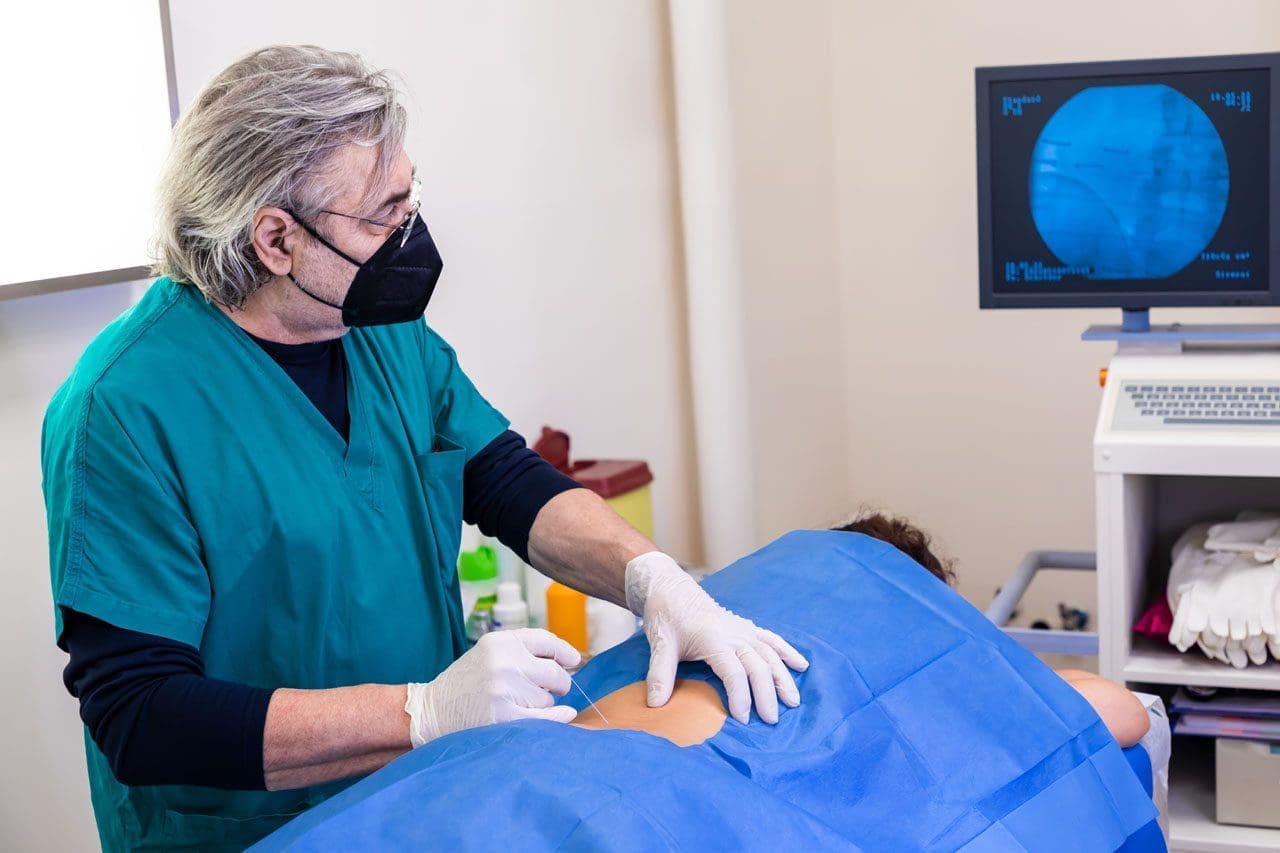
Acupuncture represents yet another tool in Dr. Jimenez’s comprehensive treatment arsenal. By incorporating this ancient healing practice, he helps patients modulate immune function, reduce inflammation, and alleviate pain through mechanisms distinct from but complementary to chiropractic adjustments (A4M, 2016).
Patient-Centered Care and Education
Dr. Jimenez’s commitment to patient education sets his practice apart. His comprehensive website, dralexjimenez.com, offers evidence-based information on health conditions, treatment options, and wellness strategies (A4M, 2016). By fostering health literacy, Dr. Jimenez enables patients to make informed decisions and take charge of their well-being.
The practice accepts major insurances, including Aetna, Blue Cross Blue Shield, Cigna, and First Health, making integrative care accessible to a broad patient base (A4M, 2016). Patients can visit in person at 11860 Vista Del Sol, Suite 128, El Paso, TX 79936, or engage through telehealth consultations (A4M, 2016).
Dr. Jimenez’s functional medicine series educates patients on holistic health principles, covering topics such as spinal health and metabolic optimization (A4M, 2016). This educational focus empowers patients to become active participants in their healing journey rather than passive recipients of care.
Through his dual expertise and integrative philosophy, Dr. Jimenez demonstrates how combining chiropractic care, functional medicine, targeted exercise, massage therapy, and acupuncture can address the complex needs of patients with autoimmune conditions, promoting natural healing and preventing long-term complications.
Acupuncture: Ancient Wisdom for Modern Autoimmune Challenges
Acupuncture, a traditional Chinese medicine practice with over 2,000 years of history, has gained recognition for its potential benefits in managing autoimmune disorders (Carey, n.d.; PubMed, 2025). This ancient healing modality offers a unique approach to restoring balance and supporting immune health.
The Mechanisms Behind Acupuncture’s Effectiveness
The fundamental principle of acupuncture centers on Qi (pronounced “chee”), the vital life force that flows through the body along specific pathways known as meridians (Carey, n.d.). When this flow becomes disrupted, it can lead to imbalances and health issues. By inserting thin needles into strategic points along these meridians, acupuncture aims to restore Qi balance, promoting overall health and wellness.
Research supports acupuncture’s efficacy in modulating the immune system, particularly relevant for those dealing with autoimmune disorders (Carey, n.d.; PubMed, 2025). Acupuncture can activate the vagal-adrenal axis, resulting in decreased systemic inflammation (QJM, 2024; PubMed, 2025). Studies indicate that acupuncture may help reduce inflammation and regulate immune responses, potentially leading to fewer flare-ups and improved quality of life (Carey, n.d.).
Evidence-Based Benefits for Autoimmune Conditions
A comprehensive narrative review of experimental and clinical evidence for acupuncture in autoimmune diseases, based on randomized controlled studies, systematic reviews, and meta-analyses from 2000 to 2023, revealed significant findings (PubMed, 2025). Acupuncture in experimental models of rheumatoid arthritis, multiple sclerosis, psoriasis, and ulcerative colitis downregulated inflammatory cytokine expression, increased IL-10 expression, improved regulatory T-cell differentiation, and modulated macrophage polarization (PubMed, 2025).
The anti-inflammatory effect of acupuncture in autoimmune disorders has been demonstrated to involve vagal-adrenal and cholinergic anti-inflammatory pathways (PubMed, 2025). The analgesic effect involves both peripheral and central anti-nociceptive mechanisms (PubMed, 2025).
Randomized controlled studies support the use of acupuncture in rheumatoid arthritis, fibromyalgia, Crohn’s disease, and Sjögren’s syndrome (PubMed, 2025). Evidence indicates that acupuncture may benefit as a symptomatic treatment for multiple sclerosis, myasthenia gravis, psoriasis, and ankylosing spondylitis (PubMed, 2025).
Immune Modulation and Regulation
One of the primary benefits of acupuncture involves its ability to modulate the immune system (Carey, n.d.). Autoimmune diseases often result from an overactive immune response where the body mistakenly attacks its own tissues. Acupuncture helps restore balance by stimulating the production of regulatory T cells, which play a crucial role in controlling immune responses (Carey, n.d.).
This regulation can reduce symptom severity and flare-ups associated with conditions such as rheumatoid arthritis, lupus, and multiple sclerosis (Carey, n.d.). Studies have shown that acupuncture can reduce antibody levels and decrease cytokine production—chemical messengers that can worsen autoimmune symptoms (Dr. Guan PTAP, 2023).
Acupuncture has the potential to regulate inhibition in the management of certain hyperimmune diseases by modulating Th1, Th2, and Th17 immunity and regulatory T-cell homeostasis, thereby restoring immune homeostasis (QJM, 2024). Research found that needling at acupoint ST36 can activate corticotropin-releasing hormone neurons in experimental models and significantly reduce signs of disease and demyelination while restoring the balance of Th1/Th2/Th17/Treg cellular responses (QJM, 2024).
Reducing Inflammation
In addition to immune modulation, acupuncture has been shown to reduce inflammation, a common feature of autoimmune conditions (Carey, n.d.; PubMed, 2025). Inflammation contributes to pain, fatigue, and other debilitating symptoms. Research has shown that acupuncture can reduce pro-inflammatory cytokines and promote the release of anti-inflammatory substances (Carey, n.d.).
Studies have shown that acupuncture primarily modulates the polarization of M1/M2 macrophages by altering cytokine production and recruitment to inflammatory injury sites, thereby enhancing anti-inflammatory effects to reduce inflammatory pain and promote tissue repair (Frontiers in Immunology, 2023; PMC, 2021).
This dual effect of reducing inflammation while enhancing pain relief makes acupuncture an appealing option in a comprehensive management plan (Carey, n.d.). Animal studies show that acupuncture reduced inflammation and pain in affected joints and changed the levels of inflammatory cytokines (Global Autoimmune Institute, 2025). Another study showed that acupuncture had anti-inflammatory and pain-reducing effects, possibly by preventing macrophages from turning into pro-inflammatory cells in inflamed joints (Global Autoimmune Institute, 2025).
Mental and Emotional Well-Being
Acupuncture can significantly impact mental well-being, which is often compromised in those with autoimmune conditions (Carey, n.d.). The stress and anxiety associated with managing chronic illness can exacerbate symptoms and create a cycle of discomfort. Acupuncture has been shown to stimulate the release of endorphins and other neurotransmitters, promoting relaxation and reducing stress (Carey, n.d.).
By addressing both physical and emotional aspects of illness, acupuncture fosters a sense of harmony, making it easier for individuals to cope with their conditions and maintain a positive mindset (Carey, n.d.). Regular acupuncture sessions not only help with physical symptoms but also contribute to a more positive outlook, essential when navigating the challenges of autoimmune disorders.
Improving Circulation and Overall Function
One of the main ways acupuncture helps with autoimmune diseases is by increasing circulation throughout the body (Dr. Guan PTAP, 2023). By stimulating acupuncture points, the treatment increases blood and oxygen flow, which helps regulate the immune system and relieves inflammation associated with autoimmune disorders (Dr. Guan PTAP, 2023).
Studies have found that patients with rheumatoid arthritis experienced reduced pain and improved mobility with regular acupuncture treatments (Dr. Guan PTAP, 2023). Another study observed that patients with multiple sclerosis who received acupuncture experienced improvements in cognitive functioning and speech (Dr. Guan PTAP, 2023).
Integration with Other Therapies
Integrating acupuncture with other lifestyle practices such as nutrition, sleep, and supplements can create a synergistic effect in managing autoimmune conditions (Carey, n.d.). Nutrition plays a critical role in supporting the immune system, while adequate sleep is essential for recovery and overall health. Mindset cultivation also proves pivotal; a positive outlook and resilience can enhance the effectiveness of all healing modalities (Carey, n.d.).
By embracing acupuncture as part of a multifaceted approach, individuals can empower themselves to take control of their health and improve their quality of life while managing autoimmune conditions (Carey, n.d.).
Massage Therapy: Healing Through Touch
Massage therapy offers another powerful tool in the comprehensive management of autoimmune conditions. By addressing muscle tension, improving circulation, and promoting relaxation, therapeutic massage provides both physical and emotional benefits for those living with chronic inflammatory conditions.
How Massage Helps Chronic Pain and Inflammation
Chronic pain often stems from muscle tension, nerve compression, or inflammation, and massage therapy directly targets these underlying causes (Renton Chiropractic Care, 2025). When muscles are tight and overworked from autoimmune-related inflammation, they can lead to discomfort, restricted movement, and pain. Massage therapy works by gently stretching and loosening muscles, relieving tightness and improving flexibility (Physician Partners of America, n.d.; Renton Chiropractic Care, 2025).
Increased blood flow is crucial for the healing process, as it delivers oxygen and nutrients to damaged tissues and helps flush out metabolic waste products such as lactic acid (Renton Chiropractic Care, 2025). Massage therapy stimulates circulation, ensuring that blood and lymph fluid can flow more freely through the body. This enhanced circulation not only promotes healing but also helps reduce swelling and inflammation in affected areas (Renton Chiropractic Care, 2025; Mayo Clinic Health System, 2022).
Breaking the Pain-Inflammation Cycle
Chronic pain and muscle tension can lead to painful bundles of tissue and the buildup of inflammatory toxins (Physician Partners of America, n.d.). If left untreated, these issues can worsen pain and reduce flexibility and mobility in the area. Regular massage therapy can break up these trigger points and promote nutrient-rich blood flow to the area, helping break up toxins (Physician Partners of America, n.d.).
When muscles or tissues press on nearby nerves, they can cause pain, numbness, or tingling (Renton Chiropractic Care, 2025). Therapeutic massage can help reduce pressure on nerves, providing relief from these symptoms and promoting better nerve function, particularly beneficial for conditions like sciatica and nerve-related autoimmune complications (Renton Chiropractic Care, 2025).
Natural Pain Relief and Stress Reduction
Massage therapy encourages the release of endorphins, the body’s natural painkillers (Renton Chiropractic Care, 2025; Texas Health School, 2024). These “feel-good” hormones help block pain signals and provide a natural, drug-free way to alleviate discomfort. Regular massage therapy can help reduce the frequency and intensity of chronic pain by boosting the body’s natural pain management (Renton Chiropractic Care, 2025).
Chronic pain is often exacerbated by stress, anxiety, and emotional tension (Renton Chiropractic Care, 2025). Massage therapy has a profound effect on the nervous system, promoting relaxation and reducing stress levels. By calming the nervous system, massage therapy can help break the cycle of pain and tension, allowing the body to heal more efficiently (Renton Chiropractic Care, 2025).
In addition to physical benefits, massage therapy offers relief from mental stress and anxiety around pain (Physician Partners of America, n.d.). The emotional and mental health aspects of pain can be just as significant as physical symptoms. The relaxation response from massage can soothe the mind, helping people more effectively manage their pain (Physician Partners of America, n.d.).
Physical Benefits for Autoimmune Conditions
For most patients, massage therapy serves as an important part of specific medical treatment plans to help them return to daily activities (Mayo Clinic Health System, 2022). Physical benefits of massage include improved circulation, reduced muscle stiffness, reduced joint inflammation, better sleep quality, quicker recovery between activities, improved flexibility, reduced pain and soreness, and a strengthened immune response (Mayo Clinic Health System, 2022).
Research has shown specific benefits for autoimmune conditions. In a study conducted at the University of Miami, a 15-minute moderate-pressure massage daily led to reduced pain and anxiety and improved grip strength in 22 adults diagnosed with hand or wrist arthritis (Arthritis Foundation, 2021). The participants received four weekly massages from a therapist and were taught to massage their sore joints daily at home. Results showed that massage combinations could reduce hand pain by up to 57% (Arthritis Foundation, 2021).
A study found that people receiving 10 weekly sessions of either relaxation massage or structural massage had less pain and were better able to perform daily activities than those receiving usual care (Arthritis Foundation, 2021). A separate 2014 study found that deep tissue massage alone relieved back pain equally as well as the combination of massage and nonsteroidal anti-inflammatory drugs (Arthritis Foundation, 2021).
Types of Therapeutic Massage
Swedish Massage: This gentle and relaxing technique uses long, smooth strokes, kneading, and circular movements to improve circulation and relieve muscle tension (Renton Chiropractic Care, 2025). It’s ideal for people with mild to moderate chronic pain and is often used to promote relaxation and stress relief.
Deep Tissue Massage: This technique targets the deeper layers of muscle and connective tissue, making it an excellent choice for individuals dealing with chronic tension, muscle stiffness, or injuries (Renton Chiropractic Care, 2025). By applying firm pressure and slower strokes, this technique helps break up scar tissue, reduce muscle knots, and relieve pain associated with chronic conditions like fibromyalgia and chronic back pain.
Trigger Point Therapy: This approach focuses on specific areas of muscle tension, known as “trigger points” or “muscle knots” (Renton Chiropractic Care, 2025). These points can refer pain to other areas of the body, leading to discomfort in seemingly unrelated regions. By applying direct pressure to these trigger points, massage therapists can release tension and alleviate pain, often providing significant relief.
Integration into Holistic Care
For many people, therapeutic massage represents an effective part of a holistic interventional pain management program (Physician Partners of America, n.d.). Treatments can also include physical therapy, lifestyle changes such as posture improvement and nutritional counseling, therapeutic injections when necessary, and other integrative approaches.
By incorporating massage therapy into a comprehensive autoimmune management plan alongside chiropractic care, acupuncture, targeted exercise, proper nutrition, and stress management, individuals can experience synergistic benefits that address both physical symptoms and overall well-being.
Targeted Exercise and Rehabilitation: Building Strength and Resilience
Exercise and rehabilitation programs specifically designed for autoimmune conditions offer powerful tools for managing symptoms, preventing complications, and improving quality of life. When properly tailored to individual needs and limitations, targeted exercise can reduce inflammation, strengthen vulnerable areas, and enhance overall function.
The Foundation of Injury Prevention
Injury prevention hinges on proactively identifying and addressing deficits in strength, mobility, balance, and movement control (South Island Physio, 2025). Strength training, in particular, is among the most evidence-based strategies to reduce injury risk across all age groups and activity levels. Studies show that strengthening exercises can reduce sports-related injuries by up to 50% (South Island Physio, 2025; Ozark PT Specialists, 2024).
For individuals with autoimmune conditions who may already experience joint instability, muscle weakness, and increased inflammation, preventing additional injuries becomes crucial. A well-rounded exercise program should include specific exercises targeting common weak points such as hips, core, and rotator cuff; balance exercises to enhance neuromuscular control; stretching exercises and dynamic stretches to improve flexibility and range of motion; and gradually increasing training loads to build resilience and avoid overuse (South Island Physio, 2025).
Benefits of Targeted Exercise for Autoimmune Conditions
Strengthening Vulnerable Areas: Targeted exercises reduce the risk of common injuries such as sprains, strains, and tears by strengthening muscles, stabilizing joints, and improving biomechanics (Ozark PT Specialists, 2024). For those with autoimmune conditions affecting joints and connective tissue, this strengthening provides crucial support and protection.
Promoting Recovery: Exercise aids recovery by increasing blood flow, reducing inflammation, and promoting tissue repair (Ozark PT Specialists, 2024). This helps individuals bounce back quicker from disease flares or intense activity sessions. As the body transitions from the inflammatory to the repair phase, targeted exercises become critical for restoring a full range of motion, rebuilding strength in and around affected areas, preventing stiffness and muscle weakness, and improving joint stability and mobility (South Island Physio, 2025).
Enhancing Mental Focus: Targeted exercises promote mental focus and concentration, helping individuals stay present and engaged (Ozark PT Specialists, 2024). For those dealing with brain fog and cognitive difficulties common in autoimmune conditions, this mental benefit proves particularly valuable.
Reducing Inflammation: Regular physical activity has been shown to reduce systemic inflammation when performed at appropriate intensity and duration (Ozark PT Specialists, 2024). This anti-inflammatory effect complements other interventions and helps manage autoimmune symptoms.
Essential Exercise Components
Dynamic Warm-Up: Beginning workouts with dynamic movements such as leg swings, arm circles, and hip rotations increases blood flow, warms up muscles, and improves joint mobility (Ozark PT Specialists, 2024). This preparation reduces injury risk and enhances exercise effectiveness.
Strength Training: Incorporating exercises that target major muscle groups—including squats, lunges, deadlifts, push-ups, and rows—builds strength, stability, and muscular endurance (Ozark PT Specialists, 2024). For patients with autoimmune conditions, modifications may be necessary based on joint involvement and current symptom levels.
Flexibility Exercises: Performing stretches for key muscle groups involved in daily activities, focusing on areas prone to tightness and imbalances such as hamstrings, quadriceps, hip flexors, calves, shoulders, and chest, maintains and improves range of motion (Ozark PT Specialists, 2024). Gentle stretching proves particularly beneficial for those with rheumatoid arthritis and other joint-affecting conditions.
Core Stability: Strengthening core muscles with exercises like planks, bridges, Russian twists, and bicycle crunches improves posture, balance, and overall stability, reducing the risk of lower back and hip injuries (Ozark PT Specialists, 2024). Core stability training is effective in reducing injury rates (PMC, 2012).
Personalized Exercise Programs
Therapists implement targeted exercises that strengthen muscles and improve joint stability, effectively preventing common ailments and minimizing the likelihood of disease-related setbacks (Simply PT, 2025). By conducting comprehensive assessments, physical therapists identify potential vulnerabilities and provide personalized recommendations to address specific needs (Simply PT, 2025).
Customized exercise plans often include strengthening and flexibility routines that target key areas, enhancing overall performance and reducing injury risk (Simply PT, 2025). This personalized approach ensures each individual receives the specific interventions necessary to maintain optimal health and function.
Rehabilitation Strategies
Engaging in Customized Routines: Exercise routines help strengthen specific muscles and improve joint flexibility (Harter Physical Therapy, 2024). These exercises focus on strengthening weakened areas and gradually restoring strength and function.
Joint Mobilizations: Therapists use precise movements to improve joint mobility, reduce stiffness, and alleviate pain (Harter Physical Therapy, 2024). This approach proves particularly valuable for autoimmune conditions involving joint inflammation.
Corrective Exercises: These exercises address muscle imbalances and faulty movement patterns, reducing the risk of injuries (Simply PT, 2025). By targeting specific weaknesses, they ensure individuals perform activities safely and efficiently.
Functional Training: This enhances the ability to execute daily movements, aiding in effective pain management without excessive reliance on medication (Simply PT, 2025). Tailored programs empower individuals to maintain function and recover swiftly from exertion.
Integration with Professional Guidance
Assessment of movement patterns is vital in reducing injury risk, as it identifies issues and weaknesses (Simply PT, 2025). Physical therapists develop customized exercise regimens targeting specific needs to enhance coordination between the brain and muscles. Routine follow-ups and adjustments ensure ongoing effectiveness and adapt to progress, fostering sustained improvement (Simply PT, 2025).
Neglecting exercise therapy during rehabilitation can lead to persistent deficits and a higher chance of symptom recurrence (South Island Physio, 2025). By incorporating targeted exercise into comprehensive autoimmune management plans alongside chiropractic care, acupuncture, massage therapy, proper nutrition, and stress management, individuals can achieve optimal outcomes and long-term health.
Nutrition and Dietary Approaches for Autoimmune Wellness
Nutrition plays a pivotal role in managing autoimmune conditions by supporting immune function, reducing inflammation, and promoting overall health. The foods we consume can either fuel inflammation or help calm the immune response, making dietary choices a powerful tool in managing autoimmune conditions.
The Anti-Inflammatory Diet Foundation
Anti-inflammatory diets have garnered attention as a lifestyle approach to modulating unregulated immune responses (PMC, 2024). They are characterized by high consumption of fruits, vegetables, whole grains, legumes, fatty fish, nuts, olive oil, and phytochemicals, while limiting intake of foods with potentially pro-inflammatory properties such as red meat, refined carbohydrates, and alcohol (PMC, 2024).
The rationale for anti-inflammatory diets lies in their capacity to decrease levels of pro-inflammatory biomarkers and positively influence pathways involved in the inflammatory cascade (PMC, 2024). Foods rich in omega-3 fatty acids, antioxidants, and polyphenols have been shown to reduce the expression of pro-inflammatory genes and decrease circulating levels of inflammatory biomarkers (PMC, 2024).
Anti-inflammatory food plans include the Mediterranean diet, vegetarian and vegan diets, ketogenic diets, and foods rich in omega-3 fatty acids and fiber (Global Autoimmune Institute, 2025). Popular anti-inflammatory dietary patterns include the Mediterranean diet, DASH diet, and vegetarian diets (Harvard Nutrition Source, 2025).
Key Anti-Inflammatory Foods
Omega-3 Fatty Acids: Present in fatty fish like salmon, mackerel, sardines, as well as flaxseeds and walnuts, these fats help reduce inflammation (Dr. Farheen, n.d.; Harvard Nutrition Source, 2025). Studies show that consuming foods rich in omega-3 PUFAs is associated with lower levels of pro-inflammatory biomarkers (PMC, 2024).
Fruits and Vegetables: Rich in antioxidants and phytonutrients, colorful fruits and vegetables help combat oxidative stress and inflammation (Carey, n.d.; Dr. Farheen, n.d.). Berries, leafy greens like spinach, kale, and Swiss chard, and colorful vegetables are particularly beneficial due to their high levels of vitamins A, C, and K, as well as magnesium and iron (Carey, n.d.).
Whole Grains: Foods like brown rice, quinoa, and whole oats provide essential nutrients and fiber, promoting digestive health and reducing inflammation (Dr. Farheen, n.d.; Global Autoimmune Institute, 2025).
Nuts and Seeds: Almonds, chia seeds, walnuts, and sunflower seeds offer healthy fats and antioxidants that support joint health (Carey, n.d.; Dr. Farheen, n.d.).
Healthy Fats: Olive oil and avocados are excellent sources of monounsaturated fats, which can help reduce inflammation (Carey, n.d.; Dr. Farheen, n.d.; Harvard Nutrition Source, 2025).
Herbs and Spices: Turmeric and ginger contain natural anti-inflammatory compounds that can aid in pain relief and inflammation reduction (Dr. Farheen, n.d.; Harvard Nutrition Source, 2025). Components found in rosemary, basil, garlic, anise, red pepper, onions, and cloves exhibit efficacy against chronic diseases by inhibiting inflammatory pathways and reducing inflammatory cytokines (PMC, 2024).
Fermented Foods: Including yogurt, kefir, sauerkraut, and kimchi, these foods support gut health by providing probiotics (Carey, n.d.). A healthy gut microbiome is increasingly recognized for its role in immune regulation, making these foods valuable for autoimmune management.
The Autoimmune Protocol (AIP) Diet
The Autoimmune Protocol diet is an elimination diet that aims to reduce inflammation and symptoms associated with autoimmune disorders (Healthline, 2020; Cleveland Clinic, 2023). The AIP diet eliminates all possible food-based triggers for inflammation and slowly reintroduces them until individual triggers are identified (Cleveland Clinic, 2023).
Research shows promising results for the AIP diet. In an 11-week study of 15 people with inflammatory bowel disease on an AIP diet, participants reported significantly fewer IBD-related symptoms by the end of the survey (Healthline, 2020). Another study had 16 women with Hashimoto’s thyroiditis follow the AIP diet for 10 weeks. By the end, inflammation and disease-related symptoms decreased by 29% and 68%, respectively (Healthline, 2020).
Foods to Avoid
Processed Foods: These often contain additives, preservatives, and artificial ingredients that can disrupt gut health and trigger inflammation (Carey, n.d.). Common examples include packaged snacks, sugary cereals, and frozen meals. High levels of refined sugars and unhealthy fats in processed foods can lead to insulin resistance and increased inflammation (Carey, n.d.).
Gluten: A protein found in wheat, barley, and rye, gluten can provoke immune responses in individuals with celiac disease or non-celiac gluten sensitivity (Carey, n.d.). Even in those without diagnosed sensitivities, gluten can contribute to gut permeability and inflammation, making it beneficial to eliminate gluten-containing foods and explore gluten-free alternatives (Carey, n.d.).
Dairy Products: While dairy can be a good source of calcium and vitamin D, it can trigger inflammation and digestive issues in some individuals (Carey, n.d.). Many report experiencing symptoms such as bloating, gas, or fatigue after consuming dairy. Plant-based alternatives such as almond or coconut milk can serve as substitutes while still providing essential nutrients (Carey, n.d.).
High-Sugar Foods and Beverages: Excessive sugar intake can increase inflammation and raise the risk of chronic diseases (Carey, n.d.). Sugary drinks, desserts, and snacks can spike blood sugar levels, leading to energy crashes and mood swings. Focusing on natural sources of sweetness, such as fruits, and incorporating healthy fats and proteins can provide more stable energy (Carey, n.d.).
The Gut-Immune Connection
The gut microbiome plays a crucial role in immune system regulation, with research highlighting strong links between autoimmune diseases and gut microbiota (Oxford Academic, 2024). Analysis revealed that autoimmune disease had the strongest association with variations in gut microbiota compared to other diseases (Oxford Academic, 2024).
Gut dysbiosis, characterized by altered diversity and function, is consistently associated with autoimmune conditions such as rheumatoid arthritis, multiple sclerosis, and type 1 diabetes (Frontiers in Microbiomes, 2025). The microbiota exerts a protective influence on autoimmunity by promoting immune regulation, although it can also trigger pro-inflammatory immune responses when disrupted (Frontiers in Microbiomes, 2025).
A disruption to the gut microbiome or an impaired mucosal barrier can allow antigens to enter and interact with the mucosal immune system in a pro-inflammatory manner (Frontiers in Microbiomes, 2025). Over time, this dysfunction can trigger a systemic immune response, driving pathogenic immune cells and the development of autoimmune disease.
Restoring gut health through the use of probiotics, prebiotics, and dietary changes can significantly improve immune function and reduce autoimmune symptoms (Flourish Counseling, MD, 2025). By prioritizing gut health through appropriate nutrition, individuals can create a foundation for better immune regulation and reduced inflammation.
Sleep: The Underestimated Pillar of Autoimmune Health
Sleep represents a fundamental aspect of overall health, particularly for those managing autoimmune conditions. Quality sleep enables vital restorative processes, including tissue repair, muscle growth, and hormone regulation (Carey, n.d.). Disrupted sleep can exacerbate symptoms of autoimmune diseases, leading to increased fatigue, pain, and inflammation.
The Sleep-Immune Connection
Sleep exerts an immune-supportive function, promoting host defense against infection and inflammatory insults (Nature, 2021). Sleep deprivation has been associated with alterations of innate and adaptive immune parameters, leading to a chronic inflammatory state and increased risk for infectious and inflammatory pathologies, including cardiometabolic, neoplastic, autoimmune, and neurodegenerative diseases (Nature, 2021).
Research has shown a strong connection between sleep disturbances and the exacerbation of autoimmune symptoms (Carey, n.d.). Conditions such as rheumatoid arthritis, lupus, and multiple sclerosis often see a worsening of symptoms with poor sleep quality. Sleep deprivation can lead to hormonal imbalances, increased stress levels, and a weakened immune response, all of which can further complicate autoimmune disease management (Carey, n.d.).
Sleep’s Impact on Immune Function
The sleep cycle consists of several stages, including light sleep, deep sleep, and REM sleep (Carey, n.d.). Each stage plays a crucial role in physical and mental health. Deep sleep is particularly important for physical recovery and immune function, while REM sleep supports cognitive processes like memory consolidation and emotional regulation (Carey, n.d.).
Quality sleep helps regulate the immune response, promoting the production of cytokines—proteins involved in inflammation and immune responses (Carey, n.d.). Insufficient sleep can lead to an imbalance, causing the immune system to become overactive or underactive. For those with autoimmune conditions, this can mean heightened symptoms and flare-ups (Carey, n.d.).
Seven days after experimental sleep deprivation, levels of IL-17A and TNF-α remained elevated (PMC, 2022). These results suggest that some inflammatory effects of sleep deprivation may be long-lasting and possibly not easily reversible, thus impacting the risk of autoimmune diseases (PMC, 2022). A meta-analysis of more than 50,000 patients found a significant increase in CRP and IL-6 levels in subjects with sleep disturbances (PMC, 2022).
Sleep Deficiency and Autoimmune Risk
Studies demonstrate that disturbed sleep may be a trigger or risk factor for autoimmune diseases (PMC, 2015). Regulatory T cells (Treg) are key players in suppressing inappropriate immune responses and maintaining self-tolerance (PMC, 2015). Breakdown of self-tolerance is central to the pathogenesis of most autoimmune diseases. In experimentally sleep-deprived healthy persons, the suppressive activity of Treg is reduced, providing a link between sleep disorders and autoimmune diseases (PMC, 2015).
Non-apnea sleep disorders can increase the risk of developing autoimmune diseases such as rheumatoid arthritis, ankylosing spondylitis, systemic lupus erythematosus, and systemic sclerosis by disrupting immunologic self-tolerance and increasing the production of several pro-inflammatory cytokines (Nufactor, 2023). Even temporary sleep deprivation may lead to suppression of Treg cells and excessive immune response (Nufactor, 2023).
It is theorized that sleep deprivation may directly increase susceptibility to immune system diseases by elevating circulating leukocyte levels, increasing circulating pro-inflammatory markers, and reducing natural killer cell activity (Nufactor, 2023). Gene expression changes related to immune and inflammatory processes and oxidative stress were also specifically observed in those with deprived sleep (Nufactor, 2023).
Strategies for Improving Sleep Quality
Establish Consistent Sleep Schedule: Going to bed and waking up at the same time each day helps regulate the body’s internal clock (Carey, n.d.). This consistency makes it easier to fall asleep and wake up refreshed, particularly beneficial for those experiencing fatigue as a symptom of autoimmune conditions.
Create Optimal Sleep Environment: A comfortable, dark, and quiet bedroom facilitates better sleep (Carey, n.d.). Consider investing in blackout curtains to block light, using earplugs or a white noise machine to minimize noise disruptions, and keeping the room cool. Ensure the mattress and pillows provide adequate support and comfort (Carey, n.d.).
Develop Bedtime Routine: Creating a calming bedtime routine signals the body that it’s time to wind down (Carey, n.d.). This might include relaxing activities such as reading, gentle stretching, or meditation. Limiting screen time before bed is essential, as blue light from devices can interfere with melatonin production.
Practice Relaxation Techniques: Mindfulness, meditation, or deep-breathing exercises can help calm the mind and prepare it for sleep (Carey, n.d.). Journaling before bed can also be beneficial, allowing expression of thoughts and feelings, which may reduce anxiety and promote peace.
Address Nutrition: Certain foods can promote sleep, such as those rich in magnesium, tryptophan, and melatonin (Carey, n.d.). Incorporating a balanced diet that emphasizes whole foods, healthy fats, and lean proteins can support better sleep and overall health. Limiting caffeine and sugar intake, especially in the hours before bedtime, can help prevent sleep disturbances (Carey, n.d.).
Consider Sleep Supplements: Melatonin, an essential hormone for regulating sleep-wake cycles, can be particularly beneficial for those struggling with insomnia (Carey, n.d.). Magnesium supplements can also promote relaxation and improve sleep quality. Before starting any new supplements, consult a healthcare provider to ensure safety and effectiveness, especially when used in conjunction with existing treatment plans (Carey, n.d.).
Poor sleep can increase inflammation and weaken the immune system (Flourish Counseling, MD, 2025). Prioritizing quality sleep is essential for managing autoimmune conditions. By implementing these strategies and recognizing sleep as a crucial component of health, individuals can significantly improve their outcomes in managing autoimmune conditions.
Stress Management and Mindset: The Mind-Body Connection
The mind-body connection represents a profound concept that recognizes the intricate relationship between mental, emotional, and physical health (Carey, n.d.). For those managing autoimmune conditions, understanding this connection proves particularly empowering, as stress, anxiety, and emotional turmoil can exacerbate physical symptoms and lead to flare-ups.
Understanding Stress’s Impact on Autoimmune Health
Chronic stress can worsen autoimmune conditions by increasing inflammation and disrupting immune function (Flourish Counseling MD, 2025; MGI Clinic, 2025). Stress management techniques like mindfulness meditation, yoga, and biofeedback help reduce stress and its impact on the body (Flourish Counseling MD, 2025).
Stress is a common trigger for autoimmune flare-ups (Tolpa Holistic Health, 2025). Elevated cortisol over long periods creates imbalances that worsen symptoms. Holistic care emphasizes practices that calm the nervous system and restore balance, including yoga or tai chi for gentle movements that reduce tension; meditation and breathwork as tools that lower cortisol and improve focus; and journaling or nature walks as outlets for emotional release and clarity (Tolpa Holistic Health, 2025).
These practices improve more than symptoms—they strengthen resilience, enhance mood, and support better sleep (Tolpa Holistic Health, 2025). Stress management is considered a cornerstone of long-term autoimmune wellness.
The Power of Meditation for Autoimmune Healing
Meditation offers transformative healing for autoimmune disease, with science-backed symptom relief (PowersBeing, 2025). Research shows that meditation was among the most critical components in achieving remission in autoimmune diseases such as lupus and Hashimoto’s (PowersBeing, 2025).
Physically, meditation has been shown to strengthen immune function, reduce chronic pain, improve sleep, lower risk of cardiovascular disease, lower resting heart rate, and lower blood pressure (PowersBeing, 2025). Research continues to suggest meditation helps in the management of a series of conditions, including autoimmune diseases.
Meditation helps improve cognitive function, enhance stress management, lower anxiety, increase mindfulness, foster self-awareness, reduce negative emotions, increase feelings of patience, tolerance, and compassion, and improve focus (PowersBeing, 2025). With brain fog experienced with autoimmune diseases, cognitive health, and focus are compelling reasons to develop a meditation practice.
A specific type of Heart Coherence Meditation has helped individuals heal from the effects of chronic inflammation associated with autoimmune conditions (Dr. Daniel Lewis, 2021). This meditation allows the heart and brain to synchronize, creating a coherent rhythm between the organs that establishes a new baseline and leads to sustained improvements in health over time (Dr. Daniel Lewis, 2021).
Cultivating a Positive Mindset
Building a positive mindset is essential for navigating the challenges of autoimmune conditions (Carey, n.d.). A positive outlook can significantly influence not only emotional well-being but also physical health. Research shows that a positive mindset can lead to improved immune function, reduced stress levels, and greater ability to cope with symptoms of chronic illness (Carey, n.d.).
Mindfulness and Meditation Practices: These techniques encourage individuals to focus on the present moment, thereby reducing anxiety and negative thought patterns (Carey, n.d.). Simple mindfulness exercises such as deep-breathing techniques or guided meditations can be incorporated into daily routines. By dedicating just a few minutes each day to these practices, individuals can begin to rewire their brains, promoting a sense of calm and resilience (Carey, n.d.).
Supportive Community: Surrounding oneself with a supportive community can play a vital role in building a positive mindset (Carey, n.d.). Engaging with others who understand the struggles of autoimmune conditions can provide a sense of belonging and validation. Support groups, both in-person and online, offer safe spaces for sharing experiences, learning from one another, and celebrating progress (Carey, n.d.).
Gratitude Practice: Cultivating gratitude can serve as a powerful tool for building a positive mindset (Carey, n.d.). Regularly reflecting on and acknowledging things one is thankful for can shift focus from challenges to positive aspects of life. Keeping a gratitude journal, in which individuals jot down daily reflections, can reinforce this practice and offer an opportunity to celebrate small victories (Carey, n.d.).
Mind-Body Techniques for Stress Reduction
Stress management techniques are crucial for those managing autoimmune conditions (Carey, n.d.). Various approaches can be employed to create a more balanced and harmonious life:
Mindfulness Meditation: This practice encourages individuals to focus on the present moment, reducing anxiety and promoting calm (Carey, n.d.). Regular mindfulness sessions, whether through guided apps or local classes, can empower individuals to regain control over their mental well-being.
Cognitive Behavioral Strategies: These techniques help shift negative thought patterns and focus on gratitude and acceptance (Carey, n.d.). By reframing negative thoughts, individuals can move their experience of living with an autoimmune condition, thereby increasing motivation to engage in healthy behaviors.
Biofeedback: This technique helps individuals learn to control physiological functions by receiving real-time feedback about body processes (Flourish Counseling, MD, 2025). It can be particularly helpful for managing stress responses.
Creative Expression: Engaging in creative activities—whether through art, music, or writing—provides an outlet for stress relief (MGI Clinic, 2025). Creative expression has been shown to lower stress and provide emotional release, which can improve immune system regulation.
Addressing Unmet Needs
Ignoring unmet needs and allowing stress to persist can severely hinder the body’s ability to heal (MGI Clinic, 2025). Unmet needs commonly linked to autoimmune inflammation include time in nature, rest and sleep, intimacy, physical activity, creative expression, and access to healthy food (MGI Clinic, 2025).
In many cases, individuals dealing with autoimmune inflammation have multiple unmet needs that go unnoticed or unaddressed (MGI Clinic, 2025). When individuals fail to prioritize these needs due to work, family, or social obligations, the immune system continues to struggle, unable to return to a state of balance.
By taking a holistic approach that addresses the root causes of stress and unmet needs, individuals can start to manage inflammation more effectively (MGI Clinic, 2025). Autoimmune inflammation management is about more than treating symptoms; it’s about nurturing overall well-being through intentional changes that help reduce inflammation, improve immune function, and restore balance to the body.
Nutritional Supplements for Autoimmune Support
Supplementation can play a vital role in managing autoimmune conditions by addressing nutrient deficiencies, reducing inflammation, and supporting immune balance (Performance Health and Wellness, 2025). When combined with lifestyle changes and integrative care, targeted supplements can enhance healing and resilience.
Vitamin D: The Immune Regulator
Vitamin D is crucial for immune regulation, with research indicating a significant correlation between low vitamin D levels and the severity of various autoimmune diseases (Carey, n.d.). This vitamin plays a critical role in immune regulation and may help mitigate the risk of flare-ups. Women should consider getting their vitamin D levels tested and consult with a healthcare provider about appropriate supplementation, particularly during winter months or in regions with limited sunlight (Carey, n.d.).
Vitamin D supplementation for five years, with or without omega-3 fatty acids, reduced autoimmune disease by 22% (BMJ, 2022; Cooper Complete, 2024). In a large study of more than 25,000 older adults, vitamin D supplementation at 2000 IU/day for approximately 5 years was associated with a lower incidence of confirmed autoimmune disease than placebo (BMJ, 2022).
When only the last three years of the intervention were considered, the vitamin D group had 39% fewer participants with confirmed autoimmune disease than the placebo group (BMJ, 2022). Vitamin D regulates a wide array of genes involved in inflammation and immunity and has been inconsistently associated with reduced risk of several autoimmune diseases in previous observational studies (BMJ, 2022).
Omega-3 Fatty Acids: Natural Anti-Inflammatories
Omega-3 fatty acids are among the most researched supplements for autoimmune support (Carey, n.d.). Found in fish oil or algae-based sources, these fatty acids possess anti-inflammatory properties that can help alleviate symptoms of conditions such as rheumatoid arthritis and lupus. Regular intake of omega-3s may reduce joint pain and stiffness and support heart health, which is often a concern for those with autoimmune diseases (Carey, n.d.).
Omega-3 fatty acid supplementation, with or without vitamin D, reduced the rate of autoimmune disease by 15% (BMJ, 2022). Several past trials have proven the beneficial effects of omega-3 fatty acid supplementation on systemic inflammation and tender and swollen joints (PMC, 2024). Dietary marine-derived long-chain omega-3 fatty acids decrease systemic inflammation and ameliorate symptoms in some autoimmune diseases (BMJ, 2022).
A study of more than 20,000 participants showed that, two years after a randomized five-year trial, the benefits of vitamin D in preventing autoimmune diseases had waned. At the same time, those of omega-3 remained strong (Medical News Today, 2024). There were 234 confirmed autoimmune disease cases among people who received omega-3 supplements compared with 280 among those who received a placebo—a statistically significant hazard ratio of 0.83 (Medical News Today, 2024; Rheumatology Advisor, 2024).
In the two-year follow-up period after the initial trial, the protective effect of vitamin D alone diminished compared with placebo arms, whereas the effect of omega-3 fatty acids was sustained (Rheumatology Advisor, 2024; Lupus News, 2022).
Probiotics: Supporting Gut Health
Probiotics hold immense potential for managing autoimmune conditions (Carey, n.d.). These beneficial bacteria can enhance gut health, a cornerstone of immune function, increasingly recognized as such. A balanced gut microbiome can influence levels of inflammation and the overall immune response.
Choosing a high-quality probiotic supplement can promote digestive health and may help reduce the frequency and severity of autoimmune flare-ups (Carey, n.d.). Fermented foods such as yogurt and kimchi can further support this effort by providing a diverse array of beneficial bacteria.
Antioxidants: Combating Oxidative Stress
Antioxidants like vitamins C and E, as well as selenium, can provide essential support for those facing autoimmune challenges (Carey, n.d.). These nutrients help combat oxidative stress, which is often elevated in individuals with autoimmune diseases. Incorporating a range of colorful fruits and vegetables into the diet can significantly boost antioxidant intake.
Additionally, considering a comprehensive multivitamin may ensure adequate levels of these vital nutrients, contributing to a more balanced and resilient immune system (Carey, n.d.).
Targeted Supplementation
The role of supplements in managing autoimmune diseases extends beyond basic nutrition (Flourish Counseling, MD, 2025). In addition to traditional medical treatments, integrative therapies combined with targeted supplementation can significantly enhance outcomes.
It’s important to consult healthcare professionals, such as registered dietitians or naturopathic doctors specializing in autoimmune conditions, for personalized recommendations (Carey, n.d.). They can provide guidance based on unique health needs and dietary requirements. This step is particularly important because certain supplements can interact with medications or exacerbate specific symptoms (Carey, n.d.).
When choosing supplements, research the brand and its reputation (Carey, n.d.). Look for companies that prioritize transparency about sourcing, manufacturing processes, and ingredient quality. Check for third-party testing and certifications from reputable organizations to ensure supplements are free from contaminants and accurately labeled.
Creating Your Personalized Autoimmune Wellness Plan
Managing autoimmune conditions requires a comprehensive approach that addresses not just symptoms but underlying causes. By integrating the various modalities discussed—chiropractic care, acupuncture, massage therapy, targeted exercise, nutrition, sleep optimization, stress management, and appropriate supplementation—individuals can create personalized plans that support healing and enhance quality of life.
The Integrative Approach
Integrating acupuncture, nutrition, sleep, mindset, and supplements creates a holistic approach to managing autoimmune conditions (Carey, n.d.). Each of these elements plays a crucial role in overall health and well-being, influencing the body’s ability to heal and maintain balance. When these practices are combined, they can enhance each other’s effects, leading to improved symptom management and better quality of life (Carey, n.d.).
By adopting a holistic approach that integrates multiple modalities, individuals can take charge of their health and navigate the complexities of autoimmune conditions more effectively (Carey, n.d.). This comprehensive strategy addresses the whole person—physical, emotional, and mental—rather than isolated symptoms.
Assessing Your Unique Needs
Assessing unique needs is a critical first step in managing autoimmune conditions effectively (Carey, n.d.). Every person’s experience with autoimmune disorders is distinct, shaped by individual symptoms, lifestyle choices, and personal circumstances. Understanding these unique factors allows for tailoring a management plan that resonates with the body and mind.
Begin by keeping a detailed journal of symptoms, dietary habits, sleep patterns, and emotional responses (Carey, n.d.). This practice helps recognize triggers and guides more effective communication with healthcare providers. Different individuals may react differently to various interventions, making it essential to identify which approaches provide relief and which exacerbate symptoms.
Working with Healthcare Professionals
Consulting with healthcare professionals is crucial in managing autoimmune conditions (Carey, n.d.). Expert guidance can provide clarity on symptoms, treatment options, and lifestyle adjustments that significantly impact overall well-being. When seeking assistance, engage a range of professionals, including primary care physicians, specialists, nutritionists, and alternative medicine practitioners, to create a comprehensive, personalized management plan.
Open communication is essential; sharing experiences and any changes in condition helps physicians tailor their approach to individual needs (Carey, n.d.). Incorporating nutrition guidance from registered dietitians or nutritionists who specialize in autoimmune conditions offers valuable insights into dietary choices that may reduce inflammation and support immune function.
Alternative therapies such as acupuncture can be beneficial when coordinated with the healthcare team (Carey, n.d.). Discussing these options ensures a coordinated approach that considers all aspects of health. Maintaining consistent dialogue with healthcare professionals about mental health and mindset is also essential, as chronic health conditions can lead to feelings of anxiety or depression that may exacerbate physical symptoms (Carey, n.d.).
Tracking Progress and Adjustments
Tracking progress when managing autoimmune conditions is crucial for understanding what strategies are effective and what may need adjustment (Carey, n.d.). Keeping a journal documenting daily symptoms, diet, sleep patterns, and emotional states helps identify trends and triggers that impact health. This information provides valuable data for discussing with healthcare providers, enabling more personalized and effective treatment plans.
Incorporating various therapies into a wellness routine can lead to significant improvements, but progress may vary (Carey, n.d.). Noting changes in symptoms after interventions helps identify which approaches yield better results. If symptoms persist despite adjustments, it may be necessary to consult specialists to refine strategies.
Regularly assessing mental well-being through self-reflection or mindfulness practices helps track emotional fluctuations (Carey, n.d.). If negative emotions persist, consider engaging in therapy or support groups. Monitoring progress in these areas not only enhances resilience but also empowers individuals to take an active role in health management.
Celebrating Achievements
Celebrating achievements, no matter how small, is an essential part of managing autoimmune conditions (Carey, n.d.). Recognizing and celebrating progress can help shift mindset, reinforce positive behaviors, and motivate continued effort on the healing journey. Whether it’s a successful treatment session, improved habits, better sleep patterns, or a positive change in mindset, acknowledging these milestones can significantly enhance well-being.
Keeping a journal dedicated to the health journey can serve as a reflection of progress (Carey, n.d.). Writing down accomplishments allows tracking of growth over time and provides a tangible reminder of how far one has come. Sharing achievements with a support network—friends, family, or a community of others who understand the unique challenges—can foster connection and encouragement.
By cultivating self-compassion and recognizing that progress is not always linear, individuals can remain grounded and realistic about their journey (Carey, n.d.). Celebrating achievements includes recognizing the effort put into managing health, regardless of outcomes. By nurturing a supportive inner dialogue, individuals can propel themselves forward, celebrating victories today while looking forward to future possibilities.
Conclusion: Embracing a New Path to Wellness
Autoimmune conditions present complex challenges that affect millions of people, particularly women, impacting virtually every aspect of life. However, as research and clinical experience demonstrate, these conditions need not define or limit one’s quality of life. Through functional wellness approaches that address root causes rather than merely suppressing symptoms, individuals can achieve significant improvements in health and well-being. The comprehensive management of autoimmune conditions requires understanding the intricate workings of the immune system, recognizing how inflammation drives disease processes, and appreciating the multifaceted nature of these disorders. By addressing the whole person—body, mind, and spirit—through integrative approaches, individuals can restore balance and promote natural healing.
Chiropractic care, as exemplified by Dr. Alexander Jimenez’s integrative practice in El Paso, offers powerful tools to improve nervous system function, reduce inflammation, enhance mobility, and support overall health. When combined with acupuncture’s immune-modulating effects, massage therapy’s ability to reduce pain and promote relaxation, targeted exercise programs that strengthen vulnerable areas, anti-inflammatory nutrition that supports gut health, restorative sleep practices, stress management techniques, and appropriate supplementation, a synergistic healing effect emerges. The clinical correlations observed by practitioners like Dr. Jimenez—who bridges the worlds of chiropractic medicine and functional medicine through his dual training—demonstrate that addressing injury causes and enhancing health through targeted, nonsurgical interventions can produce remarkable results. By combining spinal adjustments, acupuncture, massage therapy, and rehabilitative exercises, practitioners can help reduce overlapping pain symptoms, improve function, and enhance quality of life while addressing the underlying imbalances contributing to autoimmune dysfunction.
Perhaps most importantly, the functional wellness approach empowers individuals to become active participants in their healing journey. Rather than feeling helpless in the face of chronic illness, people can make informed choices about nutrition, exercise, stress management, and other lifestyle factors that profoundly influence autoimmune disease progression. This sense of agency and control, combined with comprehensive professional support, creates an optimal environment for healing and long-term wellness. As we continue to deepen our understanding of autoimmune diseases and refine integrative treatment approaches, the future holds promise for even more effective management strategies. For now, those living with autoimmune conditions can take heart knowing that multiple safe, effective, and natural interventions exist to support their health, reduce symptoms, and enhance their quality of life.
The journey toward wellness may be gradual and require patience, but with the right combination of professional guidance, lifestyle modifications, and holistic therapies, individuals can experience profound improvements. By embracing functional wellness and working with knowledgeable practitioners who understand the interconnected nature of body systems, those with autoimmune conditions can move beyond mere survival to truly thrive.
Faster Recovery After Spine Surgery: Enhanced Surgical Recovery (ESR) Programs at El Paso Back Clinic® in El Paso, TX
The doctor administers a local anesthetic into the patient’s affected area, using ultrasound to visualize the spine’s anatomical components.
Spine surgery can help treat serious back problems, such as pain from injuries, disc issues, or aging. At El Paso Back Clinic® in El Paso, TX, we focus on helping patients recover faster and more safely through modern methods. Enhanced Surgical Recovery (ESR), also called Enhanced Recovery After Surgery (ERAS), is a team-based plan that reduces the need for strong pain medications, shortens hospital stays, and lowers the risk of readmission. Led by Dr. Alexander Jimenez, DC, APRN, FNP-BC, our clinic combines chiropractic care, nurse practitioner expertise, and new tools to support healing. This article explains the main parts of ESR for spine surgery, how it cuts opioid use, shortens hospital stays, and reduces readmissions. We also cover the big roles of integrative chiropractic care and nurse practitioners, plus exciting new tech like virtual reality (VR) for building strength after surgery.
Many people in El Paso face back pain from work, accidents, or daily life. Surgery may be necessary, but traditional methods can make recovery challenging. ESR improves this process by planning care before, during, and after the operation. It uses simple steps, such as teaching patients, eating better, and moving early. Studies show these measures can cut opioid use a lot and help people go home sooner (Dagal et al., 2023). At El Paso Back Clinic®, we work with surgeons to add non-drug options for even better results.
What Is Enhanced Surgical Recovery (ESR)?
ESR is a proven plan to make surgery recovery easier and quicker. It started in other surgeries, but now helps a lot with spine operations, such as fusions or disc repairs. The idea is to lower body stress and speed natural healing. Instead of staying in bed and taking many pain pills, patients move soon and use gentler pain control.
Key parts of ESR include:
Team Approach — Doctors, nurses, chiropractors, and therapists all work together.
Step-by-Step Care — Planning starts before surgery and continues at home.
Personal Plans — Care fits each person’s health needs.
Research shows ESR helps with many spine issues, from small fixes to big ones (Zaed et al., 2023). Reviews find that most programs use around 12 key steps, such as better pain management and early walking (Berk et al., 2025).
Main Components of ESR for Spine Surgery
ESR has steps before, during, and after surgery to make things smoother.
Before Surgery (Pre-Op)
Getting ready early helps avoid problems.
Teaching Patients: Learn what to expect, how to manage pain, and why moving matters. This lowers worry and helps follow the plan (Zaed et al., 2023).
Better Nutrition: Check for low energy or anemia. Eat protein and carbs to build strength. Nutritious food helps healing (Soffin et al., 2022).
Pain Prep: Start gentle meds like acetaminophen. Quit smoking to lower risks (American Association of Nurse Anesthesiology, n.d.).
Prehab Exercises: Build strength with walks or stretches.
These make surgery safer.
During Surgery (Intra-Op)
The team uses ways to protect the body.
Better Anesthesia: Short drugs to wake up fast. Add non-opioid options (Dagal et al., 2023).
Careful Fluids: Just the right amount to avoid issues.
Small Cuts: Less muscle damage for quicker recovery (Dietz et al., 2019).
Pain Blocks: Numb the area for hours after.
Patients feel better right away.
After Surgery (Post-Op)
Focus on rapid healing.
Early Walking: Get up soon and walk daily (Zaed et al., 2023).
Mixed Pain Control: Use non-opioids, ice, and movement.
Quick Eating: Start foods and drinks early.
Checks for Safety: Watch for clots or other issues.
These steps lower risks.
How ESR Reduces Opioid Use
Strong pain drugs like opioids help, but can lead to problems like addiction. ESR cuts its use by at least half (Dagal et al., 2023). At El Paso Back Clinic®, we add chiropractic methods for even less need.
Mixed Pain Options: Non-opioids first, like NSAIDs and nerve meds. Some programs use almost no IV opioids (HCA Healthcare, n.d.).
Teaching Non-Drug Ways: Ice, breathing, and adjustments.
Blocks and Early Move: Numb areas and walk to ease pain.
In fusions, opioids dropped considerably without worse pain (Dagal et al., 2023). This helps avoid side effects and promotes natural healing.
Shortening Hospital Stays with ESR
Long hospital time raises costs and risks. ESR cuts stay by 1-2 days (HCA Healthcare Today, 2022).
Early Movement: Prevents issues and builds strength.
Fast Nutrition: Energy for recovery.
Good Pain Control: Less bedtime.
Team Reviews: Go home when ready.
One example shows noticeable shortened stays (Dagal et al., 2023). Patients heal better at home.
Lowering Readmission Rates
Going back to the hospital is tough. ESR lowers this risk (HCA Healthcare Today, 2022).
Home Care Teaching: Know warning signs.
Follow-Ups: Calls from our team at El Paso Back Clinic®.
Fewer Problems: Better prep means fewer infections.
Full Care: Controls swelling early.
Fewer complications overall (Berk et al., 2025).
Integrative Chiropractic Care at El Paso Back Clinic®
Chiropractic care fits perfectly with ESR. At our clinic, Dr. Jimenez uses hands-on adjustments to align and relieve symptoms.
Before Surgery: Improve posture and movement.
Pain Without Drugs: Soft tissue work eases tension.
After Surgery: Reduce scar tissue and build mobility (New York City Spine, n.d.).
Nerve Help: Better signals for less pain.
We complement therapy for smoother recovery (Active Health Center, n.d.).
Role of Nurse Practitioners
Nurse practitioners (NPs) like Dr. Jimenez coordinate care.
Team Links: Connect everyone.
Teaching and Meds: Focus on safe, non-opioid options.
Tracking Progress: Adjust plans.
NPs help stick to ESR paths (American Association of Nurse Anesthesiology, n.d.).
New Tech: Virtual Reality (VR) for Recovery
VR uses games and guides to make rehab more enjoyable. It helps spine patients build strength.
Fun Exercises: Improves engagement and movement.
Less Pain Feel: Distraction helps.
Strength Gains: Tailored for muscles and focus.
Home Options: Practice alone.
Recent studies show VR speeds recovery after spine issues, like in cervical cases or general neurorehab (Bolton et al., 2025; various 2025 trials).
Insights from Dr. Alexander Jimenez at El Paso Back Clinic®
Dr. Alexander Jimenez, DC, APRN, FNP-BC, leads El Paso Back Clinic® with dual expertise in chiropractic and nursing. He uses team care for pain management and rehab after injuries or surgery. His plans include adjustments, nutrition, and integrative methods for better mobility without heavy drugs. He stresses whole-body healing for lasting results (Dr. Alex Jimenez, n.d.; LinkedIn, n.d.).
Conclusion
ESR programs accelerate spine surgery recovery and make it safer. With education, nutrition, movement, and team support, they reduce opioids, shorten stays, and lower readmissions. At El Paso Back Clinic® in El Paso, TX, we add chiropractic care and NP guidance for full support. New VR tech adds exciting ways to build strength. If facing spine surgery, ask about ESR and our integrative options. Contact us at 915-850-0900 for help.
Understand the importance of pain management in a clinical setting in fostering recovery and improving patients’ overall health.
Understanding Pain: Causes, Categories, and Effective Management Strategies
Pain is a universal experience that can range from mild discomfort to debilitating agony, affecting millions worldwide. Environmental factors often play a key role in how pain develops, particularly in muscles and joints, where things like weather changes or stress can trigger or worsen symptoms. This comprehensive guide explores the origins of pain, its various categories with real-life examples, and how healthcare professionals manage it in clinical settings through both surgical and non-surgical therapies. Drawing from expert insights, including those from Dr. Alexander Jimenez, DC, APRN, FNP-BC, we delve into integrative approaches that promote natural healing and prevent long-term issues.
Research indicates that pain isn’t solely from physical trauma; external influences like humidity or pollution can heighten sensitivity in the body. It appears that addressing these factors through lifestyle adjustments and targeted therapies can significantly improve outcomes. The evidence points toward a balanced approach that combines medical interventions with natural methods to help individuals regain control over their health.
Key Insights on Pain and Its Management
Environmental Triggers Are Common: Factors such as cold temperatures or air pollution can trigger inflammation in muscles and joints, increasing the risk of pain.
Pain Comes in Many Forms: From acute, sharp stabs to chronic, dull aches, understanding the categories helps choose the right treatment.
Clinical Care Varies: Specialists use non-surgical options such as exercise and acupuncture for many cases, reserving surgery for severe cases.
Integrative Methods Work Well: Experts like Dr. Jimenez show how chiropractic care and massage can address root causes, fostering natural recovery.
Pain management in clinics follows guidelines that prioritize patient safety and effectiveness, as outlined in resources on defining and managing pain (U.S. Department of Justice, Drug Enforcement Administration, 2023).
—
Pain affects everyone differently, but understanding its roots can empower better handling. This article expands on the biology of pain, environmental influences, categories, and management techniques, incorporating clinical observations from professionals like Dr. Alexander Jimenez. We’ll cover detailed examples, case studies, and tables to make the information accessible and actionable.
The Biology of Pain: How It Develops in the Body
Pain starts as a protective mechanism. When the body detects harm, nerves send signals to the brain, which processes them as pain to prompt action, like pulling away from heat. However, this system can go awry, especially with environmental factors involved.
Nociceptors, the body’s pain sensors, are found in skin, muscles, joints, and organs. They respond to stimuli such as temperature and pressure. When activated, they trigger inflammation, which can swell tissues and press on nerves, amplifying discomfort (International Association for the Study of Pain, 2022). In muscles, this might cause tightness or spasms; in joints, it leads to stiffness or swelling.
Chronic pain, lasting over three months, often persists beyond the initial injury due to sensitized nerves. This sensitization lowers the pain threshold, making everyday activities hurtful (International Association for the Study of Pain, 2022). For instance, a minor joint strain can progress to ongoing arthritis if environmental stressors, such as humidity, exacerbate inflammation.
How Pain Affects Muscles and Joints Specifically
Muscles, made of fibers that contract for movement, can develop pain from overuse or tension. Environmental factors cause micro-tears or inflammation, leading to conditions like myalgia. Joints, cushioned by cartilage and synovial fluid, suffer when pressure changes cause fluid shifts, resulting in arthritis-like pain (Arthritis Foundation, 2024).
Case Study: A 45-year-old office worker experiences shoulder pain from poor ergonomics (static posture) combined with stress, leading to muscle knots and joint misalignment. Over time, this evolves into chronic upper back pain, affecting daily life.
Environmental Factors Contributing to Pain Development
Environmental factors are crucial in the onset and progression of pain, especially in muscles and joints. These factors interact with biology, making some people more susceptible.
Weather and Climate Influences
Weather changes significantly impact pain. Low temperatures constrict blood vessels, reducing flow to muscles and causing stiffness. High humidity increases joint fluid pressure, leading to swelling and ache (Arthritis Foundation, 2024). Barometric pressure drops before storms can trigger migraines or joint pain by altering tissue expansion.
Examples:
In osteoarthritis, patients report worse knee pain during cold, damp weather due to increased joint rigidity (PMC, 2025a).
Fibromyalgia sufferers experience muscle flares from temperature swings, with cold lowering pain thresholds by 11.3°C compared to healthy individuals (PMC, 2025a).
Studies show modest correlations between pain and humidity, pressure, and wind speed (Arthritis Foundation, 2024). For muscles, cold induces spasms; for joints, humidity exacerbates inflammation.
Stress and Psychosocial Elements
Stress releases cortisol, promoting inflammation that affects muscles and joints. Chronic stress from work or life events heightens pain perception, leading to tension headaches or back pain (MDPI, 2022). Low social support or discrimination correlates with thicker brain structures involved in pain processing, such as the insula, making discomfort more intense (Nature, 2024).
Examples:
Job insecurity causes muscle tension in the neck and shoulders, evolving into chronic pain.
Discrimination experiences are associated with greater hippocampal volume and greater pain in patients with knee osteoarthritis (Nature, 2024).
Pollution and Toxins
Air pollution, including particulates and toxins such as acrolein, increases inflammation, worsening joint pain in rheumatic diseases (ScienceDirect, 2024a). Smoking aggravates arthritis by activating immune cells, predicting higher pain in spinal injuries (ScienceDirect, 2024a).
Examples:
Urban dwellers exposed to pollution have more emergency visits for joint pain.
Vitamin D deficiency due to reduced sunlight exposure is associated with muscle hypersensitivity (ScienceDirect, 2024a).
Work and Lifestyle Environments
Poor ergonomics, such as prolonged sitting, strains muscles and joints, leading to musculoskeletal pain (MDPI, 2022). Repetitive tasks lead to back pain by reducing movement variability (IASP, n.d.a).
Examples:
Factory workers develop joint pain from repetitive lifting.
Sedentary lifestyles in air-conditioned offices can cause dry-air-related stiffness.
Sociocultural Factors
Lower income and education are associated with higher pain levels due to limited access to healthy environments (Nature, 2024). Household size and employment status explain variance in pain-related brain structures.
To mitigate, strategies include weather-appropriate clothing, stress management, and pollution avoidance. Tables below summarize factors.
Environmental Factor
Description
Impact on Muscles
Impact on Joints
Examples
Weather (Temperature)
Changes in ambient heat/cold
Constriction, spasms
Stiffness, reduced mobility
OA knee pain in cold
Humidity
High moisture levels
Swelling, tension
Fluid pressure increase
Arthritis flares in damp weather
Stress
Psychosocial pressures
Tension, knots
Inflammation from cortisol
Neck pain from job stress
Pollution
Air toxins
Inflammation, hypersensitivity
Rheumatic exacerbations
Joint pain in urban areas
Work Conditions
Ergonomic issues
Strain, fatigue
Misalignment
Back pain from sitting
Deeper Dive into Physicochemical Factors
Physicochemical factors, such as pollution and toxins, directly alter pain pathways. Air pollution exacerbates neuropathic pain by sensitizing nerves (ScienceDirect, 2024a). Toxic compounds such as 4-HNE activate receptors, triggering neurogenic inflammation in joints.
Biological factors, such as viral infections, lead to arthritis-like joint pain (ScienceDirect, 2024a). Smoking induces hyperalgesia through serotonergic changes.
Psychosocial factors, such as stress, promote chronicity, while environmental enrichment reduces pain by lowering stress (ScienceDirect, 2024a).
Case Study: A patient with rheumatoid arthritis experiences worse joint pain during pollution spikes, managed by indoor air filters and an anti-inflammatory diet.
Categories of Pain: Descriptions and Examples
Pain is classified by duration, cause, and location to guide treatment (Healthline, 2018).
Acute Pain
Short-term, lasting days to weeks, from injury. Sharp or intense, it alerts the body (Healthline, 2018).
Examples:
Muscle strain from lifting heavy objects.
Joint pain from a sprained ankle.
Chronic Pain
Lasts months or years, often without a clear cause. Mild to severe, impacting life (Healthline, 2018).
Examples:
Low back pain from poor posture.
Arthritis causes ongoing joint pain.
Nociceptive Pain
From tissue damage, activating nociceptors. Acute or chronic (WebMD, 2025).
Subtypes:
Somatic: Skin, muscles, bones. Aching or throbbing.
Examples: Muscle pull, joint fracture.
Visceral: Organs. Dull, cramping.
Examples: Appendicitis, but it can also refer to muscle pain.
Neuropathic Pain
From nerve damage. Burning, tingling (WebMD, 2025).
Examples:
Diabetic neuropathy in the feet (joint-related).
Sciatica from spinal nerve compression (muscle/joint).
Other Categories (IASP Definitions)
Allodynia: Pain from non-painful stimuli, e.g., light touch on sunburned muscle (IASP, 2022).
Hyperalgesia: Amplified pain from normal stimuli, e.g., pinprick on an inflamed joint.
Nociplastic Pain: Altered nociception without damage, e.g., fibromyalgia muscle pain.
Category
Duration
Cause
Sensation
Muscle/Joint Example
Acute
Short
Injury
Sharp
Strained hamstring
Chronic
Long
Ongoing
Dull
Chronic knee arthritis
Nociceptive Somatic
Varies
Tissue
Aching
Bone fracture joint pain
Neuropathic
Varies
Nerve
Burning
Sciatica leg muscle
Nociplastic
Chronic
Altered processing
Widespread
Fibromyalgia joint tenderness
Case Study: An athlete with acute nociceptive pain from a joint sprain transitions to chronic pain if left untreated, demonstrating category evolution.
Exploring Integrative Medicine- Video
Pain Management in Clinical Settings
Healthcare specialists follow evidence-based rationale for pain management, emphasizing multimodal approaches to minimize risks like addiction (SAMHSA, 2024). The MATE Act requires training on safe prescribing, focusing on opioid use disorders and pain treatment (DEA, 2023).
Non-Surgical Therapies
These are first-line for many, using meds, therapy, and complementary methods.
Medications: NSAIDs for inflammation, acetaminophen for mild pain (NEJM, 2019).
Physical Therapy: Exercises strengthen muscles and improve joint mobility.
Complementary: Acupuncture and massage reduce tension (PMC, 2024).
Examples: Massage post-injury eases muscle tension; breathing techniques lower anxiety in the clinic.
Surgical Therapies
For severe cases, such as joint replacement. Post-op management includes multimodal analgesia (JAMA, 2021).
Opioids: Short-term for breakthrough pain.
Non-Drug: Music therapy reduces opioid needs by 31% (PMC, 2024).
Rationale: Balances relief with safety, per guidelines (DEA, 2023).
Therapy Type
Examples
Benefits
Clinical Rationale
Non-Surgical Meds
NSAIDs
Reduce inflammation
Low risk for chronic pain
Physical Therapy
Exercises
Strengthen muscles
Prevents long-term weakness
Surgical Post-Op
Opioids + Music
Pain relief
Minimizes addiction risk
Case Study: Patient with joint pain undergoes non-surgical acupuncture, avoiding surgery.
Insights from Dr. Alexander Jimenez
Dr. Jimenez, with 30+ years in chiropractic and functional medicine, observes correlations such as perimenopausal estrogen drops causing joint pain or TBI leading to posture issues and muscle aches (LinkedIn, n.d.; DrAlexJimenez.com, n.d.).
His integrative approach addresses causes:
Targeted Exercise: Rehab programs build strength and prevent recurrence.
Massage Therapy: Relieves soft tissue tension.
Acupuncture: Promotes healing in sciatica.
Prevents long-term problems through nutrition and monitoring (DrAlexJimenez.com, n.d.).
Case Study: TBI patient regains mobility via chiropractic adjustments and exercises.
Prevention and Future Trends
Prevention involves environmental awareness, regular physical activity, and a healthy diet. Future trends include wearables for trigger monitoring.
This guide, drawing from diverse sources, shows pain as manageable with informed care.
Conclusion: Embracing a Future Free from Chronic Pain
Pain is a complex signal influenced by biology, environment, and lifestyle, as we have covered in this lengthy piece. It is not only a transient discomfort. Understanding these things, such as the distinctions between nociplastic, neuropathic, and nociceptive pain, and how pollution and barometric pressure may cause muscles and joints to expand, empowers us to take control. The 2024 recommendations (American College of Surgeons et al., 2024) stress that proactive, multimodal approaches in both surgical and non-surgical settings are highly valued for treating pain to reduce symptoms, prevent worsening, and avoid the onset of chronic illnesses.
Health care providers are crucial in this situation. They use ERAS protocols to support rehabilitation after surgery and integrative therapies to help individuals feel better every day. El Paso chiropractor Dr. Alexander Jimenez, DC, APRN, FNP-BC, has shown that addressing the root causes with acupuncture, massage therapy, chiropractic adjustments, and targeted workouts promotes the body’s natural healing process and prevents long-term issues. His case correlations demonstrate that while environmental stresses, including metabolic imbalances or repetitive work strains, often cause chronic pain, these effects may be reduced with evidence-based, individualized therapies.
Restoring equilibrium and enhancing general health, rather than masking the issue, are the ultimate objectives of effective pain management. You may end the cycle of suffering by recognizing the environmental sources of your pain, appropriately categorizing it, and looking for all-encompassing answers. You may still have a more mobile, healthy, and contented life if you make the proper decisions and seek professional help, regardless of how long you’ve experienced joint pain or how recently you were injured. Speak with a professional as soon as possible, implement your own strategy, and begin a better, less painful future.
Self-Massage Tools That Support Your Care at El Paso Back Clinic
Using A Percussive Massager Correctly: El Paso Back Clinic
At El Paso Back Clinic, patients do not just get an adjustment and leave. The team, led by Dr. Alexander Jimenez, DC, APRN, FNP-BC, focuses on full-body recovery, including what you do at home between visits. El Paso Back Clinic® • 915-850-0900+1
One of the simplest ways to support your spine, joints, and muscles is with self-massage tools. When used correctly and with guidance, tools like foam rollers, massage balls, massage guns, and trigger point devices can:
Ease muscle tension
Improve circulation and tissue recovery
Help your adjustments “hold” longer
Support better posture and movement
However, not every tool is right for every person. The doctors, nurse practitioners, and rehab team at El Paso Back Clinic help patients decide which devices are safe for their bodies and how to use them without causing harm. El Paso Back Clinic® • 915-850-0900+1
Integrative Chiropractic Care at El Paso Back Clinic
Because Dr. Jimenez is both a chiropractor and a family nurse practitioner, he views your body from both structural and medical perspectives. This dual training helps him safely combine: El Paso, TX Doctor Of Chiropractic+1
Spine and joint alignment
Muscle and fascia recovery
Nerve health
Whole-person wellness, including nutrition and lifestyle
Self-massage devices fit into this model as home-care tools that extend the benefits of what happens in the clinic.
Why Self-Massage Tools Help Your Spine and Muscles
Most self-massage tools work by applying controlled pressure to muscles and fascia (the thin connective tissue around muscles). This pressure can:
Loosen tight areas that restrict movement
Improve local blood flow
Help your body remove waste products after activity
In simple terms, self-massage tools can help your body feel “less stuck” and more able to move. When your muscles and fascia move more freely, your joints can do the same, which supports your chiropractic adjustments.
Foam Rollers: A Core Tool for El Paso Back Clinic Patients
Foam rollers are one of the most recommended self-massage tools in chiropractic and rehab settings. They are firm foam cylinders you use under your back, hips, or legs as you slowly roll over them.
What Foam Rolling Does
Chiropractic and rehab sources describe foam rolling as a type of self-myofascial release that can:
Improve circulation and tissue oxygenation
Reduce muscle tightness and soreness
Support better posture by opening the chest and upper back
A chiropractic clinic article notes that foam rollers, when used properly, can enhance circulation and “support preventive chiropractic treatment,” while also helping with posture and movement. King Chiropractic Hand & Foot
Important: Foam rollers are usually not rolled directly over the lower back for patients with certain spine problems unless a provider has shown a safe method.
Basic Foam Rolling Tips
Your El Paso Back Clinic team may teach you:
Go slow. Roll slowly along the muscle, pausing on tender spots for 20–30 seconds.
Breathe. Relax your breathing instead of tensing up.
Control pressure. Use your arms and opposite leg to reduce weight if it is too intense.
Aim for “good discomfort.” If the pain is sharp, electric, or burning, stop and tell your provider.
Short sessions—5–10 minutes a day—can be enough to make a difference when done consistently.
Massage Balls and Spheres: Targeting the Tough Spots
Massage balls (such as lacrosse balls, rubber balls, or specialized therapy balls) deliver more precise pressure than a foam roller. They are very helpful for small or hard-to-reach areas. Articles on self-massage tools note that balls are especially useful for the feet, hips, and muscles around the spine. RAD Roller+3High Amplitude Health Chiropractic+3IDEA Health & Fitness Association+3
Areas Where Massage Balls Shine
Between the shoulder blades
Back of the shoulders and rotator cuff
Glutes and deep hip muscles
Bottom of the feet (plantar fascia)
Small tight spots along the spine (used carefully)
How Your Chiropractor Might Have You Use Them
Examples your provider might show you:
Wall technique:
Place the ball between your upper back and a wall.
Gently lean into it and roll up, down, or side-to-side until you find a tight spot.
Floor technique (hips):
Sit or lie with the ball under one buttock.
Slowly shift your weight until you feel a trigger point, then hold and breathe.
Foot rolling:
Stand or sit and roll the ball under your foot from heel to toes.
Use light to moderate pressure, not sharp pain.
Because these points can be very sensitive, Dr. Jimenez and his team usually suggest short, frequent sessions rather than long, aggressive work—especially in people with nerve irritation or high pain sensitivity. El Paso Back Clinic® • 915-850-0900+1
Percussion Massage Guns: High-Tech Help for Sore Muscles
Percussion massage guns use rapid pulses to work into muscle tissue. Articles reviewing these devices note that they can improve local blood flow, reduce muscle soreness, and assist recovery when used properly. Allure+3BarBend+3BarBend+3
Massage Guns vs. Foam Rollers
Fitness and recovery experts have compared massage guns with foam rollers: BarBend+1
Massage guns
More targeted
Easier to use while standing or sitting
Adjustable speeds and attachments
Can be very intense if used on high settings
Foam rollers
Broader, more gentle pressure
Less expensive
Great for overall mobility and posture work
At El Paso Back Clinic, a massage gun may be recommended for:
Large muscle groups like the quadriceps, hamstrings, and glutes
Athletes or highly active patients who need a quick recovery
Patients who struggle to get on and off the floor to use a foam roller
Safe Use Tips for Massage Guns
Based on physical therapy and recovery guidance: BarBend+1
Start with the lowest speed.
Move slowly over the muscle, not the bones.
Limit each area to about 1–2 minutes.
Avoid the front of the neck, directly over the spine, or areas with swelling or bruising.
Do not use directly over recent injuries or unhealed surgical sites, or if you have vascular conditions, unless your provider clears it.
The team at El Paso Back Clinic may show you which muscles are safe to massage with a massage gun and which areas to avoid.
Manual Trigger Point Tools and Massage Sticks
Manual tools like massage sticks, canes, and handheld knobs are popular because they let you apply deep pressure without overworking your hands. Chiropractic and massage supply companies offer many options, including neck supports, rollers, and trigger-point tools. Redison Tech LLC+3ScripHessco+3RAD Roller+3
Common Manual Tools
Massage sticks: Rolled along muscles in the legs and back
Trigger point canes: The Hooked shape allows you to press knots between the shoulder blades
Handheld knobs: Designed to mimic a therapist’s thumb or elbow
When Dr. Jimenez Might Suggest These
Long-standing muscle knots that flare between visits
Old injuries with scar tissue
Posture correction programs that need focused daily soft-tissue work
Often, these tools are paired with corrective exercises right after use. For example:
Use a trigger point cane on the upper back
Then do posture drills, band work, or thoracic mobility exercises
Many patients ask about back massager chairs, cushions, or handheld units for home use. Consumer guides and chiropractic associations discuss how these devices can provide gentle, hands-free relief for general muscle tension. The Spruce+2ACA Today+2
Possible Benefits
Soothing end-of-day relaxation
Heat plus massage to ease stiffness
Helpful for people who sit long hours or drive frequently around El Paso
However, these devices do not replace a full evaluation at El Paso Back Clinic, especially if you have:
Radiating pain, numbness, or tingling down the arms or legs
Known disc herniations, spinal stenosis, or severe arthritis
Recent injuries from car accidents, sports, or falls
In those cases, the clinic team may only clear gentle back massagers after imaging, testing, and a clear plan.
Myofascial Release and Why Guidance Matters
The deeper goal behind many of these tools is myofascial release—loosening tight fascia and muscle layers so they can move freely again. Educational articles on myofascial release stress that: Spine & Health Co+2Spine & Health Co+2
Fascia can become tight due to injury, overuse, or prolonged sitting.
Skilled manual therapy can teach you how to extend these techniques at home safely.
Poor technique or excessive pressure can irritate tissues and sometimes worsen pain.
That is why the El Paso Back Clinic team often:
Demonstrates tool use in the office
Gives written or video instructions
Uses telemedicine follow-ups to review technique
Adjust your plan if your symptoms change
Guided self-massage is much safer and more effective than guessing on your own.
When to Be Careful or Avoid Self-Massage Tools
Self-massage tools are not for everyone, nor for every situation. Always speak with your chiropractor, nurse practitioner, or medical provider first if you have:
Recent fractures or major sprains
Recent surgery
Active infection, fever, or unexplained weight loss
History of blood clots or bleeding disorders
Cancer, especially in bone
Severe osteoporosis
Stop and call your provider or seek emergency care if you notice:
Sudden, sharp, or electric pain
New numbness or weakness in arms or legs
Loss of bowel or bladder control
Also, avoid using tools directly over:
Joints and bony areas
Open wounds or rashes
Areas with obvious swelling or strong bruising
The El Paso Back Clinic team will clearly explain what is safe for your specific diagnosis.
Simple Self-Massage Routines for El Paso Patients
Below are example routines that Dr. Jimenez and the team might customize for different patient groups. These are not medical advice; they show how tools can be used when approved by your provider.
1. Desk and Driver Routine
Goal: Reduce neck and upper-back tension from screens and driving.
Tools: Foam roller, massage ball
3–5 minutes foam rolling mid-back against the floor or wall
2 minutes lying lengthwise on the roller to open the chest
2–3 minutes with a massage ball against the wall between the shoulder blades
Follow with simple chin tucks and shoulder blade squeezes
2. Post-Workout Recovery Routine
Goal: Help muscles recover after sports or gym workouts.
5–10 minutes foam rolling quads, hamstrings, glutes, and calves
1–2 minutes per muscle group with a massage gun on low speed
3–5 minutes of light stretching and mobility drills after using the tools
3. Gentle Routine for Chronic Back Pain
Goal: Support mobility without overloading sensitive tissues.
Tools: Soft foam roller, massage ball, possibly a gentle back cushion
2–3 minutes foam rolling glutes and upper back (avoiding painful low back areas)
2 minutes of gentle ball work for glutes and hips
Short session with a low-intensity back cushion, if cleared
Follow with core stability exercises prescribed by the clinic
4. Mobility and Posture Routine
Goal: Improve posture and spinal mobility for daily life.
Tools: Foam roller, trigger point cane
3–5 minutes of foam rolling the upper back and sides of the rib cage
3–5 minutes using a trigger point cane on knots between the shoulder blades
Then, posture drills, band pulls, and breathing exercises are prescribed
These routines are most powerful when combined with the chiropractic adjustments, rehab exercises, and nutrition plans created for you at El Paso Back Clinic. El Paso Back Clinic® • 915-850-0900+1
Telemedicine visits for follow-up and problem-solving
Integration with exercises, nutrition support, and lifestyle changes
The goal is simple:
Make home care safe, effective, and easy to follow so your body keeps healing between visits.
If you are a current or new patient in the El Paso area and want to know which self-massage tools are right for you, contact El Paso Back Clinic® (915-850-0900) to schedule an in-person or telemedicine consultation and get a plan that matches your spine, lifestyle, and goals. El Paso Back Clinic® • 915-850-0900+1
How Telemedicine Can Assist in the Management of Sciatica (with Integrative Chiropractic Care)
A man at home consults a chiropractor via telemedicine for back pain and sciatica.
Sciatica can make even simple tasks—like getting out of bed, sitting at a desk, or driving—feel almost impossible. When pain shoots down your leg or feels like burning, stabbing, or tingling, the idea of driving across town to sit in a waiting room can be overwhelming.
Telemedicine offers a way to get expert help for sciatica without leaving home. Telemedicine can significantly improve the quality of life for many individuals experiencing limited mobility or frequent flare-ups of pain. Spine specialists and integrative chiropractic teams now use secure video visits to evaluate symptoms, design treatment plans, and follow patients through recovery. UT Southwestern Medical Center+1
Dr. Alexander Jimenez, DC, APRN, FNP-BC, is a dual-licensed chiropractor and nurse practitioner in El Paso, Texas. His integrative model combines medical decision-making (such as imaging and prescriptions) with chiropractic and functional medicine. This blended approach fits perfectly with telemedicine because it allows him to assess nerve pain, guide movement, and adjust treatment plans over time—even when the patient is at home. El Paso, TX Doctor Of Chiropractic
What Is Sciatica?
Sciatica is not a disease by itself. It is a pattern of symptoms caused by irritation or compression of the sciatic nerve. This nerve starts in the lower back, runs through the hips and buttocks, and travels down each leg.
Common symptoms include:
Sharp or burning pain in the lower back, buttocks, and legs
Numbness, tingling, or “pins and needles” in the leg or foot
Weakness when trying to stand, walk, or lift the leg
Pain that worsens with sitting, coughing, or bending
Sciatica is usually caused by:
Herniated or bulging discs pressing on a nerve root
Spinal stenosis (narrowing of the spinal canal)
Degenerative disc disease
Muscle or joint dysfunction in the pelvis and lower back
Less commonly, tumors, infections, or serious conditions
Because sciatica can have many causes, proper evaluation and treatment planning are very important—this is where telemedicine can help you start sooner and stay on track.
What Is Telemedicine and How Does It Work for Back and Nerve Pain?
Telemedicine (also called telehealth) is health care delivered via secure video or phone rather than an in-person visit. You use a smartphone, tablet, or computer to speak with your provider, similar to a video call with family or friends.
Clinics that treat spine and nerve problems have made telemedicine a core part of their care model. They use it for first visits, follow-ups, second opinions, and surgical planning, especially for conditions like back pain, neck pain, and sciatica. UT Southwestern Medical Center+1
During a typical telemedicine visit for sciatica, your provider can:
Ask detailed questions about your pain pattern
Watch how you move on camera
Guide simple movement and strength tests
Review MRI, X-ray, or CT results
Explain treatment options, including chiropractic, physical therapy, injections, or surgery if needed
Many clinics report that they can accurately diagnose spine issues through video visits and that most telemedicine-based surgical plans do not require major changes after in-person exams. UT Southwestern Medical Center
Why Telemedicine Is Especially Helpful for Sciatica
People with sciatica often have trouble sitting, driving, or walking long distances. Telemedicine meets them where they are—literally.
Key benefits for sciatica patients
Less travel and less pain getting to care
No long car rides or sitting in waiting rooms
Easier for patients who have mobility issues or rely on others for transportation Southeast Texas Spine+1
Faster access to evaluation and treatment
Many clinics can schedule telemedicine visits sooner than in-person visits
You can start treatment earlier instead of waiting weeks to be seen
Better continuity of care
Telemedicine makes it easier to attend follow-ups, especially during long recovery plans
Providers can adjust medications, exercises, and activity limits in real time Southeast Texas Spine+1
Home-based evaluation of your real environment
Your provider can see your work setup, couch, bed, or home office
Straight-leg raise or seated leg raise while on camera
Heel and toe walking to assess nerve strength
Balance and gait observation
Imaging and tests
Your nurse practitioner or physician can order MRI, X-rays, or CT scans when needed
They may also recommend nerve tests (EMG/NCS) through in-person referrals
Spine centers and orthopedic clinics report that telemedicine visits can help determine when conservative care is sufficient and when urgent in-person care or surgery is needed. UT Southwestern Medical Center+1
Integrative Chiropractic Telemedicine for Sciatica
Integrative chiropractic telemedicine combines:
Medical care—history, diagnosis, imaging orders, prescriptions, and referrals
Chiropractic care—movement analysis, spinal and pelvic mechanics, and guided home-based therapies
Dr. Jimenez’s dual-scope role as a chiropractor and nurse practitioner is a strong example of this model. In his practice, he uses telemedicine to:
Review MRI and other imaging results with patients
Coordinate conservative care (chiropractic, physical therapy, massage, acupuncture, and functional medicine)
Monitor nerve symptoms and red flags that require fast in-person intervention
Looks for patterns of dysfunction in the lower back, pelvis, and hips
Guides you through gentle tests and movements
Designs a home exercise and stretching plan
Educates you about ergonomics, sleep positions, and movement habits
Even without hands-on adjustments, chiropractic expertise is used to understand mechanics and guide safe self-care at home. Evolve Chiropractic+2HealthCentral+2
Telemedicine and Medication Management for Sciatica
Telemedicine is also useful for medication oversight and pain management. Virtual pain management services can:
Review current medications and supplements
Start or adjust anti-inflammatory drugs, muscle relaxers, or nerve pain medications when appropriate
Help taper short-term medications to avoid long-term dependence
Coordinate with other therapies like physical therapy and chiropractic care Everlywell+1
This is important because the goal is not just to reduce pain for a few days but to manage it safely while addressing the underlying cause.
Guided Home Exercises and Self-Care for Sciatica via Telemedicine
A large part of sciatica management involves what you do every day at home. Telemedicine allows your integrative provider to coach you in real time.
Types of exercises a provider may guide over video
Always follow your own provider’s instructions. The list below is for education, not a personal prescription.
An integrative chiropractor, such as Dr. Jimenez, will often blend chiropractic reasoning (how joints and muscles are moving) with physical therapy-style exercise progressions to build strength and reduce nerve irritation over time. Integrative Medical of DFW+1
Telemedicine and Physical Therapy for Sciatica
Physical therapy is a key part of long-term sciatica care. Telemedicine makes it easier for your team to coordinate and supervise this care.
An NP–chiropractor team can:
Refer you to in-person physical therapy when you need hands-on manual work
Work with therapists to align goals: pain reduction, nerve mobility, strength, and posture
Review PT progress notes with you by video
Add or modify home exercises between in-person therapy visits
Modern integrative clinics describe physical therapy as treatment focused on your goals, your function, and your time—whether you are recovering from an acute episode of sciatica or managing long-term spine issues. Integrative Medical of DFW+1
Telemedicine for Office Workers and Remote Workers with Sciatica
Many people with sciatica sit for long periods at desks or work remotely at kitchen tables, couches, or beds. Poor ergonomics can worsen nerve pain.
Telemedicine allows providers to see your real work setup and give specific advice.
They may help you:
Adjust chair height, screen level, and keyboard position
Chiropractic-based telemedicine visits for office workers often focus on spinal alignment, hip position, and load sharing between joints — even if the provider cannot physically adjust the spine during the visit, they can teach you how to move better and reduce pressure on the sciatic nerve. tigardchiropracticautoinjury.com+1
How to Prepare for a Telemedicine Visit for Sciatica
Preparing well can make your telemedicine visit smoother and more helpful.
Before your appointment
Check your technology
Test your camera, microphone, and internet connection
Charge your device and have a backup (like a phone) ready
Choose your space
Find a quiet, private room
Make sure you have enough room to stand, walk, and lie down if needed
Gather information
List your current medications and supplements
Have your medical history and imaging reports handy
Dr. Jimenez’s clinical experience shows that when patients feel seen and supported—through regular check-ins, education, and coordinated care—they are more likely to stay consistent with their home program and achieve better long-term outcomes. El Paso, TX Doctor Of Chiropractic+1
Practical Tips for Getting the Most from Telemedicine for Sciatica
Here are some simple strategies to make telemedicine work for you:
Treat the visit like an in-person appointment
Show up on time and minimize distractions
Have a notebook handy for instructions
Be specific about your goals
“I want to sit for 30 minutes without pain”
“I want to walk around the block again”
Clear goals help your provider design better plans
Use photos or videos
Take a short video of how you walk or how you get out of a chair during painful times
Share this with your provider if their platform allows
Stay consistent with home exercises
Put reminders in your phone
Tie exercises to habits (after brushing teeth, after lunch, etc.)
Ask for a written or emailed summary
Many clinics send a visit summary through the patient portal
This can include your diagnosis, exercise plan, and red-flag symptoms
The Future: Telemedicine, Sciatica, and Integrative Care
Telemedicine is no longer just an emergency backup plan—it is a core part of modern spine and pain care. Spine centers, pain clinics, and integrative practices across the country use telemedicine to: UT Southwestern Medical Center+2NJ Spine & Orthopedic+2
Speed up diagnosis and treatment
Improve convenience for patients in pain
Coordinate care between specialists, therapists, and primary providers
Support long-term recovery with flexible follow-ups
For people with sciatica, this means you can:
Get expert guidance without leaving your home
Partner with an integrative chiropractor and nurse practitioner who can see both the nerve problem and the whole person
Combine remote consultations, at-home exercises, and lifestyle changes into a comprehensive plan
Under the care of a dual-licensed provider like Dr. Alexander Jimenez, telemedicine becomes more than a video call. It becomes a bridge between medical science, chiropractic biomechanics, and day-to-day life—helping you move from intense nerve pain toward safer movement, better function, and long-term relief. El Paso, TX Doctor Of Chiropractic+2Evolve Chiropractic+2
IFM's Find A Practitioner tool is the largest referral network in Functional Medicine, created to help patients locate Functional Medicine practitioners anywhere in the world. IFM Certified Practitioners are listed first in the search results, given their extensive education in Functional Medicine