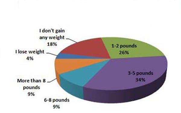
Chiropractic recommendations to avoid holiday weight gain and back pain. All the food, gravy, pies, cookies, candy, chocolate, wine, eggnog, hot cocoa, and whipped cream are great, but moderation is key to keeping unwanted weight gain that can lead to back pain. Research has found that up to 70% of individuals typically gain weight during the holidays.
Holiday weight gain
Also known as the seasonal seven or a month-long celebration with the diet starting in the new year. Any type of extra weight can contribute to back discomfort and pain. A body that is overweight can begin to overwork the spine, as it tries to adjust to bear the extra weight. This can contribute to injury and back pain. When the body is overweight, the potential for a back injury increases and can be more difficult to recover from, which can result in chronic back pain. Too much weight can exacerbate and/or lead to symptoms of:
Degenerative disc disease
Compression fracture
Osteoporosis
Osteoarthritis
Spinal stenosis
Spondylolisthesis
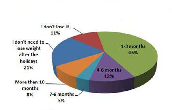
Losing the weight
However, most individuals struggle to lose the holiday weight. Research has found that around 10% of individuals are unable to lose the holiday weight until the following fall. Another 11% say that they never lose the weight gained. Year after year the holiday weight gain adds up.
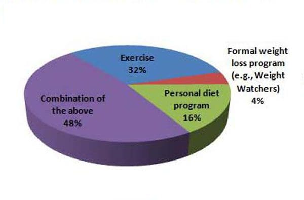
To stay healthy and protect the spine, here are some recommendation to fight the holiday weight gain:
Physical Activity
Physical activity is the best way to lose or maintain a healthy weight. Walking is a quick and perfect way to get in some quick exercise. With all the shopping, wrapping, and unwrapping, it can be challenging to find time to exercise. It does not have to be a full workout. Just a quick walk around the block is a perfect option. Don’t get frustrated up over not being able to exercise regularly. A 10-20 minute walk is better than no physical activity at all.
Enjoy in moderation
Don’t try to avoid all the food during the holidays, instead develop an action plan. The objective is to eat and drink in moderation. An example could be before eating, eat a healthy snack so there is no temptation to fill up on sweets, pastries, etc. Also at the dinner, pass on the gravy and share the dessert.
Lose weight effectively
Keeping the holiday weight off can be done with a few adjustments keeping the spine and whole-body healthy.One way is to think about keeping the spine healthy as a gift to yourself. Contact our chiropractic and health coaching team for spinal adjustments, exercises, stretches, and diet.
Weight Loss Chiropractic Treatment
�
Dr. Alex Jimenez�s Blog Post Disclaimer
The scope of our information is limited to chiropractic, musculoskeletal, physical medicines, wellness, and sensitive health issues and/or functional medicine articles, topics, and discussions. We use functional health & wellness protocols to treat and support care for injuries or disorders of the musculoskeletal system. Our posts, topics, subjects, and insights cover clinical matters, issues, and topics that relate and support directly or indirectly our clinical scope of practice.*
Our office has made a reasonable attempt to provide supportive citations and has identified the relevant research study or studies supporting our posts. We also make copies of supporting research studies available to the board and or the public upon request. We understand that we cover matters that require an additional explanation as to how it may assist in a particular care plan or treatment protocol; therefore, to further discuss the subject matter above, please feel free to ask Dr. Alex Jimenez or contact us at 915-850-0900. The provider(s) Licensed in Texas& New Mexico*
References
Diaz-Zavala RG, Castro-Cantu MF, Valencia ME, et al. Effect of the holiday season on weight gain: A narrative review.�Obesity. 2017; article ID 2085136. https://www.hindawi.com/journals/jobe/2017/2085136/ Accessed November 28, 2017.
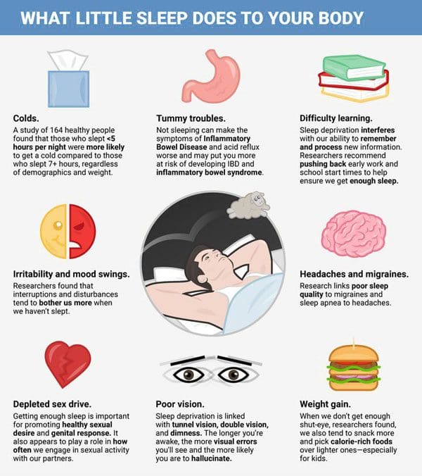
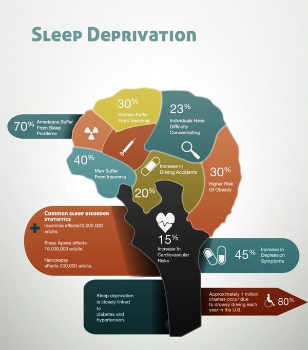
Chiropractic adjusting: Poor sleep is detrimental to the body’s general health. If it becomes chronic side effects like high blood pressure, brain fog, exacerbation or development of a disease, and chronic fatigue can set in. Getting the proper quality amount of sleep needs to become a priority. The hard work and to-do lists can only get done when the machine that is the human body is functioning well-rested and at full capacity. When the body is starved of sleep it sets in motion a downward spiral of system failure, burnout, declining health, and decreased productivity.
Assessing sleep quality
7 to 9 hours should be the objective for the quantity each night or day depending on shift work, etc. Quality of sleep is just as vital as the proper amount. Sleep quality can be affected by:
Not waking up unless to use the bathroom and being able to fall back to sleep within 30 minutes
Wake up feeling fresh and rested
Being full of energy, focused, and able to handle responsibilities
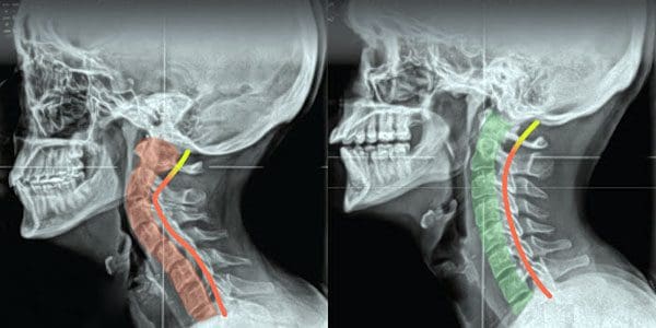
Spinal alignment affects sleep ability
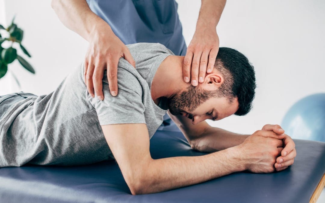
Chiropractic can improve sleep quality by adjusting the spine, bringing the body back into balance. Chiropractic is an expert-based approach that will address any underlying issues with spinal misalignment that could be affecting sleep. Spine misalignment contributes to poor nerve energy circulation that affects the body�s ability to function and recover, which occurs during the sleep cycle.
A chiropractor will restore alignment and provide expert recommendations for decreasing pain and sleeping posture optimization. Poor posture specifically when sleeping, a pain-causing injury, and poor biomechanics can all contribute to poor spinal alignment. This affects the body�s ability to complete normal tasks like sleep. Not letting the body rest properly can exacerbate other health issues and create a vicious cycle of compromised health.
Chiropractic adjusting
Restoring the body’s balance via the spine allows the body to properly move, heal, and function at full potential. Working with a chiropractic care provider can help this process and help restore refreshing and restful sleep. When sleep quality improves quality of life improves. Chiropractic practitioners can provide effective spinal treatment and optimize the quality of sleep.
Sciatica Pain Chiropractor
Dr. Alex Jimenez�s Blog Post Disclaimer
The scope of our information is limited to chiropractic, musculoskeletal, physical medicines, wellness, and sensitive health issues and/or functional medicine articles, topics, and discussions. We use functional health & wellness protocols to treat and support care for injuries or disorders of the musculoskeletal system. Our posts, topics, subjects, and insights cover clinical matters, issues, and topics that relate and support directly or indirectly our clinical scope of practice.*
Our office has made a reasonable attempt to provide supportive citations and has identified the relevant research study or studies supporting our posts. We also make copies of supporting research studies available to the board and or the public upon request. We understand that we cover matters that require an additional explanation as to how it may assist in a particular care plan or treatment protocol; therefore, to further discuss the subject matter above, please feel free to ask Dr. Alex Jimenez or contact us at 915-850-0900. The provider(s) Licensed in Texas& New Mexico*
References
Fietze, Ingo. �Sleep Applications to Assess Sleep Quality.��Sleep medicine clinics�vol. 11,4 (2016): 461-468. doi:10.1016/j.jsmc.2016.08.008
Sedentary lifestyle prevention through chiropractic is highly recommended for seniors. With advanced age, the body’s muscles, bones, and spinal system begin to wear down and need to be maintained to retain their mobility and flexibility. Regular chiropractic adjustments are recommended as part of an active/fitness lifestyle for seniors and can help older individuals maintain optimal health.
Sedentary Lifestyle Prevention
Many seniors tend to reduce physical activity after reaching retirement age. Many individuals just want to kick back and relax. However, living too laid-back can impact overall health. A lack of exercise and physical activity can cause the muscles, cardiovascular system, and skeletal system to prematurely wear out. Living an active lifestyle will keep individuals at their best when retiring and maintain a healthy quality of life.
Pain Management
Chiropractic can help individuals realize their full potential. The chiropractic approach to achieving top health means treating the source of symptoms, and not just the symptoms. This helps maintain a high quality of life. Chiropractic sedentary prevention includes:
Adjustments
Adjustments to the body are the core of chiropractic. Adjustments are utilized to realign the spine that helps treat various nervous system-related conditions. These include subluxations, slipped discs, sciatica, nerve damage, and more. Adjustments also increase the immune system’s functionality. This is extremely beneficial for older individuals.
Nutritional Recommendations
Older individuals can benefit from a customized nutrition plan to help with any deficits in their diets. Often older individuals need more calcium to help fortify bone health. Chiropractors and health coaches can provide individuals with nutritional information needed to live a healthy life.
Exercise Program
An active lifestyle is an essential component of staying healthy. Chiropractors can provide individuals with specialized exercises that take into account their specific and specialized needs.
Physical Therapy
Physical therapy is another component of chiropractic, as it helps enhance the adjustments.
Health Coaching
Health coaching can provide individuals with the tools necessary to take charge of their health. Counseling involves exercise recommendations, nutritional advice, healthy habits development, and more depending on the individual’s abilities.
Stay Moving
A sedentary lifestyle places an older individual’s spine in a compromised position. Older individuals have an increased risk of developing detrimental spinal conditions. Overexertion happens more easily in weakened bodies. The solution is to maintain physical activity with proper support. Chiropractic is designed to improve the whole-body. If you�re looking for sedentary prevention and the latest care approaches, contact us, we’re ready to help!
Chiropractic Shoulder Pain Treatment
Dr. Alex Jimenez�s Blog Post Disclaimer
The scope of our information is limited to chiropractic, musculoskeletal, physical medicines, wellness, and sensitive health issues and/or functional medicine articles, topics, and discussions. We use functional health & wellness protocols to treat and support care for injuries or disorders of the musculoskeletal system. Our posts, topics, subjects, and insights cover clinical matters, issues, and topics that relate and support directly or indirectly our clinical scope of practice.*
Our office has made a reasonable attempt to provide supportive citations and has identified the relevant research study or studies supporting our posts. We also make copies of supporting research studies available to the board and or the public upon request. We understand that we cover matters that require an additional explanation as to how it may assist in a particular care plan or treatment protocol; therefore, to further discuss the subject matter above, please feel free to ask Dr. Alex Jimenez or contact us at 915-850-0900. The provider(s) Licensed in Texas& New Mexico*
References
Hawk, Cheryl et al. �Best Practices for Chiropractic Care for Older Adults: A Systematic Review and Consensus Update.��Journal of manipulative and physiological therapeutics�vol. 40,4 (2017): 217-229. doi:10.1016/j.jmpt.2017.02.001
Chiropractic alignment is often the go-to option when injured or experiencing various types of pain. And for good reason, because it works. However, chiropractic medicine goes beyond injuries, and pain conditions. Individuals can reap the benefits of regular chiropractic for increased well being and maintaining optimal health.
Chiropractors understand the importance of balancing all areas of the body’s health. This is done through a combination of spinal adjustments and educating individuals on how to develop healthy lifestyle habits. Chiropractic care can increase positivity, overall life, and well-being.
Regular Chiropractic Alignment Benefits
Being able to enjoy a full and healthy life is more than just being free of pain or injury. It means all of the body’s functions are operating properly and cleanly. Understanding what helps the body thrive can take an individual’s quality of life to another level. Chiropractic can help guide individuals to feel their best with recommendations and regular spinal monitoring and alignment. The benefits of chiropractic include:
Energy levels increase
Anxiety, stress, and depression are reduced
Sleep quality improves
Pain reduces and alleviates
The body�s natural healing abilities are activated
Mood and positivity increases
Muscle strength increases and physical performance improves
Inflammation is reduced
Spinal Alignment and Increased Positivity
The science behind chiropractic is based on the health of the nervous system. A well-balanced nervous system keeps the body functioning at an optimal level. The primary focus of chiropractic is making sure the spine is in proper alignment. Once a balance has been achieved further recommendations and treatment options can be incorporated into a full maintenance regimen.
Spine misalignment occurs with the regular stress placed on the body. Poor posture, too much sitting, and developing injury are a few reasons the spine slips out of alignment. These misalignments are hard to detect without the help of a trained professional chiropractor. Spinal misalignment causes the neural tissues and energy to become compromised and progresses little by little to health degradation.
Health to the Next Level
Chiropractic medicine specializes in the best possible spinal alignment treatment. An in-depth science-based approach focuses on whole-body results to help an individual feel their best. Get in touch with a chiropractor to see how they can help boost health.
Migraine Treatment
��
Dr. Alex Jimenez�s Blog Post Disclaimer
The scope of our information is limited to chiropractic, musculoskeletal, physical medicines, wellness, and sensitive health issues and/or functional medicine articles, topics, and discussions. We use functional health & wellness protocols to treat and support care for injuries or disorders of the musculoskeletal system. Our posts, topics, subjects, and insights cover clinical matters, issues, and topics that relate and support directly or indirectly our clinical scope of practice.*
Our office has made a reasonable attempt to provide supportive citations and has identified the relevant research study or studies supporting our posts. We also make copies of supporting research studies available to the board and or the public upon request. We understand that we cover matters that require an additional explanation as to how it may assist in a particular care plan or treatment protocol; therefore, to further discuss the subject matter above, please feel free to ask Dr. Alex Jimenez or contact us at 915-850-0900. The provider(s) Licensed in Texas& New Mexico*
References
Ernst, Edzard. �Chiropractic: a critical evaluation.��Journal of Pain and symptom management�vol. 35,5 (2008): 544-62. doi:10.1016/j.jpainsymman.2007.07.004
Individuals with neck and back pain should consider adding a few pain-relieving therapeutic tools to the holiday wish list. Spine specialists/experts have some tools for their patients and others who are dealing with back and neck pain. Looking at various points, these therapeutic tools offer the gift of helping to reduce neck and back pain, when unable to see a chiropractor or physical therapist.
Foam Rollers
Foam rolling is effective for different types of aches and pains, especially backaches. Foam rolling benefits include:
Releasing muscle knots and tension
Reduces inflammation
Decreases pain
Improves range of motion
Returns flexibility
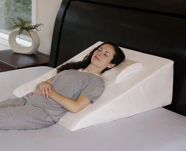
Wedge Pillow
A wedge pillow for the back is a necessity. A wedge pillow removes the stress from the spine and neck when lying down. Flipped around will take the tension off the legs also bringing back pain relief.
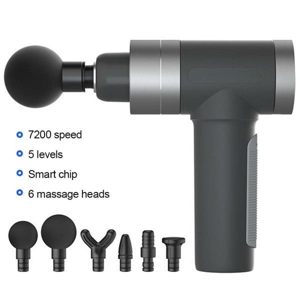
Deep Percussive Massager
Percussive massagers can provide a deep massage to various areas of the body especially the lower back. There are a variety of brands available with different levels of technology. However, careful use of these instruments must be exercised. This is because the massage can be intense and can exacerbate or cause further injury, and individuals can develop a tolerance making the massage no longer effective.
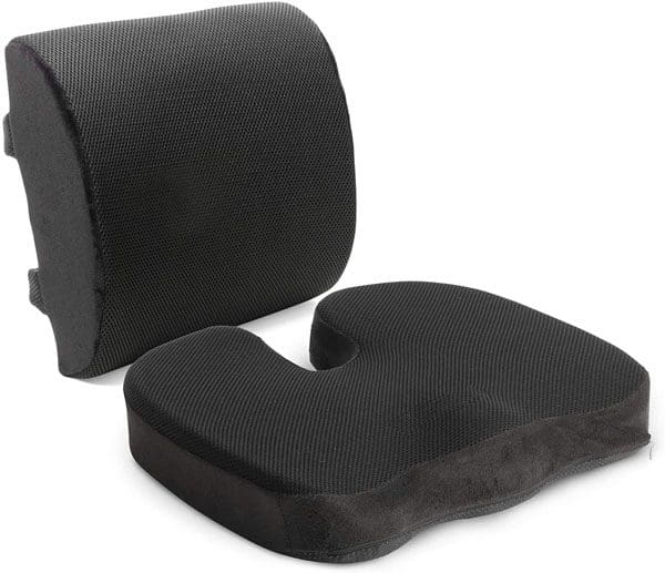
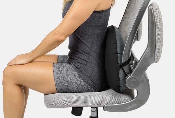
Seat Cushion
If sitting at a desk throughout the day or working from home a proper seat cushion is mandatory. Many individuals who sit the majority of their day utilize a combination cushion that includes the seat cushion with lower back support. Individual cushions are great because they can be moved easily and adjusted to fit where needed. Therapeutic seat cushions come with various features available, here are a few to keep in mind. Memory foam and air cells offer the most pressure relief. If there is tailbone pain, focus on a seat cushion with the tailbone cut out for extra relief. An office chair with these features should also be considered.
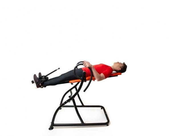
Inversion Table
Inversion tables are available at reasonable prices, starting around $100. Used correctly this therapeutic tool can successfully help relieve back pain. Inversion tables and cervical traction provide decompression and postural alignment for the spine helping with pain relief. These devices offer gentle decompression through the angle used. Wider angles or full inversion provides more decompression on the back. Individual spinal needs should be discussed with a chiropractor, physical therapist, or physician before using this therapeutic tool.
Pain Patches and Topical Agents
Pain-relieving patches like Lidocaine, IcyHot, and Salonpas patches are widely recommended for tight and sore areas of the body.
Sitting Standing Desk
A sitting and standing desk can be highly beneficial to back pain. In addition to burning off bonus calories throughout the day, Changing positions and postures throughout the day are recommended. This is to keep the muscles, ligaments, tendons moving, and not in a static position for too long. Changing every 20 to 30 minutes is the recommended time. Sitting and standing desks can provide positional changes that will help with posture, core stability, and circulation. This will help reduce and alleviate pain in the low back, neck, and shoulders. However, the desk needs to be stable and adjusted to the proper height.
Lower Back Sitting Support
These therapeutic tools help reinforce the low back region when seated. Most of us start to slouch forward with the head and shoulders hunched forward after some time at the computer. This strains the whole body, specifically the low back. Lower back supports can help maintain proper alignment of the spine when seated.
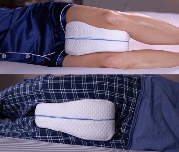
Knee, Thigh, Pelvis Pillow
These pillows have different names but are used in the same way. This is a pillow that can be placed between the legs while sleeping takes the pressure off the pelvis and spine. These types of pillows are great for individuals that sleep on their side. This is because the top leg often shifts down, leading to increased stress on the hips and low back. These pillows help keep the legs aligned during sleep relieving pressure on the low back.
How To Self-Care for Back Pain Books
There are a variety of books that offer tips, and therapies for self-care. These products are not a cure-all. They are intended to help in combination with proper treatment, especially for certain spinal conditions. If pain is limiting daily function, consult a chiropractor, physical therapist, or physician about using the above therapeutic tools.
Doctor of Chiropractic Near Me
Dr. Alex Jimenez�s Blog Post Disclaimer
The scope of our information is limited to chiropractic, musculoskeletal, physical medicines, wellness, and sensitive health issues and/or functional medicine articles, topics, and discussions. We use functional health & wellness protocols to treat and support care for injuries or disorders of the musculoskeletal system. Our posts, topics, subjects, and insights cover clinical matters, issues, and topics that relate and support directly or indirectly our clinical scope of practice.*
Our office has made a reasonable attempt to provide supportive citations and has identified the relevant research study or studies supporting our posts. We also make copies of supporting research studies available to the board and or the public upon request. We understand that we cover matters that require an additional explanation as to how it may assist in a particular care plan or treatment protocol; therefore, to further discuss the subject matter above, please feel free to ask Dr. Alex Jimenez or contact us at 915-850-0900. The provider(s) Licensed in Texas& New Mexico*
References
Furlan, Andrea D et al. �Massage for low-back pain.��The Cochrane database of systematic reviews,9 CD001929. 1 Sep. 2015, doi:10.1002/14651858.CD001929.pub3
The nervous system helps regulate the body’s immunity. Any dysfunction can initiate or aggravate issues associated with autoimmune disease. Chiropractic medicine focuses on the body�s ability to heal itself. These natural abilities function optimally when the body, and the nervous system, are well-balanced. Total body homeostasis elevates:
Mood
Immunity
Overall health
Quality of life
Autoimmune Disease
The immune system goes after any foreign bodies that the body comes in contact with to combat/prevent illness and disease. This could be:
Bacteria
Cancer cells
Viruses
However, the system can over-activate and start attacking its own cells and organs. This can manifest as an autoimmune disease. The type of disease depends on the specific area of the body that is being attacked. Common autoimmune diseases include:
Rheumatoid arthritis
Lupus
Multiple sclerosis
Irritable bowel syndrome
Psoriasis
A chiropractic approach to addressing autoimmune disease will usually begin with stress reduction. Therapies include:
Chiropractic adjustments to get the body balanced
Massage to loosen and relax the body’s tissues
Meditation to help manage stress, learn strategies of healthy coping
Yoga to keep the body limber
Exercise to strengthen the body inside and out
Diet to help with gut health and inflammation
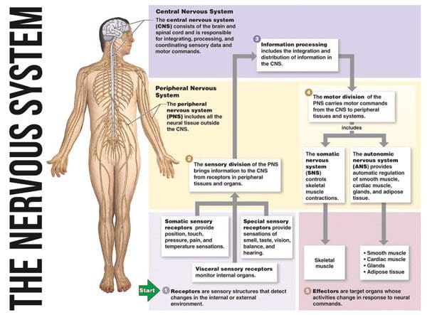
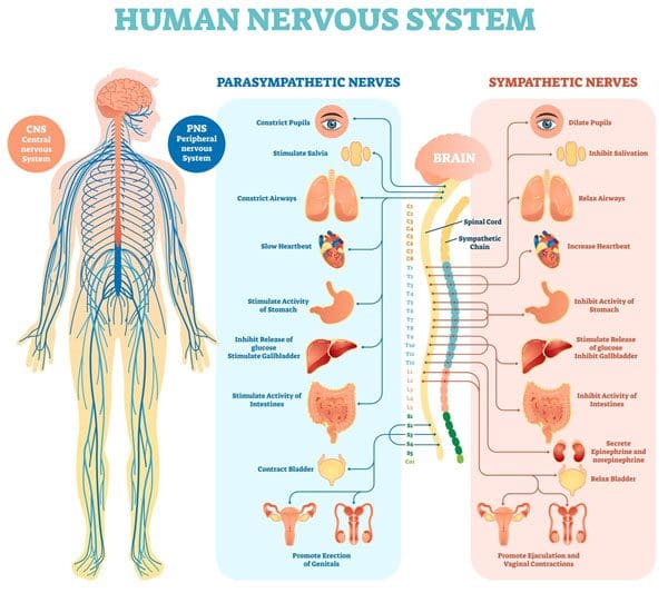
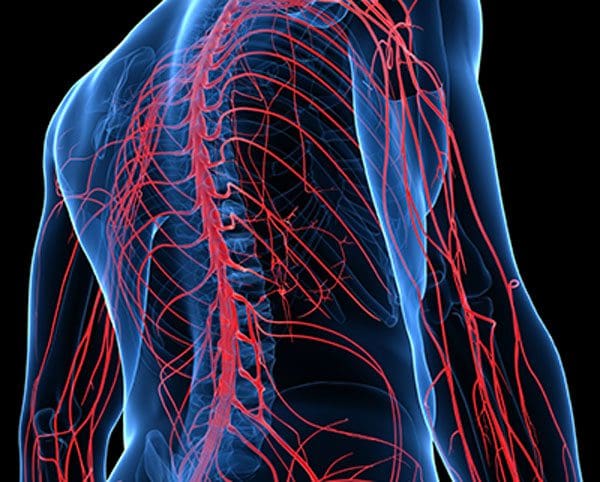
The Nervous System and Immunity
The nervous system has a significant role in the body’s immunity. The system transmits signals when to battle illness when to engage in the healing process, and so on. If the nerve’s path is compromised it can interfere with the brain�s ability to properly function and regulate immunity. This can lead to an underactive or overactive immune system that translates into an autoimmune disease.
Proper spinal alignment is vital for maintaining optimal nervous system function. When the spine is out of alignment/balance, the communication highway/s are inhibited, deteriorating the body’s health and neural tissue response. Spinal misalignment restoration is a chiropractic specialty that will increase the body’s natural healing power and promote whole-body balance. The nerve circulation is optimized and can properly communicate without sending improper/damaged signals.
Chiropractic
Any type of misalignment can be overlooked as a cause for body dysfunction. Chiropractic is about naturally sustained spinal and whole-body health. It will optimize the body to manage the disease and possibly reverse it. Injury Medical and Functional Chiropractic Clinic can offer a variety of customized treatment plans to achieve optimal health and maximum quality of life.
Back Pain Treatment
��
Dr. Alex Jimenez�s Blog Post Disclaimer
The scope of our information is limited to chiropractic, musculoskeletal, physical medicines, wellness, and sensitive health issues and/or functional medicine articles, topics, and discussions. We use functional health & wellness protocols to treat and support care for injuries or disorders of the musculoskeletal system. Our posts, topics, subjects, and insights cover clinical matters, issues, and topics that relate and support directly or indirectly our clinical scope of practice.*
Our office has made a reasonable attempt to provide supportive citations and has identified the relevant research study or studies supporting our posts. We also make copies of supporting research studies available to the board and or the public upon request. We understand that we cover matters that require an additional explanation as to how it may assist in a particular care plan or treatment protocol; therefore, to further discuss the subject matter above, please feel free to ask Dr. Alex Jimenez or contact us at 915-850-0900. The provider(s) Licensed in Texas& New Mexico*
References
Stojanovich, Ljudmila, and Dragomir Marisavljevich. �Stress as a trigger of autoimmune disease.��Autoimmunity reviews�vol. 7,3 (2008): 209-13. doi:10.1016/j.autrev.2007.11.007
Weakness, pain, and numbness can find their root in the spine. It is known as radiculopathy. If left untreated, health problems will only continue to get worse, with the potential to become a chronic condition. When it comes to degenerative pain conditions they usually start small. There is occasional discomfort that gradually develops into pain, then weakness and numbness.
Usually, by the time an individual seeks help, the original condition has devolved into radiating pain. This is why it is important to address the issue right away.Chiropractic works with individuals to help them understand the degenerative nature of pain and how to address and prevent it. Early intervention will prevent minor discomfort from turning into debilitating, chronic pain.
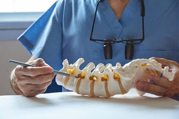
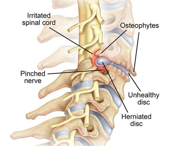
Radiculopathy
Radiating pain root cause is a compressed or inflamed nerve. It happens at the site of compression and spreads outward growing larger with time. It can be a catalyst for different pain conditions and syndromes like sciaticaor complex regional pain syndrome. There are a variety of terms for nerve pain, they are typically a form of radiculopathy.
Stages
The pain can spiral as fast as the underlying condition/s that is causing it. A compressed nerve that generates pain but does not become worse in severity is usually because the compression stays the same. Conversely, minimal discomfort brought on from shifted vertebrae can rapidly progress into weakness, numbness, and reduced mobility as a nerve is continually and severely getting compressed. Radiculopathy pain usually follows a pattern. Understanding the signs and symptoms will help determine to what extent the condition has progressed, and how it can develop into a worsening nerve injury:
Discomfort is the first stage. Subluxation, rotation, or a spinal shift is what is occurring, with the nerve not yet affected.
The pain signals that come from nerve compression along with the severity can help determine the cause of the condition.
Weakness usually follows pain. The nerve that is affected begins to take on permanent damage and cannot function properly.
Numbness follows the weakness reaching the most severe level. Mobility is limited along with a high increase for permanent nerve pain.
Being aware of these radiculopathy symptoms will help an individual stay ahead of nerve injury. Acting on discomfort can prevent progression into pain, addressing pain can stave off weakness, and acting upon weakness may prevent permanent nerve damage. The sooner an individual seeks help for any type of pain, the better chance they have to prevent degeneration.
Long-Term Prevention
It is crucial to consult with a chiropractor at the first sign of discomfort in the spine or if there is radiating/spreading back pain. A chiropractor will be able to provide decisive treatment that will bring relief and prevent the pain from worsening. The pain, weakness, and numbness can be avoided, along with long-term damage to the nerves.
Sciatica Pain Therapy
Dr. Alex Jimenez�s Blog Post Disclaimer
The scope of our information is limited to chiropractic, musculoskeletal, physical medicines, wellness, and sensitive health issues and/or functional medicine articles, topics, and discussions. We use functional health & wellness protocols to treat and support care for injuries or disorders of the musculoskeletal system. Our posts, topics, subjects, and insights cover clinical matters, issues, and topics that relate and support directly or indirectly our clinical scope of practice.*
Our office has made a reasonable attempt to provide supportive citations and has identified the relevant research study or studies supporting our posts. We also make copies of supporting research studies available to the board and or the public upon request. We understand that we cover matters that require an additional explanation as to how it may assist in a particular care plan or treatment protocol; therefore, to further discuss the subject matter above, please feel free to ask Dr. Alex Jimenez or contact us at 915-850-0900. The provider(s) Licensed in Texas& New Mexico*
References
Stochkendahl, Mette Jensen et al. �National Clinical Guidelines for non-surgical treatment of patients with recent-onset low back pain or lumbar radiculopathy.� The European spine journal: official publication of the European Spine Society, the European Spinal Deformity Society, and the European Section of the Cervical Spine Research Society�vol. 27,1 (2018): 60-75. doi:10.1007/s00586-017-5099-2
IFM's Find A Practitioner tool is the largest referral network in Functional Medicine, created to help patients locate Functional Medicine practitioners anywhere in the world. IFM Certified Practitioners are listed first in the search results, given their extensive education in Functional Medicine