These assessment and treatment recommendations represent a synthesis of information derived from personal clinical experience and from the numerous sources which are cited, or are based on the work of researchers, clinicians and therapists who are named (Basmajian 1974, Cailliet 1962, Dvorak & Dvorak 1984, Fryette 1954, Greenman 1989, 1996, Janda 1983, Lewit 1992, 1999, Mennell 1964, Rolf 1977, Williams 1965).
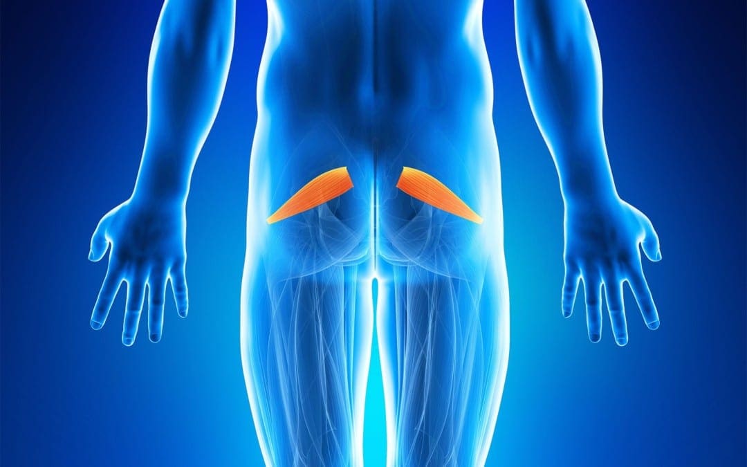
Clinical Application of Neuromuscular Techniques: Piriformis
Assessment of Shortened Piriformis
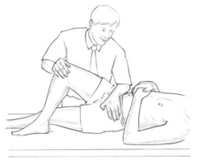
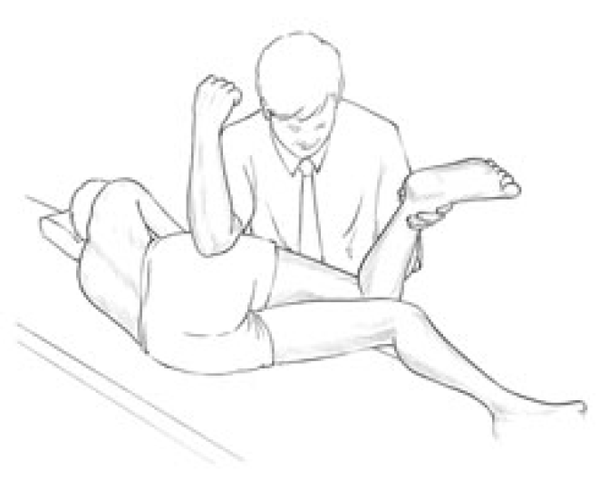
Test (a) Stretch test. When short, piriformis will cause the affected side leg of the supine patient to appear to be short and externally rotated. With the patient supine, the tested leg is placed into flexion at the hip and knee so that the foot rests on the table lateral to the contralateral knee (the tested leg is crossed over the straight non-tested leg, in other words as shown in Fig. 4.17). The angle of hip flexion should not exceed 60� (see notes on piriformis in Box 4.6).
Figure 4.17 MET treatment of piriformis muscle with patient supine. The pelvis must be maintained in a stable position as the knee (right in this example) is adducted to stretch piriformis following an isometric contraction.
The non-tested side ASIS is stabilised to prevent pelvic motion during the test and the knee of the tested side is pushed into adduction to place a stretch on piriformis. If there is a short piriformis the degree of adduction will be limited and the patient will report discomfort behind the trochanter.
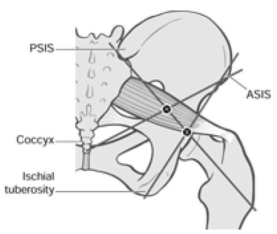
Test (b) Palpation test (Fig. 4.18) The patient is side-lying, tested side uppermost. The practitioner stands at the level of the pelvis in front of and facing the patient, and, in order to contact the insertion of piriformis, draws imaginary lines between:
ASIS and ischial tuberosity, and
PSIS and the most prominent point of trochanter.
Where these reference lines cross, just posterior to the trochanter, is the insertion of the muscle, and pressure here will produce marked discomfort if the structure is short or irritated.
Figure 4.18 Using bony landmarks as coordinates the commonest tender areas are located in piriformis, in the belly and at the attachment of the muscle.
If the most common trigger point site in the belly of the muscle is sought, then the line from the ASIS should be taken to the tip of the coccyx rather than to the ischial tuberosity. Pressure where this line crosses the other will access the mid-point of the belly of piriformis where triggers are common. Light compression here which produces a painful response is indicative of a stressed muscle and possibly an active myofascial trigger point.
Piriformis Strength Test
The patient lies prone, both knees flexed to 90�, with practitioner at foot of table grasping lower legs at the limit of their separation (which internally rotates the hip and therefore allows comparison of range of movement permitted by shortened external rotators such as the piriformis).
The patient attempts to bring the ankles together as the practitioner assesses the relative strength of the two legs. Mitchell et al (1979) suggest that if there is relative shortness (as evidenced by the lower leg not being able to travel as far from the mid-line as its pair in this position), and if that same side also tests strong, then MET is called for. If there is shortness but also weakness then the reasons for the weakness need to be dealt with prior to stretching using MET.
Box 4.6 Notes on Piriformis
Piriformis paradox. The performance of external rotation of the hip by piriformis occurs when the angle of hip flexion is 60� or less. Once the angle of hip flexion is greater than 60� piriformis function changes, so that it becomes an internal rotator of the hip (Gluck & Liebenson 1997, Lehmkuhl & Smith 1983). The implications of this are illustrated in Figures 4.17 and 4.19.
This postural muscle, like all others which have a predominence of type l fibres, will shorten if stressed. In the case of piriformis, the effect of shortening is to increase its diameter and because of its location this allows for direct pressure to be exerted on the sciatic nerve, which passes under it in 80% of people. In the other 20% the nerve passes through the muscle so that contraction will produce veritable strangulation of the sciatic nerve.
In addition, the pudendal nerve and the blood vessels of the internal iliac artery, as well as common perineal nerves, posterior femoral cutaneous nerve and nerves of the hip rotators, can all be affected.
If there is sciatic pain associated with piriformis shortness, then on straight leg raising, which reproduces the pain, external rotation of the hip should relieve it, since this slackens piriformis. (This clue may, however, only apply to any degree if the individual is one of those in whom the nerve actually passes through the muscle.)
The effects can be circulatory, neurological and functional, inducing pain and paraesthesia of the affected limb as well as alterations to pelvic and lumbar function. Diagnosis usually hinges on the absence of spinal causative factors and the distributions of symptoms from the sacrum to the hip joint, over the gluteal region and down to the popliteal space. Palpation of the affected piriformis tendon, near the head of the trochanter, will elicit pain and the affected leg will probably be externally rotated.
The piriformis muscle syndrome is frequently characterised by such bizarre symptoms that they may seem unrelated. One characteristic complaint is a persistent, severe, radiating low back pain extending from the sacrum to the hip joint, over the gluteal region and the posterior portion of the upper leg, to the popliteal space. In the most severe cases the patient will be unable to lie or stand comfortably, and changes in position will not relieve the pain. Intense pain will occur when the patient sits or squats since this type of movement requires external rotation of the upper leg and flexion at the knee.
Compression of the pudendal nerve and blood vessels which pass through the greater sciatic foramen and re-enter the pelvis via the lesser sciatic foramen is possible because of piriformis contracture. Any compression would result in impaired circulation to the genitalia in both sexes. Since external rotation of the hips is required for coitus by women, pain noted during this act could relate to impaired circulation induced by piriformis dysfunction. This could also be a basis for impotency in men. (See also Box 4.7.)
Piriformis involvement often relates to a pattern of pain which includes: pain near the trochanter; pain in the inguinal area; local tenderness over the insertion behind trochanter; SI joint pain on the opposite side; externally rotated foot on the same side; pain unrelieved by most positions with standing and walking being the easiest; limitation of internal rotation of the leg which produces pain near the hip; and a short leg on the affected side.
The pain itself will be persistent and radiating, covering anywhere from the sacrum to the buttock, hip and leg including inguinal and perineal areas.
Bourdillon (1982) suggests that piriformis syndrome and SI joint dysfunction are intimately connected and that recurrent SI problems will not stabilise until hypertonic piriformis is corrected.
Janda (1996) points to the vast amount of pelvic organ dysfunction to which piriformis can contribute due to its relationship with circulation to the area.
Mitchell et al (1979) suggest that (as in psoas example above) piriformis shortness should only be treated if it is tested to be short and stronger than its pair. If it is short and weak (see p. 110 for strength test), then whatever is hypertonic and influencing it should be released and stretched first (Mitchell et al 1979). When it tests strong and short, piriformis should receive MET treatment.
Since piriformis is an external rotator of the hip it can be inhibited (made to test weak) if an internal rotator such as TFL is hypertonic or if its pair is hypertonic, since one piriformis will inhibit the other.
Box 4.7 Notes on Working and Resting Muscles
Richard (1978) reminds us that a working muscle will mobilise up to 10 times the quantity of blood mobilised by a resting muscle. He points out the link between pelvic circulation and lumbar, ischiatic and gluteal arteries and the chance this allows to engineer the involvement of 2400 square metres of capillaries by using repetitive pumping of these muscles (including piriformis).
The therapeutic use of this knowledge involves the patient being asked to repetitively contract both piriformis muscles against resistance. The patient is supine, knees bent, feet on the table; the practitioner resists their effort to abduct their flexed knees, using pulsed muscle energy approach (Ruddy�s method) in which two isometrically resisted pulsation/contractions per second are introduced for as long as possible (a minute seems a long time doing this).
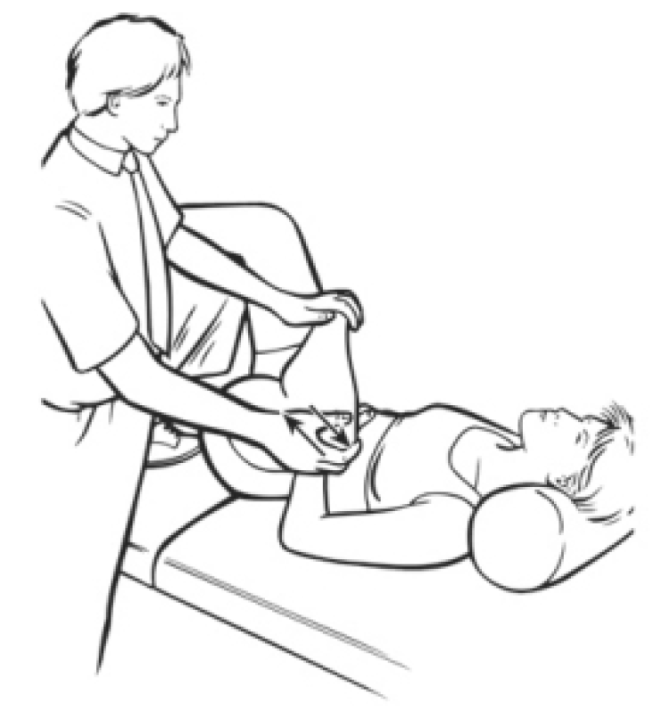
Figure 4.19 MET treatment of piriformis with hip fully flexed and externally rotated (see Box 4.6, first bullet point).
Figure 4.20 A combined ischaemic compression (elbow pressure) and MET side-lying treatment of piriformis. The pressure is alternated with isometric contractions/stretching of the muscle until no further gain is achieved.
MET Treatment of Piriformis
Piriformis method (a) Side-lying The patient is side-lying, close to the edge of the table, affected side uppermost, both legs flexed at hip and knee. The practitioner stands facing the patient at hip level.
The practitioner places his cephalad elbow tip gently over the point behind trochanter, where piriformis inserts. The patient should be close enough to the edge of the table for the practitioner to stabilise the pelvis against his trunk (Fig. 4.20). At the same time, the practitioner�s caudad hand grasps the ankle and uses this to bring the upper leg/hip into internal rotation, taking out all the slack in piriformis.
A degree of inhibitory pressure (sufficient to cause discomfort but not pain) is applied via the elbow for 5�7 seconds while the muscle is kept at a reasonable but not excessive degree of stretch. The practitioner maintains contact on the point, but eases pressure, and asks the patient to introduce an isometric contraction (25% of strength for 5�7 seconds) to piriformis by bringing the lower leg towards the table against resistance. (The same acute and chronic rules as discussed previously are employed, together with cooperative breathing if appropriate, see Box 4.2.)
After the contraction ceases and the patient relaxes, the lower limb is taken to its new resistance barrier and elbow pressure is reapplied. This process is repeated until no further gain is achieved.
Piriformis method (b)1 This method is a variation on the method advocated by TePoorten (1960) which calls for longer and heavier compression, and no intermediate isometric contractions.
In the first stage of TePoorten�s method the patient lies on the non-affected side with knees flexed and hip joints flexed to 90�.The practitioner places his elbow on the piriformis musculotendinous junction and a steady pressure of 20�30 lb (9�13 kg) is applied. With his other hand he abducts the foot so that it will force an internal rotation of the upper leg.
The leg is held in this rotated position for periods of up to 2 minutes. This procedure is repeated two or three times. The patient is then placed in the supine position and the affected leg is tested for freedom of both external and internal rotation.
Piriformis method (b)2 The second stage of TePoorten�s treatment is performed with the patient supine with both legs extended. The foot of the affected leg is grasped and the leg is flexed at both the knee and the hip. As knee and hip flexion is performed the practitioner turns the foot inward, so inducing an external rotation of the upper leg. The practitioner then extends the knee, and simultaneously turns the foot outward, resulting in an internal rotation of the upper leg.
During these procedures the patient is instructed to partially resist the movements introduced by the practitioner (i.e. the procedure becomes an isokinetic activity). This treatment method, repeated two or three times, serves to relieve the contracture of the muscles of external and internal hip rotation.
Piriformis method (c) A series of MET isometric contractions and stretches can be applied with the patient prone and the affected side knee flexed. The hip is rotated internally by the practitioner using the foot as a lever to ease it laterally, so putting piriformis at stretch. Acute and chronic guidelines described earlier are used to determine the appropriate starting point for the contraction (at the barrier for acute and short of it for chronic).
The patient attempts to lightly bring the heel back towards the midline against resistance (avoiding strong contractions to avoid knee strain in this position) and this is held for 7�10 seconds. After release of the contraction the hip is rotated further to move piriformis to or through the barrier, as appropriate. Application of inhibitory pressure to the attachment or belly of piriformis is possible via thumb, if deemed necessary.
Piriformis method (d) A general approach which balances muscles of the region, as well as the pelvic diaphragm, is achieved by having the patient squat while the practitioner stands and stabilises both shoulders, preventing the patient from rising as this is attempted, while the breath is held. After 7�10 seconds the effort is released; a deeper squat is performed, and the procedure is repeated several times.
Piriformis method (e) This method is based on the test position (see Fig. 4.17) and is described by Lewit (1992). With the patient supine, the treated leg is placed into flexion at the hip and knee, so that the foot rests on the table lateral to the contralateral knee (the leg on the side to be treated is crossed over the other, straight, leg). The angle of hip flexion should not exceed 60� (see notes on piriformis, Box 4.6, for explanation).
The practitioner places one hand on the contralateral ASIS to prevent pelvic motion, while the other hand is placed against the lateral flexed knee as this is pushed into resisted abduction to contract piriformis for 7�10 seconds. Following the contraction the practitioner eases the treated side leg into adduction until a sense of resistance is noted; this is held for 10�30 seconds.
Piriformis method (f) Since contraction of one piriformis inhibits its pair, it is possible to self-treat an affected short piriformis by having the patient lie up against a wall with the non-affected side touching it, both knees flexed (modified from Retzlaff 1974). The patient monitors the affected side by palpating behind the trochanter, ensuring that no contraction takes place on that side.
After a contraction lasting 10 seconds or so of the non-affected side (the patient presses the knee against the wall), the patient moves away from the wall and the position described for piriformis test (see Fig. 4.17) above is adopted, and the patient pushes the affected side knee into adduction, stretching piriformis on that side. This is repeated several times.
Dr. Alex Jimenez offers an additional assessment and treatment of the hip flexors as a part of a referenced clinical application of neuromuscular techniques by Leon Chaitow and Judith Walker DeLany. The scope of our information is limited to chiropractic and spinal injuries and conditions. To discuss the subject matter, please feel free to ask Dr. Jimenez or contact us at 915-850-0900 .
By Dr. Alex Jimenez
Additional Topics: Wellness
Overall health and wellness are essential towards maintaining the proper mental and physical balance in the body. From eating a balanced nutrition as well as exercising and participating in physical activities, to sleeping a healthy amount of time on a regular basis, following the best health and wellness tips can ultimately help maintain overall well-being. Eating plenty of fruits and vegetables can go a long way towards helping people become healthy.
Suffer Sciatica: Are you experiencing pain along one side of your body from your lower back down through your hip and the back of your leg? If so, you could be suffering from a condition called sciatica.
According to the Mayo Clinic, sciatica can best be described as “most commonly occurring when a herniated disk or a bone spur on the spine compresses part of the nerve. This causes inflammation, pain and often some numbness in the affected leg.”
A variety of issues weigh in on an individual’s likelihood of ending up with sciatica. Most of them deal with increased pressure on the spine.
Suffer Sciatica: Causes
Obesity: carrying too much weight is instrumental in bringing on a number of health related issues. Extra pounds overload the spine, causing damage that results in sciatica.
Improper Lifting: Individuals who frequently twist the bodies and lift heavy loads are more likely to suffer from sciatica. Certain jobs that require these movements are a key cause of the condition.
Sedentary Lifestyle. A person’s job does not have to involve lifting to be responsible for this condition. Sitting for extended periods without stretching or standing puts excess pressure on the spine and can cause sciatica.
Too Many Birthdays. Getting older can affect all of our body’s joints and bones in a negative manner, especially if we never committed to an exercise routing. An individual’s back often deteriorates with age, causing bone spurs and herniated disks that sometimes result in sciatica.
Treatment options for sciatica are varied, and the choice depends on the severity of the condition.
Pain Medication: A common and easy way to treat sciatica is with drug therapy. Anti-inflammatory drugs are frequently used to reduce�the inflammation around the nerve, which is a big contributor of the pain. Over-the-counter pain medicines, as well as codeine, may also help with pain management.
Acupuncture. Alternative therapies like acupuncture have shown positive results in the treatment of sciatica. If a drug-free treatment option appeals to you, find an experienced acupuncturist in your area and talk to them about treatment options.
Strengthening Exercises. A consistent exercise program strengthens your muscles and helps the body function effectively. Ask your doctor which exercises assist the body with bouncing back from sciatica.
Supplements. Supplying the body with vital vitamins and minerals assists in overall health in general, including improvement from sciatica. Daily doses of supplements such as calcium, magnesium, St. John’s Wort, and Vitamin B12 have shown to treat sciatica effectively.
Chiropractic Care. Chiropractors understand all things spine-related, and work with the body as a whole to help it heal itself. Chiropractic treatment for sciatica works to align the spine and reduce the stress to the lower back. Treatment helps alleviate the underlying causes of the condition, and shows positive results in a short amount of time.
Cortisone Injections. Most of the time, sciatica can be treated by the less invasive measures mentioned above. However, severe bouts of sciatica may require a shot of cortisone directly into the inflamed area. Individuals generally choose this option when other treatments have garnered no relief.
Dealing with sciatica is painful and irritating, as the condition often sidelines the sufferer from daily activities. By knowing the treatment options that are effective in combating both the underlying causes and the pain of sciatica, sufferers can begin a regimen that will help them get back on their feet, pain-free in the shortest period of time possible and no longer have to suffer.
If you are suffering from sciatica and would like to talk to an experienced chiropractor about how to treat the condition, contact us today.
Sciatica
This article is copyrighted by Blogging Chiros LLC for its Doctor of Chiropractic members and may not be copied or duplicated in any manner including printed or electronic media, regardless of whether for a fee or gratis without the prior written permission of Blogging Chiros, LLC.
If you are among the 45 million Americans who suffer headaches regularly, you are undoubtedly familiar with the traditional methods that people use to treat them, including taking some type of over the counter medication that is supposed to eliminate the pain and reduce any associated swelling. There are many other types of treatment that you may have attempted as well, including taking pharmaceutical concoctions designed to treat pain. However, none of these solutions provide permanent relief that addresses the problem at the core.
In the past few decades, there have been an increasing number of patients throughout the country that have begun to seek alternative forms of therapy for all types of conditions, including headaches. One form of treatment that is very promising for physical as well as other types of pain is chiropractic.
This form of alternative care has been used successfully for over a hundred years and has become a regular part of the American healthcare delivery system. In fact, there are an increasing number of insurance providers that are willing to pay for chiropractic because of its effectiveness.
When a person goes to school to learn about chiropractic, they begin by studying the human anatomy in great detail, just like other medical professionals. However, in addition to looking at how all of the parts work together physically, their training primarily revolves around the diagnosis and treatment of misalignments in the spine known as subluxations.
Theses subluxations compress nerve tissue that affects organ function, soft tissue like muscle, ligaments and tendons and can eventually manifest as other health problems if not treated.
Once the nerves are disrupted pain will result. While it usually manifests itself as physical pain, this is not always the case. In some instances, the person may experience difficulties with sleep or other routine habits.
On the first visit with a chiropractor, a review of previous health issues will be completed, including x-rays (if needed) to determine what types of nerve blockage may be occurring. They will listen to the patient attentively and make an assessment, including determining what types of treatment will best suit the patient.
In the case of headaches, the patient usually has misalignment (subluxations) in the cervical spine. This may be accompanied by muscles that are unduly tight in the neck, shoulders and nearby areas. Pressure on the nerves may cause sharp stabbing pains or there may be a continual dull throb in the region.
The chiropractor will assess the area and then move forward with treatment known as a chiropractic adjustment in order to relieve the pressure and pain. The relief is usually instantaneous, with an increase in positive symptoms for the following few hours.
However, chiropractors don�t just focus on relieving symptoms but rather correcting the problem; therefore, additional treatments will follow to help correct the cause. In addition, the chiropractor will take the time to educate you on the importance of chiropractic, especially if you�ve never been to one before.
If you need further help with your headaches or are ready to see how beneficial chiropractic care can be to your overall health, please give us a call so that you can schedule an appointment with our Doctor of Chiropractic.
This article is copyrighted by Blogging Chiros LLC for its Doctor of Chiropractic members and may not be copied or duplicated in any manner including printed or electronic media, regardless of whether for a fee or gratis without the prior written permission of Blogging Chiros, LLC.
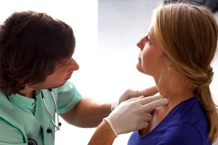
Hypothyroidism is evaluated and diagnosed by a physician, your primary care doctor or an endocrinologist. Many factors, signs, and symptoms are taken into consideration when hypothyroidism is diagnosed.
How is hypothyroidism diagnosed?
A diagnosis is reached after a thorough review of the patient’s symptoms, family and medical history, risk factors, physical examination, and effectively, a blood test. There are many types of blood tests, which the most authoritative one is known as the TSH test (thyroid-stimulating hormone). However, in some cases, healthcare professionals may refer patients to receive a total T4 or T4, free T4 index, or even thyroxine to aid in the diagnosis.
Why Hypothyroidism is not Diagnosed on Symptoms Alone
Lots of the signs of hypothyroidism are fairly frequent complaints found in people with a normally functioning thyroid gland, so it can be tough to decipher if the symptoms are linked to the thyroid gland. Among the best ways to find out whether your symptoms might be related to a thyroid condition is to consider how long you have been experiencing them. For example, have you felt cold when others were warm? Did you just begin to notice decreased energy? It might be associated with a thyroid issue if you are beginning to notice new signs and symptoms. But only a specialized healthcare professional (eg, endocrinologist) can diagnose a thyroid issue.
Medical and Family History
It is important to give your doctor as many details as you can about your own personal medical history, in addition to family history (eg, mom had eczema). Make sure you talk about:
Your overall state of health, particularly any changes you’ve noticed on your general well-being.
Your family’s health history, especially if a near relative was diagnosed with hypothyroidism (or any other thyroid-related issues).
Whether you’ve ever had thyroid surgery, or radiation into your own neck to deal with cancer.
Any medications you could be taking that could cause hypothyroidism (eg, amiodarone, lithiumion, interferon alpha, interlukin-2, or even earlier chemotherapy).
Physical Evaluation
Your doctor will perform a thorough examination and look for physical signs of hypothyroidism, such as:
Proof of dry skin
Swelling around the eyes and legs
Slower reflexes
Slower heartbeat
Blood Tests
Hypothyroidism can be diagnosed using different blood tests such as:
TSH Evaluation
A thyroid-stimulating hormone or TSH is a blood test that measures the amount of T4 (thyroxine) that the thyroid gland has been indicated to create. In case you have an abnormally significant degree of TSH, it might indicate you have hypothyroidism.
T4 (thyroxine) Evaluation
The thyroid gland produces T4 (thyroxine). The T4 along with the free T4 index are blood tests which, in conjunction with a TSH test, can let your doctor know your thyroid is functioning.
The adrenal gland tells the thyroid how much thyroxine to produce through signaling by TSH. There are cells from the pituitary gland that determine what your body’s “set point” is. Your collection point is that the normal array of TSH as determined by your thyroid gland that your body needs.
As blood flows throughout the pituitary gland, the very same cells detect if there are sufficient T4 levels in the body. The pituitary sends the amount of TSH into the thyroid to maintain levels in the standard range in case your T4 amount is sufficient. If your level is too low, the pituitary sends TSH outside telling the thyroid to make more T4. In case your T4 level is too high, the pituitary sends TSH that is less out, then telling the thyroid to make less T4.
Normal and Abnormal TSH Ranges
0.4 mU/L to 4.0 mU/L is considered the reference array (there may be slight variation depending on the laboratory), and people that have a normally functioning thyroid gland usually fall within this range.
If TSH measures > 4.0 mU/L, a second evaluation (T4) is done to verify the results. TSH p4.0/mU/L using a very low T4 level indicates hypothyroidism.
If your TSH is > 4.0 mU/L along with your T4 level is normal, this may prompt your physician to test your serum anti-thyroid peroxidase (anti-TPO) antibodies. When these antibodies are found, it may signal an autoimmune thyroid disease, which is a risk factor for developing hypothyroidism. In case you have those anti-bodies, your doctor will perform and TSH test at least once each year.
An easy way to remember how the thyroid works is to think about supply and demand. The TSH rises as the T4 level drops. The TSH drops as the T4 level rises. But not everyone with hypothyroidism has elevated levels of TSH. If the pituitary is not working properly, perhaps it does not send out regular TSH levels. But if the quantity of TSH is off, the thyroid will not make the perfect quantity of T4. This is rare and is called secondary or central hypothyroidism.
The scope of our information is limited to chiropractic and spinal injuries and conditions. To discuss options on the subject matter, please feel free to ask Dr. Jimenez or contact us at 915-850-0900 .
By Dr. Alex Jimenez
Additional Topics: Wellness
Overall health and wellness are essential towards maintaining the proper mental and physical balance in the body. From eating a balanced nutrition as well as exercising and participating in physical activities, to sleeping a healthy amount of time on a regular basis, following the best health and wellness tips can ultimately help maintain overall well-being. Eating plenty of fruits and vegetables can go a long way towards helping people become healthy.
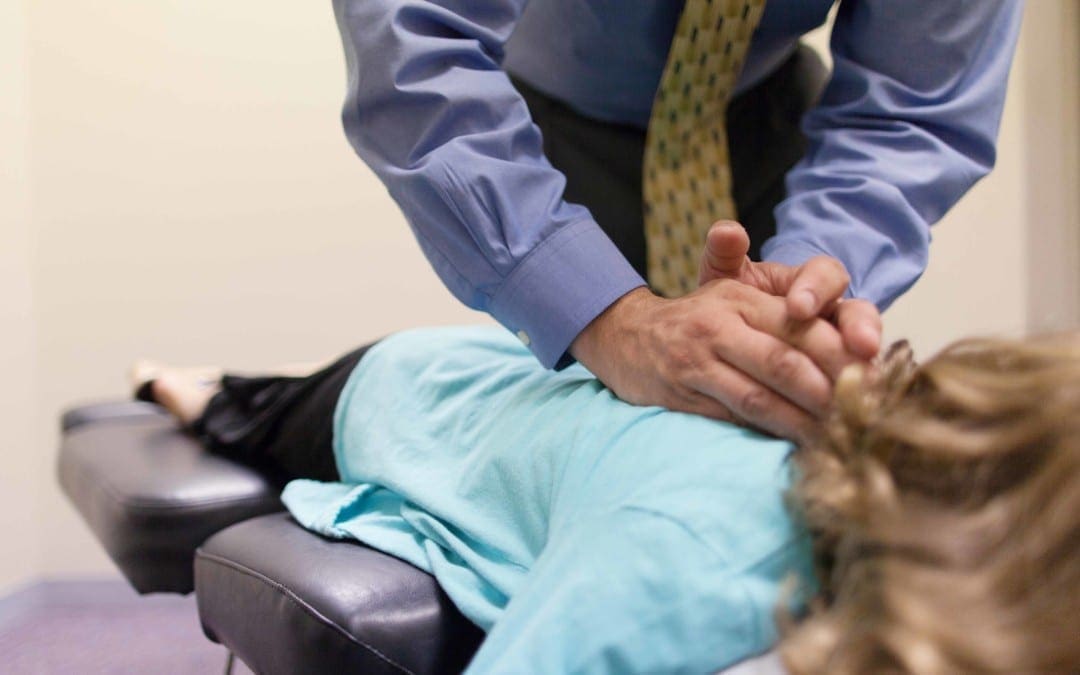
A chiropractor is a healthcare professional who focuses on the diagnosis, treatment and prevention of neuromuscular and musculoskeletal disorders through the use of adjustments and manipulations of the spine.
What are a chiropractor’s treatment goals and beliefs?
Chiropractors seek to reduce pain and enhance the performance of patients as well as to instruct them on how they can account for their health via ergonomics, exercise and other therapies to deal with their pain. Chiropractic is usually categorized as alternative medicine or complementary medicine.
Fundamental Chiropractor Beliefs and Goals
Chiropractors focus on the intimate relationship between the nervous system and the spine, to provide overall health and wellness to the human body. They also hold accurate the following beliefs:
Biomechanical and structural derangement of the spine can affect the nervous system
For many conditions, chiropractic treatment may restore the structural integrity of the spine, reduce stress on the sensitive adrenal gland, and thus improve the wellness of the individual.
The treatment concept of chiropractic is to re-establish normal spinal mobility, which in turn alleviates the irritation to the spinal nerve or re-establishes altered folds, to reduce painful symptoms affecting the individual.
Conditions Treated by Chiropractic
Chiropractors use a number of non-surgical treatment modalities to treat patients with certain types of:
Lower back pain and/or leg pain (sciatica)
Neck pain
Repetitive strains
Headaches
Sports injuries
Car accident injuries
Arthritic pain
While primarily focusing on fixing neuromusculoskeletal disorders, chiropractors aren’t exclusively confined to problems with the nervous system and musculoskeletal system. If appropriate, these healthcare professionals will refer patients to medical doctors or other healthcare practitioners for treatment of lower back pain, or other injuries and conditions. Chiropractors have a local referral network or function collectively with other spinal experts.
Chiropractic Examination
In most regards, a chiropractic evaluation is quite much like conventional assessment procedures administered by all health care providers. With that said chiropractors examine function and the arrangement of the spine and then determine chiropractic therapies separates attention.
Chiropractic Exam of Back Pain
A first chiropractic examination for back pain will generally have three parts: a consultation, case history, and physical examination. Laboratory investigation and X-ray examination may be done if needed.
Consultation. The chiropractor meets with the patient and provides a synopsis of their back pain, such as:
Duration and frequency of symptoms
Description of these symptoms (e.g. burning, throbbing)
Areas of pain
What makes the pain feel better (e.g. sitting, extending)
What makes the pain feel worse (e.g. standing, lifting)
Case history. The chiropractor identifies the area(s) of complaint and the nature of the spine pain by asking questions and learning more about different regions of the patient’s background, including:
Family background
Dietary customs
Past background of other therapies (chiropractic, osteopathic, medical and other)
Occupational history
Psychosocial history
Other places to probe, frequently based on responses to preceding questions
Physical evaluation. A chiropractor may use a variety of methods to determine the spinal sections which require chiropractic treatments, including but not limited to, static and motion palpation techniques ascertaining spinal segments which are hypo cellular (restricted in their motion) or fixated. Depending on the results of the evaluation, extra diagnostic tests may be used by a chiropractor, for example:
X-ray to locate subluxations (the altered position of the vertebra)
A device that detects the temperature of their skin in the paraspinal area to identify spinal areas having a substantial temperature variance which needs manipulation.
Many chiropractors use a holistic, biomechanical concept of treating the bipedal structure completely, in an effort to balance the arrangement from the feet upwards.
Chiropractors are usually trained in multiple procedures of evaluating lower back pain, for example:
Evaluation and management solutions. Chiropractors are trained in examining the joints, bones, muscles and tendons of the body with the objective of imagining tenderness any misalignment, asymmetry, defects or other issues.
Neurologic and other common physical examination procedures. Chiropractors are trained to perform a variety of neurologic tests (nerve root compression/tension, engine strength, coordination, deep tendon and pathological reflexes, etc.) and are proficient in doing orthopedic, cardiovascular and several other frequent assessments.
Specialized assessment. Chiropractors are trained to assess range of motion, stability, muscle strength, muscle tone along with other assessments.
Common diagnostic studies. Chiropractors are trained in use of diagnostic tools and studies like radiography (X-rays), laboratory diagnostics and neurodiagnostics.
The scope of our information is limited to chiropractic and spinal injuries and conditions. To discuss options on the subject matter, please feel free to ask Dr. Jimenez or contact us at 915-850-0900 .�
By Dr. Alex Jimenez
Additional Topics: Wellness
Overall health and wellness are essential towards maintaining the proper mental and physical balance in the body. From eating a balanced nutrition as well as exercising and participating in physical activities, to sleeping a healthy amount of time on a regular basis, following the best health and wellness tips can ultimately help maintain overall well-being. Eating plenty of fruits and vegetables can go a long way towards helping people become healthy.
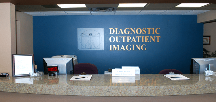
Dr. Alex Jimenez collaborates with top rated diagnosticians and imaging specialists. We are blessed to have in our association, imaging specialists that provide fast, courteous & premiere board certified specialists. In collaboration with our offices we can provide the quality of service our patients mandate and deserve.
Who We Are
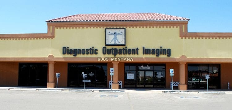
Diagnostic Outpatient Imaging (DOI) is a state-of-the-art Radiology center in El Paso, TX. It is the only center of its kind in El Paso, owned and operated by a Radiologist.
This means when you come to DOI for a radiologic exam, every detail, from the design of the rooms, the choice of the equipment, the hand-picked technologists, and the software which runs the office, is carefully chosen or designed by the Radiologist and not by an accountant.
Our market niche is one center of excellence. Our values related to patient care are: We believe in treating patients the way we would treat our family and we will do our best to ensure that you have a good experience at our clinic.
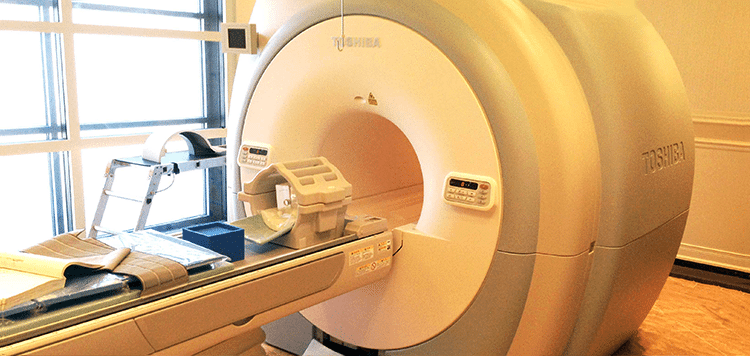
Dear Doctors,
We are pleased to inform you of the arrival of our Titan 3-Tesla MRI at Diagnostic Outpatient Imaging. This is El Paso’s only radiology imaging center that offers this technology. Patients do not always realize how important image quality is: It can make the difference in the diagnosis.
3-Tesla MRI is like HD TV and once you try it, you will not want to go back. The increased magnet strength gives us many benefits at no additional expense to the patient. It gives us the ability to scan faster or to scan with higher detail. An MRI of the brain can take 20 minutes and have exceptional quality, or we can perform the scan in less time, with better quality that is achieved on most 1.5 Tesla “high field” MRIs. This is incredibly useful for children.
Our 3T MRI can perform Diffusion Tensor Imaging, MRI Spectroscopy and CSF flow studies to name just a few of its possibilities.
This scanner is not only very fast, it is very large. Our open MRI has a clearance of 35 cm. The 3T has a diameter of 71 cm! This is welcome news for nervous or claustrophobic patients, and combined with its speed, it can actually eliminate the need for sedation for some patients. 3T MRI is faster, clearer, and has more diagnostic possibilities. We are certain you and your patients will notice the difference.
Our Services
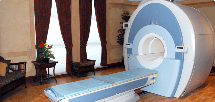
MRI’s:
DOI has three MRI’s under one roof. All are American College of Radiology (ACR) Certified.
Good
Open MRI (0.35 Tesla): This MRI perfect for claustrophobic and very large patients. There is no table weight limit on this MRI
Better
High Field 1.5 Tesla MRI- This is a eight channel MRI with high end image quality. It is in a beautiful room and has ‘pianissimo’ technology, which makes the MRI relatively quiet. This machine has been the best MRI in private practice in El Paso for years. It will soon be eclipsed by our new 3.0 Tesla MRI.
Best
High Field 3.0 Tesla MRI- This is the only 3.0 Tesla MRI in private practice in El Paso. This technology can deliver stunning image quality, which can actually make a difference in your diagnosis. The increased magnet strength gives us many benefits at no additional expense to the patient.�??It gives us the ability to scan faster, or to scan with higher detail. This is welcome news for nervous or claustrophobic patients, and as well as for children as it can actually eliminate the need for sedation in some patients. 3T is faster, clearer, more diagnostic for a better for MRI. It is like HD TV. Once you have tried it, you won’t want to go back. This MRI effectively doubles our MRI capacity. If needed most exams can be completed in under 5 minutes, instead of the normal 30-45 minutes.
Breast MRI:
DOI began Breast MRI in July 2007, being the first facility in El Paso to perform the exam. We have now performed over 2500 breast MRI’s and many MRI-guided breast biopsies. All have been interpreted and/or performed by Dr. Boushka, making him the most experienced radiologist in the city with this exam. This is the most powerful tool for the detection of Breast cancer to date.
Hours: Monday to Thursday 7 am to 9 pm Friday 7 am to 5 pm Saturday 8 am to 4 pm
Prostate MRI:
Guys, you need great medical care also. We are the only facility in El Paso performing this leading edge exam. MRI can see cancers when other imaging methods cannot. Not only can we see prostate cancers with MRI, we can perform MRI-guided prostate biopies for pathologic (definitive) diagnosis.
Monday to Thursday 7 am to 9 pm Friday 7 am to 5 pm Saturday 8 am to 4 pm
CT:
We have a 16 slice Toshiba Aquillion CT scanner, with newly updated in Dec 2013. The upgrade allows for reduced X-ray dose, higher resolution, more patient comfort, shorter breath holds and doubles the speed of the scanner. This scanner performs CT X-ray exams as helical volume acquisitions in 3D from a single patient exam. Most exams are finished in under 60 seconds, unless delayed images with contrast are indicated. Additionally we have a powerful 3D post processing workstation.
Hours Monday to Friday 7 am to 6 pm
Ultrasound:
DOI has just doubled our Ultrasound capacity with newly purchased Philips 34 XRL scanner. We have Three certified Ultrasonographers with cumulative experience of 45 years. We are confident you will find them professional and compassionate. Beverly Bruner RDMS, Sonographer, formally of Desert Imaging has joined our team.
3D OB Ultrasounds:
You better believe it. Available whenever our US department is open. No referral necessary. Images are reviewed by an actual radiologist.
Ultrasound Hours: Monday, Tuesday, Thursday 8 am to 5 pm Wednesday 8 am to 8 pm Friday 8 am to 5 pm Saturday 8 am to 12 pm
Digital Mammography
DOI was the first facility in El Paso to acquire Hologic Full Field Digital Mammography and thus we have more experience with this technology than any facility in El Paso. Our Mammographer has 20 years of experience and has her own following of patents who seek her out to perform their mammograms because of her excellent and compassionate care. Our private pay screening mammography price of $90, including the interpretation is an unbeaten price in El Paso.
Hours Mon – Fri 8am to 4pm Extended hours Wednesday until 8pm) Saturdays 8am to 12pm
Bone Denisity (DEXA)
We have a brand new, Hologic Discovery CI bone densitometer scanner. This is the latest technology.
X-Ray
Our digital computed radiography was just updated February 2014. No appointments are necessary.
Many Americans in the United States will visit a healthcare professional’s office reporting some type of pain. While most cases of pain are considered acute, or temporary, resolving after the injury or condition causing the symptoms has healed, a large percentage of individuals will still report pain long after the source has disappeared. This is known as chronic pain. Fortunately, there are a variety of treatment methods which can also help ease these symptoms.
Are injections used to treat chronic pain?
From physical therapy and chiropractic care, to drugs and medications, numerous types of treatment methods and therapies can be used to treat chronic pain, each more beneficial to certain people than others. Epidural steroid injections and facet joint injections are some of the most common types of injections utilized to ease chronic pain symptoms. For some individuals, injections may be more useful than other forms of treatment. As with any medical procedure, however, it’s important to understand how helpful these can be for each, individual patient.
Epidural Corticosteroid Injections for Chronic Pain
Although epidural steroid injections (also called epidural corticosteroid injections) can be helpful to confirm a diagnosis, they should be used primarily after a specific presumptive diagnosis has been established. Additionally, injections shouldn’t be used in isolation, but rather in combination with a program strengthening, stressing muscle flexibility, and operational recovery, most commonly associated with chronic pain, in this case.
Appropriate follow-up after shots to rate ability and the individual’s treatment response to progress in the rehabilitation program is indispensable. Observation of this response is necessary prior to a second or third shot, although a number of injections can be attempted to decrease pain. Epidural steroid injections are an adjunct treatment, which facilitates participation in an active exercise program and may assist in avoiding the need for surgical intervention.
Treatment Rationale
The rationale for the use of epidural corticosteroid injection has enhanced with the signs of an inflammatory basis for radicular pain from disc herniation. Although prospective trials are lacking, epidural steroids have been proven to be effective in pain reduction in patients with referred pain. If used in the initial weeks after onset the efficacy is increased.
The goal of these injections would be to facilitate an active exercise program and also to progress sufferers through the pain and inflammation phase of healing as quickly as possible. As with all injections, it needs to be a part of a comprehensive treatment plan involving active exercise programs.
How the Injection Is Applied
To ensure proper needle placement of corticosteroids, fluoroscopic guidance is recommended. Meaning a healthcare professional will use special imaging gear during the injection to be sure the needle is going in at the right place. Some patients may require more than one injection. Repeat shots should be based on goals and the response after the injection. It is not necessary for many patients to experience a set number or “series” of injections. If minimal to no advancement is found following two shots, then further similar shots aren’t warranted. The recent usage of the approach allows the medicine to be delivered in a fashion to the ventral part of the spinal canal. All patients must be followed by consecutive injections (10-14 days later) to assess therapeutic reaction.
Utilization of Epidural Steroid Injections
Epidural shots and intradiscal injections have been used in treating non-radicular degenerative disc disorder with limited success. In addition, epidural steroids are used in patients with neurogenic claudication from spinal stenosis with mixed outcomes. A number of shots can be tried to decrease pain thought to be at least in part mediated by inflammation.
Facet Joint Injections for Chronic Pain
The therapeutic advantage of facet injections remains controversial. The controversy starts with the significance of the background and examination with lower back pain. Many patients will complain of back and lower extremity pain with standing, walking, and extension-type pursuits. The examination is normal, and also tests for nerve root inflammation are often negative. Many patients may have increased pain on passive expansion, or extension and rotation.
Additionally, radiographic and bone scanning imaging hasn’t been useful in selecting appropriate patients for facet injections. Consequently, the primary job of facet injections remains diagnostic. There is support for the impact of shots or ablations of the nerves. Facet injections should be used for patients who have failed a guided non-operative treatment program that incorporates various manipulation/mobilization methods. They should be done under fluoroscopic guidance and are not suggested in the initial four to six weeks of treatment.
Goal of Facet Joint Injections
The goal of facet injections is to verify the diagnosis and perhaps assist with pain reduction to be able to alleviate an active physical treatment program. If prior injections were helpful and there’s a recurrence of pain, they can be replicated replicate injections should be limited. This process should be used only in people failing a comprehensive application and in no manner should be considered at the initial management of an incident of acute low back pain.
Be sure to seek the proper guidance from an experienced and qualified healthcare professional before attempting any medical procedure, method or therapy. Injections for chronic pain are only one form of treatment used for the mentioned symptoms. Other treatment options can be used alongside these or in place of the above.
The scope of our information is limited to chiropractic and spinal injuries and conditions. To discuss options on the subject matter, please feel free to ask Dr. Jimenez or contact us at 915-850-0900 .
By Dr. Alex Jimenez
Additional Topics: Wellness
Overall health and wellness are essential towards maintaining the proper mental and physical balance in the body. From eating a balanced nutrition as well as exercising and participating in physical activities, to sleeping a healthy amount of time on a regular basis, following the best health and wellness tips can ultimately help maintain overall well-being. Eating plenty of fruits and vegetables can go a long way towards helping people become healthy.
IFM's Find A Practitioner tool is the largest referral network in Functional Medicine, created to help patients locate Functional Medicine practitioners anywhere in the world. IFM Certified Practitioners are listed first in the search results, given their extensive education in Functional Medicine


 Please note that we can answer general questions, but anything specific to your medical case should be discussed with your physician.