Healing After a Head Injury: How Your Body Can Recover with the Right Team
Your son sustained a severe blow during Friday-night football. Your wife walked away from a three-car pile-up. Your husband fell 12 feet off a scaffold. All three left the hospital with the same three letters: TBI – traumatic brain injury. The doctor said, “Go home and rest.” But two weeks later, the headaches, foggy thinking, and stomach troubles are worse. You feel lost. This article is written for you – the person healing, the family member searching at 2 a.m., and the nurse, coach, or therapist who wants to help.
We will walk through:
What really happens inside the skull in the first minutes, hours, and weeks?
Why does the damage keep spreading if no one stops the “second wave”
How a whole-body team – including chiropractic nurse practitioners (CNPs) – can turn the tide.
Simple daily steps you can start tomorrow.
Let’s begin where the injury begins.
The Two Waves That Steal Recovery
Wave 1: The First Hit (Primary Injury)
A helmet-to-helmet tackle, a steering wheel to the forehead, or a beam to the hard hat cracks open brain cells in an instant. Blood vessels tear. The skull may stay whole, but the soft brain bounces like gelatin in a jar. This is the damage everyone sees on the CT scan (Missouri University of Science and Technology, 2025).
Wave 2: The Hidden Fire (Secondary Injury)
The real thief shows up later. Four chemical storms start inside the brain and body:
Excitotoxicity – Too much glutamate (brain messenger chemical) pours out. Neurons fire relentlessly until they burn out (Waters, 2023).
Oxidative stress – Tiny sparks called reactive oxygen species (ROS) act like rust on brain wires (Gharavi et al., 2023).
Neuroinflammation – Immune cells rush in to help, but stay too long and attack healthy tissue (Simon et al., 2017).
Gut-brain meltdown – The gut lining leaks, bad bacteria cross into the blood, and the brain swells even more (Heuer Fischer, 2024).
These four storms can last weeks, not hours. One mouse study showed that brain toxins still remained elevated 7 days after the crash (Missouri University of Science and Technology, 2025). That is why “I feel worse at week three” is so common.
Real People, Real Storms
Jake, 17, linebacker – Cleared to play after 10 days. By week four, he had forgotten his homework, snapped at his mom, and thrown up every morning. Gut-brain tests showed that no good bacteria remained.
Maria, 34, Uber driver – Whiplash plus airbag to the temple. Doctors only checked her neck X-ray. Six months later, she still can’t balance her checkbook. Blood tests revealed extremely high levels of inflammation markers.
Carlos, 42, roofer – The hard hat saved his life, but it could not protect him from the diffuse twist inside his body. His wife noticed he cried at commercials and slept 14 hours a day. His oxidative stress score was triple the norm.
All three were told, “It’s just a concussion. Wait it out.” Waiting lets the second wave win.
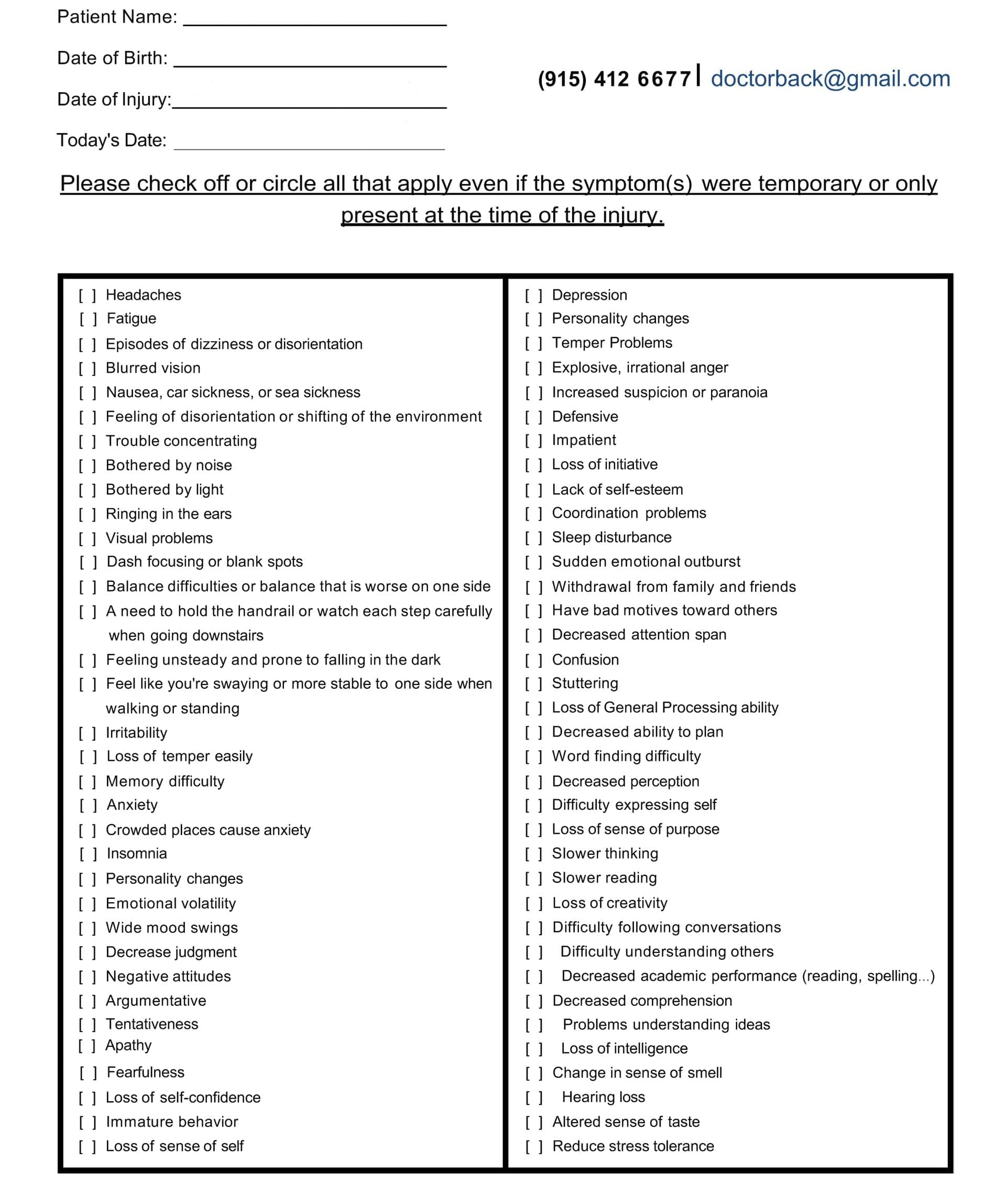
Symptom Questionnaire:
The Blood-Brain Barrier: Your Skull’s Broken Gate
Think of the blood-brain barrier (BBB) as a velvet rope around a VIP club. After TBI, it rips. Proteins and water leak in, causing brain swelling (edema). Two kinds matter:
Cytotoxic edema – Cells drink too much water and burst.
Vasogenic edema – The rope is cut; everything floods the dance floor (Kuriakose & Uzunova, 2023).
Swelling squeezes healthy areas. Memory, mood, and movement shut down. MRI may still look “normal” because standard scans miss these tiny leaks.
The Gut-Brain Highway No One Talks About
Your gut has more nerve endings than your spinal cord. After TBI:
Stress hormones crash.
Good bacteria die.
The gut wall gets holes.
Toxins ride the vagus nerve straight to the brain.
Result? Anxiety, constipation, and brain fog that no pill fixes (Heuer Fischer, 2024). Heal the gut, calm the brain.
Meet the Team That Sees the Whole Picture
The Chiropractic Nurse Practitioner (CNP)
A CNP is a registered nurse with extra doctoral training in chiropractic neurology and functional medicine. Dr. Alexander Jimenez, DC, APRN, FNP-BC, in El Paso, Texas, has treated more than 4,000 TBI patients. Dr. Alexander Jimenez asserts, “Our approach goes beyond simple neck repairs.” We reset the entire nervous system dashboard” (Jimenez, 2025).
The Core Four Tests Every TBI Patient Needs
qEEG brain map – Shows which brain waves are stuck.
Ice for 10 minutes on / 20 minutes off, behind the neck.
Zero screens after 7 p.m. Blue light feeds excitotoxicity.
Sip bone broth; it contains glycine, nature’s brake pedal on glutamate.
Week 2 – Feed the Repair Crew
20 g collagen + 500 mg vitamin C before breakfast.
Walk 10 minutes outside; sunlight reboots the circadian rhythm.
Family rule: No yelling. Loud voices re-trigger fight-or-flight.
Week 3 – Reboot the Gut-Brain Highway
One new fermented food daily: sauerkraut, kimchi, kefir.
4-7-8 breathing with kids: In 4, hold 7, out 8. Calms the vagus nerve.
Week 4 – Gentle Brain Games
Lumosity 10 min/day.
Chiropractic CNP checks the pupil’s response and the balance board.
Month 2 – Return-to-Life Checklist
Driver’s test with an occupational therapist.
Coach reviews film for neck-safe tackling.
The employer receives a light-duty note based on the HRV score.
Stories That Prove It Works
Jake – After 6 weeks of CNP care plus fermented foods, his qEEG looked like his pre-season map. He started in the playoffs.
Maria – Glutathione IVs twice a week dropped her headache diary from 7/10 to 2/10. She passed the driving retest on her first try.
Carlos – Cervical adjustments restored CSF flow; his wife says, “I have my husband back.” He returned to framing houses with a new hard-hat liner.
Why Insurance Is Starting to Pay
Medicare and most Blue Cross plans now cover:
Chiropractic neurology E/M codes 99xxx
IV antioxidant therapy under “medically necessary”
qEEG as diagnostic code R94.01
Request Dr. Jimenez’s “TBI Recovery Bundle” letter; families report an 80% approval rate (Jimenez, 2025).
Your 3-Minute Action Plan Tonight
Text your CNP: “Can we do the Core Four tests?”
Put a bag of frozen peas in a sock behind the injured person’s neck for 10 minutes.
Open the fridge, eat one spoon of yogurt, and hum “Happy Birthday” out loud.
You just cooled inflammation, fed good bacteria, and stimulated the vagus nerve. That is real medicine.
The Promise We Make to Families
No one should feel alone in the dark after a head injury. The brain wants to heal. Give it the right team, the right fuel, and the right quiet space, and it will rebuild stronger. You are not “just concussed.” You are a whole person with a whole team ready to walk the road with you.
Discover the relationship between cognitive impairment and traumatic brain injury to improve awareness and prevention strategies.
Understanding Traumatic Brain Injury and Cognitive Impairment: An Integrative Approach to Recovery
Introduction
Brain injuries represent one of the most significant health challenges of our time, affecting millions of people annually and creating long-lasting impacts that extend far beyond the initial injury. Traumatic brain injuries stand as a major cause of death and disability around the world, with more than thirty percent of injury-related deaths in the United States involving some form of brain trauma (Bailes & Borlongan, 2020). These injuries trigger a complex cascade of events that affect not only brain function but also create widespread changes throughout the entire body.missionlegalcenter When someone experiences a traumatic brain injury, the damage occurs in two distinct phases. The primary injury happens at the moment of impact when external forces cause immediate mechanical damage to brain tissue. This is followed by a secondary injury phase, during which biochemical processes, including inflammation, cell death, oxidative stress, and other harmful changes, lead to further brain deterioration over days, weeks, and even months following the initial trauma (Bailes & Borlongan, 2020). Understanding these injury patterns helps healthcare providers develop more effective treatment approaches that address both immediate concerns and long-term recovery needs.missionlegalcenter Cognitive impairment represents one of the most common and challenging consequences of traumatic brain injury. Problems with attention, memory, and executive functioning emerge as the primary neurocognitive consequences across all levels of injury severity (Cognitive Impairment Following Traumatic Brain Injury, 2002). These cognitive disruptions profoundly affect daily life, making it difficult for individuals to work, maintain relationships, manage household tasks, and participate fully in their communities. Because attention and memory serve as foundational cognitive abilities, their disruption can trigger additional problems with executive function, communication, and other complex mental processes (Cognitive Impairment Following Traumatic Brain Injury, 2002).pubmed.ncbi.nlm.nih
The connection between brain and body becomes especially important when considering traumatic brain injury recovery. The brain controls virtually every function in the human body through an intricate network of nerves and chemical signals. The central nervous system, comprising the brain and spinal cord, regulates awareness, movement, sensation, thought, speech, and memory (Anatomy and physiology of the nervous system, 2020). When injury disrupts these control centers, the effects ripple throughout the entire body, affecting muscles, bones, organs, and metabolic processes.cancer An integrative approach that combines chiropractic care with nurse practitioner oversight offers promising possibilities for individuals recovering from traumatic brain injuries. This collaborative model addresses the neurological, musculoskeletal, cognitive, emotional, and metabolic aspects of recovery. Chiropractic care focuses on restoring nervous system function through spinal adjustments, soft-tissue therapies, and targeted exercises, while nurse practitioners provide comprehensive medical management, cognitive support, and coordination of overall health needs. Together, these providers can create comprehensive treatment plans that support the brain’s natural healing processes and help patients regain function and improve their quality of life.
What is a Traumatic Brain Injury?
Traumatic brain injury refers to brain damage caused by an outside force that disrupts normal brain function. This external force can take many forms, including a forceful bump, blow, or jolt to the head or body, or an object penetrating the skull and damaging brain tissue (Traumatic Brain Injury, 2023). The injury occurs when the brain moves violently inside the skull or when an external object breaks through the skull barrier. Common causes include motor vehicle accidents, falls, sports injuries, violence, and blast exposures from explosions (Types of Traumatic Brain Injury, 2024).ninds.nih+1 Healthcare providers classify traumatic brain injuries according to their severity, which helps guide treatment decisions and predict outcomes. The three main categories include mild, moderate, and severe traumatic brain injury. Medical professionals use several measures to determine severity, including the Glasgow Coma Scale score, duration of loss of consciousness, length of post-traumatic amnesia, and results from brain imaging studies (Criteria used to classify TBI severity, 2012).ncbi.nlm.nih+1
Mild traumatic brain injury, often called a concussion, generally does not cause prolonged loss of consciousness. If unconsciousness occurs, it typically lasts less than thirty minutes. The Glasgow Coma Scale score ranges from thirteen to fifteen for mild injuries. Common symptoms include headaches, dizziness, confusion, nausea, vision problems, difficulty thinking clearly, balance issues, sleep disturbances, sensitivity to light and sound, problems with attention and concentration, fatigue, anxiety, irritability, and emotional changes (4 Types of Brain Injuries and 3 Levels of Severity, 2021). Memory loss associated with mild traumatic brain injury usually lasts less than twenty-four hours. Most people with mild injuries recover within a few days to weeks with appropriate rest and management (4 Types of Brain Injuries and 3 Levels of Severity, 2021).missionlegalcenter
Moderate traumatic brain injury involves unconsciousness lasting more than thirty minutes but less than twenty-four hours. The Glasgow Coma Scale score falls between nine and twelve. Individuals with moderate injuries experience all the symptoms associated with mild traumatic brain injury, plus additional concerning signs. These include headaches that worsen or do not improve, seizures or convulsions, numbness or weakness in the arms and legs, repeated vomiting, inability to wake from sleep, and slurred speech (4 Types of Brain Injuries and 3 Levels of Severity, 2021). Post-traumatic amnesia lasts more than one day but less than seven days. Brain imaging may show abnormalities such as bleeding, bruising, or swelling (Criteria used to classify TBI severity, 2012).ncbi.nlm.nih+1
Severe traumatic brain injury represents the most serious category, with loss of consciousness exceeding twenty-four hours. The Glasgow Coma Scale score ranges from three to eight. Post-traumatic amnesia persists for more than seven days. Individuals with severe injuries often require intensive medical care and extended rehabilitation. They may experience altered consciousness states, including coma, vegetative state, or minimally conscious state (Traumatic Brain Injury, 2023). Brain imaging typically reveals significant abnormalities including bleeding within the brain tissue, bleeding over the brain surface, bleeding in the brain’s ventricles, swelling, and tissue damage (Types of Traumatic Brain Injury, 2024).medschool.ucla+1
The type of traumatic brain injury also provides important classification information. Closed head injuries occur when the head experiences impact or rapid movement without skull penetration. Concussions, diffuse axonal injury, and contusions fall into this category. Diffuse axonal injury, one of the most common types, involves widespread damage to the brain’s white matter, which contains nerve fibers that facilitate communication between different brain regions. This type of injury commonly happens in auto accidents, falls, and sports-related trauma (Traumatic Brain Injury, 2023). Penetrating brain injuries occur when an object breaks through the skull and enters brain tissue, as seen with gunshot wounds or impalement injuries (Types of Traumatic Brain Injury, 2024).ninds.nih+1 Understanding whether an injury is primary or secondary helps guide treatment approaches. Primary traumatic brain injury refers to the immediate structural damage inside the brain resulting directly from the initial impact. This includes bruising, bleeding, and tearing of brain tissue and blood vessels. Secondary traumatic brain injury describes complications that develop after the initial trauma and cause additional damage to an already compromised brain. Secondary injury mechanisms include increased pressure inside the skull, progressive brain swelling, damage to blood vessels triggering stroke or seizures, and lack of oxygen related to blood pressure drops or breathing difficulties (Types of Traumatic Brain Injury, 2024).medschool.ucla
How TBI Causes Cognitive Impairment
Traumatic brain injury triggers a complex series of events that disrupt normal brain function and lead to cognitive impairment. Understanding these mechanisms enables healthcare providers to develop targeted interventions that support recovery and effectively manage symptoms.
The physical damage from traumatic brain injury affects brain structure and function in multiple ways. When the brain experiences trauma, nerve cells can be stretched, torn, or destroyed. The white matter tracts that connect different brain regions become damaged, disrupting the communication networks essential for coordinated brain function. Diffuse axonal injury particularly affects these communication pathways, as the nerve fibers that transmit signals between brain cells break down and lose their ability to conduct information efficiently (Traumatic Brain Injury, 2023).ninds.nih
Bleeding within the brain creates additional problems. When blood vessels rupture, blood accumulates in spaces where it does not belong, creating pressure that compresses surrounding brain tissue. This compression damages cells both directly through physical pressure and indirectly by reducing blood flow to affected areas. Swelling further compounds these problems, as increased fluid within the rigid skull creates mounting pressure that can damage brain tissue and reduce oxygen delivery (Types of Traumatic Brain Injury, 2024).medschool.ucla
At the cellular level, traumatic brain injury initiates harmful biochemical cascades. Cell membranes become disrupted, allowing excessive calcium and sodium to enter neurons. This triggers a series of destructive processes including activation of enzymes that break down cellular components, production of free radicals that damage cell structures, mitochondrial dysfunction that impairs energy production, and release of inflammatory molecules that promote further injury (Bailes & Borlongan, 2020). These processes can continue for days, weeks, or even months after the initial injury, explaining why symptoms sometimes worsen or new problems emerge well after the traumatic event.missionlegalcenter
Inflammation plays a particularly important role in post-traumatic brain injury cognitive impairment. Within seconds after trauma, inflammatory responses activate in the brain. The blood-brain barrier, which normally protects the brain from harmful substances in the bloodstream, becomes damaged and allows inflammatory cells and molecules to enter brain tissue. While some inflammation helps with healing and clearing damaged tissue, excessive or prolonged inflammation damages healthy brain cells and interferes with recovery. Inflammatory molecules affect neurotransmitter systems, disrupt nerve signaling, and impair the formation of new neural connections needed for cognitive recovery (Mesenchymal stem cell therapy alleviates the neuroinflammation, 2020).medicine.washu
Different brain regions show varying vulnerability to traumatic injury, which explains the specific cognitive impairments that develop. The frontal lobes, responsible for executive functions such as planning, decision-making, impulse control, and working memory, are particularly susceptible to damage from trauma. The temporal lobes, involved in memory formation and language processing, also commonly sustain injury. Damage to the hippocampus, a structure critical for forming new memories, explains why memory problems rank among the most frequent cognitive complaints after traumatic brain injury (Cognitive Problems After Traumatic Brain Injury, n.d.).uwmsktc.washington
Attention and concentration problems emerge as foundational deficits following traumatic brain injury. Individuals may struggle to focus, pay attention to relevant information while filtering out distractions, or attend to more than one task at a time. This leads to restlessness, easy distractibility, difficulty finishing projects, problems carrying on conversations, and trouble sitting still for extended periods. Because attention skills serve as building blocks for higher-level cognitive abilities, people with attention problems often develop additional difficulties with memory, reasoning, and problem-solving (Cognitive Problems After Traumatic Brain Injury, n.d.).uwmsktc.washington
Processing speed commonly slows after brain injury. Individuals take longer to understand what others are saying, need more time to follow directions, struggle to keep up with television shows or movies, require additional time to read and comprehend written material, and show delayed reactions to stimuli. This slowed processing affects everyday activities and can make tasks that were once automatic feel laborious and exhausting. The reduced reaction time poses particular concerns for activities requiring quick responses, such as driving (Cognitive Problems After Traumatic Brain Injury, n.d.).uwmsktc.washington
Memory impairments manifest in various ways after traumatic brain injury. Short-term memory problems make it difficult to hold information in mind temporarily, such as remembering a phone number long enough to dial it or recalling items on a shopping list. Long-term memory difficulties affect the ability to store and retrieve information over extended periods. People may struggle to remember recent events, learn new information, or recall facts and procedures they previously knew well. Working memory, which involves holding and manipulating information simultaneously, becomes compromised, affecting complex cognitive tasks like mental arithmetic, following multi-step directions, and reasoning (Cognitive Impairment Following Traumatic Brain Injury, 2002).pubmed.ncbi.nlm.nih
Executive function deficits represent another hallmark of traumatic brain injury cognitive impairment. Executive functions include the mental processes that help people plan activities, organize information, initiate tasks, monitor performance, shift between tasks flexibly, solve problems, make decisions, and control impulses. When these abilities become impaired, individuals struggle with goal-directed behavior, adapting to new situations, regulating emotions, and functioning independently in daily life (Cognitive Problems After Traumatic Brain Injury, n.d.).uwmsktc.washington
The Brain-Body Connection
The relationship between the brain and body represents one of the most fundamental aspects of human physiology. This intricate connection enables all body functions, from voluntary movements to unconscious processes that sustain life. Understanding this connection becomes especially important when considering how traumatic brain injury affects not just cognitive abilities but overall physical health and function.
The central nervous system serves as the command center for the entire body. Made up of the brain and spinal cord, this system controls awareness, movements, sensations, thoughts, speech, and the five senses of seeing, hearing, feeling, tasting, and smelling (Central nervous system function, 2025). The brain manages most body functions by processing information from sensory receptors throughout the body and sending out instructions through an extensive network of nerves. The spinal cord acts as an extension of the brain, carrying messages between the brain and peripheral nerves that reach every part of the body (Central nervous system function, 2025).healthdirect
The peripheral nervous system complements the central nervous system by connecting the brain and spinal cord to the rest of the body. This network of nerves and ganglia sends signals to and receives signals from the central nervous system, enabling two-way communication between the brain and body tissues. The peripheral nervous system divides into the somatic nervous system, which controls voluntary movements like walking and grasping objects, and the autonomic nervous system, which manages involuntary functions that the body performs automatically, such as breathing, heartbeat, digestion, and blood pressure regulation (Anatomy and physiology of the nervous system, 2020).cancer
The autonomic nervous system further separates into two complementary branches that maintain balance in body functions. The sympathetic nervous system prepares the body for situations requiring strength, heightened awareness, or rapid response, commonly known as the fight-or-flight response. Activation of this system increases heart rate, elevates blood pressure, speeds breathing, dilates pupils, and increases metabolic rate. The parasympathetic nervous system creates opposite effects, returning heart rate and breathing to normal, constricting pupils, and slowing metabolism to conserve energy and promote rest and recovery (Anatomy and physiology of the nervous system, 2020).cancer
Research demonstrates that the brain and body maintain constant, bidirectional communication through multiple pathways. Recent studies show that parts of the brain area controlling movement connect directly to networks involved in thinking, planning, and control of involuntary body functions such as blood pressure and heartbeat. This literal linkage of body and mind in brain structure helps explain phenomena like why anxiety makes people pace, why vagus nerve stimulation can alleviate depression, and why regular exercise improves mental outlook (Mind-body connection is built into brain, 2023).medicine.washu
The vagus nerve exemplifies this brain-body connection. This cranial nerve carries signals between the brain and internal organs, providing information about organ function and regulating processes like digestion and heart rate. Signals traveling through the vagus nerve are coded independently by specialized neurons, allowing the brain to discriminate precisely among various body signals and respond appropriately. This sophisticated communication system enables the brain to monitor and adjust organ function continuously based on changing body needs and environmental demands (Revealing Communications Between Brain and Body, 2022).medicine.yale
Blood flow represents another critical aspect of brain-body connection. The brain, despite constituting only about two percent of total body mass, consumes over twenty percent of the body’s glucose-derived energy. Continuous glucose metabolism supports neuronal signaling, as adenosine triphosphate, the cell’s energy currency, powers action potentials, maintains ionic gradients, and supports synaptic transmission. Because the brain cannot synthesize or store glucose independently, it depends entirely on glucose from dietary intake and blood circulation. Any disruption to blood flow or energy metabolism can significantly impair brain function (Metabolic hormones mediate cognition, 2009).sciencedirect
The musculoskeletal system connects intimately with brain function through sensory feedback and motor control. Muscles contain specialized receptors that constantly send information to the brain about body position, movement, and force. This proprioceptive feedback allows the brain to coordinate movement, maintain posture, and adjust to environmental demands. The brain processes this information and sends motor commands back to muscles, enabling precise, coordinated movement. When traumatic brain injury disrupts these communication pathways, both sensory perception and motor control become impaired (Nervous System Function, 2024).clevelandclinic
Hormonal systems provide another dimension of brain-body connection. The hypothalamus and pituitary gland, located deep within the brain, regulate hormonal signals that control growth, metabolism, reproduction, stress response, and many other functions. These structures form a feedback loop, with the hypothalamus releasing hormones that signal the pituitary gland, which then distributes hormones to various body systems including the adrenal glands, thyroid, reproductive organs, skin, bone, and muscle. This hormonal regulation affects mood, memory, metabolism, muscle mass, energy levels, stress response, and reproductive function (Neuroendocrine Disturbances Following TBI, 2023).biausa
The immune system also maintains constant communication with the brain. Immune cells and inflammatory molecules can cross from the bloodstream into brain tissue, particularly when the blood-brain barrier becomes damaged following injury. The brain, in turn, can influence immune function through neural and hormonal signals. This bidirectional communication becomes particularly important following traumatic brain injury, when both local brain inflammation and systemic immune responses affect recovery and long-term outcomes (Multiorgan Dysfunction After Severe TBI, 2021).pmc.ncbi.nlm.nih
Causes and Symptoms of Cognitive Impairment
Cognitive impairment following traumatic brain injury arises from multiple interrelated causes that affect brain structure and function. Understanding these causes enables healthcare providers to identify risk factors, develop effective prevention strategies, and tailor targeted treatment approaches.
The primary cause of cognitive impairment stems from direct damage to brain tissue at the moment of injury. When the brain experiences sudden acceleration, deceleration, or rotational forces, nerve cells stretch and tear, blood vessels rupture, and tissue bruises. The specific location and extent of damage determine which cognitive functions become impaired. Injuries to the frontal lobes typically affect executive functions, attention, and working memory. Damage to the temporal lobe disrupts memory formation and language processing. Parietal lobe injuries interfere with sensory processing and spatial awareness, while occipital lobe damage affects visual processing (Traumatic Brain Injury, 2023).ninds.nih
Secondary injury mechanisms compound the initial damage. Swelling increases pressure within the rigid skull, compressing brain tissue and reducing blood flow. Bleeding creates masses that displace normal brain structures and increase intracranial pressure. Chemical imbalances develop as damaged cells release excessive amounts of neurotransmitters, particularly glutamate, which overstimulates neighboring neurons and triggers cell death. Free radicals produced during cellular metabolism damage cell membranes and DNA. Mitochondrial dysfunction impairs energy production, leaving neurons unable to maintain normal function. These secondary processes continue for days to weeks after the initial injury, explaining why cognitive symptoms may worsen or emerge gradually (Bailes & Borlongan, 2020).missionlegalcenter
Inflammation represents a major contributor to cognitive impairment following traumatic brain injury. The inflammatory response activates within seconds after trauma and can persist for months or even years. While acute inflammation helps remove damaged tissue and initiate healing, chronic inflammation damages healthy neurons and interferes with recovery. Inflammatory molecules disrupt neurotransmitter systems, impair synaptic plasticity, reduce the production of growth factors needed for neural repair, and contribute to the ongoing death of brain cells. This persistent inflammation particularly affects cognitive functions requiring complex neural networks and plasticity, such as learning, memory consolidation, and executive function (Mesenchymal stem cell therapy alleviates the neuroinflammation, 2020).medicine.washu
Disrupted blood flow contributes to cognitive impairment by reducing oxygen and nutrient delivery to brain tissue. Traumatic brain injury can damage blood vessels directly, alter blood pressure regulation, and trigger vasospasm where blood vessels constrict excessively. The brain requires constant, abundant blood supply to meet its high metabolic demands. Even brief or partial reductions in blood flow can impair neural function and contribute to cell death. Chronic reductions in cerebral blood flow may explain some persistent cognitive deficits that remain long after the initial injury (Long-term Consequences of TBI in Bone, 2018).pmc.ncbi.nlm.nih
Hormonal disruptions following traumatic brain injury affect cognition through multiple pathways. The hypothalamus and pituitary gland, structures that regulate hormonal systems, are particularly vulnerable to traumatic injury due to their location and delicate structure. Damage to these areas causes hypopituitarism, a condition where insufficient hormone production affects growth, metabolism, stress response, and reproduction. Growth hormone deficiency, thyroid hormone deficiency, and sex hormone deficiencies all contribute to cognitive impairment, affecting memory, attention, processing speed, and executive function (Neuroendocrine Disturbances Following TBI, 2023).biausa
The symptoms of cognitive impairment following traumatic brain injury vary widely depending on injury severity, location, and individual factors. Attention and concentration problems rank among the most common complaints. Individuals struggle to focus on tasks, become easily distracted by environmental stimuli, have difficulty filtering out irrelevant information, and cannot maintain attention for extended periods. These problems make it challenging to follow conversations, complete work tasks, read for comprehension, or perform activities requiring sustained mental effort (Cognitive Problems After Traumatic Brain Injury, n.d.).uwmsktc.washington
Memory impairments manifest in various ways. Short-term memory problems make it difficult to remember recent events, conversations, or instructions. People may repeatedly ask the same questions, forget appointments, or lose track of items. Long-term memory difficulties affect the ability to recall past events, previously learned information, or familiar procedures. Working memory deficits interfere with tasks requiring simultaneous information holding and manipulation, such as mental calculations, following multi-step directions, or reasoning through problems (Cognitive Impairment Following Traumatic Brain Injury, 2002).pubmed.ncbi.nlm.nih
Processing speed reductions cause delays in understanding and responding to information. Individuals take longer to comprehend spoken or written language, need extra time to formulate responses, show slowed reaction times, and struggle to keep pace in conversations or fast-moving situations. This slowed processing affects virtually all cognitive tasks and creates frustration when individuals recognize their difficulties but cannot overcome them through effort alone (Cognitive Problems After Traumatic Brain Injury, n.d.).uwmsktc.washington
Executive function deficits create problems with higher-order cognitive processes. People struggle with planning and organizing activities, initiating tasks without prompting, maintaining focus on long-term goals, shifting flexibly between tasks or mental sets, monitoring their own performance, solving novel problems, making sound decisions, and controlling impulses. These difficulties severely impact independence, as they interfere with managing finances, maintaining employment, keeping appointments, completing household tasks, and regulating behavior in social situations (Cognitive Problems After Traumatic Brain Injury, n.d.).uwmsktc.washington
Communication problems often accompany cognitive impairment. Individuals may have difficulty finding the right words, organizing their thoughts coherently, following complex conversations, understanding nonliteral language like sarcasm or idioms, interpreting social cues, or maintaining appropriate topics in conversation. These challenges affect relationships and social participation, contributing to isolation and reduced quality of life (Cognitive Impairment Following Traumatic Brain Injury, 2002).pubmed.ncbi.nlm.nih
Learning difficulties emerge when cognitive impairment affects the ability to acquire new information or skills. People need more repetition to learn new material, struggle to transfer learned skills to new situations, have difficulty recognizing patterns, and cannot efficiently organize information for storage and retrieval. These learning problems affect vocational rehabilitation, academic pursuits, and adaptation to life changes necessitated by the injury (Cognitive Impairment Following Traumatic Brain Injury, 2002).pubmed.ncbi.nlm.nih
Effects on Musculoskeletal and Neurological Systems
Traumatic brain injury creates widespread effects throughout the musculoskeletal and neurological systems, affecting movement, coordination, sensation, and physical integrity. These effects arise from both direct injury to neural structures that control these systems and secondary changes that develop over time. The musculoskeletal system experiences significant impacts following traumatic brain injury through multiple mechanisms. Spasticity, characterized by increased muscle tone and involuntary muscle contractions, develops in a substantial proportion of individuals with moderate to severe traumatic brain injury. The degree of spasticity varies from mild muscle stiffness to severe, painful, uncontrollable muscle spasms. Affected muscles may resist passive stretching, contract involuntarily, and develop shortened resting length over time. Spasticity interferes with movement, positioning, comfort, and functional activities. It can lead to joint contractures, pain, skin breakdown, and difficulty with daily care (TBI-Induced Spasticity, 2015).ncbi.nlm.nih
Muscle weakness and paralysis occur when traumatic brain injury damages motor cortex areas or descending motor pathways that transmit movement commands from brain to muscles. The pattern and severity of weakness depend on injury location. Hemiparesis, weakness affecting one side of the body, develops when injury occurs to motor areas in one brain hemisphere. Quadriparesis involves weakness in all four limbs. Even mild weakness significantly impacts function, affecting walking, reaching, grasping, and other essential movements. Muscle atrophy, or wasting, develops over time when muscles cannot be used normally due to weakness or inactivity (Physical effects of brain injury, n.d.).headway
Balance and coordination problems represent common musculoskeletal consequences of traumatic brain injury. Damage to the cerebellum, a brain structure that coordinates movement, causes ataxia characterized by unsteady gait, difficulty with fine motor tasks, tremor during purposeful movements, and impaired ability to judge distances. Balance problems also arise from vestibular system damage, proprioceptive deficits, visual processing impairments, and motor control difficulties. These balance and coordination deficits increase fall risk, limit mobility, and reduce independence in daily activities (Physical effects of brain injury, n.d.).headway
Post-traumatic seizures develop in some individuals following traumatic brain injury, representing neurological system dysfunction. Seizures can occur immediately after injury, within the first week, or months to years later. They result from abnormal electrical activity in damaged brain tissue. The risk increases with injury severity, presence of bleeding in the brain, skull fractures, and penetrating injuries. Seizures interfere with daily activities, increase injury risk, and may worsen cognitive impairment if not well controlled (Traumatic Brain Injury, 2023).ninds.nih
Sensory disturbances commonly accompany traumatic brain injury. Individuals may experience numbness, tingling, burning sensations, or altered temperature perception. Pain syndromes develop, including headaches, neck pain, and widespread body pain. These sensory changes result from damage to sensory processing areas in the brain, peripheral nerves, or spinal structures often injured concurrently with traumatic brain injury. Chronic pain significantly affects quality of life, mood, sleep, and rehabilitation participation (Pain and Traumatic Brain Injury, 2024).health
Vestibular dysfunction affects up to fifty percent of traumatic brain injury patients at five years post-injury. The vestibular system, which controls balance and spatial orientation, can be damaged at the peripheral level in the inner ear, at the central level in the brain, or both. Common vestibular diagnoses following traumatic brain injury include benign paroxysmal positional vertigo, where calcium crystals in the inner ear become displaced causing brief spinning sensations with position changes; acute unilateral peripheral vestibular loss, where one inner ear loses function; and migraine-associated vertigo. Vestibular dysfunction causes dizziness, vertigo, imbalance, nausea, and difficulty with activities requiring head movement. Interestingly, many individuals with objective vestibular dysfunction do not report symptoms, likely because traumatic brain injury affects perceptual mechanisms (Vestibular dysfunction in acute TBI, 2019).pmc.ncbi.nlm.nih
Vision and eye movement problems affect up to ninety percent of traumatic brain injury patients. These problems include difficulty tracking moving objects smoothly, impaired ability to shift gaze rapidly between targets, reduced convergence ability needed for near vision tasks, double vision from misalignment of the eyes, difficulty focusing, reduced visual field, and light sensitivity. These visual disturbances result from damage to cranial nerves that control eye muscles, brain areas that process visual information, or brain regions that coordinate eye movements. Visual dysfunction significantly impacts reading, driving, balance, and participation in rehabilitation activities (Eye Movement Problems After Brain Injury, 2021).optometrists+1
The skeletal system experiences long-term consequences from traumatic brain injury that are less obvious but clinically significant. Research shows that traumatic brain injury patients have increased risk of osteopenia and osteoporosis, conditions characterized by reduced bone mineral density and increased fracture risk. Bone loss occurs through multiple mechanisms, including reduced physical activity, hormonal disruptions affecting bone metabolism, vitamin D deficiency, inflammation, and altered bone formation and resorption signaling. Adults with traumatic brain injury show accelerated bone mineral density loss in the femur, particularly within the first year after injury. This increased skeletal fragility raises concern for future fractures that could complicate recovery and independence (Long-term Consequences of TBI in Bone, 2018).pmc.ncbi.nlm.nih
Heterotopic ossification, the formation of bone in soft tissues where bone should not normally exist, develops in some traumatic brain injury patients. This condition commonly affects muscles and soft tissues around major joints, particularly the hips, knees, elbows, and shoulders. Heterotopic ossification causes pain, limits joint range of motion, and interferes with positioning and movement. The mechanisms involve altered signaling from the injured brain that activates bone-forming cells in abnormal locations, increased inflammation, and changes in local blood flow (Long-term Consequences of TBI in Bone, 2018).pmc.ncbi.nlm.nih
Effects on Vital Organs
Traumatic brain injury extends its impact beyond the brain to affect vital organs throughout the body. This multiorgan dysfunction occurs through autonomic nervous system disruption, inflammatory mediators, hormonal changes, and metabolic alterations that the injured brain cannot properly regulate.
The cardiovascular system experiences significant effects following traumatic brain injury. Severe injuries trigger massive catecholamine release and autonomic nervous system activation, leading to elevated heart rate, increased blood pressure, and altered heart rhythm. While these changes may initially help maintain blood flow to the injured brain, they can become harmful if excessive or prolonged. Cardiac complications include neurogenic stress cardiomyopathy, where the heart muscle weakens temporarily; cardiac arrhythmias; and increased myocardial oxygen demand that can trigger ischemia in vulnerable individuals. Blood pressure dysregulation complicates management, as both very high and very low blood pressure can worsen brain injury outcomes (Multiorgan Dysfunction After Severe TBI, 2021).pmc.ncbi.nlm.nih
The pulmonary system suffers frequent complications after traumatic brain injury. Acute lung injury develops in many patients with severe brain trauma due to neurogenic pulmonary edema, where fluid accumulates in the lungs from autonomic nervous system dysfunction and altered blood vessel permeability. Pneumonia occurs frequently due to impaired ability to protect the airway, reduced cough effectiveness, and prolonged mechanical ventilation when required. Acute respiratory distress syndrome, a severe form of lung injury, can develop. These pulmonary complications reduce oxygen delivery to the injured brain and other organs, potentially worsening outcomes (Multiorgan Dysfunction After Severe TBI, 2021).pmc.ncbi.nlm.nih
The gastrointestinal system demonstrates vulnerability to traumatic brain injury effects. Autonomic nervous system disruption alters gut motility, reduces blood flow to intestinal tissues, and changes the gut microbiome composition. These changes increase intestinal permeability, potentially allowing bacteria and bacterial products to enter the bloodstream. Stress ulcers develop in the stomach and duodenum from reduced mucosal blood flow and altered protective mechanisms. Feeding intolerance complicates nutritional support. Gastrointestinal complications affect nutrient absorption, contribute to systemic inflammation, and may influence brain recovery (Multiorgan Dysfunction After Severe TBI, 2021).pmc.ncbi.nlm.nih
Kidney function becomes impaired in many traumatic brain injury patients through multiple mechanisms. Sympathetic nervous system activation reduces blood flow to the kidneys, decreasing glomerular filtration. Inflammatory mediators released from the injured brain affect kidney cells directly. Acute kidney injury develops in a significant proportion of patients with severe traumatic brain injury, potentially requiring dialysis and affecting long-term kidney function. Impaired kidney function complicates medication dosing, fluid management, and elimination of metabolic waste products (Multiorgan Dysfunction After Severe TBI, 2021).pmc.ncbi.nlm.nih
The liver, which synthesizes proteins and lipids crucial for brain recovery, experiences altered function following traumatic brain injury. Inflammatory signals affect hepatic protein synthesis, lipid metabolism, and glucose production. The liver may become a source of inflammatory mediators that worsen brain injury. Liver dysfunction affects drug metabolism, coagulation factor production, and nutritional status. Recent research suggests the liver plays a crucial role in traumatic brain injury pathogenesis through its metabolic and inflammatory functions (Traumatic brain injury from a peripheral axis perspective, 2025).sciencedirect
Metabolic and endocrine systems show widespread dysfunction after traumatic brain injury. The hypothalamic-pituitary axis, which regulates hormonal systems, commonly sustains damage. This results in deficiencies of growth hormone, thyroid hormone, adrenal hormones, and sex hormones. Growth hormone deficiency contributes to muscle wasting, bone loss, fatigue, and cognitive impairment. Thyroid hormone deficiency slows metabolism, affects mood and cognition, and impairs recovery. Adrenal insufficiency compromises stress response and blood pressure regulation. Sex hormone deficiencies affect mood, energy, muscle mass, and bone density. These hormonal disturbances can develop acutely or emerge months to years after injury, emphasizing the need for ongoing monitoring (Neuroendocrine Disturbances Following TBI, 2023).biausa
Blood sugar regulation becomes disrupted following traumatic brain injury, with both hyperglycemia and hypoglycemia occurring. The injured brain has altered glucose metabolism and increased metabolic demands. Insulin resistance can develop, affecting cellular energy metabolism throughout the body. These metabolic changes complicate nutritional management and may affect recovery outcomes. Evidence suggests that metabolic dysregulation contributes to cognitive impairment, as insulin and other metabolic hormones influence neuroplasticity and synaptic function (Metabolic hormones mediate cognition, 2009).sciencedirect
A TBI Symptom Questionnaire Example:
Detailed History and Questioning by Providers
Comprehensive assessment through detailed history-taking and systematic questioning forms the foundation of effective traumatic brain injury care. Both chiropractors and nurse practitioners use specific strategies to uncover cognitive impairment and identify the full scope of injury-related problems. A thorough history begins with understanding the mechanism of injury. Providers need detailed information about how the traumatic event occurred, including the forces involved, direction of impact, presence of acceleration or deceleration, rotational forces, and any loss of consciousness. This information helps predict injury patterns and potential complications. For example, motor vehicle accidents often cause both brain injury and cervical spine trauma, blast injuries affect multiple organ systems, and falls in older adults carry high risk for bleeding complications (Survey of chiropractic clinicians on MTBI, 2018).pmc.ncbi.nlm.nih
Timeline documentation provides essential context for symptom development. Providers should ask when symptoms first appeared, whether they emerged immediately after injury or developed gradually, how symptoms have changed over time, and whether any factors make symptoms better or worse. Some traumatic brain injury symptoms appear immediately, while others develop days, weeks, or months later. This temporal pattern helps distinguish primary injury effects from secondary complications and guides treatment planning (Survey of chiropractic clinicians on MTBI, 2018).pmc.ncbi.nlm.nih Cognitive symptoms require detailed exploration through specific questioning. Providers should systematically assess attention and concentration by asking about distractibility, ability to complete tasks, difficulty maintaining focus during conversations or activities, and need for frequent breaks. Memory problems should be explored across multiple domains, including difficulty remembering recent events, appointments, or conversations; problems with learning new information; struggles with recalling previously known facts or procedures; and concerns expressed by family members about changes in memory. Executive function difficulties often manifest as problems with planning, organizing, initiating tasks, managing time, making decisions, solving problems, and regulating emotions (Cognitive Problems After Traumatic Brain Injury, n.d.).uwmsktc.washington
Musculoskeletal symptoms deserve thorough investigation because they often accompany cognitive impairment and affect rehabilitation. Providers should ask about neck pain, back pain, headaches, dizziness, balance problems, muscle weakness, numbness or tingling, muscle stiffness or spasms, and changes in coordination or movement. The cervical spine frequently sustains injury concurrently with traumatic brain injury, and cervical dysfunction can contribute to headaches, dizziness, and cognitive symptoms through its effects on blood flow and proprioceptive input (Chiropractic Management of Post Traumatic Vertigo, 2004).pmc.ncbi.nlm.nih Vestibular symptoms require specific questioning because they are common but often underreported. Providers should directly ask about dizziness, vertigo, lightheadedness, imbalance, motion sensitivity, visual disturbances with movement, and situations that provoke symptoms. Many traumatic brain injury patients have vestibular dysfunction but do not report symptoms spontaneously, possibly because brain injury affects symptom perception. Direct questioning reveals these problems that might otherwise remain unidentified (Vestibular dysfunction in acute TBI, 2019).pmc.ncbi.nlm.nih
Visual symptoms affect the majority of traumatic brain injury patients and significantly impact function. Providers should systematically assess blurred vision, double vision, difficulty focusing, eye strain, light sensitivity, problems tracking moving objects, difficulty with reading, visual field deficits, and eye misalignment. Because visual dysfunction contributes to balance problems, reading difficulties, and participation limitations, thorough visual assessment guides appropriate referrals and treatment planning (Eye Movement Problems After Brain Injury, 2021).optometrists Sleep disturbances occur in thirty to seventy percent of traumatic brain injury patients and affect recovery. Providers should ask about difficulty falling asleep, frequent nighttime awakenings, early morning awakening, excessive daytime sleepiness, prolonged sleep need, nightmares, and changes in sleep schedule or quality. Sleep disruption worsens cognitive function, mood, pain perception, and overall recovery. Identifying sleep problems allows targeted interventions that may improve multiple outcome domains (Sleep Disorders After Brain Injury, 2025).practicalneurology
Mood and emotional symptoms commonly develop after traumatic brain injury and require sensitive, direct questioning. Depression affects forty to sixty percent of individuals with moderate to severe traumatic brain injury. Symptoms include persistent sadness, loss of interest in previously enjoyed activities, feelings of hopelessness, changes in appetite, sleep disturbances, fatigue, difficulty concentrating, and suicidal thoughts. Anxiety disorders affect eleven to seventy percent of traumatic brain injury patients, with symptoms including excessive worry, restlessness, tension, hypervigilance, and panic attacks. Emotional dysregulation may manifest as irritability, anger outbursts, emotional lability, or apathy (Mood Disorders Following TBI, 2025).practicalneurology
Functional impacts should be thoroughly explored to understand how symptoms affect daily life. Providers should ask about changes in work or school performance, difficulty managing household tasks, problems maintaining relationships, challenges with self-care activities, driving limitations, and overall quality of life. Understanding functional limitations helps prioritize treatment goals and measure progress over time. Family member or caregiver input provides valuable perspective on functional changes that patients may not fully recognize (Strategies Nurses Use when Caring for Patients with TBI, 2019).pmc.ncbi.nlm.nih Inquiry about significant others’ observations proves particularly valuable, as cognitive impairment can affect self-awareness. Studies show that seventy to eighty-eight percent of healthcare providers inquire about family members’ observations of cognitive changes. Family members often notice personality changes, memory problems, emotional shifts, and functional declines that patients minimize or do not recognize (Survey of chiropractic clinicians on MTBI, 2018).pmc.ncbi.nlm.nih
Associated Symptoms from TBI
Beyond cognitive impairment, traumatic brain injury produces a constellation of associated symptoms that significantly affect quality of life and recovery. Understanding these symptoms helps providers develop comprehensive treatment approaches and set realistic expectations for recovery.
Fatigue represents one of the most common and debilitating symptoms after traumatic brain injury. Research indicates that as many as ninety-eight percent of people who have experienced traumatic brain injury have some form of fatigue. This fatigue differs from normal tiredness in that it does not improve adequately with rest, appears disproportionate to activity level, and significantly limits function. Physical fatigue manifests as muscle weakness, reduced endurance, and increased need for rest. Mental fatigue involves reduced ability to sustain cognitive effort, difficulty concentrating as the day progresses, and overwhelming sense of mental exhaustion. Fatigue worsens other symptoms, including pain, cognitive problems, and mood disturbances (Fatigue After Brain Injury, 2021).biausa
Headaches affect up to eighty percent of traumatic brain injury survivors and may persist for months or years. Post-traumatic headaches take various forms, including tension-type headaches characterized by band-like pressure, migraine-type headaches with throbbing pain and associated symptoms, cervicogenic headaches originating from neck dysfunction, and neuralgic headaches involving specific nerve distributions. Headaches interfere with concentration, sleep, mood, and participation in rehabilitation activities. The mechanisms involve inflammation, altered pain processing, muscle tension, cervical spine dysfunction, and vascular changes (Traumatic Brain Injury, 2023).ninds.nih
Sleep disorders affect thirty to seventy percent of traumatic brain injury patients and take various forms. Insomnia, characterized by difficulty initiating or maintaining sleep, affects approximately twenty-nine percent of patients. Sleep apnea, where breathing repeatedly stops during sleep, occurs in about twenty-five percent. Hypersomnia, excessive sleepiness or prolonged sleep need, affects twenty-eight percent. Narcolepsy develops in approximately four percent. These sleep disturbances result from damage to brain structures regulating sleep-wake cycles, hormonal disruptions affecting sleep, pain interfering with rest, and mood disturbances. Poor sleep quality worsens cognitive function, mood, pain, fatigue, and overall recovery (Impact of TBI on sleep, 2019).pmc.ncbi.nlm.nih
Depression emerges as a frequent complication, affecting thirteen to fifty-three percent of traumatic brain injury survivors. Post-traumatic depression may result from direct brain damage affecting mood-regulating circuits, particularly in frontotemporal regions, or from psychological response to injury-related losses and life changes. Symptoms include persistent sadness, loss of interest, feelings of worthlessness, guilt, changes in appetite and sleep, fatigue, difficulty concentrating, psychomotor agitation or retardation, and suicidal ideation. Depression significantly impairs rehabilitation participation, functional recovery, and quality of life. It increases caregiver burden and raises risk of suicide (Mood Disorders Following TBI, 2025).pmc.ncbi.nlm.nih+1
Anxiety disorders develop in eleven to seventy percent of traumatic brain injury patients. Post-traumatic stress disorder occurs particularly in those whose injuries resulted from violence, combat, or accidents. Generalized anxiety disorder involves excessive, uncontrollable worry about multiple life domains. Panic disorder includes unexpected panic attacks with physical symptoms like rapid heartbeat, sweating, trembling, and fear of dying. Social anxiety involves fear of social situations and negative evaluation. Anxiety often co-occurs with depression and exacerbates cognitive symptoms, sleep problems, and pain (Anxiety and Depression Following TBI, 2023).connectivity+1
Irritability and emotional dysregulation commonly follow traumatic brain injury, resulting from damage to frontal lobe regions that regulate emotions and control impulses. Individuals may experience frequent anger, decreased frustration tolerance, emotional outbursts disproportionate to triggers, rapid mood shifts, and difficulty calming down once upset. These symptoms strain relationships, interfere with community reintegration, and may limit employment options (Traumatic brain injury and mood disorders, 2020).pmc.ncbi.nlm.nih
Sensory sensitivities develop in many traumatic brain injury survivors. Light sensitivity, or photophobia, makes normal lighting uncomfortable and can trigger headaches. Noise sensitivity causes ordinary sounds to seem overwhelmingly loud or irritating. Some individuals develop increased sensitivity to touch, temperature, or smells. These sensitivities result from altered sensory processing in the injured brain and often accompany headaches and cognitive symptoms. They limit participation in bright or noisy environments and affect quality of life (Traumatic Brain Injury, 2023).ninds.nih
Beyond the Surface: Understanding the Effects of Personal Injury- Video
Integrative Approach: Chiropractic and Nurse Practitioner Care
An integrative approach combining chiropractic care with nurse practitioner oversight offers comprehensive support for individuals recovering from traumatic brain injuries. This collaborative model addresses the complex, multifaceted nature of brain injury by bringing together complementary expertise and treatment approaches. Chiropractic care focuses on the nervous system and musculoskeletal health through manual therapies, rehabilitative exercises, and supportive interventions. Chiropractors assess and address spinal alignment, particularly in the cervical spine which commonly sustains injury alongside traumatic brain injury. Cervical spine dysfunction contributes to many post-traumatic brain injury symptoms, including headaches, neck pain, dizziness, balance problems, and potentially cognitive symptoms through effects on blood flow and proprioceptive input (Chiropractic Care Supports TBI Healing, 2009).pinnaclehealthchiro
Spinal adjustments form a core component of chiropractic care for traumatic brain injury patients. These precise, controlled movements applied to spinal joints aim to restore proper alignment, reduce nerve interference, and optimize nervous system function. In the cervical spine, adjustments may improve blood flow to the brain by reducing compression on vertebral arteries. Research demonstrates that correction of cervical lordosis associates with immediate increases in cerebral blood flow, suggesting that biomechanical improvements can positively affect brain perfusion (Cervical lordosis correction increases cerebral blood flow, 2019).pmc.ncbi.nlm.nih
Chiropractic care addresses cerebrospinal fluid circulation, which plays important roles in brain health and recovery. Cerebrospinal fluid protects and nourishes the brain, removes metabolic waste products, and facilitates nutrient delivery. Traumatic brain injury can disrupt normal cerebrospinal fluid flow. Manual chiropractic techniques and spinal adjustments may help restore optimal cerebrospinal fluid dynamics, supporting brain healing processes (Chiropractic Care Supports TBI Healing, 2009).pinnaclehealthchiro
Soft tissue therapies complement spinal adjustments by addressing muscle tension, fascial restrictions, and myofascial pain that commonly develop after trauma. Techniques include manual therapy, instrument-assisted soft tissue mobilization, therapeutic massage, and trigger point release. These interventions reduce pain, improve tissue quality, restore range of motion, and promote healing. Addressing soft tissue dysfunction proves particularly important for cervical and upper thoracic regions where muscle tension contributes to headaches and affects cervical spine mechanics (Chiropractic Care Supports TBI Healing, 2009).elpasochiropractorblog+1
Rehabilitative exercises form an essential component of chiropractic traumatic brain injury care. Exercise programs address specific deficits identified through comprehensive assessment. Cervical strengthening and stabilization exercises improve neck muscle function and support spinal structures. Vestibular rehabilitation exercises help retrain balance systems through specific movements and visual tasks. Oculomotor exercises address eye tracking and coordination problems. Proprioceptive training improves body position awareness and motor control. Progressive strengthening builds overall fitness and supports functional recovery (Therapeutic Interventions for TBI, 2024).physio-pedia
Chiropractic neurologists employ specialized techniques to stimulate nervous system recovery. These approaches use specific sensory inputs, including light, sound, movement, and other stimuli, to activate targeted brain regions and promote neuroplasticity. The principle recognizes that the brain responds to appropriate stimulation similarly to how muscles respond to exercise, strengthening neural pathways through repeated activation. This non-invasive approach may benefit patients who have not responded well to traditional treatments (Chiropractic Neurology Supports Brain Healing, 2025).hmlfunctionalcare+1 Nurse practitioners provide medical oversight and management that complements chiropractic interventions. As board-certified family practice nurse practitioners with advanced training, they perform comprehensive health assessments, order and interpret diagnostic tests, prescribe medications when appropriate, manage medical comorbidities, and coordinate care across specialties. This medical oversight ensures that serious complications are identified and addressed promptly while supporting the body’s natural healing processes (Dr. Alex Jimenez’s Integrative Practice, n.d.).missionlegalcenter
Cognitive support represents an important aspect of nurse practitioner care for traumatic brain injury patients. Nurse practitioners assess cognitive function using standardized tools, provide education about cognitive symptoms and compensatory strategies, recommend cognitive rehabilitation services, and monitor cognitive recovery over time. They help patients and families understand cognitive changes and develop practical approaches to manage daily activities despite cognitive limitations (Strategies Nurses Use when Caring for Patients with TBI, 2019).nursing.duke+1
Metabolic and nutritional support provided by nurse practitioners addresses the altered metabolic demands following traumatic brain injury. The injured brain has increased energy requirements and specific nutritional needs. Nurse practitioners assess nutritional status, develop individualized nutrition plans, recommend supplements when indicated, and monitor response to nutritional interventions. Emerging research suggests that specific nutrients, including omega-3 fatty acids, vitamin D, magnesium, and amino acids, may support brain recovery when provided during the acute and subacute phases after injury (Nutritional interventions to support acute mTBI recovery, 2022).frontiersin
Emotional and psychological support forms another critical component of nurse practitioner care. They screen for depression, anxiety, post-traumatic stress disorder, and other mood disturbances, provide counseling and supportive therapy, prescribe psychotropic medications when appropriate, and refer to mental health specialists when needed. Addressing emotional health proves essential for overall recovery, as mood disturbances affect rehabilitation participation, cognitive function, and quality of life (Mood Disorders Following TBI, 2025).practicalneurology
Sleep management represents an area where nurse practitioner expertise particularly benefits traumatic brain injury patients. Nurse practitioners assess sleep quality and quantity, identify specific sleep disorders, recommend sleep hygiene improvements, prescribe sleep aids when appropriate, and coordinate sleep studies when indicated. Improving sleep quality enhances cognitive function, mood, pain management, and overall recovery (Sleep Disorders After Brain Injury, 2025).practicalneurology
The integrative model exemplified by Dr. Alexander Jimenez’s dual-scope practice demonstrates how chiropractic and nurse practitioner expertise can be combined within a single provider or collaborative team. Dr. Jimenez’s approach incorporates functional medicine principles, detailed health assessments, spinal and musculoskeletal care, nutritional support, exercise therapy, and comprehensive medical case management. This holistic model addresses the person as a whole rather than treating isolated symptoms, potentially leading to more complete and sustained recovery (Dr. Alex Jimenez’s Integrative Practice, n.d.).elpasochiropractorblog+1
Holistic Treatment Plans
Comprehensive, holistic treatment plans for traumatic brain injury incorporate multiple therapeutic approaches that address physical, cognitive, emotional, and metabolic aspects of recovery. These plans recognize that healing requires supporting the body’s natural recovery mechanisms while addressing specific symptoms and functional limitations.
Initial assessment forms the foundation of effective treatment planning. Comprehensive evaluation includes detailed history of the injury and symptom development, neurological examination assessing mental status, cranial nerves, motor function, sensory function, reflexes, coordination, and gait. Cervical spine assessment evaluates posture, range of motion, segmental mobility, muscle tone and strength, and joint function. Vestibular and oculomotor testing examines balance, eye movements, and visual-vestibular integration. Cognitive screening identifies attention, memory, processing speed, and executive function deficits. Mood and sleep questionnaires quantify emotional and sleep-related symptoms (Hidden TBI Symptoms: Integrative Model, 2025).elpasochiropractorblog
Spinal care targets cervical and upper thoracic dysfunction that commonly accompanies traumatic brain injury. Treatment begins with gentle mobilization techniques before progressing to specific adjustments as tolerated. The approach remains cautious, individualized, and responsive to patient symptoms, as excessive or aggressive treatment could worsen symptoms in vulnerable patients. Spinal adjustments aim to restore proper alignment, reduce nerve interference, improve proprioceptive input, and enhance blood flow to the brain. Treatment frequency and intensity adapt based on patient response, with some individuals benefiting from frequent initial visits that taper as function improves (Hidden TBI Symptoms: Integrative Model, 2025).zakerchiropractic+1
Soft tissue interventions address muscle tension, trigger points, fascial restrictions, and movement dysfunction. Manual therapy techniques include ischemic compression for trigger points, myofascial release for fascial restrictions, instrument-assisted soft tissue mobilization to address tissue quality, and therapeutic massage for overall relaxation and pain relief. These interventions reduce pain, improve tissue flexibility, restore normal movement patterns, and support overall healing. Treatment focuses particularly on cervical, upper thoracic, and cranial regions where soft tissue dysfunction contributes to headaches, neck pain, and dizziness (Hidden TBI Symptoms: Integrative Model, 2025).elpasochiropractorblog
Exercise therapy progresses systematically based on symptom tolerance and functional goals. Early-phase exercises focus on gentle range of motion, postural awareness, and basic strengthening within symptom limits. As tolerance improves, exercises advance to include cervical stabilization training to support injured structures, vestibular rehabilitation exercises to retrain balance systems, oculomotor exercises to improve eye coordination and tracking, proprioceptive training to enhance body position awareness, and progressive aerobic and strengthening exercises to build overall fitness. Exercise prescription follows graduated principles, starting with brief, low-intensity activities and progressing gradually while monitoring for symptom exacerbation (Hidden TBI Symptoms: Integrative Model, 2025).elpasochiropractorblog
Nutritional support addresses the increased metabolic demands and specific nutrient needs following traumatic brain injury. The injured brain requires adequate calories, high-quality protein for tissue repair, essential fatty acids particularly omega-3s for neural membrane health and anti-inflammatory effects, antioxidants to combat oxidative stress, vitamins and minerals for metabolic processes and neurotransmitter synthesis, and adequate hydration for optimal brain function. Nutritional assessment identifies deficiencies and guides supplement recommendations. Evidence suggests that omega-3 fatty acids, vitamin D, magnesium, and certain amino acids may support brain recovery when provided during acute and subacute phases (Nutritional interventions to support acute mTBI recovery, 2022).pmc.ncbi.nlm.nih+1
Cognitive rehabilitation strategies help individuals compensate for cognitive impairments and retrain affected abilities. Techniques include external memory aids such as written schedules, calendars, lists, and electronic reminders; attention strategies like reducing distractions, taking breaks, and focusing on one task at a time; organization systems that simplify and structure tasks; time management tools that help with planning and prioritization; and specific cognitive exercises that challenge and strengthen affected abilities. Education helps patients and families understand cognitive changes and develop realistic expectations while maintaining hope for continued improvement (Strategies Nurses Use when Caring for Patients with TBI, 2019).nursing.duke+1
Sleep optimization represents a crucial treatment component that affects multiple outcome domains. Sleep hygiene education covers maintaining consistent sleep-wake schedules, creating optimal sleep environments that are dark, quiet, and cool, limiting screen time before bed, avoiding caffeine and alcohol, and using relaxation techniques. Treatment of specific sleep disorders may include continuous positive airway pressure for sleep apnea, repositioning techniques for positional sleep disorders, or medications when appropriate. Improving sleep quality enhances cognitive function, mood, pain management, and overall recovery (Sleep Disorders After Brain Injury, 2025).practicalneurology
Pain management employs multiple modalities to address headaches, neck pain, and other pain complaints. Non-pharmacological approaches include manual therapy, therapeutic exercise, heat or cold application, relaxation techniques, and biofeedback. Pharmacological options when needed include appropriate analgesics, muscle relaxants, or nerve pain medications, used cautiously to avoid medication overuse and unwanted side effects. The goal emphasizes restoring function and reducing pain interference rather than complete pain elimination, which may not be achievable (Pain and Traumatic Brain Injury, 2024).health
Mood and emotional support acknowledges the profound psychological impacts of brain injury and chronic symptoms. Interventions include supportive counseling addressing adjustment to injury and life changes, cognitive-behavioral therapy to modify unhelpful thought patterns and behaviors, stress management techniques, mindfulness and relaxation training, support groups connecting individuals with others facing similar challenges, and psychiatric medications when appropriate. Family involvement and education play a crucial role in creating a supportive home environment that fosters recovery from mood disorders following traumatic brain injury (TBI) (Mood Disorders Following TBI, 2025).practicalneurology
Coordination across providers ensures comprehensive, efficient care. Regular communication between chiropractors, nurse practitioners, physical therapists, occupational therapists, speech-language pathologists, neuropsychologists, and other specialists involved in care promotes integrated treatment planning. Case conferences discuss patient progress, treatment responses, and plan modifications. This team approach prevents fragmented care and ensures all providers work toward shared goals. Dr. Alexander Jimenez’s practice exemplifies this coordination by offering multiple services within an integrated setting while maintaining referral relationships with specialists for needs beyond the clinic’s scope (Dr. Alex Jimenez’s Integrative Practice, n.d.).missionlegalcenter
Progress monitoring uses both objective measures and subjective reports to assess treatment effectiveness. Standardized outcome measures track symptom severity, functional abilities, and quality of life over time. Regular reassessments identify improvements, plateaus, or declines that necessitate adjustments to treatment. Flexible treatment plans adapt to changing needs as recovery progresses, with some interventions becoming less necessary as function improves, while others may need to be added or intensified. This responsive approach ensures that treatment remains appropriate throughout the recovery trajectory.
Conclusion
Traumatic brain injury represents a complex medical condition that affects not only the brain but the entire body through disrupted neural control, inflammatory processes, hormonal imbalances, and metabolic changes. Cognitive impairment emerges as one of the most significant consequences, affecting attention, memory, processing speed, executive function, and other mental abilities essential for independent living. The impacts extend to the musculoskeletal system through spasticity, weakness, coordination problems, and balance deficits, and to vital organs through autonomic dysfunction and systemic inflammatory responses. Understanding the brain-body connection proves essential for comprehensive traumatic brain injury care. The intricate communication networks linking the brain to muscles, bones, organs, and metabolic systems become disrupted by injury, creating widespread effects that require multifaceted treatment approaches. Detailed history-taking and systematic questioning by healthcare providers help uncover the full scope of injury-related problems, including cognitive impairments that patients may not spontaneously report and musculoskeletal or systemic symptoms that significantly affect recovery.
An integrative approach combining chiropractic care with nurse practitioner oversight addresses the multidimensional nature of traumatic brain injury recovery. Chiropractic interventions restore nervous system function through spinal adjustments, soft tissue therapies, and rehabilitative exercises that improve biomechanics, reduce pain, and support neuroplasticity. Nurse practitioners provide medical oversight, cognitive support, nutritional guidance, mood management, and coordination of comprehensive care. This collaboration, exemplified by Dr. Alexander Jimenez’s dual-scope practice, creates holistic treatment plans that address physical, cognitive, emotional, and metabolic aspects of recovery. Holistic treatment plans incorporate multiple therapeutic modalities tailored to individual needs and adjusted based on response. These plans recognize that effective recovery requires supporting the body’s natural healing processes while addressing specific symptoms and functional limitations. Spinal care, soft tissue work, exercise therapy, nutritional support, cognitive rehabilitation, sleep optimization, pain management, and emotional support work synergistically to promote healing and restore function. Progress monitoring and treatment flexibility ensure that interventions remain appropriate throughout the recovery journey. The path to recovery from traumatic brain injury varies considerably among individuals, depending on injury severity, location, age, pre-injury health, and many other factors. While some people experience relatively rapid and complete recovery, others face persistent symptoms and long-term functional limitations. An integrative, holistic approach offers hope for improved outcomes by addressing the whole person rather than isolated symptoms and by supporting the brain’s remarkable capacity for healing and adaptation. Continued research, improved diagnostic tools, and refined treatment approaches promise to further enhance recovery possibilities for individuals facing the challenges of traumatic brain injury.
Collins, M. E., & Misukanis, T. M. (2004). Chiropractic Management of a Patient with Post-Traumatic Vertigo of Complex Origin. Journal of Chiropractic Medicine, 4(4), 32-38. https://pmc.ncbi.nlm.nih.gov/articles/PMC2647031/
Figueiredo, T. H., et al. (2024). An integrative, holistic treatment approach for veterans with chronic traumatic brain injury and associated comorbidities: case report. Frontiers in Neurology. https://pmc.ncbi.nlm.nih.gov/articles/PMC12081389/
Kanayama, G., et al. (2019). Increase in cerebral blood flow indicated by increased cerebral arterial area and pixel intensity on brain magnetic resonance angiogram following correction of cervical lordosis. Brain Circulation, 5(1), 19-26. https://pmc.ncbi.nlm.nih.gov/articles/PMC6458772/
Oyesanya, T. O., & Thomas, M. A. (2019). Strategies Nurses Use when Caring for Patients with Moderate-to-Severe Traumatic Brain Injury who have Cognitive Impairments. Journal of Clinical Nursing, 28(23-24), 4277-4289. https://pmc.ncbi.nlm.nih.gov/articles/PMC6800766/
Willing, A. E., et al. (2020). Potential of mesenchymal stem cells alone, or in combination, to treat traumatic brain injury. CNS Neuroscience & Therapeutics, 26(6), 616-627. https://pmc.ncbi.nlm.nih.gov/articles/PMC7248541/
How Traumatic Brain Injuries Affect Posture: From Balance Issues to Abnormal Stiffening and How Chiropractic Care Can Help
The doctor consults with the patient using the medical traumatic brain injury symptom questionnaire.
Traumatic brain injuries, or TBIs, happen when a sudden bump or blow to the head damages the brain. These injuries can range from mild, like a concussion, to severe, where the brain suffers major harm. One key way TBIs impact the body is through changes in posture—the way we hold and balance ourselves. In mild cases, individuals may experience persistent balance issues that make standing or walking difficult. In severe cases, the body can lock into stiff, unnatural positions known as abnormal posturing. TBIs can also cause the spine to shift out of place, especially in the neck and upper back areas, leading to more issues like headaches and dizziness.
This article explores these effects in detail. It examines how damage to specific brain areas, such as those involved in sensory processing, vision, or inner ear balance, can disrupt postural control. We will also discuss how chiropractic care, which focuses on fixing spine alignments, can help by improving nerve signals, blood flow, and body coordination. Drawing from medical sources and experts, such as Dr. Alexander Jimenez, a chiropractor with over 30 years of experience, we see how integrated treatments support recovery without the need for drugs or surgery.
Understanding Traumatic Brain Injuries and Their Range
TBIs affect millions each year, from sports accidents to falls or car crashes. Mild TBIs, often called concussions, might seem minor at first but can have a lasting effect on how the body maintains balance. Severe TBIs, on the other hand, can cause immediate and life-threatening changes, including rigid body positions that signal deep brain damage. Balance relies on the brain working with muscles, eyes, and the inner ear systems to keep us steady. When a TBI affects these areas, posture suffers, making everyday tasks more challenging.
Research shows that even after mild TBIs heal, people can have subtle shifts in how they control their posture. For example, studies using special tests found that those with past concussions show different patterns in body sway compared to healthy people. These changes may not be apparent in basic balance checks but become visible in more detailed analyses. This means that the brain’s ability to adapt and remain stable can weaken over time, increasing the risk of falls.
In severe TBIs, the damage often hits deeper brain parts, leading to reflexive stiffening. This is the body’s way of reacting without conscious control, and it requires prompt medical attention to prevent worse outcomes.
Symptom Questionnaire:
Subtle and Long-Lasting Balance Problems from Mild TBIs
Mild TBIs don’t always cause obvious symptoms right away, but they can quietly change postural control for months or years. Postural control refers to the brain’s ability to maintain the body’s upright and balanced position during movement or rest. It involves integrating signals from the eyes (visual system), inner ear (vestibular system), and body sensors (sensory system). A TBI can disrupt any of these, leading to dizziness, unsteadiness, or trouble walking straight.
For instance, about 30% to 65% of people with brain injuries report balance issues. These problems stem from weakened muscle strength, poor coordination, or faulty signals from the central nervous system. In one study of over 900 adults with TBIs, only 16% had normal standing balance soon after injury, showing how common these issues are. Factors like age play a role—younger people under 50 tend to recover balance more quickly—but severe injuries or complications, such as infections, can worsen the condition.
These balance changes can be subtle. People might lean to one side without noticing or feel dizzy in crowded places. Over time, this affects daily life, from climbing stairs to playing sports. The brain’s vestibular system, which regulates head position and motion, is often the most severely affected, leading to ongoing instability. Visual problems, such as blurry vision, add to the mix, making it harder for the brain to process where the body is in space.
Dr. Alexander Jimenez, a chiropractor and family nurse practitioner in El Paso, Texas, notes in his clinical work that hidden TBI symptoms, such as these balance shifts, are often overlooked. He emphasizes the importance of early checks and integrative care to identify and address issues before they worsen. His approach combines chiropractic adjustments with nutrition to support long-term healing.
Severe TBIs and Abnormal Posturing: Decorticate and Decerebrate Types
In severe TBIs, the body can exhibit abnormal posturing—stiff, reflexive poses that indicate serious brain damage. These aren’t voluntary; they’re automatic responses from damaged areas of the brain. Two main types are decorticate and decerebrate posturing, both of which require urgent care.
Decorticate posturing happens when damage affects the brain’s outer layers or midbrain pathways. The arms bend inward toward the chest, with clenched fists and curled wrists. Legs stay straight and extended, toes pointing down. The flexed upper body and rigid lower body give the person a stiff and unbalanced appearance. It’s often a sign of a head injury and can happen on one or both sides. The survival rate is about 37%, which is better than the other type, but it still means coma or deep unconsciousness.
Decerebrate posturing is more severe and often associated with deeper brain damage, such as in the brainstem. Here, arms and legs extend straight out, toes point down, and the head arches back with the neck stiff. Muscles tighten rigidly, sometimes with severe spasms called opisthotonos. This extended pose signals life-threatening issues and has only a 10% survival rate. Both types respond to pain or stimuli in unconscious people and require immediate help, like breathing support or ICU care.
These postures render balance impossible, as the body becomes rigid and inflexible. They result from disrupted nerve paths that control movement. Quick treatment focuses on stabilizing the brain to stop more damage.
Spinal Misalignments from TBIs: Adding to Posture Problems
TBIs not only damage the brain, but they can also cause spinal misalignment, particularly in the cervical and thoracic regions of the neck. This occurs due to the force of the injury, such as in whiplash resulting from a car accident. Misalignments, or subluxations, mess up posture by causing uneven weight distribution and muscle strain.
Poor spinal alignment worsens TBI symptoms. Headaches intensify from pinched nerves, and dizziness increases because blood and fluid flow to the brain gets blocked. Studies show TBIs often lead to ongoing musculoskeletal pain, like in the back or shoulders, from these shifts. In one hospital study, 85% of brain injury patients had pain, often from spine issues.
Dr. Jimenez notes a connection between TBIs and spinal issues such as disc herniations, which lead to inflammation and pain. He uses chiropractic to realign the spine and reduce these effects, promoting better posture and less dizziness.
Chiropractic Integrated Care: A Path to Recovery
Chiropractic care provides a natural approach to addressing TBI-related posture issues. It begins with spinal adjustments—gentle movements to correct misalignments. This optimizes the nervous system, as the spine protects the nerves that link the brain to the body. Better alignment means clearer signals, helping with balance and coordination.
For TBIs, chiropractic care can enhance blood and cerebrospinal fluid flow to the brain, facilitating healing and reducing inflammation. This reduces headaches, dizziness, and fatigue. Integrated care incorporates sensory and motor therapies, such as balance exercises or eye movement drills, to retrain the brain. These help rebuild postural control by strengthening muscles and improving proprioception—the sense of body position.
One approach includes soft tissue massage to ease tension and rehab exercises for strength. Lifestyle tips, such as eating anti-inflammatory foods and managing stress, support overall recovery.
Dr. Jimenez’s work highlights this integration. At his clinic, he combines chiropractic with functional medicine for TBIs, focusing on root causes like spinal shifts. His insights demonstrate how adjustments can restore mobility and reduce pain, often in conjunction with a balanced diet to combat inflammation. Patients experience improved posture and reduced symptoms through personalized plans.
Chiropractic care isn’t a cure-all, but it works well in conjunction with medical care. For mild TBIs, it alleviates ongoing balance issues; for severe cases, it supports recovery after acute treatment. The costs of TBIs run high—over $13 billion annually—so non-invasive options like this can be beneficial.
Wrapping Up: Hope Through Awareness and Care
TBIs change posture in ways big and small, from wobbly balance in mild cases to rigid posturing in severe ones. Spinal misalignments add layers of trouble, worsening headaches and instability. But chiropractic integrated care provides tools to fight back—adjustments for alignment, therapies for coordination, and holistic steps for healing.
Experts like Dr. Jimenez remind us that early action and combined treatments make a difference. If you or someone you know has a TBI, talk to a doctor about these options. With the right support, achieving better posture and an improved quality of life is possible.
Detecting Hidden Traumatic Brain Injury: How Chiropractors and Nurse Practitioners Work Together for Better Recovery
Patient answers the doctor’s traumatic brain injury questions.
Traumatic brain injuries (TBIs) affect millions each year, often in ways that are hard to spot right away. These injuries happen from falls, car crashes, sports hits, or other sudden jolts to the head or body. While severe cases show clear signs like unconsciousness or seizures, milder ones can hide in plain sight. This can lead to ongoing problems with thinking, feelings, and daily life if not caught early. Healthcare providers like chiropractors and nurse practitioners play key roles in spotting these hidden signs through careful talks with patients. By asking the right questions, they uncover subtle clues that point to brain damage.
An integrative approach combines chiropractic adjustments with nurse-led medical care. This team effort targets both the body’s frame and the brain’s wiring, helping people heal better after a TBI. Treatments include gentle spine work, muscle therapies, and custom exercises to fix imbalances and boost brain power. Nurse practitioners add layers of support for mood, energy, and overall health. Advanced tools help confirm diagnoses, starting from simple checks to high-tech scans. Missing a TBI is easy because symptoms mimic stress or fatigue, but thorough checks and treatments can change lives. This article dives into how to detect, treat, and recover from these “invisible” wounds.
The Sneaky Nature of Traumatic Brain Injuries
TBIs disrupt brain function through direct hits or shaking forces. Mild forms, like concussions, might seem minor at first but can linger. According to the Mayo Clinic, symptoms can start right after the injury or pop up days later (Mayo Clinic, 2023). This delay makes them tricky. For example, someone might walk away from a car accident feeling fine, only to struggle with focus at work weeks later. Without prompt care, these issues can worsen, leading to long-term changes in how a person thinks or feels.
The brain controls everything from movement to emotions, so damage shows up in varied ways. Physical signs include headaches that won’t quit or a constant feeling of tiredness. Cognitive hints involve forgetting simple things or zoning out during talks. Emotional shifts, like sudden anger bursts, strain relationships. Sensory changes, such as a weird metallic taste in food or a loss of smell, add to the puzzle. These aren’t always dramatic, which is why they’re often ignored. Friends or family might notice first, saying, “You’ve been off since the fall.” Early spotting is vital because the brain can rewire itself if helped soon (Hauger et al., 2024).
Statistics paint a stark picture: About 1.7 million TBIs occur yearly in the U.S., with many going undiagnosed (Reis, 2022). Military personnel and athletes face higher risks, but anyone can be hit. Children and older adults show unique signs, such as kids losing interest in play or elders stumbling more often. The key? Listen to the body’s quiet signals. Untreated TBIs raise the chances of depression, job loss, or even repeat injuries. But with awareness, recovery paths open up.
Spotting Hidden Signs: A Step-by-Step Guide for Providers
Chiropractors and nurse practitioners are on the front lines for catching TBIs. They start with a deep dive into the patient’s story. This isn’t a quick chat—it’s a series of targeted questions to peel back layers. For instance, a chiropractor might ask, “Have you noticed foods tasting off, or smells fading since your accident?” This uncovers sensory shifts linked to brain areas involved in taste and smell (Fisher Stark, P.C., n.d.). Such details often slip past standard checkups.
A thorough history covers the injury event, immediate aftermath, and ongoing quirks. Questions probe cognitive slips: “Do tasks that used to take 10 minutes now drag on for hours?” This flags concentration woes common in TBIs (BrainLine, 2023). Emotional probes include, “Have small frustrations sparked big anger lately?” Irritability or mood swings signal disrupted emotional centers (U.S. Air Force, 2017). Balance checks ask, “Do you feel wobbly in crowds or after standing quickly?” These point to coordination hits.
Symptom questionnaire:
Nurse practitioners layer in health checks, asking about sleep: “Are you crashing early but waking foggy?” Disturbed rest is a red flag (Mayo Clinic, 2023). Fatigue questions follow: “Does everyday stuff leave you wiped out?” Together, these inquiries build a full picture. Dr. Alexander Jimenez, a chiropractor and nurse practitioner, stresses personalized histories in his practice. He notes that linking subtle symptoms to past traumas helps tailor care, drawing from his work in integrative injury recovery (Jimenez, n.d.a). By cross-checking answers, providers spot patterns missed in rushed visits.
This method shines in real cases. One patient described headaches as “just stress,” but questions revealed post-accident timing, leading to TBI confirmation (Denver Chiropractic, LLC, n.d.). Multiple angles—physical, mental, sensory—ensure nothing hides. It’s simple yet powerful: Listen actively, ask openly, and connect dots.
Nuanced Symptoms: What to Watch For
TBI signs aren’t always obvious; they whisper through daily life. Cognitive disturbances top the list. Trouble focusing might mean rereading the same page over and over or missing deadlines. Memory lapses, like forgetting recent chats, disrupt routines (BrainLine, 2023). These stem from damaged brain pathways handling attention and recall.
Emotional turbulence follows closely. Anger flares over minor things, or flat moods replace joy. Depression creeps in, with unexplained sadness (U.S. Air Force, 2017). Loved ones often spot this first, as the injured person might downplay it. Dr. Jimenez observes in his clinic that these shifts strain families, urging early emotional screens (Jimenez, n.d.b).
Sensory oddities add intrigue. Taste changes turn meals bland or metallic; smells vanish, making cooking a chore (Fisher Stark, P.C., n.d.). Vision blurs, or lights sting eyes. These links to brain regions processing senses are hit hard by jolts.
Physical clues include relentless headaches, pulsing behind the eyes. Fatigue hits like a wall, even after rest. Sleep goes haywire—too much or too little, with nightmares or insomnia (Mayo Clinic, 2023). Balance falters, turning walks into teeters; coordination slips, fumbling keys or tripping.
In kids, signs differ: cranky spells, play changes, or nap shifts (BrainLine, 2023). Elders might show confusion mimicking dementia. These nuances demand vigilance. As one source notes, symptoms overlap with those of stress, delaying help-seeking (Reis, 2022). Recognizing them early prevents cascades, like poor sleep fueling more fatigue.
Building an Integrative Recovery Team
Recovery thrives on teamwork. Chiropractic care fixes body mechanics, while nurse practitioners oversee whole-body health. This blend heals the brain’s hardware and software. Spinal tweaks ease nerve pressure, boosting signals to damaged areas (Northwest Florida Physicians Group, LLC, n.d.). Nurses handle meds, nutrition, and mood support, creating balance.
Dr. Jimenez embodies this, merging his DC and APRN roles for seamless care. His clinic uses functional assessments to link spine health to brain function, echoing collaborative models (Jimenez, n.d.a). Studies back this: Teams reduce recovery time by addressing root causes (Holden et al., 2010).
Chiropractic Tools for Brain and Body Healing
Chiropractors target the spine-brain link. Adjustments realign vertebrae, improving fluid flow and nerve flow (Pinnacle Health Chiropractic, 2023). This cuts headaches and fog. Soft-tissue work kneads tight muscles, easing tension from impacts (Calibration Mansfield, n.d.).
Targeted exercises build strength and balance. Simple drills, like ball tosses, retrain coordination (HML Functional Care, n.d.). Vestibular therapies steady dizziness. Low-level lasers reduce swelling, aiding neuron repair (Reis, 2022).
Benefits stack: Better circulation delivers oxygen to the brain; balanced nerves sharpen focus (Apex Chiropractic, n.d.). One case saw a survivor return to EMT work after such care (Reis, 2022). It’s drug-free, focusing on the body’s own fixes.
Nurse Practitioners: Holistic Health Guardians
Nurses round out the plan with metabolic and emotional aid. They monitor blood sugar and electrolytes, preventing crashes that slow healing (Kim et al., 2024). Nutrition plans help fight inflammation, a key factor in TBI recovery.
Mental support includes therapy referrals and family coaching. Sedation eases agitation; family visits calm nerves (Kim et al., 2024). Dr. Jimenez integrates this, using coaching for stress and hormones (Jimenez, n.d.b). This full-spectrum care lifts mood, energy, and resilience.
Diagnostic Tools: From Basics to High-Tech
Diagnosis starts simple, scaling with needs. Basic tools include the Glasgow Coma Scale (GCS), scoring eye, verbal, and motor responses for quick severity checks (Mayo Clinic, 2023). Vital signs track blood pressure and heart rate, spotting brain pressure rises.
Next, CT scans image bleeds or swells—fast for emergencies (Mayo Clinic, 2023). MRIs detail soft tissue damage, revealing axon tears invisible on CT.
Advanced options: EEGs catch hidden seizures via brain waves (Kim et al., 2024). Neuropsychological tests probe memory and focus. Functional MRIs map active brain areas; diffusion tensor imaging traces nerve paths (HML Functional Care, n.d.).
Eye-tracking tools like RightEye assess gaze for subtle deficits (Calibration Mansfield, n.d.). Blood tests measure biomarkers for the extent of damage. Order matters: Basics rule out crises; advanced confirm and guide plans.
Why TBIs Slip Through Cracks—and How to Fix It
Traumatic Brain Injuries (TBIs) often go unnoticed because their symptoms can be mistaken for everyday life stresses. A headache? Blame work. Irritability? “Bad day.” This oversight delays care, worsening outcomes (BrainLine, 2023). Providers miss them without deep histories, as one study shows that up to 50% of mild cases go undiagnosed (Reis, 2022).
Meticulous evaluation changes this. Multi-question intakes and team huddles catch clues. Treatment urgency: Early adjustments prevent scarring; holistic plans build lasting gains. Paula’s story proves it— from bedbound to marathon training via integrated care (Reis, 2022).
In military settings, recognition cuts long-term woes (U.S. Air Force, 2017). For all, it means reclaiming life. Push for thorough checks; the brain deserves it.
Holden, C. Q., et al. (2010). Chiropractors and collaborative care: An overview illustrated with a case report. Chiropractic & Osteopathy, 18(21). https://doi.org/10.1186/1746-1340-18-21
Kim, J., et al. (2024). Nursing interventions to prevent secondary injury in critically ill patients with traumatic brain injury: A scoping review. Journal of Clinical Medicine, 13(8), 2396. https://doi.org/10.3390/jcm13082396
Chiropractic & Manual Therapies. (2018). Be good, communicate, and collaborate: A qualitative analysis of stakeholder perspectives on adding a chiropractor to the multidisciplinary rehabilitation team. Chiropractic & Manual Therapies, 26(17). https://doi.org/10.1186/s12998-018-0200-4
Gut Neuropathies: Understanding Nerve Damage in Digestion and Integrative Relief Options
Damage to the nerves controlling the digestive system results in gut neuropathies, also known as enteric or autonomic neuropathies. These conditions disrupt the normal movement of food through the stomach and intestines, leading to symptoms like gastroparesis (delayed stomach emptying), chronic constipation, or frequent diarrhea. Often linked to underlying issues such as diabetes, autoimmune disorders, or physical trauma, gut neuropathies can significantly affect daily life with persistent digestive discomfort. This article reviews the causes, symptoms, diagnostic approaches, and management strategies for gut neuropathies, highlighting how integrative chiropractic care, as practiced at El Paso Back Clinic® in El Paso, TX, supports nerve health and overall recovery.
The Enteric Nervous System and Neuropathy
The digestive tract relies on the enteric nervous system (ENS), a complex network of neurons embedded in the gut wall. Often called the “second brain,” the ENS manages digestion by coordinating muscle contractions, enzyme secretion, and nutrient absorption. When these nerves are damaged, known as enteric neuropathy, digestion falters, causing food to move too slowly or too quickly. Autonomic neuropathy, which affects involuntary functions like heart rate and digestion, can make gut motility even worse by damaging nerves like the vagus nerve, which controls how quickly the stomach empties (Stanford Health Care, n.d.).
Damage to enteric neurons or glia—support cells in the ENS—alters signaling, leading to issues like bloating or malabsorption. Inflammation, oxidative stress, or immune attacks often drive this damage, weakening the gut barrier and increasing infection risks (McClurg et al., 2024). These changes can ripple outward, affecting overall health and complicating conditions like diabetes.
Causes of Gut Neuropathies
Gut neuropathies stem from various sources, with diabetes being a primary driver. Elevated blood sugar levels damage nerve fibers and their blood supply, particularly in the gastrointestinal tract, impairing motility and causing conditions like gastroparesis (Meldgaard et al., 2015). This nerve damage makes it hard for the stomach to contract properly, which slows down digestion.
Autoimmune disorders also play a significant role. The immune system may produce antibodies that attack gut nerves, resulting in motility issues or pseudo-obstruction—a condition mimicking a physical blockage (Camilleri et al., 2021). Diseases like Sjögren’s syndrome or paraneoplastic syndromes can trigger such responses, inflaming nerves and altering function.
Infections, including bacterial or viral gastroenteritis, can cause severe nerve damage. Post-infectious neuropathy may cause persistent diarrhea or constipation, as viruses like Epstein-Barr or toxins disrupt neural pathways (Caula et al., 2018). Medications, such as chemotherapy drugs or certain antibiotics, and environmental toxins like heavy metals further contribute by directly harming neurons.
Inflammatory conditions, such as Crohn’s disease or celiac disease, inflame the gut, leading to nerve dysfunction through malabsorption of nutrients like vitamin B12 or E (Zhang et al., 2024). Trauma or spinal misalignments from injuries can indirectly affect gut nerves by compressing autonomic pathways, exacerbating symptoms in vulnerable individuals (Kumar, n.d.). Aging also reduces enteric neuron density, increasing risks for constipation or motility issues (McClurg et al., 2024).
Symptoms of Gut Neuropathies
The symptoms of gut neuropathies depend on which part of the digestive system is affected. In the upper gut, gastroparesis is common, causing nausea, vomiting, bloating, and early fullness after meals. Patients may struggle with appetite loss or weight changes due to poor nutrient absorption (NIDDK, n.d.). Lower gut involvement leads to constipation, diarrhea, or alternating patterns, often with abdominal pain or urgency.
Small intestine dysfunction results in bloating, cramping, and malabsorption, which can cause fatigue, anemia, or vitamin deficiencies (Pathways Consult Service, n.d.). Severe cases may present as pseudo-obstruction, where the gut stops moving effectively, mimicking a blockage (Camilleri et al., 2021). Other signs include acid reflux, trouble swallowing, or fecal incontinence, which can get worse when you’re stressed or have a systemic condition like diabetes, because irregular digestion makes it harder to control blood sugar (NIDDK, n.d.).
Some patients experience systemic autonomic symptoms, such as dizziness upon standing or abnormal sweating, indicating broader nerve involvement (Stanford Health Care, n.d.). These symptoms often overlap with other neuropathic conditions, making diagnosis critical.
Diagnosing Gut Neuropathies
Diagnosis begins with a thorough medical history to identify risk factors like diabetes, autoimmune diseases, or recent infections. Physical exams assess abdominal symptoms, while blood tests check for glucose levels, autoantibodies, or nutrient deficiencies like B12.
Specialized tests evaluate gut motility. Gastric emptying scintigraphy uses a radioactive meal to measure digestion speed via imaging. Breath tests detect bacterial overgrowth or slow transit. Wireless motility capsules track movement through the digestive tract, providing detailed data (Meldgaard et al., 2015). Endoscopy or manometry measures muscle and nerve function, while biopsies, though less common, can confirm nerve damage.
For suspected autoimmune causes, antibody tests target specific markers like anti-Hu or ganglionic receptors (Camilleri et al., 2021). Advanced imaging, such as MRI or digital motion X-rays, assesses spinal contributions to autonomic dysfunction, especially in trauma cases (Jimenez, n.d.a). Skin biopsies may detect small fiber neuropathy linked to gut issues (Pathways Consult Service, n.d.).
Traditional Treatment Approaches
Managing gut neuropathies focuses on addressing causes and relieving symptoms. For diabetic patients, strict blood sugar control slows nerve damage progression (NIDDK, n.d.). Dietary changes—small, frequent meals for gastroparesis or high-fiber diets for constipation—support motility. Prokinetic drugs like erythromycin enhance stomach emptying, while laxatives or antidiarrheals regulate bowel habits (Stanford Health Care, n.d.).
Autoimmune neuropathies may respond to immunosuppressive therapies, such as corticosteroids or intravenous immunoglobulin (IVIG) (Caula et al., 2018). Nutritional supplements address deficiencies, improving nerve health. In severe cases, gastric electrical stimulation or feeding tubes manage intractable symptoms. Pain relief often involves medications targeting neuropathic discomfort (Kumar, n.d.).
Lifestyle adjustments, including hydration, stress management, and moderate exercise, support nerve repair and symptom control. Since many cases are chronic, long-term management is key (Piedmont Healthcare, n.d.).
Integrative Chiropractic Care for Nerve Health and Recovery
Integrative chiropractic care takes a whole-person approach to treating gut neuropathies by looking at the neuromusculoskeletal factors that affect nerve function. At El Paso Back Clinic® in El Paso, TX, practitioners combine spinal adjustments, soft tissue therapies, and rehabilitation exercises to enhance nerve communication, reduce inflammation, and promote recovery.
Spinal manipulations correct misalignments that exert pressure on autonomic nerves, such as the vagus nerve, which directly influences bodily functions. Soft tissue methods, like myofascial release or using tools to help with movement, reduce muscle tightness and increase blood flow to the nerves. Targeted exercises strengthen core and pelvic muscles, supporting digestive mechanics and preventing symptom flares.
Dr. Alexander Jimenez, DC, APRN, FNP-BC, at El Paso Back Clinic®, integrates his chiropractic and nurse practitioner expertise to provide comprehensive care. His clinic evaluates patients with advanced neuromusculoskeletal imaging, such as MRI or digital motion X-rays, to identify nerve compression from injuries like motor vehicle accidents (MVAs), workplace strains, sports impacts, or personal falls (Jimenez, n.d.a). These injuries can exacerbate gut neuropathy by disrupting spinal nerve pathways linked to digestion.
Dr. Jimenez’s clinical observations highlight connections between injuries and gut dysfunction. For example, MVAs causing whiplash may inflame cervical nerves, impairing vagus nerve function and worsening gastroparesis. Work-related repetitive strains misalign the thoracic spine, affecting enteric signals. Sports injuries compressing lumbar nerves contribute to lower gut dysmotility. Personal accidents, like slips, trigger systemic inflammation that mimics autoimmune neuropathy effects.
Treatment protocols at the clinic include chiropractic adjustments, therapeutic ultrasound, acupuncture, and functional rehabilitation tailored to the injury type. For people with diabetic neuropathy, nutritional counseling focuses on reducing inflammation and oxidative stress by giving them supplements like omega-3s or B vitamins. The clinic also provides detailed medical-legal documentation for insurance, workers’ compensation, or personal injury cases, which makes it easier for everyone to work together (Jimenez, n.d.b).
This integrative approach reduces pain, enhances mobility, and supports digestive health by optimizing nerve function. Patients say they can handle their symptoms better and are less likely to have problems again, which fits with the clinic’s focus on functional medicine and wellness.
The Spine-Gut Connection
The spine plays a critical role in gut health, as autonomic nerves, including the vagus, pass through spinal regions. Misalignments or trauma-induced inflammation can disrupt these pathways, aggravating neuropathy symptoms (Kumar, n.d.). Chiropractic care restores alignment, potentially easing gut motility issues by improving nerve signaling.
Dr. Jimenez notes that patients with spinal injuries often report digestive complaints, such as bloating or irregular bowels, which improve with targeted adjustments and rehab. Functional medicine tests at the clinic find signs of inflammation, helping create personalized plans that include nutrition, exercise, and chiropractic care to support the connection between the gut and brain.
Preventing Gut Neuropathies
Prevention involves managing risk factors: maintaining stable blood sugar, eating nutrient-rich foods, and avoiding neurotoxic substances like alcohol or certain medications. Regular exercise promotes nerve health, while stress reduction techniques, such as mindfulness, support autonomic balance (Piedmont Healthcare, n.d.).
Early intervention after injuries is crucial. Integrative care at El Paso Back Clinic® addresses trauma promptly, preventing chronic nerve dysfunction through holistic strategies that blend chiropractic, nutrition, and rehabilitation.
Conclusion
Gut neuropathies make it difficult to digest food because of nerve damage from diabetes, autoimmune issues, or trauma, but knowing how they work Symptoms like gastroparesis or dysmotility require precise diagnosis and tailored treatments. Integrative chiropractic care, as practiced at El Paso Back Clinic®, enhances recovery by addressing spinal and nerve health, offering a patient-centered path to relief. By combining medical insights with holistic methods, individuals can achieve better digestive function and overall wellness.
Caula, C., et al. (2018). Peripheral neuropathy and gastroenterologic disorders: An overview on an underrecognized association. European Journal of Gastroenterology & Hepatology. https://pmc.ncbi.nlm.nih.gov/articles/PMC6502186/
Overcoming Sciatic Nerve Pain: Expert Insights from El Paso Back Clinic® in El Paso, TX
At El Paso Back Clinic®, located in the heart of El Paso, Texas, we specialize in helping people get back to their active lives without the constant burden of pain. Led by Dr. Alexander Jimenez, DC, APRN, FNP-BC, our team combines chiropractic care with modern wellness strategies to treat conditions like sciatic nerve issues. Whether you’re dealing with sharp leg pain from a work injury or numbness after a car accident, our clinic offers personalized plans to ease your symptoms and promote long-term health. We focus on non-invasive methods that address the root causes, not just the signs. If you’re in El Paso and searching for effective sciatica relief, our integrated approach could be the key to feeling better.
Sciatica isn’t just a back problem—it’s a nerve issue that can disrupt your daily routine. The sciatic nerve, which is like a thick cable running from your lower spine down each leg, gets irritated or squeezed, causing discomfort that travels far. At our clinic, we’ve seen how this affects everyone from athletes to office workers. In this guide, we’ll dive into what sciatica really is, why it happens, and how our team at El Paso Back Clinic® uses proven techniques to help. We’ll cover the physical side of nerve damage, common triggers, signs to watch for, and recovery steps. Plus, we’ll share how our chiropractic integrative care stands out in treating these issues right here in El Paso.
The Basics of the Sciatic Nerve and How Pressure Affects It
The sciatic nerve is your body’s main pathway for signals between the brain and legs. It begins at the lower back, where several nerve roots join, then branches through the buttocks and down to the feet. This nerve handles movement in your hamstrings, calves, and feet, as well as sensation in those areas. When something, like a slipped disk or tight muscle, presses on it, problems start.
When the sciatic nerve is compressed, pinched, or crushed, it suffers physical damage that disrupts its ability to transmit signals, leading to pain, numbness, and muscle weakness. The severity of the physical changes depends on the nature and duration of the pressure. Inside the nerve, axons carry messages, protected by myelin sheaths for fast travel. Pressure squishes these, causing swelling and blocking blood flow, which starves cells of oxygen—a state called ischemia (Verywell Health, 2023). In mild squeezes, like poor posture, the myelin gets worn but regrows. But harder crushes break axons, triggering degeneration where the nerve falls apart below the spot (Menorca et al., 2013).
Think of it like a garden hose: a light kink slows water, but a stomp cuts it off. Short pressure might cause temporary numbness, but ongoing force leads to scarring and chronic issues. At El Paso Back Clinic®, we use advanced checks to spot these changes early, helping prevent lasting harm.
Recognizing the Signs of Sciatic Nerve Trouble
Sciatica shows up in ways that can sneak up on you. The classic sign is radiating pain—starting in the lower back and zipping down one leg like an electric shock. It might burn, tingle, or feel sharp, worse when you sit, stand, or sneeze (Penn Medicine, n.d.). Numbness follows, making parts of your leg feel asleep or prickly. Weakness hits muscles, causing limps or trouble with stairs (Align Wellness Center, n.d.).
In our El Paso clinic, patients often describe it as a “leg giving out” or a constant ache. Severe cases bring muscle shrinking or even bladder issues if nerves are badly pinched (ADR Spine, n.d.). We see this in folks from all walks—drivers with long hauls, athletes pushing limits, or those in desk jobs. Early signs? Pay attention to one-sided pain that doesn’t fade with rest.
What Causes Sciatic Nerve Compression in Everyday Life
Life in El Paso means active days, but that can lead to sciatica triggers. A herniated disk, where the cushy part bulges and presses on the spinal cord roots, is common from lifting heavy boxes at work (Mayo Clinic, 2023). Aging narrows the spine in stenosis, squeezing nerves (Physio Pretoria, n.d.). Tight piriformis muscles in the butt can pinch the sciatic nerve, too, especially in runners or cyclists.
Accidents amp it up—car crashes crush nerves directly (MedStar Health, n.d.). Bone growths from arthritis add pressure, and extra weight strains everything (Advanced Orthopaedics & Sports Medicine, n.d.). Double crush? That’s when two spots squeeze, such as the back and leg, worsening the condition (Southwest Regional Wound Care Center, n.d.). At our clinic, we trace these in locals from border commutes to sports fields.
How We Diagnose Sciatica at El Paso Back Clinic®
Getting the right diagnosis is step one to relief. At our El Paso locations, we start with a full chat about your history—when the pain started and what makes it worse. Then, during hands-on exams, we test reflexes, strength, and perform the leg raise to pinpoint nerve irritation (Penn Medicine, n.d.).
We use top tools like MRI for disk views, EMG for signal checks, and X-rays for bones (ICliniq, 2023). Dr. Jimenez’s dual training as a chiropractor and nurse practitioner allows for a dual-scope diagnosis—blending medical tests with chiropractic insights for a full picture (Jimenez, n.d.). This helps link injuries to symptoms accurately, especially in complex cases from MVAs or sports.
Physical Damage from Compression: A Closer Look
Diving deeper, compression physically alters the nerve. Mild pressure causes neurapraxia: myelin dents, slowing signals without axon breaks. You feel weak but recover fast (Menorca et al., 2013). Stronger pinches lead to axonotmesis: axons snap, walls degenerate, and swelling builds. Healing takes time as new growth crawls along (Horton Mendez, n.d.).
Crushes bring neurotmesis: full sever, with scars blocking regrowth (Bhatia, 2023). Pressure cuts off blood, causing ischemia and cell death (Verywell Health, 2023). Chronic? Fibrosis hardens tissue (Mackinnon, 1998). Our clinic spots these via imaging, guiding treatments to reduce pressure and aid repair.
Integrative Chiropractic Care: Our Approach at El Paso Back Clinic®
We believe in whole-body healing. Our chiropractic integrative care combines spinal adjustments, soft tissue work, and exercises to correct alignment, enhance muscle function, and improve nerve signals. Adjustments ease pressure on the sciatic nerve, while massage loosens tight spots (AMTA, n.d.). Rehab builds strength to prevent repeats (Byington, n.d.).
This holistic method tackles pain now and builds stability for tomorrow. We add nutrition and stress tips, as gut health and emotions affect recovery. For El Paso folks, it’s about getting back to hikes or family time pain-free.
Dr. Alexander Jimenez’s Clinical Observations and Clinic Breakdown
With over 30 years, Dr. Jimenez brings unique insights. His dual credentials allow clinical correlations: linking injuries to assessments via advanced imaging like MRI for neuromusculoskeletal views (LinkedIn, n.d.). We treat work strains, sports sprains, personal slips, and MVAs with tailored plans.
Our process: Dual-scope diagnosis merges chiropractic and medical for thorough checks. Treatments include adjustments, non-surgical decompression, acupuncture, and functional medicine. We handle legal documents for accident claims, ensuring smooth care (Jimenez, n.d.). Observations? Stress worsens posture, and gut issues slow healing—we address all.
Recovery and Prevention Tips from Our El Paso Team
Recovery varies: Mild cases heal in weeks with rest and therapy, while severe cases require months (ADR Spine, n.d.). We guide with exercises, avoiding surgery when possible.
Prevent? Good posture, regular moves, and weight control. At our clinic, we teach these in sessions.
In El Paso, sciatica doesn’t have to hold you back. El Paso Back Clinic® offers expert, local care to restore your life.
Understanding Core Overtraining Injuries: Wellness Strategies and Chiropractic Solutions at El Paso Back Clinic
A man is training in a gym to build strong and healthy muscles.
Pushing your core muscles too far without proper rest can create big issues for your overall health, especially your back. At El Paso Back Clinic® in El Paso, TX, we specialize in wellness chiropractic care that helps people recover from these problems and stay strong. Core overtraining affects the muscles around your midsection, which support your spine and daily movements. This article breaks down the injuries that can happen, why they occur, ways to stop them, and how our chiropractic methods provide relief. We focus on natural wellness to keep your back and body in balance.
The Role of Core Muscles in Back Health and Overtraining Basics
Your core is like the foundation of a house—it holds everything together. It includes muscles in your stomach, sides, lower back, and hips. These help with bending, twisting, and standing straight. When you overtrain, you repeat exercises like sit-ups or lifts too much, without breaks. This wears down tissues faster than they can heal. Wellness experts note that this leads to lasting soreness, reduced energy, and risks to your spine.
At our clinic, we treat many cases where core issues cause back pain. Overtraining disrupts the natural alignment, pulling on the back. Science shows repetitive stress changes how muscles and bones work together, leading to problems.
Everyday Muscle Strains from Pushing the Core Too Hard
Strains are pulls or small tears in muscles. They are common when the core gets overworked and can’t support the body well.
Strains Around the Groin
These happen in the inner thigh muscles tied to the core. Quick stops and starts in activities like hiking or playing kickball can trigger them. If the core is weak from too much training, it adds extra pull. You feel a sudden sharp pain, maybe see bruising, and have trouble moving your legs inward. In the back, this strain can tug on the lower spine, causing aches there too.
Strains in the Stomach Area
Abdominal strains come from forceful turns, like swinging a racket or carrying heavy bags. Overdoing core workouts builds up damage over time. Pain hits when you tense up or laugh, and the area feels sore to the touch. This connects to back health because weak abs force the back muscles to overcompensate, leading to stiffness.
Problems with Hip Flexors
These muscles help raise your legs and link directly to the core. Running uphill or doing too many leg raises without rest inflames them. Symptoms include a tight feeling in the front of the hip and pain when stepping up. Poor core balance makes the back arch unnatural, adding pressure.
Our wellness approach at El Paso Back Clinic uses gentle checks to find these strains early and guide healing without harsh methods.
Deeper Issues: Fractures and Bone Stress from Core Overuse
When overtraining goes on, it can harm bones, which support the core and back.
Fractures in the Ribs
Rib stress fractures are tiny breaks from constant tugging by core muscles. This shows up in paddling sports or even heavy coughing fits from overuse. The muscles contract hard, stressing the bone until it cracks. Pain sharpens with deep breaths or twists, and it can feel tender. Since ribs protect the upper back area, this injury often leads to posture problems and back discomfort.
Other Stress Fractures
These small cracks appear in weight-bearing bones like the pelvis or lower spine from ongoing impact. Walkers or dancers who ignore rest will experience issues when bone repair lags behind damage. Early signs are dull aches that worsen with activity. In young active people, it might involve cartilage issues, too. Back clinic patients often report these symptoms linked to core weakness, causing spinal instability.
Healing takes rest, but our chiropractic wellness plans speed it up safely.
Extra Effects Like Ongoing Pain, Loss of Power, and Stiffness
Overtraining doesn’t stop at big injuries—it brings smaller but nagging problems.
Lasting Pain and Rigid Muscles
You might wake up stiff or feel constant soreness in the core. This spreads to the back, making sitting or standing tough. It’s a sign the body is inflamed and needs recovery time.
Weaker Muscles Overall
Tired core muscles can’t hold strong, leading to drops in power. One side might become too tight while the other weakens, throwing off balance. This imbalance pulls on the back, increasing the risk of slips or strains during daily tasks.
Tight Spots in the Legs and Sides
Muscles like the back of the thighs (hamstrings) or the outer thigh band (IT band) tighten to make up for a worn-out core. This causes knee or hip issues that refer pain to the lower back. Wellness care addresses these chains of problems.
Signs also include getting sick more or feeling down, as the body fights overload.
How These Injuries Develop and What Increases the Risk
Injuries build from too much activity without balance. Body mechanics play a part—bad posture during exercises adds uneven stress. Muscles need time to fix small wear, but skipping rest lets damage grow.
In jobs with lifting or sports with jumps, core pulls transfer to bones. Poor shoes or low nutrients weaken things further. At our back clinic, we look at the whole picture, including how back alignment affects core strain.
Smart Ways to Avoid Core Overtraining Problems
Wellness starts with prevention. Ramp up workouts slowly, adding just a bit more each week. Switch activities to give muscles variety. Take full rest days and stretch gently.
Eat foods rich in vitamins for strong bones, and use supportive gear. Pay attention to body signals like unusual tiredness. Our clinic offers wellness checks to catch risks early.
Chiropractic Wellness Care for Healing Core Injuries
At El Paso Back Clinic, we use integrated chiropractic to resolve core issues and boost back health.
Adjustments to the Spine
These hands-on moves realign the back, easing nerve pressure and helping muscles relax. It improves how the core and back communicate, reducing pain fast.
Therapies for Soft Tissues
Massage-like techniques release tight spots, increase blood flow, and calm inflammation. This works well for strains and stiffness.
Our methods treat current pain while building wellness to avoid repeats. They enhance flexibility, strength, and nerve health for long-term back care.
Expertise from Dr. Alexander Jimenez at El Paso Back Clinic
Dr. Alexander Jimenez, DC, APRN, FNP-BC, leads our wellness efforts with deep knowledge in chiropractic science.
Linking Injuries to Patient Histories
He connects core overtraining to back pain through detailed stories, exams, and imaging. For instance, work lifts or sports twists can lead to patterns in muscle and nerve issues.
Diagnosis with Dual Expertise
Using his skills as a nurse practitioner and chiropractor, he does thorough assessments. Advanced scans reveal hidden bone or tissue problems in core areas.
Step-by-Step Treatments
Procedures include spine adjustments, tissue work, and guided exercises. For sports or personal injuries, he adds strength training. In car accidents, focus is on impact-related core and back strains.
Care for Medical and Legal Needs
The clinic manages full treatment plans, including reports for insurance or court in injury cases. This covers work, auto, or everyday accidents with clear documentation.
Dr. Jimenez promotes education on wellness to empower patients.
Bringing It All Together for Better Health
Core overtraining brings strains, fractures, pain, and imbalances that hit the back hard. Prevention through smart habits and chiropractic care keeps you moving well. At El Paso Back Clinic®, our wellness focus helps restore balance naturally.
IFM's Find A Practitioner tool is the largest referral network in Functional Medicine, created to help patients locate Functional Medicine practitioners anywhere in the world. IFM Certified Practitioners are listed first in the search results, given their extensive education in Functional Medicine