Fibromyalgia is one of the most common chronic pain conditions in the United States. It is believed to affect one in fifty people in the U.S. alone, according to the American College of Rheumatology.
This condition is very problematic because it is extremely difficult to diagnose. It is also difficult to treat because it tends to be a chronic condition. Many doctors use pain medication that treats the symptoms, but there is evidence that shows chiropractic treatment as a very effective way to treat fibromyalgia, minimize the pain, and even control.
Contents
What Is Fibromyalgia?
Doctors don�t really know what fibromyalgia is or what causes it, but researchers believe that that the condition affects the way that the brain processes pain, causing it to be amplified and spread throughout the body. Symptoms of fibromyalgia include pain, excessive sleeping, mood swings, fatigue, memory loss, fuzziness, and depression. It seems to be more prevalent in women than in men.
The condition may develop suddenly after severe psychological stress or physical trauma such as infection or surgery. However, it may also develop gradually with no known triggers. Often people who suffer from fibromyalgia also have irritable bowel syndrome, tension headaches, anxiety, temporomandibular joint (TMJ), and depression.
4 Ways That Chiropractors Can Help Fibromyalgia Patients
There are four primary ways that a chiropractor can help patients with fibromyalgia. Chiropractic focuses on whole body wellness and has been shown to be very effective in treating the condition.
Chiropractic Adjustments
This chiropractic technique helps to realign your body and re-establish the connection between the nervous system and nerves throughout the body. It essentially resets the nervous system so that the brain can process pain more accurately. In fibromyalgia the pain is believed to be caused by the central nervous system receiving faulty or inaccurate pain signals and the result is pain felt throughout the body.
Physical Therapy
This is an important part of treatment that your chiropractor will recommend. Fibromyalgia can cause severe muscular pain and patient stop moving or exercising because it simply hurts too much. When the patient stops moving they lose muscle tone which actually exacerbates the effects of the condition.
Lifestyle Adjustment Recommendations
The chiropractor may also make recommendations regarding lifestyle adjustments. This can include incorporating exercise into the patient�s daily routine or finding ways to control the pain without medication. A patient who spends most of their day sitting behind a desk may be advised to take periodic walks. The recommendations will depend on the patient�s everyday activities and lifestyle.
Nutritional Recommendations
Nutrition plays a large part in every aspect of the body. It can cure conditions and help alleviate symptoms. The chiropractor will often make recommendations regarding changes to the diet and supplements. It helps both the psychological aspects of the condition as well as they psychological ones. This type of treatment is often recommended in conjunction with chiropractic treatments as part of a whole body wellness approach.
Chiropractic treatments have been shown to be very successful in treating fibromyalgia patients, particularly in alleviating pain. Patients report a significant decrease in neck, back, and leg pain after just one visit. Because it provides immediate relief without medication, chiropractic is regarded as one of the best treatments for the condition. When the spine is properly aligned the entire body functions more efficiently and effectively.
One of the most attractive benefits of chiropractic treatments is that it treats pain and mobility issues without the use of medications. In the case of fibromyalgia, because so little is actually known about it, chiropractic is an optimal treatment method because it does work but it does not put an additional burden on the body by introducing medications.
Chiropractic Clinic Extra: Fibromyalgia Care & Treatment
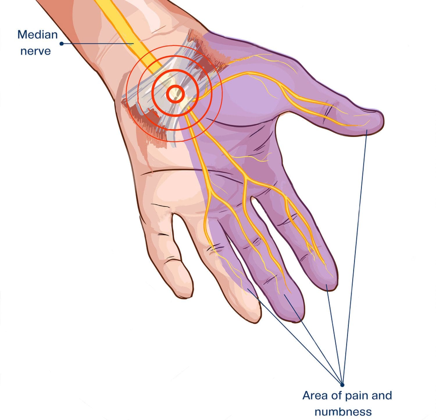
Many people think of chiropractic as a treatment for neck and back problems, but in truth it is a whole body wellness practice and the neck and back are just one aspect. One application that is garnering a great deal of attention is the treatment of carpal tunnel syndrome (CTS). Chiropractic is an effective, non-invasive treatment for CTS, relieving the pain and promoting healing without the use of medications. There are three ways the chiropractic has been proven to be effective in the treatment of CTS.
Contents
Carpal Tunnel Syndrome
Carpal tunnel syndrome is a repetitive stress injury, a condition that affects the hands and wrists. The most common symptoms include tingling, numbness, and weakness in the middle finger, index finger, and thumb as well as pain in the wrist or hand that gets worse at night.
It is typically found in hairdressers, typists, mechanics, cashiers, and other people who perform repetitive hand motions for long periods of time and it is estimated that about 3 percent of adults in the United States will deal with CTS at some time in their lives.
About 50 percent of workplace injuries in the United States are attributed to CTS. There was a time when it was believed to affect women more than men, but research has shown that a person�s occupation plays a large part in how it is contracted and gender only plays a small role � if any at all.
The carpal tunnel is a band of fibrous tissue that lies on the front side of the wrist. It holds the blood vessels, nerves, and tendons that are necessary for the hand to move and work as it should. When that carpal tunnel becomes inflamed, damaged, or pressure on that area compresses the arteries and nerves, CTS develops. It usually starts slowly and the symptoms gradually worsen as the condition progresses.
How Chiropractic Helps CTS/Carpal Tunnel Syndrome
There are three primary ways that chiropractors treat CTS. These three common treatments have been shown to be very effective in treating the pain and the condition as a whole.
Adjustments of the upper spine, arm, and wrist � When the spine is not in alignment, it could exacerbate carpal tunnel syndrome. A chiropractor will use spinal manipulation to realign the spine to its correct position. These manipulations, or adjustments, are often performed by hand, but at times he or she may use special devices or a special table made specifically for chiropractic treatments. In addition to the spine, the chiropractor may make adjustments to other areas of the body such as the arm and wrist.
Ultrasound therapy � This is another type of therapy employed by chiropractors. It uses low energy sound or high energy sound � both outside the range of a human�s ability to hear. The device that is used emits these sound waves that are extremely focused and penetrate the body�s tissue at a very deep level. These sound waves can be very effective in reducing inflammation, alleviating pain, and relaxing muscles. It may be used as a stand-alone treatment or with chiropractic treatments.
Wrist Supports � The purpose of wrist supports is to keep the wrist properly aligned with the rest of the arm to reduce pressure and compression. It is often used as a preventative measure against carpal tunnel. The chiropractor may choose this option if the patient does repetitive work that may cause the condition. This is often combined with regular chiropractic treatments such as spinal alignment and is used to prevent conditions such as CTS from developing.
When treating carpal tunnel, the chiropractor may also suggest specific exercises as well as a dietary program to promote overall wellness. This is intended to keep the patient from experiencing carpal tunnel as well as other conditions.
Low back pain is a common condition which occurs as a result of a lumbar sprain, spinal stenosis, disc herniation, and due to various other degenerative spinal disorders. The topic of discussion today, however, focuses on discogenic lower back pain; a degenerative condition. Discogenic low back pain refers to painful symptoms caused by the degeneration, damage or injury of one or more intervertebral discs along the lumbar spine.
Contents
Symptoms of Discogenic Low Back Pain
As we get older, our bodies undergo numerous changes. For instance, our hair may begin to turn gray or thin. Similar changes have an effect on the complex structures of the spine, specifically on the intervertebral discs. Disc degeneration doesn’t necessarily cause severe pain or any other symptoms for that matter, but back pain may occur, if the degeneration becomes too advanced. Typically, discogenic pain is related to activities that increase the pressure within the intervertebral disc, known as intradiscal pressure.
Sitting, bending forward, coughing and sneezing can increase discogenic pain.
Leg pain caused by pinching of the nerves in the lumbar spine, called radiculopathy, may also accompany low back discogenic pain; particularly while sitting, walking or standing.
Discogenic low back pain is generally a chronic disorder.
How Discs Cause Pain
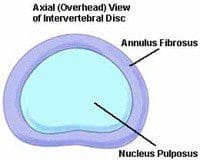
The same as other areas of the human body, each intervertebral disc has a nerve supply. Discs are constituted of 2 parts; the annulus fibrosus, an outer ring-like structure, and nucleus pulposus, a gel-like interior. The nucleus pulposus is void of nerves. However, nerve fibers are contained by the outer third of the annulus fibrosus.
One type of discogenic disorder is medically referred to as an Internal Disc Disruption, or IDD. An IDD takes place when the disc tears or cracks, creating a fissure which enables the nucleus pulposus to come in contact with the annulus fibrosus. While this happens, a chemical called a protecogylcan might be flooded into the nucleus pulposus. The annular nerves may then become irritated causing an inflammatory response as well as pain and discomfort. For reasons that are unknown, some people have annular tears and can remain symptom free.
Diagnosing Discogenic Low Back� Pain
Degenerative disc changes can best be observed through magnetic resonance imaging (MRI). If a couple of spinal discs are suspected as the pain source, the doctor may order a discography or a discogram. During this procedure, the suspected discs are injected with a contrast dye to make each disk visible under fluoroscopy. Provocative discography helps the doctor see the form and dimensions of the intervertebral discs. The pressure is altered by the injection of the contrast dye within the disc and may ‘excite’ or replicate the patient’s pain pattern helping to isolate which disc may be the source of their symptoms.
Non-Surgical Treatments
There are different types of treatments to help alleviate low back pain and radiating symptoms. Frequently, treatments are combined for symptom relief or control.
Physical Therapy: passive therapies such as ultrasound, transcutaneous nerve stimulation, or TENS, and massage could be used with a disciplined program of strengthening exercises. Core strengthening, or the strengthening of the abdominal and low back muscles, is often helpful in relieving pain in degenerative disc disease. When the muscles around the disc become stronger, they may protect the intervertebral discs and might reduce pain.
Spinal Injections: local anesthetics combined with long-acting corticosteroid injections. This medication combination may be injected into the facet joints or around the nerves of the back to reduce symptoms.
Bracing: braces, or orthoses, help support the spine and limit movement which may provoke painful episodes. Rarely is bracing a treatment for back pain. Long-term bracing may lead to weakened back and abdominal muscles that might provoke muscle strain.
Alternative Therapies: acupuncture, yoga.
Lifestyle Modifications: Nutrition and dieting to reach a more ‘back friendly’ body weight, smoking cessation, and physical activity help keep a healthy spine.
Minimally Invasive Procedures
Many spine surgical procedures can be performed with minimally invasive techniques. For instance, spine surgeons utilize these methods to correct scoliosis, treat herniated discs, and perform spinal fusion. The advantages to the individual may be significant and include smaller incisions, shorter time hospitalized, less post-operative pain, and a quicker healing. By replacing the disc with cages and bone, related back pain may be alleviated by spinal fusions. Artificial cervical and lumbar discs have become another option to replace damaged intervertebral discs. Depending on your type of symptoms and spinal health issue, it’s essential to talk to a healthcare professional to properly determine the most appropriate type of treatment for your discogenic low back pain.
Chiropractic Care for Discogenic Low Back Pain
Chiropractic can be another non-surgical treatment for discogenic low back pain. Chiropractic care is a well-known alternative treatment option which focuses on the diagnosis, treatment and prevention of a variety of injuries and/or condition associated with the musculoskeletal and nervous system. Correcting discogenic lower back pain for a chiropractor is similar to treating several musculoskeletal disorders; it is about reducing the inflammation from around the disc but most importantly, it is about restoring the muscles to take the pressure off the disc so that the same forces are not going through it.
Chiropractors and physical therapists, or physiotherapists, specialize in the non-surgical treatment of discogenic low back pain.�Chiropractors and physical therapists concentrate on functional improvement and pain reduction. A chiropractor, or doctor of chiropractic, commonly utilizes spinal adjustments and manual manipulations to carefully correct any spinal misalignment, or subluxation, which may be affecting the natural integrity of the spine. Other common treatment methods include mechanical diagnosis, the McKenzie Method, as well as nutritional and fitness exercise programs and advice. By realigning the spine, chiropractic care can help reduce pressure around the affected intervertebral discs, decreasing inflammation and improving circulation to eliminate the compression of the soft tissues, ultimately improving back pain symptoms. Furthermore, stretches and exercises recommended by a chiropractor can help improve strength, mobility and flexibility in order to speed up the rehabilitation process.
Physical therapy can also be useful for correcting discogenic lower back pain. Physical therapy focuses on restoring the patient’s original strength. Strengthening the core, the lower abdominal muscles, including the pelvis, as well as stretching the hip flexors can restore the original integrity of the spine, reducing additional stress on the spine and decreasing back pain symptoms. Strengthening the muscles of the back as well as restoring the aligned of the spine will allow the intervertebral discs to begin healing themselves naturally. Utilizing chiropractic care together with physical therapy can ultimately help decrease the pain and discomfort associated with discogenic low back pain.
Dr. Alex Jimenez’s Insight
Low back pain is a common, modern day health issue which carries a tremendous burden on those who suffer from it. With more cases of low back pain being diagnosed every year, understanding the causes of low back pain can be essential towards its proper treatment. Discogenic low back pain refers to symptoms of pain and discomfort along the lumbar spine caused by the degeneration of the intervertebral discs. A variety of treatment methods, including non-surgical procedures, can help improve the symptoms associated with this degenerative condition. Chiropractic care is a safe and effective alternative treatment option which can help restore the original integrity of the spine, reducing pain caused by discogenic low back pain.
Oral drugs and/or medications are often used to help control symptoms while the patient engages in several of the treatment methods mentioned above. While these don’t treat the problem, they can help to control the inflammation as well as the pain and discomfort while a patient corrects their biomechanics and participates in stretches and exercises or physical therapy. Within the treatment itself, there are some treatment modalities, such as massage, ultrasound, traction, and electric stimulation that could help to control the pain and symptoms without the use of drugs and/or medications. If chiropractic care or physical therapy doesn’t improve a patients symptoms, the healthcare professional can refer the patient to another back pain specialist which can work alongside their current treatment to help improve discogenic low back pain. The scope of our information is limited to chiropractic as well as to spinal injuries and conditions. To discuss the subject matter, please feel free to ask Dr. Jimenez or contact us at 915-850-0900 .
Curated by Dr. Alex Jimenez
Additional Topics: Back Pain
According to statistics, approximately 80% of people will experience symptoms of back pain at least once throughout their lifetimes. Back pain is a common complaint which can result due to a variety of injuries and/or conditions. Often times, the natural degeneration of the spine with age can cause back pain. Herniated discs occur when the soft, gel-like center of an intervertebral disc pushes through a tear in its surrounding, outer ring of cartilage, compressing and irritating the nerve roots. Disc herniations most commonly occur along the lower back, or lumbar spine, but they may also occur along the cervical spine, or neck. The impingement of the nerves found in the low back due to injury and/or an aggravated condition can lead to symptoms of sciatica.
New Patient Intake Form: Truide Torres, office manager at Injury Medical Clinic with Dr. Alex Jimenez, discusses some of the most common questions patients have when they come in for their first office visit. Patients can save time and fill most of the required forms online by visiting https://elpasobackclinic.com/patient-intake-form. If you’ve been involved in an automobile accident or a work-related accident, Truide Torres describes which type of insurance can be used to provide you with the healthcare benefits you deserve. Dr. Alex Jimenez is the recommended non-surgical choice for well-being.
New Patient Intake Form Explained
Personal injury is a valid term for a physical, mental or emotional injury to yourself, as opposed to damage to property. The expression is commonly used to refer to a type of tort lawsuit where the individual has suffered harm. Personal injury lawsuits are filed against the person or entity that caused the injury through negligence, gross negligence, reckless conduct, or misconduct, and in some instances on the grounds of strict liability. Common types of personal injury claims include automobile accidents, work-related accidents, slip-and-fall injuries, assault claims, and product defect accidents (product liability).
Our team has takes great�pride in bringing our families and injured patients only�clinically proven treatments protocols. �By teaching complete holistic wellness as a lifestyle,�we also change not only our patients lives but their families as well.� We do this so that we may reach as many El Pasoans who need us, no matter the affordability issues.
There is no reason we cannot help you.�
If you have enjoyed this video and/or we have helped you in any way please feel free to subscribe and share us.
Allergy Sufferers!�As winter gives way to spring, seasonal allergies can really get you down. Whether you get a few sniffles and some sneezing or you are down for the count with every terrible allergy symptom known to man, it can make spring pretty unbearable.
There is no shortage of allergy medications on the market, but they come with their own issues. The majority of them cause drowsiness and other unpleasant side effects, leaving you barely able to function. Those that are made from a �non drowsy� formula sound great, but if you have certain health conditions, like high blood pressure, you are out of luck � and stuck either taking the ones that make you sleep sucking it up and dealing with your allergies sans medication.
That�s no way to live.
Contents
What Are Allergies?
When your immune produces histamines in response to an allergen that you encounter the physiological reaction that you experience is broadly referred to as allergies or hay fever. The allergens may be simple substances that normally do not affect people, but when your body is out of balance, it can cause a variety of problems.
Symptoms of allergies include:
Runny nose
Stuffy nose
Headache
Sneezing
Itchy eyes
Coughing or scratchy throat
Skin Rash or Hives
Swelling
Diarrhea
Nausea
Fatigue
Anaphylaxis, severe, life threatening allergies can include swelling of the airways, tongue, and throat, inability to breathe due to blocked airway, and other dangerous symptoms.
The allergens can be something you come in contact with, like poison ivy, something you breathe in, like mold or dust, or it can be something you ingest, like strawberries or peanuts. Different people will have different allergies, but those who are allergic to the same things may not have the same reaction. Often a doctor or allergist will diagnose your allergies.
Chiropractic Care For Allergy Sufferers
Chiropractic treatments have been found to be very effective for relieving allergy symptoms and even stopping allergies at their source. It reduces the severity of allergy symptoms as well as the frequency of occurrence. It does not work like allergy medications which have an anti-histamine effect and only work as a short term fix for your allergy symptoms.
Chiropractic treatments help your body become more balanced so that it is better equipped for combating allergies at the source. When your spine is not aligned it can impact your nervous system leading to a variety of problems � including allergies. Your immune system can be affected, causing it to malfunction.
A chiropractor can help relieve the stress on your nervous system by aligning your spine. This takes the pressure off of nerves, allowing your immune system to function at a more optimal level. This makes it easier for your body to ward off infections while recognizing allergens as harmless.
When your immune system encounters allergens it doesn�t overreact to them. Instead, the reaction is much more subdued, or even nonexistent. Chiropractic has also been found to help asthma patients breathe easier. Asthma symptoms are diminished.
Chiropractic care is more than just spinal manipulation, though. It promotes whole body wellness. Patients are taught exercise, stress relief, and nutrition so that the entire system is treated. The whole body treatment plan for chiropractic patients will help you be allergy free in a short time.
It is important to follow your chiropractic plan thoroughly and consistently. Get plenty of rest and take time to destress. The more you can relax and take care of yourself, the healthier you will be overall. Chiropractic care can help so many health conditions; it can actually make you healthier. Allergy sufferers or if you are struggling with allergies for the first time, give chiropractic care a try you just might be surprised.
Back pain sometimes strikes without warning. One minute you’re bending or lifting something heavy; the next minute you’re unable to move. Sudden onset of muscle spasm from the back is common. Roughly 8 out of 10 adults will experience back pain at some point in their lives. Generally, the origin of back pain and muscle spasm can be credited to overuse, an accident or a sports injury. But more often than not, the origin of muscle spasm is the effect of damage to a structure within the lumbar spine.
One matter is clear: if you have had one or more episodes of muscular strain in the back, chances are it’ll happen again. The muscles in the back function together. Without all the muscles of the back working together, no lateral and extension motion of the spine would be impossible. Stability is also added by the muscles of the back in order to keep the spine erect and maintain equilibrium. When the muscles are in spasm due to a spinal health issue, that balance can be compromised.
Contents
What are Muscle Spasms?
Muscle spams are involuntary contractions of a muscle. Although “back attacks” seem to happen out of nowhere, the motion that triggers the episode is generally preceded by a collection of health issues to the structures of the spine that develops gradually, over time. Inflammation sets in once injured. This, in turn, sensitizes the nerves, causing the muscle/s to contract and spasm.
Disc Disorders and Muscle Spasms
Conditions, such as degenerative disc disease or a herniated disc, may cause an acute episode of low back pain. A disc bulge or disc herniation may also compress a spinal nerve root causing irritation and inflammation. The body tries to immobilize the affected area by tightening the musculature to stop pain and as a result, debilitating muscle spasms occur.
Muscles can get too tight due to insufficient exercise, a lot of exercise, structural imbalances, dehydration and electrolyte loss, or any mix thereof. By comparison, some muscle bands are weak. When muscle imbalances become persistent, aberrant forces are transmitted into the spine. Consequently, an accident can be triggered by one movement out of the norm to a joint, ligament, or disk resulting in spasm and back pain. As these structures are already “primed,” the event that activates the spasm is nothing more than an effect of an underlying health issue. Muscle spams in the back are frequently painful. Here are several remedies which can help you get moving.
Remedies for Back Spasms
Initial 48 to 72 hours: Apply ice for 20 minutes every 2 hours while lying on your back. Constantly use an ice pack, never apply ice directly to skin.
After 72 hours: Apply moist warmth. A heating pad is ideal. You might find relief by soaking in a bathtub of warm water.
Whereas ice reduces redness, warmth relaxes muscles and increases blood flow to the area and nerves that are irritated.
By elevating your legs, pressure is taken off the spine and may help relieve pain.
Aspirin or ibuprofen can help reduce inflammation and alleviate pain. Consult with your doctor or healthcare provider regarding the dosing and drug regimen most acceptable for your problem. With reduced back muscle spasms, combination remedies (ie, remainder, ice/heat and drugs) generally yield much better outcomes than one therapy alone.
Back Spasm Prevention
When the back spasm episode has passed, and you’ve allowed enough time for the inflammation to subside, begin focusing on what you can do to keep it from happening again.
Start stretching: Incorporate stretching exercises in your daily routine. Muscle fibers gain from stretching and so will you. Consider taking yoga courses or Pilates; always stretch before exercise.
Get in shape:�Now’s the time to get started in case you do not take part in routine physical activity. Exercise confers advantages too important to ignore and too numerous to mention. Join a fitness center. Start playing a sport. The key to any exercise routine is that it be done consistently.
Strength training: A�significant part of any exercise regimen, strength training not only builds muscle, it can reduce imbalances. Remember: muscles operate with one another, so be sure to balance your strengthening regularly.
Make preventing another incident of muscular spasms your priority. It’s never too late to start increasing your strength, flexibility and mobility. Choose activities that you enjoy and commit to performing them. In addition, several healthcare professionals can help decrease muscles spasms. Chiropractic care is a well-known alternative treatment option which utilizes a variety of treatment methods to help ease muscle spasms caused by spinal health issues.
How a Chiropractor Can Help
Muscle spasms both disrupt your life and can be painful. There are several ways in which a chiropractor can help you find relief for your muscle spasms. Here’s how chiropractic care can help to decrease the severity and frequency of your muscle spasms.
Chiropractic care, can go a long way toward assisting you to avoid tightness in your muscles that result in constantly painful symptoms, when you get regular spinal adjustments and manual manipulations. Chiropractic treatment includes spinal adjustments and manual manipulations which help align your body to take pressure off of soft tissues within the body and nerves, reducing the nerve signals your brain might be receiving that cause an involuntary contraction. Manual manipulations and spinal adjustments also allows your spine and joints to restore normal motion, which in turn contributes to a decrease in muscle spasms and pain.
Your chiropractor may also utilize massage treatment to help relax the muscles in your body which are prone to spasm. These kinds of treatments help to decrease fluid, reduce scar tissue and supply relief. Massage may also help to reduce any inflammation that communicates with the cramps and spasms your own muscles. Your chiropractor can also provide nutritional advice to help put an end to chronic spasms. If you are dehydrated you could have an electrolyte imbalance which may be causing your cramping and muscle spasms. Making certain modifications to your diet and drinking more water can help restore balance and decrease the number or spasms you’re experiencing as well.
Moreover, the chiropractor may use associated therapies for the treatment of muscle spasms. This can include hot and cold treatments, ultrasound and electric stimulation of the muscle. These therapies help greatly to increase flow and also prevent swelling or scarring of the muscle. Associated treatments are a means for prevention of further damage and injury recurrences as well as to reduce pain. What’s more, a doctor of chiropractic may suggest a series of appropriate stretches and exercises to speed up the recovery process. Certainly, the ultimate objective is to alleviate the muscle spasms.
Dr. Alex Jimenez’s Insight
Muscle spasms are common symptoms caused by a variety of injuries and/or aggravated conditions which can result in back pain. Regardless of the cause of your back pain, muscle spasms can often become a constant and debilitating health issue that can affect your overall quality of life. Chiropractic care is a common treatment option utilized to help ease soft tissue injuries caused by accident and/or sports injuries. Through the use of spinal adjustments and manual manipulations, a chiropractor can restore the original alignment of the spine, helping to reduce stress and tension build up in the soft tissues surrounding the spine, ultimately improving muscle spams.
Remember, it’s not normal to experience muscle spasms. If you do, then it is likely you have an underlying problem that has to be diagnosed and treated. You don’t have to suffer from the pain and discomfort of muscle spasms, your chiropractor can be your partner.�The scope of our information is limited to chiropractic as well as to spinal injuries and conditions. To discuss the subject matter, please feel free to ask Dr. Jimenez or contact us at 915-850-0900 .
Curated by Dr. Alex Jimenez
Additional Topics: Back Pain
According to statistics, approximately 80% of people will experience symptoms of back pain at least once throughout their lifetimes. Back pain is a common complaint which can result due to a variety of injuries and/or conditions. Often times, the natural degeneration of the spine with age can cause back pain. Herniated discs occur when the soft, gel-like center of an intervertebral disc pushes through a tear in its surrounding, outer ring of cartilage, compressing and irritating the nerve roots. Disc herniations most commonly occur along the lower back, or lumbar spine, but they may also occur along the cervical spine, or neck. The impingement of the nerves found in the low back due to injury and/or an aggravated condition can lead to symptoms of sciatica.
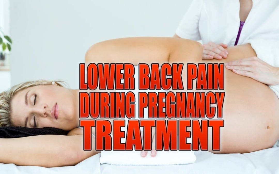
About 50 to 80 percent of pregnant women experience some type of pain back during their pregnancy. Access to the right treatment can be challenging because the connection between pregnancy and back pain is not fully comprehended. Luckily, back pain is typically short-term where most cases will go away shortly after the birth of your baby, however, waiting for the symptoms to go away on their own isn’t much of an appealing option. Back pain associated with pregnancy is often localized to a specific region of the spine rather than widespread. Back pain tends to arise between the fifth and seventh months of pregnancy, even though it can begin sooner.
Contents
Pregnancy-Related Back Pain
Women generally experience pregnancy-related back pain in the lower back, pelvic and sacrum regions of the spine. Pain along the pelvic area, for which a straightforward diagnosis isn’t yet determined, is referred to as peripartum pelvic pain. Peripartum refers to the period of time surrounding childbirth, which is usually several weeks prior to birth and a few weeks after birth.
Back pain associated with pregnancy presents itself most commonly in the following regions:
Sacroiliac joints in the posterior superior iliac spine
The groin areas
Coccyx
Pubic symphysis anteriorly
Furthermore, other areas of the pelvis and thighs may change and cause painful symptoms, however, radiating pain rarely occurs below the knees. Back pain during pregnancy tends to be associated with posture and can by influenced by a waddling gait.
What Increases Back Pain Symptoms During Pregnancy?
While several factors, including age and smoking status, have not been necessarily demonstrated to affect symptoms of back pain, a history of pain during pregnancy and a history of back pain in general, along with increased body mass, have already been linked to an increase in peripartum pain. Additionally, younger women often have more extreme pain compared to older women. Studies have reported that approximately 10 percent of women said back pain during pregnancy prevented them from engaging in their work and more than 80 percent stated that their ability to perform daily tasks was tremendously impacted.
What Causes Back Pain During Pregnancy?
The cause of pregnancy-related back pain is likely associated with a combination of metabolic, circulatory, and psychosocial factors. However, most of the causes can be grouped into these areas:
Weight gain: Women typically gain between 20 and 40 pounds throughout pregnancy, which puts pressure on the spine. This added pressure might result in lower back pain and other painful symptoms.
Shift of gravity in center: As you gain weight along while your belly grows, your ability to keep proper posture becomes contested. Posture changes in pregnancy have been connected to spine issues such as herniated discs and lordosis, which might contribute to back pain.
Hormonal changes: Some women report back pain at the first trimester even though the majority of women begin experiencing pain back during the fifth and seventh months. Since the baby isn’t large enough to cause any stress to the spine, it may be hormonal changes causing the symptoms. Relaxin, a hormone that relaxes pelvic and spinal joints as well as the ligaments to ease childbirth is produced by pregnant women. Relaxin might cause some spinal distress and may result in lower back pain. In reality, sacroiliac dysfunction can be caused by certain hormones produced during pregnancy.
Stress: Pregnancy can be an exciting and special time for many women, but it can also be stressful. Stress can worsen back pain or even cause it. Finding ways to manage stress while pregnant may help ease your back pain.
Treatment
Treatments for pregnancy-related back pain involve lifestyle modifications, for example:
Avoiding excessive weight gain: A nutritious diet is one of the very best ways to maintain a healthy weight during pregnancy, and some foods have been associated with easing spinal distress (a frequent cause of low back pain). Eating 5 or more daily servings of veggies and fruits will provide nutrients. Other good options include fatty fish and nuts, such as mackerel, which pack a healthy dose of omega-3 fatty acids and inflammation fighting agents.
Exercising to reinforce the core and back muscles: In general, pregnant women should avoid the extremes when it comes to action: little and too much action can possibly cause an increase in back pain. Walking, swimming, and yoga are great ways to condition yourself but always remember how to safely exercise while pregnant.
Reducing stress: Finding methods to manage your stress during your pregnancy has psychological in addition to physical benefits. A massage, getting lots of rest, and relaxing with a heating pad against your back are excellent ways to manage stress while helping your spine.
Posture: Talk to your doctor about ways you can keep good posture as your pregnancy progresses.
Investing in a pregnancy pillow to give support during sleep:�Pregnancy and sound sleep don’t always mix, but a supporting pillow may deliver more comfy mornings.
Wearing sensible shoes: Spine maintenance and footwear are all connected. Avoid high heels and flip flops, if your shoe size changes during pregnancy purchase new footwear.
If your back pain does not respond to these treatments, your doctor may suggest a prescription pelvic belt, medicine therapy, injection therapy, or bed rest. In addition, chiropractic care is a safe and effective alternative treatment option for back pain during pregnancy.�Fortunately, back pain may disappear within 6 months following birth, letting you focus on the addition to your own life, your baby. Speak with your doctor about which treatment will be best for you if your pain does not subside after your baby arrives.
Chiropractic Care During Pregnancy: Benefits And Safety
Chiropractic care is the health care of the spine, discs, related nerves and bone geometry without the use of surgery or drugs and medications. It involves science and the art of adjusting and manipulating the joints of the spine, which reduces spinal nerve stress and promotes health and wellness throughout the entire body.
Is Chiropractic Care During pregnancy safe?
There are no known contraindications to chiropractic care throughout pregnancy. All chiropractors are trained to use spinal adjustment and manual manipulations on women who are pregnant. Investing in the maternity and fertility wellness of women who are pregnant or trying to conceive is a top priority for chiropractors. Some chiropractors take a particular interest in prenatal and postnatal care and seek out additional training. Below represents designations of chiropractors that have taken these additional steps.
DACCP � Diplomate with ICPA reflecting highest level of advanced training
CACCP � Certified with the ICPA reflecting advanced training
Member of ICPA reflecting special interest
Webster Certified � trained to work specifically with pelvic balance in pregnancy
Chiropractors that have been trained to work with pregnant women can use tables that adjust to a pregnant female’s body, and they will use treatment methods and techniques that prevent unnecessary pressure on the abdomen. A doctor of chiropractic, or chiropractor, who is trained in the requirements of pregnant women may also supply you with exercises and stretches that are safe to participate in when pregnant.
Why Should I Have Chiropractic Care During Pregnancy?
During pregnancy, there are several physiological and endocrinological changes that occur in preparation for establishing the right environment for your growing baby. However, these changes can result in a misaligned spine or joints, including:
Protruding abdomen and increased spine
Pelvic changes
Postural adaptations
Establishing pelvic balance and orientation is just another reason to consider chiropractic care during pregnancy. When the pelvis is misaligned, or subluxated, it may reduce the amount of space available for the growing baby. This limitation is called intrauterine constraint. A misaligned pelvis may also make it hard for the baby to get in the best possible position for delivery. This may affect the mother’s ability to have a natural, non-invasive birth. Breech and posterior positions lead to interventions such as c-sections and can interfere with the ease of labor. The nervous system is the master communication system to all the body systems including the reproductive system. Keeping the spine healthy assists the whole body to function more effectively.
What are the Benefits of Chiropractic Care During Pregnancy?
Chiropractic care during pregnancy can offer benefits for women that are pregnant. Possible advantages of chiropractic care throughout pregnancy include:
Maintaining a healthier pregnancy
Controlling symptoms of nausea
Reducing the time of labor and delivery
Relieving back, neck or joint pain
Preventing a potential cesarean delivery
Chiropractic Care and Breech Deliveries
The late Larry Webster, D.C., Creator of the International Chiropractic Pediatric Association (ICPA), developed a specific chiropractic evaluation and modification which empowers chiropractors to establish balance in the pregnant female’s pelvis to reduce undue stress for the uterus and supporting ligaments of pregnant women. This balanced state in the pelvis has been clinically demonstrated to allow for optimal fetal positioning. The technique is known as the Webster Technique.
It is considered ordinary by a few for a baby to present breech until the next trimester. Most birth professionals aren’t concerned with breech presentations until a patient is 37 months into their pregnancy. Roughly 4 percent of all pregnancies lead to a breech presentation. The Journal of Manipulative and Physiological Therapeutics reported in the July/August 2002 issue an 82 percent success rate of babies turning vertex when doctors of chiropractic used the Webster Technique. Furthermore, the results from the study suggest it may be beneficial to use the Webster Technique, in the 8th month of pregnancy, even when a female has a breech presentation.
Currently the International Chiropractic Pediatric Association (ICPA) recommends women receive chiropractic care during pregnancy to set up pelvic balance and optimize the area in which a baby needs for growth during pregnancy. With a balanced pelvis, babies have a greater prospect of moving to the correct place for arrival, and the crisis and worry related to breech and posterior presentations may be avoided altogether. Optimal infant positioning in the right time of birth also removes the potential for dystocia (hard labou) and, therefore, results in simpler and safer deliveries for both the mother and baby.
Dr. Alex Jimenez’s Insight
During pregnancy, the overall health and wellness of the mother is essential towards the proper development of the baby. Because the body of a woman goes through many changes throughout their pregnancy, however, some changes to the human body may cause health issues if not properly checked by a healthcare professional. Back pain is a common health issue reported by many pregnant women. Fortunately, safe and effective alternative treatment options for both mothers and their babies are available to help ease their painful symptoms. Chiropractic care is a well-known treatment which can be used efficiently to help ease back pain in pregnant women.
Talk to a Healthcare Professional
As more women are seeking the benefits of chiropractic care throughout pregnancy, more health care providers are seeking trained doctors of chiropractic in their own communities to refer their pregnant patients to. Discuss these options with your healthcare provider. Ask them to find out more about its benefits, if they are not yet knowledgeable about chiropractic care in pregnancy. Above all, seek options that support your body’s natural abilities to function and find a team of healthcare professionals that are respectful of your options. The scope of our information is limited to chiropractic as well as to spinal injuries and conditions. To discuss the subject matter, please feel free to ask Dr. Jimenez or contact us at 915-850-0900 .
Curated by Dr. Alex Jimenez
Additional Topics: Back Pain
According to statistics, approximately 80% of people will experience symptoms of back pain at least once throughout their lifetimes. Back pain is a common complaint which can result due to a variety of injuries and/or conditions. Often times, the natural degeneration of the spine with age can cause back pain. Herniated discs occur when the soft, gel-like center of an intervertebral disc pushes through a tear in its surrounding, outer ring of cartilage, compressing and irritating the nerve roots. Disc herniations most commonly occur along the lower back, or lumbar spine, but they may also occur along the cervical spine, or neck. The impingement of the nerves found in the low back due to injury and/or an aggravated condition can lead to symptoms of sciatica.
Chiropractic care is generally the first choice of treatment for back pain as well as for a variety of other injuries and/or aggravated conditions associated with the musculoskeletal and nervous system.�Chiropractic care has numerous health benefits that can focus on helping patients of all ages. But, what many people don’t realize is that chiropractic care was not designed for only a certain person or body type, instead, a chiropractor can adjusts their treatment techniques to match each person’s specific needs. Doctors of chiropractic, or chiropractors, feel strongly about improving the overall health and wellness of their patients. In the tradition of chiropractic care, a chiropractor will treat the body of a patient as a whole, rather than focusing on a single injury and/or condition.
A doctor of chiropractic can treat many of the health issues that may be causing a patient’s back pain, however, what if the patient’s back pain is caused by obesity? The topic between whether chiropractic care can be used to treat obesity is frequently discussed among healthcare professionals and the patient. Many people are not aware of the benefits chiropractic care can have on obesity. Read below to find out how chiropractic care can help improve back pain as well as help manage obesity.
Contents
Chiropractic Care and Obesity
Obesity can affect more than just the way a person feels cosmetically. It is a health issue that may ultimately affect the individual’s skin, organs, joints, muscles, and even the spine. Excess weight can place unnecessary amounts of stress on the spine, joints and muscles, which can commonly lead to back pain, among other health issues. Its an individual’s constant struggle between managing their weight as well as coping with the symptoms manifesting as a result of the weight gain that can make weight loss difficult for many people without the proper treatment. Fortunately, chiropractic care is a safe and effective, alternative treatment option which can help diagnose, treat and prevent a variety of health issues while helping to improve overall health and wellness.
Because chiropractic care focuses on both the body and mind, the purpose of the spinal adjustment and manual manipulation in the treatment of obesity is to help improve symptoms of back pain by carefully correcting the alignment of the spine in order to reduce pressure on the spine as well as to decrease stress which may be affecting the individual’s mood. Once the patient has been geared towards a healthier body and mind, a chiropractor can also recommend a series of lifestyle modifications, such as nutritional and fitness advice, which can help a patient manage their excess weight.�The largest connection in your body is the one between your brain and the rest of the body through the communication of the nervous system. When the connection between the brain and the body is interrupted as a result of a spinal misalignment, or subluxation, it can lead to a variety of mental and physical health issues that may result in painful symptoms as well as stress, anxiety and depression, all of which have been associated with weight gain and obesity.
Furthermore, chiropractic care can also help throughout the process of weight loss. Because your body will be continuously changing as you lose weight, your spine and joints will need to be accordingly maintained to keep up with the ongoing changes. By receiving regular chiropractic care, a patient participating in a weight loss program or simply following the chiropractor’s nutritional and fitness advice will be able to fully engage in their exercise and physical activity routines due to the reduced back pain and other symptoms. In order to understand how chiropractic care can work towards excess weight and obesity, its essential to first comprehend the relationship between back pain and obesity as well as what type of treatment methods can benefit weight management.
Back Pain and Obesity
Obesity is defined by doctors as a disease. Being overweight or obese is a serious disorder that can affect children and adults. Many healthcare professionals know that obesity contributes to the development of high blood pressure, diabetes, coronary heart disease, and even colon cancer. But were you aware that obesity is a common contributing factor for back pain? Being overweight or obese may significantly contribute to symptoms associated with osteoarthritis, osteoporosis, rheumatoid arthritis, degenerative disc disease, spinal stenosis, and spondylolisthesis.
Has your primary care physician suggested you lose weight to reduce the severity of your back pain? Perhaps you have back pain, but have not considered extra body weight to be a possible cause. Even an extra 10 pounds to your average weight can eventually lead to back pain. The outcomes of a big cross-sectional population-based research study confirmed the link between obesity and back pain. The analysis involved 6,796 adults where researchers found that the risk for back pain increases as body mass index, or BMI, does. The probability of low back pain among adults who are obese is four times larger than among adults with an average weight.
BMI and What It Means
BMI is a number based on your weight and height. In general, the higher the number, the more body fat a person has. There are four categories of BMI:
Normal weight�BMI less than 25
Overweight�BMI of 25 to 30
Obese�BMI of 31 to 35
Extremely obese�BMI of 36 or higher
For instance, someone who is 5�10� tall and weighs 174 pounds has a BMI of 25, while a person who is 5�10� and weighs 251 pounds has a BMI of 36.
Obesity and Risk for Low Back Pain by the Numbers
2.9% for people of normal weight
5.2% for overweight adults
7.7% for obese adults
11.6% for extremely obese adults
The study did not address why obesity increases the risk of low back pain. But, additional body weight can contribute to how the spine works and its mechanical well-being.
Small Changes
Modest changes in the degree of physical activity can substantially lower the risk for back pain. Individuals with extreme obesity (BMI 36+) who increase their time in moderate actions by at least 17 minutes every day can reduce their risk for low back pain by approximately 32 percent. Moderate activities may include briskly walking, performing water aerobics, riding a bike, ballroom dancing, and gardening.
How Obesity Can Impact the Spine
The spine is designed to carry your body’s weight and distribute the loads encountered during rest and action. When excess weight is carried, the spine is made to assimilate the burden, which may lead to structural undermining and harm, as in the case of injury, or sciatica. One area of the spine that is most vulnerable to the consequences of obesity is the lower back, or the lumbar spine.
Why Exercise is Essential
Lack of exercise may lead to poor mobility and flexibility as well as weak muscles, especially in the back, core, pelvis and thighs. This may raise the curve of the lower spine, causing the pelvis to tilt too far ahead. Further, this is detrimental to proper posture as well as posture, causing health issues along other regions of the spine, such as the neck, and resulting in debilitating symptoms. You might attempt to dismiss the reason behind some of these spinal health issues to the practice of normal aging. It’s true that to anatomy, structural and functional changes can be caused by the degeneration of the body with age. However, if you are obese or overweight, you likely have, or may have, back pain. You may also have or develop a few of the following conditions:
Posture: Unhealthy posture accounts for neck and back pain. A level of physical fitness is necessary to properly support the spine.
Low Back Pain: Obesity may aggravate an existing low back problem and contribute to recurrence of the condition.
Osteoporosis: A sedentary lifestyle coupled with an unbalanced diet can affect the density, or strength of the bones (spinal vertebrae). When the structural architecture of a vertebral body is compromised, it is at risk for fracture. Vertebral fractures can be painful and disabling. If you have been diagnosed with osteoporosis, you have probably lost between 25% to 30% of desirable bone density.
Osteoarthritis (OA) and Rheumatoid Arthritis (RA): The joints in the spine are called facet joints. Excessive body weight places unnatural pressure and stress on the joints during movement and at rest.
Development of Obesity
Industrialization and modernization has had a huge effect on the food we eat today. Food can be bought just about everywhere. No more is it necessary to expend effort to forage and hunt for food. There are vast numbers of processed food items available and devices which require little use of labor like microwave ovens to cook meals. The market for kitchen devices and several convenience foods came about when women entered the workforce. For the time period 2011-2012, the following statistics were published:
34.9% of adults (age 20 and older) were obese
16.9% of children and adolescents (ages 2-19) were obese
Dr. Alex Jimenez’s Insight
A healthy weight is important towards many aspects of overall well-being, including for the wellness of the spine. Because the spine is the main source of support for the human body’s weight, obesity or excess weight can place great amounts of stress on the complex structures surrounding the spine, resulting in a variety of health issues. As a matter of fact, many cases of back pain have been previously attributed to obesity. Chiropractic care can benefit patients with back pain and obesity. Through the use of chiropractic treatment methods, a chiropractor can help reduce symptoms of back pain as well as recommend nutritional and fitness advice to help with weight management.
There are many tools available that could help people lose and maintain a healthy body weight. Speak with a chiropractor to find out how to begin a weight loss program alongside back pain treatment. This is important since in the event that you have spinal health issues, your exercise program will be different compared to a person without back pain. Bear in mind, no two individuals are the same, and believing that obesity is a disease, obtaining professional help might be the initial step for you. The scope of our information is limited to chiropractic as well as to spinal injuries and conditions. To discuss the subject matter, please feel free to ask Dr. Jimenez or contact us at 915-850-0900 .
Curated by Dr. Alex Jimenez
Additional Topics: Back Pain
According to statistics, approximately 80% of people will experience symptoms of back pain at least once throughout their lifetimes. Back pain is a common complaint which can result due to a variety of injuries and/or conditions. Often times, the natural degeneration of the spine with age can cause back pain. Herniated discs occur when the soft, gel-like center of an intervertebral disc pushes through a tear in its surrounding, outer ring of cartilage, compressing and irritating the nerve roots. Disc herniations most commonly occur along the lower back, or lumbar spine, but they may also occur along the cervical spine, or neck. The impingement of the nerves found in the low back due to injury and/or an aggravated condition can lead to symptoms of sciatica.
Sleeping: Lower back pain makes it hard to fall asleep, and the pain can awake anyone any hour of the night.
To help reclaim your sleep schedule, here are some simple guidelines to sleeping with lower back pain:
Contents
Sleeping With Lower Back Pain Guidelines
Sleep On Your Side To Relieve Pain
One of the most common causes of lower back pain is a pulled back muscle. This occurs when a muscle in the lower back is strained or torn as a result of being over stretched. Symptoms typically resolve within a few days, but the intense pain can make it difficult to fall asleep. The longer you lie in bed, the more unconditioned the body becomes, the worse the symptoms become.
No single sleeping position works for everybody with a pulled back muscle. But a good place to start is to test sleeping on your side. When sleeping on your side, try the following:
Avoid a tight curled-up fetal position (knees pulled in toward the body), and instead sleep with your body slightly elongated.
Slip a slim pillow between your knees to support the natural curvature of your spine.
Find a head pillow that holds your head midway between each shoulder. If your pillow is too thin or too thick it can bend your neck at an uncomfortable angle.
There is benefit from wearing a disposable heat wrap to bed, which can help alleviate the pain from a pulled back muscle. These wraps deliver muscle relaxing, low-level heat over the course of several hours. They may help to fall asleep and stay asleep.
Soothing Audio Relaxes The Mind & Body
When the lights go out, almost all of the stimuli that held your attention during the day dissipates. People tend to focus more on their back pain, and as one pays more attention to the pain, the anxiety can rise, which, makes it harder to fall asleep.
Listening to various soothing audio can relieve anxiety and the experience of back pain by redirecting the focus away from symptoms. Nighttime audio options include:
Audio Books For Children
Classical Music
Relaxation Podcasts
Regardless of what kind of audio chosen, make sure it is free of harsh sounds or intense plots. Otherwise there won’t be any sleep.
Mattress Quality Matters
On the internet one can discover all sorts of suggestions for extending the life of a sagging mattress. These methods include
Sliding Plywood Under The Mattress
Ditching The Box Spring
These tricks can work for some, but the best approach is to replace a worn out mattress.
It is important not to neglect the mattress because a sagging mattress can exacerbate lower back pain by placing additional stress on the spinal structures. This can make it harder to fall asleep.
When sleeping with lower back pain, the most expensive mattress is not always the best. Instead, the best mattress is ultimately one that provides the best sleep.
Here are a few tips to help get you started for a proper mattress:
The mattress needs to support the natural curvature of the spine. This means the spine should look similar when lying on you’re back or side as when you�re standing with good posture.
Visit the local mattress store and try out various mattresses. After 15 minutes on a mattress, one can tell if it is a good fit.
Don�t be afraid to take your time.
Sleeping with a partner, consider a larger-sized mattress. This will allow both room to sleep without startling the other.
Hopefully, the aforementioned advice will help you find relief from lower back pain and enjoy more restful sleep.
Chiropractic Clinic Extra: Back Pain Care & Treatments
Whiplash Massage: Sandra Rubio describes how whiplash-associated disorders resulting from an automobile accident can cause symptoms of neck pain. An injury to the cervical spine can damage the complex structures of the neck, including vertebrae, intervertebral discs and soft tissues like tendons, ligaments and muscles. Dr. Alex Jimenez, doctor of chiropractic, is a non surgical choice which provides several treatment methods, such as deep-tissue massage, which can help improve neck pain associated with whiplash from an auto accident.
Massage therapy�is the evaluation and manipulation of cells and joints of the human body to effect a curative response in the�prevention and treatment of physical dysfunction. It may be curative or preventative, helping to rehabilitate, to preserve, strengthen bodily function or relieve pain. Massage therapy has established its function as it achieves outcomes that were undeniable, as a wellness option used to alleviate an assortment of physical discomforts.
Massage helps alleviate the soft tissue discomfort associated with everyday stress, muscular overuse and lots of chronic pain syndromes. Massage treatment can decrease the development of painful muscular patterning if used early enough after accidents involving trauma and injury.
Whiplash Massage Therapy
Neck pain can come from various structures in the neck including: vascular, nerve, airway, digestive, and musculature or it can originate from other areas of the human body. Although the causes are many, most are easily rectified by either assistance or using self help suggestions and techniques. Treatment of neck pain is dependent upon the reason. For the vast majority of individuals, neck pain may be treated conservatively. Recommendations in conservative treatment include applying cold or heat. Other frequent treatments could include chiropractic care, physical therapy, body mechanics training, reform that is ergonomic, and drugs and/or medication.
If you have enjoyed this video and/or we have helped you in any way please feel free to subscribe and share us.
IFM's Find A Practitioner tool is the largest referral network in Functional Medicine, created to help patients locate Functional Medicine practitioners anywhere in the world. IFM Certified Practitioners are listed first in the search results, given their extensive education in Functional Medicine

 Doctors don�t really know what fibromyalgia is or what causes it, but researchers believe that that the condition affects the way that the brain processes pain, causing it to be amplified and spread throughout the body. Symptoms of fibromyalgia include pain, excessive sleeping, mood swings, fatigue, memory loss, fuzziness, and depression. It seems to be more prevalent in women than in men.
 There are four primary ways that a chiropractor can help patients with fibromyalgia. Chiropractic focuses on whole body wellness and has been shown to be very effective in treating the condition.
 How Chiropractic Helps CTS/Carpal Tunnel Syndrome


 Chiropractic Care For Allergy Sufferers