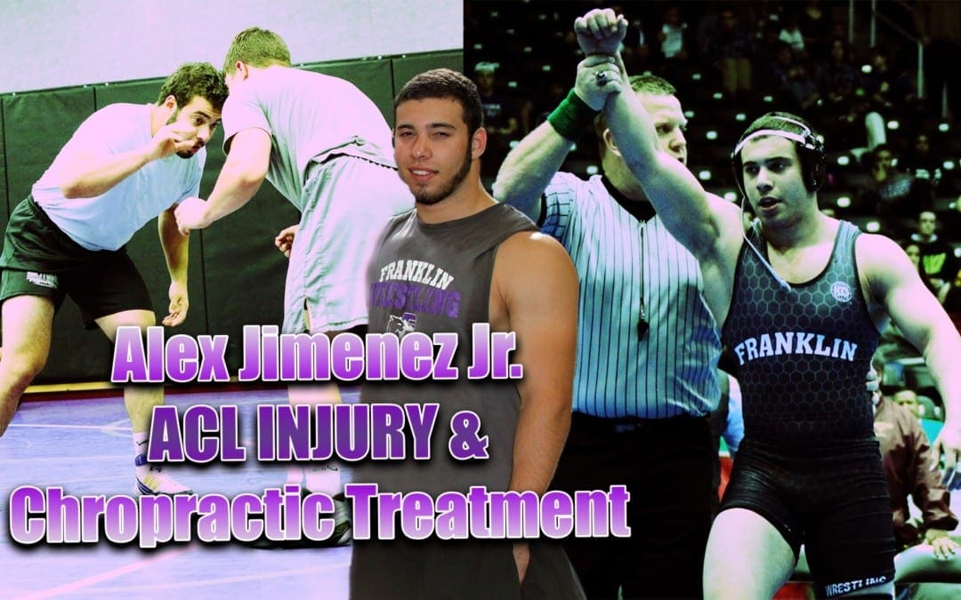
ACL Sports Injury: Alexander Jimenez, champion wrestler, trained hard at Push-as-Rx, Crossfit & Athlete Rehabilitation, to gradually become the athlete he is today. When he suffered an ACL tear, Alexander Jimenez had to undergo surgery, where he then started his rehabilitation process at Push-as-Rx. With a clear focus in mind, Alexander Jimenez was able to overcome his difficulties and he became a champion wrestler. For Alexander Jimenez, Push-as-Rx provided him with the mental and physical edge he needed in order to become the best athlete he can be. Alexander Jimenez recommends Push-as-Rx to younger athletes and athletes who are looking to achieve their fitness goals.
ACL Sports Injury Therapy
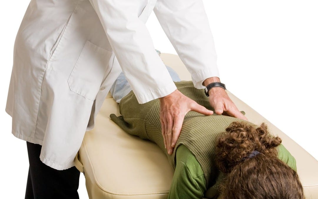
Anterior cruciate ligament injury is when the anterior cruciate ligament (ACL) is either stretched, partially torn, or totally torn. The most common injury is a tear that is complete. Symptoms include pain, a popping sound during trauma, instability of the knee, and joint swelling. Swelling generally appears within a couple hours. In approximately 50 percent of cases, other structures of the knee, for example surrounding cartilage, ligaments, or the meniscus, are damaged. The underlying mechanism frequently involves a rapid change in direction, abrupt stop, landing after a jump, or direct contact into the knee. ACL tears are most common in athletes.
If you have enjoyed this video and/or we have helped you in any way please feel free to subscribe and share us.
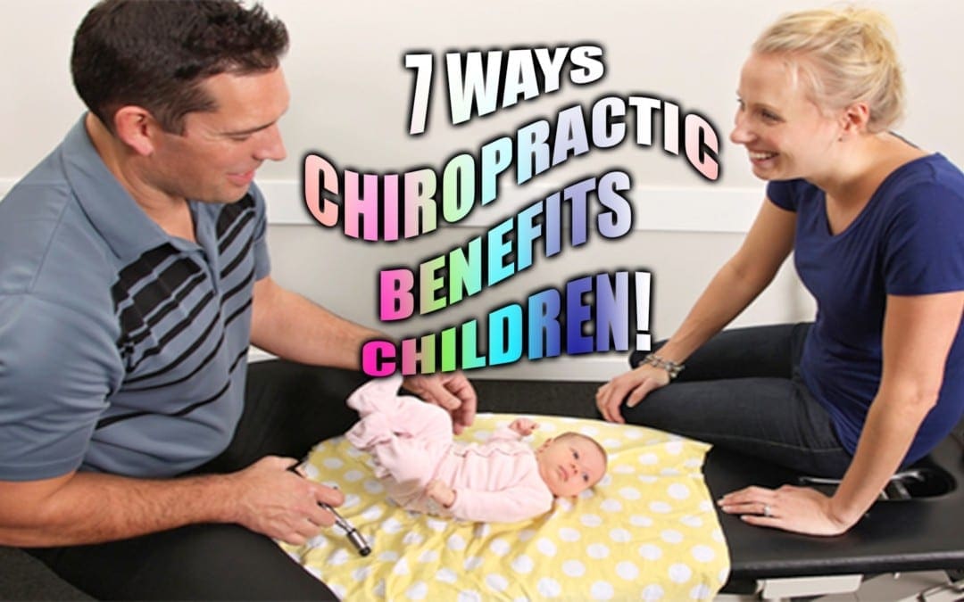
7 Ways:Chiropractic�traditionally is considered by many to be a treatment for adults. However, research shows that the treatment can be very beneficial for children. It can make birth easier and help give babies a healthy start in life. Young children can benefit from many of the childhood conditions that chiropractic helps treat. If you haven�t thought about chiropractic are for your child, take a look at these benefits of treatment; bet you�ll be convinced.
Contents
7 Ways Chiropractic Benefits Children
Promotes Optimal Health & Wellness
A properly aligned spine promotes good posture which promotes better health. However, it doesn�t stop there. When the spine is properly aligned the neural pathways are clear, allowing the central nervous system to function as it should. Additionally, the whole body approach that chiropractic takes helps provide a good foundation for healthy eating and regular exercise in children.
Helps Treat Asthma, Colic & Other Childhood Issues
There are numerous studies that show chiropractic is a viable treatment option for a variety of childhood conditions and illnesses. Chiropractic for asthma has long been discussed and is being embraced by more and more parents as well as the medical community as people are seeing first hand that it really works. There have also been studies that show chiropractic for colic is a very effective remedy, particularly for chronic colic in babies.
Helps Treat Birth Trauma
Birth trauma occurs more often than many people realize. It is typically only recognized or acknowledged when it is significant and causes severe damage or distortions. Some experts regard it as an underpublicized problem which means it is an undertreated problem.
Some studies show that using chiropractic to treat birth trauma can be very beneficial for the child. It addresses musculoskeletal injuries as well as problems with the brain stem and spinal cord caused by birth trauma.
Boosts Immunity
A flu epidemic in 1917 and 1918 was killing people � but the chiropractic patients were coming out much better than those who did not receive chiropractic treatments. They had a 5% death rate while those who were under chiropractic care had a .25% death rate. That is significant.
Regular chiropractic care can help boost immunity in children and adults. The spine plays a major part in health and the immune system. The body is better able to fight off infection, and if that isn�t enough to convince you it is also very effective in treating allergies.
Treats ADHD/ADD
Chiropractic does not �treat� ADHD/ADD but its positive effects on children who have been diagnosed with this condition has been documented in several studies. There have also been findings that suggest chiropractic may be more effective than medication.
Children who undergo chiropractic for ADHD have exhibited improvement in behavior and parental ratings of the child�s hyperactivity have also improved.
Aids In Resolving Breastfeeding Issues
Several studies have shown that chiropractic helps resolve breastfeeding problems in babies. Babies who have trouble latching on or staying on the breast and other problems can lead to breastfeeding failure. These studies assert that the outcome doesn�t have to be complete failure.
The birthing process can be very difficult on a baby, particularly when the baby is large or has large shoulders and traction is required to birth the child. Chiropractic care can help realign the baby�s spine so they are more comfortable during feeding and have better flexibility.
Decrease Risk Of Injury
The bones of babies and toddlers are more flexible than adult bones but they can still sustain injury. Regular chiropractic care helps keep the ligaments and joints supple which, in turn, helps prevent injury. This is particularly beneficial once the child becomes more mobile and begins turning over, sitting up, crawling, pulling up, and walking. It can help the child transition to each of these phases much easier and smoother.
So there you have it� 7 ways chiropractic benefits children. Ready to get your family started under chiropractic care? Then give us a call today!
Injury Medical Chiropractic Clinic: Sport Injury Treatments
Sciatica is characterized by pain in the lower back and gluteal region. This pain can radiate down one or both legs into the thigh, calf, ankle, and foot. Genuine sciatica occurs when pain travels beneath the knee.
Sciatic nerve pain results when the base of the spine is compressed or irritated and/or if trauma from an injury or an aggravated condition have compressed the spinal segments surrounding the sciatic nerve. The sciatic nerve is located at the sacral areas of the spine and the lumbar spine. Sciatic nerve pain or sciatica can be described as sharp, dull, burning, tingly, numb, continuous, or intermittent and generally affects only one side of the body. It can radiate throughout the entire length of the nerve, in some cases all of the way down to the feet.
Sciatic nerve pain is most often the result of a bulging or herniated disc, spinal stenosis, or in extremely rare instances, infection or tumor. The cause of your pain determines what are your treatment options to relieve sciatica.
Contents
Sciatica Treatment Options
The following article lists several common sciatica treatment options. You may want to read about:
People who have lower back pain have been prescribed bed rest so as to offer relief for aching bones and joints. Research in recent years has suggested that bed rest alone won’t offer relief for those suffering from sciatica or sciatic nerve pain.
Staying active may be more beneficial for people who suffer with back pain due to the compression or irritation of their sciatic nerve. Not to say that you ought to be running marathons. Activity means being mobile and up for long periods of time which are not sufficient to cause further injury and aggravation for your back pain. Some healthcare professionals may prescribe certain exercises, or some could simply indicate walking.
Sciatic Nerve Pain Relief
Pain is often treated with a non-steroidal anti-inflammatory medication (NSAID) such as ibuprofen or codeine (in severe cases).
In some cases a cortisone-like drug could be injected into the epidural space surrounding the spinal column. This process is like the epidural used during childbirth, and it’s called an epidural steroid injection. A course of this type of treatment may offer temporary relief, however, it doesn’t address the root of the problem. Treatment options like chiropractic care can help treat symptoms of sciatica at the source.
Chiropractic Treatment for Sciatic Nerve Pain
A chiropractor frequently treats people with sciatica. Their adjustments will aim to realign the spine, taking off the pressure of the sciatic nerve and often bringing rapid sciatica relief. When the stress is off, the body can begin to heal itself. While adjustments are probably most often used, other remedies may be given, especially if an adjustment is not recommended for the patient’s health issue. Other chiropractic treatment options might include using ice/cold treatment, ultrasound, a TENS (transcutaneous electric nerve stimulation) device or rehabilitative exercises. Ultrasound warms the region and increases circulation, which can reduce the swelling and muscular strain. A TENS device brings relief by using a slight electric current to relax muscle spasms and to increase endorphins. Additionally, a chiropractor might frequently consist of rehabilitative exercises so as to accelerate the recovery process. Massage therapy might also assist.
Ice/Cold Therapy reduces inflammation and aids restrain from sciatic nerve pain.
Ultrasound is gentle heat created by sound waves which penetrates deep into tissues. As mentioned previously, it increases circulation and reduces muscle spasms, cramping, swelling or inflammation, stiffness, and pain.
Adjustments (Spinal Manipulations). Spinal adjustments are in the heart of Chiropractic care. Manipulation supports restricted motion of the spine and helps to restore misaligned vertebral bodies with their proper position in the spinal column. Adjustment techniques differ from a swift high velocity thrust to those that combine minimal pressure and gentle pressure. Mastery of every technique is an art which requires great skill and precision. Spinal manipulation is the remedy that distinguishes chiropractic care from other medical areas.
Rehabilitative exercises. A combination of aerobics, strength training and stretching is commonly used to unleash pain-relieving endorphins in addition to relax the muscles which may be causing the nerve compression or irritation.
In some individuals, sciatica may fix itself, maybe occurring only once or a couple of times. However, if not treated properly, sciatica can worsen. A chiropractor can help bring relief, however, several alterations, will likely be required, especially if it has been occurring for a while. Letting a chiropractor treat sciatic nerve pain provides you a no surgery and no medication choice. Exercises will most likely be advisable to strengthen the muscles in the back to help prevent sciatica from recurring, and recovery is apt to take some time.
Surgery for Sciatic Nerve Pain?
If no other alternative treatment option has provided the patient relief from their symptoms, some patients with sciatica may discover substantial relief from surgery. In cases of herniated discs, a surgical procedure called a laminectomy may be performed. In this process, a portion of the posterior arch is removed to relieve pressure on pinched nerve tissues. In cases of spinal stenosis, the portion of bone that’s putting pressure on the sciatic nerve system could be taken off. Surgery is not for everybody. But for people who have shown no sign of progress in four to six weeks and that have had CT scans (computed tomography) or MRI that reveal a herniated disc or spinal stenosis, surgery may provide substantial relief.
Dr. Alex Jimenez’s Insight
Surgical interventions are frequently discussed as a possible treatment option for a variety of injuries and/or conditions which cause sciatica, however, surgery should only be considered as a last resort after all other treatment options have been utilized without improvement. Chiropractic care is a natural treatment approach which focuses on the diagnosis, treatment and prevention of sciatic nerve pain through the use of spinal adjustments and manual manipulations, among other commonly utilized treatment methods. Through the proper alignment of the spine, chiropractic care focuses on allowing the human body to naturally heal its sciatica without the need for surgery or drugs/medications.
The scope of our information is limited to chiropractic as well as to spinal injuries and conditions. To discuss the subject matter, please feel free to ask Dr. Jimenez or contact us at 915-850-0900 .
Curated by Dr. Alex Jimenez
Additional Topics: Sciatica
Sciatica is medically referred to as a collection of symptoms, rather than a single injury and/or condition. Symptoms of sciatic nerve pain, or sciatica, can vary in frequency and intensity, however, it is most commonly described as a sudden, sharp (knife-like) or electrical pain that radiates from the low back down the buttocks, hips, thighs and legs into the foot. Other symptoms of sciatica may include, tingling or burning sensations, numbness and weakness along the length of the sciatic nerve. Sciatica most frequently affects individuals between the ages of 30 and 50 years. It may often develop as a result of the degeneration of the spine due to age, however, the compression and irritation of the sciatic nerve caused by a bulging or herniated disc, among other spinal health issues, may also cause sciatic nerve pain.
Feet are important. When you consider what your feet go through, taking 8,000 steps over the course of a day, according to the Illinois Podiatric Medical Association (IPMA), it�s easy to see how 75 percent of all Americans will have some type of foot pain at some point in their lives. Plantar fasciitis is a common and very painful foot condition that can become chronic if not treated. It is also a condition that responds very well to chiropractic care.
Contents
Plantar Fasciitis Explained
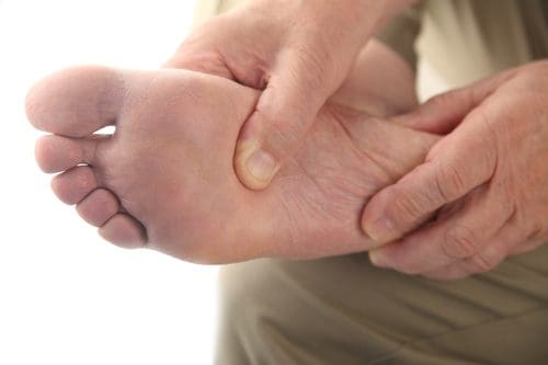
Plantar fasciitis is caused when the ligament that connects your toes to your heel (the plantar fascia) becomes inflamed, swollen, and weak. This causes the bottom of your foot or heel to hurt when you walk or stand, especially when you first wake or after sitting for a long time.
The pain tends to be sharp and stabbing. It is the most common cause of heel pain and is more prevalent among middle aged people. However, anyone can get it at any age, especially people who spend a lot of time on their feet.
How Chiropractic Helps Plantar Fasciitis
Chiropractic care is a very effective treatment for plantar fasciitis as well as the pain that is caused by the condition. Chiropractic for plantar fasciitis involves a very precise technique that involves adjustments to the feet and ankles as well as spinal alignment. This provides several benefits.
Reduces Stress in the Plantar Fascia � When a ligament is inflamed or stressed the tissue can develop very small tears that cause the pain of plantar fasciitis. Chiropractic adjustments made to the heel and foot take the pressure off of the plantar fascia, allowing it to relax.
Promotes Healing � When the stress on the plantar fascia is reduced through these chiropractic adjustments, the foot can begin to heal. The chiropractor may also recommend specific exercises that stretch the ligament and help it heal. They may also advise the patient of lifestyle changes as well as nutritional adjustments that can help with the pain and condition.
Provides Effective Pain Management � Chiropractic is a very effective way to manage pain throughout the body. Spinal adjustments allow better communication between the brain and nerves, allowing the central nervous system to function more effectively. Condition specific adjustments speak to the root of the problem, not just the symptoms. This means a more effective form of pain management that is longer lasting.
Reduces the Risk of Further Injury � When a person has a condition like plantar fasciitis, they will often adjust their gait in an effort to avoid the pain. This puts stress on other parts of the body and can lead to back pain, sore joints, strained muscles, and other problems. Chiropractic�s whole body approach helps the person realign their body properly so that they stand and walk properly. This helps them avoid further injury and discomfort.
Chiropractic Complements Other Treatments
While chiropractic care can be an effective treatment for plantar fasciitis on its own, it is also a very good complement to other treatments for the condition. Patients may use chiropractic in conjunction with physical therapy, massage, and even injections to manage the pain and treat the condition. It can also help with speeding healing and helping to provide better mobility.
Plantar fasciitis can take several months to heal, but by adding chiropractic treatments to your recovery plan, you can feel better faster while more effectively managing your pain. Regular chiropractic treatments can also keep the condition from becoming chronic. By working with your chiropractor and following their recommendations you can reduce your pain and shorten your healing time.
Injury Medical Chiropractic Clinic & Chronic Pain & Treatments
Children with cerebral palsy have various needs. Some children have problems with motor skills and spasticity, but normally pick up things pretty fast. Others have a full range of issues from motor skills to esophageal and respiratory problems. Since so many kids with cerebral palsy have an array of different medical needs, there isn’t one particular type of treatment which may help each and every child. Luckily, there are a number of different therapeutic remedies to select from, which range from holistic care, water therapy, and much more.
Contents
Acupuncture
Whilst generally not adopted in Western Medicine, acupuncture has been used for centuries by Asian countries and is viewed as a medicinal art. Some families with children who have cerebral palsy take their kids to an acupuncturist to try and relieve the frequent pain related to the disorder. Other kids find relief in acupuncture for painful birth injuries such as spina bifida, Erb’s palsy, and brain damage. Acupuncture uses needles to ease pain, often instead of medication.
Aquatherapy
Aquatherapy is among the most popular and beneficials form of treatment for children with cerebral palsy, as they suffer from limb maladiess, but this might also be advantageous for children who suffer with Erb’s palsy and are trying to regain movement in their arm.
Under the supervision of a trained and experienced professional therapist, kids may gain from the strength exercise and training afforded by the anti-gravity character of a pool. In this soothing environment, a child can have a respite from some of the pain which comes with the disability (occasionally cerebral palsy causes stress on the musculoskeletal frame by simply gravity and body weight), and they can still work through the natural curative and restorative nature of water.
Behavioral Therapy (Psychotherapy)
Some birth injuries involve an intellectual disability that impacts how kids interact in social scenarios. Other children might have had physical constraints that included them being house-bound for a long time, causing them to have a deficiency in social skills or cues. Behavioral therapy, also known as psychotherapy, allows patients to work through problems they may have within their social and mental health with a behavioral health professional.
Chiropractic Care and Massage Therapy
Children with cerebral palsy may benefit from chiropractic care and massage therapy for a few different reasons. Because some children with cerebral palsy may have experienced lots of strain or stress on their musculoskeletal system as a result of the disorder, requiring chiropractic care may ultimately be fundamental towards their proper spinal alignment as well as for their overall health and wellness.
Chiropractic care is a well-known alternative treatment option which utilizes spinal adjustments and manual manipulations to treat a variety of injuries and/or conditions associated with the musculoskeletal and nervous system, including back pain.
Another reason that a patient with cerebral palsy might need chiropractic care or massage treatment is for the basic goal of extending and stretching muscles. When muscles relax as they perform through such therapies, they are more inclined to become stronger and healthier which is needed if they are going to correctly learn how to walk. This kind of treatment isn’t generally suggested for kids suffering from spina bifida because the raw exposed nerves could be inadvertently mishandled, causing more problems.
Furthermore, chiropractic care can be used to help treat other, less noticeable aspects of cerebral palsy. The theory of chiropractic care is that by healing the central area around the spine, the extremities and other parts of the body affected by the disorder can become more normalized, allowing for improved function and quality of life. Chiropractic care can also help improve strength, mobility and flexibility in children with cerebral palsy and its associated symptoms.
Conductive Education
Some children with neurological or mobility impairment found in almost any brain-related birth trauma need help performing activities that regular people learn through daily exercise, learning, and experience. Since these children don’t often have the same sorts of experiences that non-disabled people have, conductive education is a form of special education that functions as a kind of study group for life.
Conductive education provides opportunities of every day learning experiences so that kids can have the exact same general education that non-disabled individuals do.
Hippotherapy
Using equine motion and connections with horses, children with all kinds of birth injuries could learn basic occupational and speech therapy. Hippotherapy is not therapeutic horseback riding, but rather a trained practitioner introduces the child to the horse and uses the horse to access the child in ways which were previously thought of as unconventional.
Hyperbaric Oxygen Therapy
Normally short-term treatment and frequently only experienced once or twice, Hyperbaric Oxygen Therapy is a method of fast-healing for some kids that have suffered oxygen deprivation (anoxic, hypoxic, HIE, birth asphyxia, and perinatal asphyxia). If an infant is delivered and does not breathe for the upcoming instant minutes, hyperbaric oxygen treatment is a great way to introduce a lot of oxygen into the blood stream preventing or lessening the seriousness of birth injuries such as cerebral palsy.
Occupational Therapy
Occupational therapy’s main objective is to work on creating balance, strength, and gait. An occupational therapist might consult with an orthopedic surgeon to operate on strengthening and firming muscles, in which following the occupational therapist can delegate casts and orthopedic devices which also help strengthen and form muscles. These methods are to help patients learn how to walk, and also to create control and strength to stop spasticity.
The occupational therapist also trains patients to function on decision-making, abstract reasoning, problem-solving, perception, memory, sequencing, and much more.
Play Therapy
Utilizing play with a variety of different toys in various public places, kids with all kinds of birth injuries can learn to appreciate themselves. Often children with birth injuries can feel that they’re different or that they have health issues and end up stressing about their difficulties more than having fun.
While they’re having fun in play therapy, they can learn the way to interact with other kids, learn about themselves, and to construct self-confidence.
Physiotherapy and Physical Therapy
Physiotherapy and physical therapy both operate on the rehabilitation of muscle groups. This is extremely important for children with shoulder dystocia, Erb’s palsy, Klumpke’s palsy, or Brachial Plexus palsy, and, in fact, kids suffering from these birth injuries won’t regain use of their hand or arm without physical and physiotherapy. Through this type of treatment, therapists strive to receive the perfect movement from their patients through an assortment of different challenges and exercises.
This can be like occupational therapy, though the focus is mainly on what the muscle groups are doing, and not on so many different targets like occupational therapy. A physical therapist is often like a personal trainer in a gym, training, cheering, and challenging.
Respiratory, Digestive, and Dietician Therapy
Some kids with cerebral palsy encounter respiration and esophageal problems. Consequently they can experience issues with eating, breathing, and drinking, which divides into digestive and dietician treatment, addressing what foods and drinks should be consumed. Respiratory treatment may primarily tackle breathing exercises to strengthen and optimize lung development, but may also address these other concerns.
Speech and Language Therapy
Speech and language therapy can be very important for kids with cerebral palsy and other forms of brain-related birth harm. Approximately, 1 out of every 4 patients with cerebral palsy don’t have the capability to speak. Speech and language therapy helps them to work on exercises which progress the learning of speech and get kids closer to communicating effectively.
Some speech and language therapists utilize programs that help patients understand the operation of language inside individuals, and these programs also provide communication boards using pre-formed responses so that children can get in the habit of responding with particular answers until they consider trying to verbalize these answers.
Vocational Counseling
This has many different sorts of therapists, a few children could be confused or jeopardized by visiting a lot of individuals, or, worse, by having so many people invade their home. One way of approaching treatment is by using a vocational counselor, one individual who can master several distinct types of treatment.
As vocational counselors might not have exactly the exact same depth in all of these subjects as one therapist would have regarding one subject, this might be a great first step for treatment with your little one. By getting your child to adjust to only one person interacting within their lifetime, they’re more inclined to concentrate on the subjects at hand.
Afterwards, if more obstacles and more depth is required, your kid may have more assurance in different areas (and with a few social abilities from connecting with this particular counselor) and may be able to handle other therapists more efficiently.
Yoga Therapy
Normally prescribed under the direction of an occupational or physical therapist, yoga therapy is a fantastic alternative for kids whose muscles need to be loosened or lengthened. Children with cerebral palsy suffer from particularly tight muscles, so yoga therapy helps them to work on extending and on making the muscles more limb. This type of treatment might be incorporated to other sorts of therapy, and it might also be delegated as “homework” to kids with cerebral palsy for optimal flexibility and, ultimately, optimal freedom.
Dr. Alex Jimenez’s Insight
Cerebral palsy is a lifelong set of movement disorders with no cure. However, various types of treatment options can help provide some forms of relief for individuals and children with cerebral palsy as well as help restore some function and quality of life. Because cerebral palsy can affect patients differently, people with the disorder can benefit from many different therapies, including chiropractic care and physical therapy. Chiropractic care is a popular, alternative treatment option which focuses on the diagnosis and treatment of several kinds of injuries and/or conditions, including cerebral palsy. Through the use of spinal adjustments and manual manipulations, a chiropractor can help improve strength, mobility and flexibility in people with cerebral palsy.
The scope of our information is limited to chiropractic as well as to spinal injuries and conditions. To discuss the subject matter, please feel free to ask Dr. Jimenez or contact us at 915-850-0900 .
Curated by Dr. Alex Jimenez
Additional Topics: Sciatica
Sciatica is medically referred to as a collection of symptoms, rather than a single injury and/or condition. Symptoms of sciatic nerve pain, or sciatica, can vary in frequency and intensity, however, it is most commonly described as a sudden, sharp (knife-like) or electrical pain that radiates from the low back down the buttocks, hips, thighs and legs into the foot. Other symptoms of sciatica may include, tingling or burning sensations, numbness and weakness along the length of the sciatic nerve. Sciatica most frequently affects individuals between the ages of 30 and 50 years. It may often develop as a result of the degeneration of the spine due to age, however, the compression and irritation of the sciatic nerve caused by a bulging or herniated disc, among other spinal health issues, may also cause sciatic nerve pain.
Chiropractic care, recognized as a complementary or alternative health practice in the U.S., is becoming a sought after treatment for pain control in children and adults, alike. Chiropractic interventions are used to improve forms of musculoskeletal pain, including low back, shoulder, neck, headaches, hand and foot problems, as well as for particular health conditions, such as Cerebral Palsy, fibromyalgia and attention deficit hyperactivity disorder.
The Greek word “chiropractic” means “hand practice” or therapy done by hand. Chiropractic care is a hands on approach to treatment which often centers around the adjustment to the joints and spine in a way that influences the human body’s nervous system and natural defense mechanisms for the purpose of alleviating pain and improving health and wellness.
There are 2 million children and nearly 18 million adults in the United States who’ve received chiropractic or osteopathic manipulation during a 12 month period, based on the 2007 National Health Interview Survey, or NHIS. The analysis found that children who have parents that use complementary and alternative medicine, or CAM, services are twice as likely as other children to utilize complementary health services, as well.
As a matter of fact, in 2007 the CDC National Health Statistics Report #12 indicated that rehabilitation and chiropractic services would be the next most popular form of CAM treatments used on children. CAM therapies were most widely used on children for the following purposes:
Although there is little in the way of formal studies on the effects of chiropractic care for use on individuals with Cerebral Palsy, you will find reports from the chiropractic community that demonstrated improvements for the following conditions:
Arthritis
Back pain or other problems
Breathing
Drooling (release of the TMJ-muscles)
Gait patterns
Hypertonic musculature
Joint pain or stiffness
Muscle contractures
Neck pain or other problems
Pain and tension
Scoliosis or curvature of the spine
Seizures
Sleep difficulties
Other musculoskeletal conditions
Simply explained, the brain communicates with the body. Chiropractic care is established in improving the manner in which brain control and muscles work together. The neuromuscular system sends messages from your brain, down the spine and in the nerves. When there’s interference, the body isn’t able to be effective.
Chiropractic intervention aims to enhance the structural facets of the body to clear the pathway for the brain to communicate with the nerves. This can result in improved strength, balance, flexibility and coordination abilities, especially in the extremities. One intervention does not fix all, instead the intervention chosen and the location of treatment are relative to the symptom being addressed. Since Cerebral Palsy affects people differently, assorted chiropractic treatment methods are utilized to address specific issues.
History of the Evolution of Chiropractic Care
Launched in Davenport, Iowa in the late 1890s, chiropractic care has been rooted in holistic notions that, for several decades, rendered the practice controvesial. The contention of those in the chiropractic community that the only source of pain has been spinal dysfunction called vertebral subluxation has been contested by conventional medical practitioners. Additionally, physicians and other critics have questioned the capability of chiropractic care in treating ailments which aren’t connected to the neuromusculoskeletal system.
Although chiropractic care recently has gained acceptance by the medical community because of manual therapy due to its ability to alleviate pain, the practice remains rooted in spinal adjustments and manipulations as a gateway to enhancing a person’s overall health and wellness. Currently, there are chiropractors in practice which are purists, and others that think scientific research has a place in chiropractic care.
There is evidence that chiropractic care can be helpful to children with Cerebral Palsy. Some research suggests that kids that received spinal adjustments may sit and stand with more ease. Also, the research indicated that some children became active, digested food better, slept more peacefully, and appreciated improved coordination following chiropractic care.
In the publication, “Chiropractic Care of Special Populations,” writer Robert D. Mootz reports on some special treatments which have been reported to have enhanced some circumstances of Cerebral Palsy:
Adjustment of the atlanto-occipital subluxations helped with children who had difficulty with sleeping, personality disturbances, and hypertonic musculature.
Upper cervical spine adjustments created clinical improvements in a 5-year-old male with quadriplegic Cerebral Palsy.
Adjustments can be helpful in cases of cranial dysfunction in the sphenobasilar junction in children who have a history of birth trauma or head injury where motor tracts of the medulla may be compromised.
Manual release of the TMJ-related muscles, such as the masseter and temporalis, may ease excessive drooling.
Myofascial release may be used to assist in decreasing the severity of spinal distortion and aid in stabilizing gait patterns in children with spastic Cerebral Palsy who have muscle contractures in the paraspinals, lateral thigh muscles, lower extremity abductors, Achilles tendons, and wrist extensors.
What is Chiropractic Care?
Chiropractic care is considered a manipulative and body-based therapeutic system which has an impact on the human body’s systems and structures, such as the bones, joints, soft tissues, and neuromuscular system, which are manipulated beyond their passive range of movement and with proper use of force. It is a treatment which uses the adjustment and manipulation of the spine and joints to ease pain. The spinal manipulations are made using the chiropractor’s hands, and therefore are known as “adjustments.” The dysfunctions or abnormalities at the joints of the spine are known as “vertebral subluxations.” Vertebral subluxations are a group of symptoms in the spine.
Many people seek chiropractic care to address:
Neck pain
Back pain
Spinal discomfort
Inability to sit or stand
Chiropractic care is determined by three main concepts, which are:
Reductionism: attributing the cause of pain or illness to vertebral subluxation alone.
Conservatism: committing to non-invasive interventions as a mode of treatment.
Homeostasis: emphasizing self-healing.
These three notions are heeded by both traditional, purist chiropractors and “mixers” chiropractors which are influenced by evidence-based scientific findings and fundamentals. Mixers can introduce other treatments to bring relief to people including:
Ice and heat
Vitamins and nutritional supplements
Homeopathic or holistic medicine
Herbs
However,� all chiropractors use the simple tenet of this profession, vertebral subluxation, as the centerpiece of all clinical treatments, together with a combination of other interventions.
Which are the Advantages of Chiropractic Treatment, also When is Care Advised?
Although there have been several studies that assess the impact of chiropractic care on children with Cerebral Palsy, several of which have been completed have shown that children respond well to treatment.
In a 2006 study, initially published in the Journal of Vertebral Subluxation Research suggested that kids with Cerebral Palsy that were determined to have subluxations showed improvement in their mobility after one month of chiropractic care. One child demonstrated improvement in her ability to sit up, walk, and ambulate following 22 spinal alterations.
In a research published by the Journal of Pediatric, Maternal & Family Health, it was determined that a 2-year-old boy with Cerebral Palsy was relieved of many symptoms that interfered with his freedom and ability to sleep. Following seven months of care, he was able to pull himself into an upright position and was sleeping frequently. Although, other characteristics of his condition, such as uncontrolled movements, persisted.
Individuals in several walks of life, by the elderly to children, seek chiropractic care. Many indicate that they recognize significant relief from spinal adjustments and manual manipulations. However, individual advantages are dependent on a child’s condition at the beginning of care; parents with the advice of the child’s primary care physician, will want to ascertain how chiropractic care fits in with a child’s overall treatment program.
What Happens During Chiropractic Care?
At the start of a chiropractic appointment, a complete medical history will be taken to acquaint the practitioner of the symptoms that an individual is coping with. From there, a series of exams and evaluations will happen.
Among the very first of these will be an X-ray, which ought to offer some valuable information about the condition of a child’s spinal column. This information often includes:
Curvature
Misalignments (subluxations)
Abnormalities
Muscle tone changes
Tissue abnormalities
A physical examination will assist the chiropractor to find a child’s source of pain. When the assessment is finished, the chiropractor will recommend a plan of treatment, which is very likely to include adjustments. If they suspect that another condition is causing pain or discomfort, a referral will be issued.
A chiropractor will utilize several techniques to ascertain in which a subluxation, or misalignment, is present. The most frequent procedures which will help the chiropractor decide which adjustments will be necessary to bring relief to a child are:
Static palpitation � when a practitioner uses his or her hands to detect signs of misalignment
Motion palpitation � when a doctor moves bones to separate them
Leg check � moving the legs to reveal spinal subluxation
An adjustment is finished when the joints of the spine are moved past the point at which they would normally proceed in a way where it does not damage or dislocate the joints. Doing so will require the chiropractor to use gentle force and educated expertise to finish the moves. Notice, untrained individuals should not attempt to perform these procedures on another person.
There are several specific kinds of adjustment which can be used to aid a child. They include, but are not limited to:
Diversified movement � full spine manipulation
Activator technique � using a device to adjust the spine
Cox technique � low-force adjustment
Gonstead technique � using a specific path to adjust the spine
These adjustments will be created over a period of time, comprising of several appointments, to help a patient recover motion and minimize discomfort.
Chiropractors most often operate private practices, but often, their services can be found in other medical settings such as:
Hospitals
Physician�s office
Clinics
Assisted living centers
Residential facilities and nursing homes
Who Offers Chiropractic Care?
Chiropractors offer a wide range of services and, based on where an individual resides, the scope of their duties may vary. In a small number of countries, chiropractors are allowed to perform minor surgeries and write prescriptions, for others, these functions are prohibited.
Internationally, demands to practice as a chiropractor vary. In the United States, a chiropractor needs to complete a professional degree program. Accredited programs require an applicant to complete 90 credit hours of undergraduate instruction, and many others require students to earn a bachelor’s degree.
Doctors of chiropractic, nevertheless, must complete an intensive program that revolves around healing arts that many consider to be challenging in a medical college. A doctor of chiropractic, or chiropractor generally pursues a bachelor’s of science degree prior to attending a chiropractic college.
Bachelor’s degree coursework contains:
Biology
Chemistry
Physics
Nutrition
Psychology
Anatomy
Physiology
Chiropractic college curriculums include further coursework, in addition to hands-on instruction and clinical study that lasts four or five years.
Licensure is required to practice in the USA. Most states will grant licenses for those who have successfully completed an accredited program and passed an examination administered by the National Board of Chiropractic examiners.
Healthcare practitioners that choose to provide additional services such as acupuncture or massage might have to pursue other courses of research and certificates if they intend to supply these services personally.
Are There Special Considerations or Risks for Chiropractic Therapy?
Generally, chiropractic care is considered safe. In the hands of a fully-qualified practitioner, chiropractic care may cause some mild discomfort, but it should not be painful. If a child complains that treatment is extremely uncomfortable, or painful, a parent must inquire into the issue by requesting the chiropractor why this is happening. If a parent is uncomfortable with the answer, he or she should seek the care of another healthcare practitioner or chiropractor.
Often throughout the process of making an adjustment, a child and their parents will hear a popping noise. This occurs when gases escape from fluids that surround joints. This is similar to popping that happens in the joints of the feet or ankles; it isn’t indicative of a critical illness. Also, to the untrained eye, the quick and quirky alterations could appear alarming to people unfamiliar with chiropractic interventions.
Tips for Choosing a Chiropractor
According to the National Center for Complementary and Alternative Medicine, or NCCAM, a division of the National Institute of Health, which is considered the lead agency for scientific research on diverse medical and health care practices, when choosing a chiropractor, the individual must inquire:
Their experience in coordinating care with conventional health care providers
Their experience in delivering care to children
Their education, training and licensure
You also need to inquire about their experience and expertise in treating children, or adults, with Cerebral Palsy.
NCCAM also recommends that when considering an alternative and complementary wellness approach for a child:
Make sure the child has an accurate diagnosis from a licensed healthcare provider.
Understand the potential risks, benefits and effectiveness of the specific strategy.
Discuss any and CAM approaches with the child’s primary care physician before agreeing to this therapy protocol, particularly with the physician that abroad your child’s care plan so that there’s not any conflict with other kinds of therapy.
Never use any health product or practice which hasn’t been shown safe and effective to substitute or delay conventional care or prescribed drugs.
When a health care practitioner indicates a CAM approach, don’t increase the dosage or length of this treatment beyond what is advocated without professional approval.
Discuss any and all concerns about the effects of a CAM strategy with your child’s main health care provider.
To ensure coordinated and secure care, inform all your child’s healthcare providers about any CAM strategy your child uses, giving them a full picture of what you do to manage your child’s wellbeing.
Dr. Alex Jimenez’s Insight
More so now, than ever before, people are turning to alternative and complementary medicine to treat a variety of injuries and/or conditions, especially Cerebral Palsy. An increase of CAM treatment methods can offer more therapy options for people or children with CP. Although CP has no cure, a person with Cerebral Palsy can benefit from alternative and complementary medicine. Chiropractic care has been demonstrated to help improve other symptoms associated with CP. Furthermore, chiropractic care used together with physical therapy and rehabilitation can help restore some strength, mobility and flexibility for people and children with Cerebral Palsy.
The scope of our information is limited to chiropractic as well as to spinal injuries and conditions. To discuss the subject matter, please feel free to ask Dr. Jimenez or contact us at 915-850-0900 .
Sciatica is medically referred to as a collection of symptoms, rather than a single injury and/or condition. Symptoms of sciatic nerve pain, or sciatica, can vary in frequency and intensity, however, it is most commonly described as a sudden, sharp (knife-like) or electrical pain that radiates from the low back down the buttocks, hips, thighs and legs into the foot. Other symptoms of sciatica may include, tingling or burning sensations, numbness and weakness along the length of the sciatic nerve. Sciatica most frequently affects individuals between the ages of 30 and 50 years. It may often develop as a result of the degeneration of the spine due to age, however, the compression and irritation of the sciatic nerve caused by a bulging or herniated disc, among other spinal health issues, may also cause sciatic nerve pain.
Doctors of Chiropractic (DC), or chiropractors, regularly treat sciatica. Sciatica is characterized by pain that originates in the low back or buttocks which travels down one or both legs, into the foot. Sciatic nerve pain varies in intensity and frequency; minimal, moderate, acute and chronic, intermittent, frequent or constant.
Pain is described as dull, achy, sharp, toothache-like, pins and needles or similar to electric shocks. Other symptoms associated with sciatica include numbness, burning and tingling sensations as well as weakness. Sciatica can also be called radiating or referred pain, neuropathy, or neuralgia. A common misconception, however, is that sciatica is a disease, nevertheless, sciatica is really a collection of symptoms belonging to a specific disorder.
Contents
Sciatica Is Caused by Nerve Compression
Sciatica is generally brought on by sciatic nerve compression. Disorders known to trigger sciatic nerve pain include lumbar spine subluxations, or misaligned vertebral bodies, herniated or bulging discs (slipped disks), pregnancy and childbirth, tumors, and even as a result of non-spinal disorders, such as diabetes, constipation, or sitting on one’s back pocket wallet.
One common cause of sciatica is piriformis syndrome. Piriformis syndrome is named after the piriformis muscle. The piriformis muscle is located in the lower part of the spine, connects to the thighbone, and also assists in hip rotation. The sciatic nerve runs under the piriformis muscle. This muscle is susceptible to injury from sports injuries, a slip-and-fall accident, hip arthritis, or due to a difference in leg length. Such situations can cause cramping and spasm to develop in the piriformis muscle, thus pinching the sciatic nerve and causing pain and discomfort.
Sciatic nerve compression may lead to the loss of feeling (sensory loss), paralysis of one limb or group of muscles (monoplegia), as well as sleeplessness or insomnia.
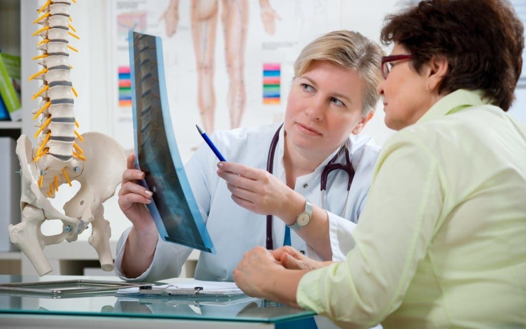
Proper Diagnosis of Sciatica Is Vital
Since there are many disorders that cause sciatica, a healthcare professional’s initial step would be to determine what is causing the patient’s sciatica symptoms. Forming a diagnosis entails a thoughtful review of the patient’s medical history, as well as a physical and neurological examination. Diagnostic testing involves a x ray, MRI, CT scan and/or electrodiagnostic tests (nerve conduction speed, electromyography). These examinations and tests help to detect possible contraindications to spinal adjustments and other chiropractic treatment methods.
Chiropractic Treatment of Sciatic Nerve Pain
The objective of chiropractic treatment for sciatic nerve pain is to assist the body’s capacity to heal itself. It is based upon the scientific principle that limited spinal movement contributes to pain and decreased function and functionality. Chiropractic care is non-invasive (nonsurgical) and drug-free.
The type of chiropractic treatment provided is dependent upon the cause of the patient’s sciatica. A sciatic nerve pain chiropractic treatment program may include several different therapies such as ice/cold treatments, ultrasound, TENS, and spinal adjustments (sometimes called spinal manipulation). Below are additional details on these chiropractic treatment modalities.
Ice/Cold therapy reduces inflammation and helps to control sciatic nerve pain.
Ultrasound is mild warmth created by sound waves which penetrate deep into cells. Ultrasound increases circulation and helps to decrease muscle spasms, cramping, swelling, stiffness, and pain.
TENS unit (transcutaneous electric nerve stimulation) is a small box-like, battery-powered, portable muscle stimulating machine. Various intensities of electrical current control acute pain and reduce muscle spasms. Larger versions of this home-use TENS units are utilized by chiropractors, physical therapists and other rehabilitation healthcare professionals.
Adjustments (Spinal Manipulation) At the heart of chiropractic care are spinal adjustments. Spinal manipulation frees restricted movement of the spine and helps to restore misaligned vertebral bodies with their appropriate position in the spine. Spinal adjustment helps to decrease nerve irritability responsible for causing inflammation, muscle soreness, pain, and other symptoms associated with sciatica. Adjustments should not be painful. Spinal manipulation is demonstrated to be safe and effective.
In college and during their training, students of chiropractic understand many different adjustment techniques permitting them to take care of various sorts of subluxations and spinal disorders. Chiropractic techniques vary in the swift high velocity thrust to people that unite minimal pressure and mild pressure. Mastery of each technique is an art which needs great precision and skill. Spinal adjustments and manipulations is the treatment that differentiates chiropractic care from other medical areas.
Dr. Alex Jimenez’s Insight
Sciatic nerve pain, or sciatica, is identified as radiating pain and discomfort along the length of the sciatic nerve, which travels from the low back down the buttocks and into one or both legs, occasionally reaching all the way down to the calf and foot. Approximately more than 3 million cases of sciatica are reported in the United States each year, where it is generally caused by the compression of the sciatic nerve as a result of a bulging or herniated disc. Chiropractic care is a well-known, alternative treatment option commonly utilized to help improve a variety of spinal health issues, including sciatica, or sciatic nerve pain.
Chiropractic’s Limitations in Treating Sciatica
Sciatica may be caused by other disorders beyond the scope of chiropractic clinic. If the doctor of chiropractic, or chiropractor, determines the patient’s disease requires treatment by another kind of doctor, then the patient is referred to another specialist. In some cases, the chiropractor can continue to treat the patient and also co-manage the patient’s care with the other specialist. The scope of our information is limited to chiropractic as well as to spinal injuries and conditions. To discuss the subject matter, please feel free to ask Dr. Jimenez or contact us at 915-850-0900 .
Curated by Dr. Alex Jimenez
Additional Topics: Sciatica
Sciatica is medically referred to as a collection of symptoms, rather than a single injury and/or condition. Symptoms of sciatic nerve pain, or sciatica, can vary in frequency and intensity, however, it is most commonly described as a sudden, sharp (knife-like) or electrical pain that radiates from the low back down the buttocks, hips, thighs and legs into the foot. Other symptoms of sciatica may include, tingling or burning sensations, numbness and weakness along the length of the sciatic nerve. Sciatica most frequently affects individuals between the ages of 30 and 50 years. It may often develop as a result of the degeneration of the spine due to age, however, the compression and irritation of the sciatic nerve caused by a bulging or herniated disc, among other spinal health issues, may also cause sciatic nerve pain.
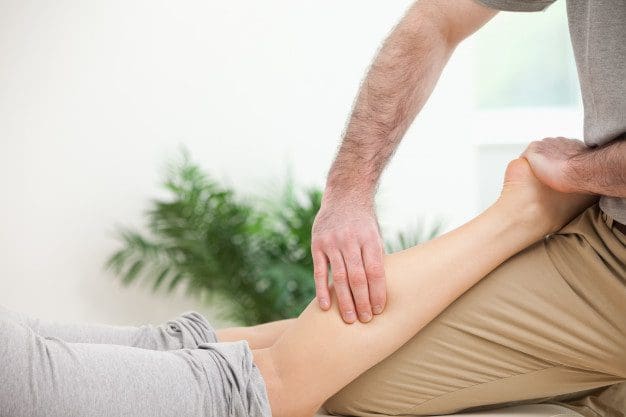
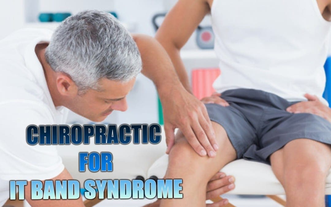
IT Iliotibial band syndrome is a very common injury among runners. If it is diagnosed early and treatment commences immediately the chances of it becoming a chronic condition are reduced. It responds very well to chiropractic since it involves the pelvis and related muscles. When pelvic mechanics are not functioning properly the muscle don�t work efficiently which hinders flexibility and mobility. This can lead to tight muscles which may inhibit motion and cause pain. Chiropractic adjustments have been proven to help with the condition.
Contents
What Is The Iliotibial Band?
The Iliotibial Band, or fasciae latae, is the outer casing of muscle that extends along the outer thigh, from the top of the hip to the outside of the knee. IT Iliotibial band syndrome occurs when that casing becomes thickened. It is flexed or tight when you stand; it is what keeps your let straight, allowing the larger thigh muscle to rest.
There are two primary muscles that are involved in iliotibial band syndrome, the buttock muscle, or gluteus maximus, and the tensor fasciae latae muscles. Sometimes Iliotibial Band Syndrome is referred to Tensor Fasciae Latae Syndrome and the two terms can be used interchangeably.
IT Iliotibial Band Syndrome Defined
As the iliotibial band thickens it pulls in the area where it connects to the knee. This results in knee pain due to the application of too much pressure on the bursa. The bursa then becomes swollen, inflamed, and painful. During activity, such as running on an incline, the glutes are heavily involved.
The other end of the iliotibial band is inserted at the glutes so as the band tightens from this activity, it can trigger iliotibial band syndrome pain. Repeated activity further aggravates it, as does running on tight indoor tracks or uneven roads as well as having collapsed arches or running it inferior or worn out running shoes.
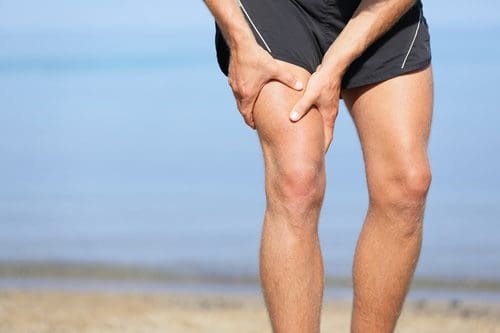
Symptoms Of Iliotibial Band Syndrome
There are several symptoms that can be used to diagnose iliotibial band syndrome. Lateral knee pain (pain on the outside of the knee) is a primary symptom and often used as a key diagnostic tool. Few conditions involve lateral knee pain. Other symptoms include:
Pain that worsens after running, particularly after running on an incline, climbing stairs, or climbing hills
There may not be any pain until you do something that aggravates it like climbing a hill.
The pain may not begin until you are mid-way through a run.
The pain can be intense and debilitating.
It can accompany a snapping hip, which occurs when the muscles that cross the outer hip may click or snap while running or walking.
The pain may be present along the lateral thigh without incorporating the knee, but it is only in very rare instances that it is concentrated on the gluteal or hip muscles.
Iliotibial band syndrome is often attributed to over training. This can mean suddenly increasing hill repeats or doubling your mileage.
Treatments For IT Iliotibial Band Syndrome
If your iliotibial band syndrome is caused by a problem with pelvic function, relieving the pain from the condition can be difficult. Stretching is not likely to bring relief � and if it does it won�t last long. If the pain from iliotibial band syndrome lasts for more than two weeks even if you are only stretching, your regular exercise routine, and ice and you don�t see much improvement, a chiropractor can help.
Even if the pain is located in the knee, the problem could originate in the pelvis. A chiropractor can assess your condition, check to see that your pelvis is functioning properly. If it isn�t, spinal adjustments and other chiropractic treatments can bring the body back into alignment and make the pelvis more functional.
Chiropractic Clinic Extra: Sport Injury Treatments
Cerebral palsy, or CP, is a medical term used to describe developmental motor conditions that cause impairment or disability. Cerebral palsy is not contagious and it isn’t regarded as a disease. Although it is not hereditary, many cases of CP are believed to be caused throughout pregnancy or birth, therefore they are typically referred to as a congenital condition. Cerebral palsy might be related to infection, radiation, or lack of oxygen during brain growth, as well as premature birth and birth trauma. Damage can occur up to age 3.
Contents
What is Cerebral Palsy?
Cerebral palsy, caused by “a permanent, non-progressive defect” in the brain, affects movement, posture, and muscle tone. Cerebral palsy is not paralysis, however, the motor centers of the brain become altered with the condition. Eyesight health issues and depth perception, cognition and communication challenges, and musculoskeletal issues can result from cerebral palsy. All cerebral palsy types involve “abnormal muscle tone”, and problems with motor development and reflexes.
Symptoms of CP include spasms, spasticity, involuntary movement, and balance and gait difficulties, including “foot walking” and “scissor walking”. The degree of disorder falls on a continuum, from “slight clumsiness” to severe impairments. Babies with the severe types of CP have bodies which may be rigid or floppy, with irregular posture. There can also be other birth defects as a result of cerebral palsy. Over the course of a child’s growth, symptoms may change or appear. It is generally when the baby becomes mobile that cerebral palsy becomes more evident. Speech problems, from respiratory and muscular difficulties, can also frequently occur.
A number of the secondary conditions related to CP include sensory impairments, eating problems, seizures, epilepsy, behavior and learning disabilities, mental retardation, and continence disorders. Speech impairments or disabilities and language delays are also commonly associated with CP. Early intervention is necessary. Patients with cerebral palsy may also have different leg lengths and shorter height, as CP affects skeletal bone growth. Spasticity and gait problems influence the vertebral development. Cerebral palsy can also interfere with sleep and cause pain and discomfort. It’s essential for individuals or families of children with cerebral treatment to seek treatment options to help relieve some of the symptoms associated with this developmental motor condition.
Traditional Treatments for Cerebral Palsy
Long-term care, from a team of healthcare professionals, can help patients with cerebral palsy properly handle their symptoms. Traditional treatments can come from physiatrists, neurologists, orthopedic surgeons, physical and occupational therapists, speech and development therapists, and from social workers, special education teachers, and mental health specialists.
Because CP patients may have tight muscles and spasticity pain, some drugs and/or medications may be prescribed. To treat “generalized spasticity”, muscle relaxants (i.e. baclofen, diazepam) could be offered. Some of the drugs/medications, however, have dependency hazards and side effects, such as nausea and sleepiness, which is why it’s important to first consult a healthcare professional who specializes in cerebral palsy, regarding the most recommended traditional treatments. To treat “isolated spasticity”, Botox injections may be used. The side effects of Botox injections include fatigue, bruising, as well as trouble swallowing and breathing. Additionally, there are also anti-drooling drugs and/or medications.
Some surgical interventions might also be proposed, such as orthopedic surgery or the severing of nerves to improve symptoms. However, other treatment options should be considered before turning to surgery for CP, unless properly recommended by a healthcare professional. Cerebral palsy patients might also have to wear braces or splints, or use canes, wheelchairs, or walkers. Muscle training and other exercises are also general prescribed as part of traditional treatments.
Chiropractic Care for Cerebral Palsy
While there is no cure for cerebral palsy, many traditional treatments can be used to help manage the symptoms associated with it. Alternative treatment options are safe and effective treatment methods utilized to help improve symptoms of cerebral palsy without the use of drugs and/or medications or surgical interventions. Chiropractic techniques are a type of health care which utilizes spinal adjustments and other bone structure associated treatment methods in order to aid a person’s body in conforming to a more normal position.
Children with cerebral palsy might benefit from chiropractic care for a few different reasons. In individuals with cerebral palsy, distinct body parts may be affected, such as one or both arms and legs. Chiropractic theory suggests that extremities and other body components can become “normalized” if the “central place around the spine” is healed and chiropractic care may be useful in assisting those limbs recover some semblance of activity. Chiropractic care can be used for the fundamental goal of extending and lengthening muscles. When muscles unwind as they do through such therapies, they’re more likely to be stronger and healthier, which is necessary if they’re likely to properly learn how to walk.
In addition, because cerebral palsy is usually caused by a brain injury, chiropractic care may be used in treating other, less noticeable, facets of the motor condition. Some individuals or children with CP have experienced a lot of strain on their musculoskeletal system, requiring the use of chiropractic techniques for basic spinal alignment and overall health and wellness. Behind the doctrine of chiropractic healing lays the idea that the brain and central nervous system control all aspects of the body’s functioning. One study showed “progress in paraspinal muscle tone” with chiropractic care, for many children who had cerebral palsy from birth injury. Another case study demonstrated marked improvement in a child with “hypotonic cerebral palsy”.
Dr. Alex Jimenez’s Insight
Cerebral palsy is believed to be a neurological disorder caused by a non-progressive brain injury or malformation which occurs during the stages of a child’s brain development. Cerebral palsy, or CP, affects body movement, muscle control, muscle coordination, muscle tone, reflex, posture and balance. It can also impact fine motor skills, gross motor skills and oral motor functioning. Although there is no cure for cerebral palsy, several traditional as well as alternative treatment options can help improve the symptoms associated with this neurological disorder. Chiropractic care is an alternative treatment option which can help return some levels of range of motion, mobility, strength and flexibility for individuals with cerebral palsy.
Chiropractic care cannot cure cerebral palsy, but it might help with some of the symptoms and its associated health issues, with no side effects and dangers of drugs/medications and surgery. Chiropractic care is gentle, and it can also improve symptoms such as seizures, spasms, and arm and leg issues. As research on the effectiveness of chiropractic techniques come to light, there are more encouraging signs for the growth of a successful course of action for individuals with cerebral palsy. The scope of our information is limited to chiropractic as well as to spinal injuries and conditions. To discuss the subject matter, please feel free to ask Dr. Jimenez or contact us at 915-850-0900 .
Curated by Dr. Alex Jimenez
Additional Topics: Sciatica
Sciatica is medically referred to as a collection of symptoms, rather than a single injury and/or condition. Symptoms of sciatic nerve pain, or sciatica, can vary in frequency and intensity, however, it is most commonly described as a sudden, sharp (knife-like) or electrical pain that radiates from the low back down the buttocks, hips, thighs and legs into the foot. Other symptoms of sciatica may include, tingling or burning sensations, numbness and weakness along the length of the sciatic nerve. Sciatica most frequently affects individuals between the ages of 30 and 50 years. It may often develop as a result of the degeneration of the spine due to age, however, the compression and irritation of the sciatic nerve caused by a bulging or herniated disc, among other spinal health issues, may also cause sciatic nerve pain.
Various exams and tests are available to diagnose the source of an individual’s sciatica symptoms. The way your healthcare professional diagnoses sciatic nerve pain may depend on their specialty as well as on the cause of sciatica.
If you suspect you may have sciatica, however, it’s essential to first contact a spine specialist who may be able to properly diagnose any spinal health issues which may be causing your sciatic nerve pain. You might suspect you’ve got sciatica if you’re experiencing shooting pain in the low back and down one or both of your legs, or if you’ve been experiencing numbness, weakness or tingling and burning sensations in your legs.
Throughout your healthcare visit, your doctor or spine specialist will ask you specific questions and also carry out a few basic examinations and tests. This is to attempt to identify the reason for your sciatica and create an appropriate treatment program for you, or a method to manage your pain and other symptoms associated with sciatica as well as to help you recover.
As you are able to learn within our previous article on the common causes of sciatica, there are numerous spinal health issues that can lead to sciatic nerve pain, or sciatica. Your treatment plan will be determined depending on the main cause of your pain, therefore it is important to get an accurate diagnosis with a qualified and experienced spine specialist.
Furthermore, your spine specialist will ask about your current symptoms and remedies you may have used to alleviate sciatica. They will also ask other common questions, such as:
When did the sciatic nerve pain start?
Where do you feel pain? Is it all of the way down your leg? Is it in both legs? Does it stop at your knees?
On a scale from 1 to 10, with 10 being the worst pain possible, rate your pain.
Are you currently experiencing tingling sensations or weakness in your legs and/or feet?
What physical activities did you recently participate in?
Does walking uphill or downhill increase pain?
What else have you done to relieve your sciatic nerve pain? Have you tried particular medications or exercises?
Does anything reduce the pain or make it worse?
Your spine specialist will even execute physical and neurological examinations to determine if any of the symptoms of sciatica may be due to these health issues.
In the physical examination, a healthcare professional will observe the patients position, range of motion, and physical condition, noting any motion which causes pain. The healthcare professional will also feel your spine, notice its curvature and alignment, and feel for muscle strain. Through the neurological exam, the spine specialist will examine your reflexes, muscle strength, and other nerve changes.
To properly diagnose the cause of your sciatica, you might need to have some imaging evaluations. You could have an x-ray or a computed tomography (CT or CAT) scan. When it’s possible that a bulging or herniated disc or spinal stenosis may be causing your sciatica, a healthcare professional may order a magnetic resonance imaging (MRI) test.
Collectively, all these exams and tests will give your spine specialist a more comprehensive view of your sciatic nerve pain. Using this information, they will most probably be in a better position to make a diagnosis of the underlying cause of your sciatica.
Contents
Chiropractic Diagnosis for Sciatica
It’s very important that sciatica be correctly diagnosed by a healthcare professional in order for it to be treated correctly. A chiropractor is a qualified and experienced spine specialist who focuses on the diagnosis, treatment and prevention of a variety of injuries and conditions associated with the musculoskeletal and nervous system, including sciatica, Because many ailments can cause symptoms of sciatica, the chiropractor’s first step before beginning treatment is to determine the reason for the patient’s sciatica.
By studying about the patient’s medical history and administering physical and neurological examination, the doctor of chiropractic, or chiropractor, can get a thorough diagnosis of a patient’s sciatica. A chiropractor can also refer patients to receive diagnostic testing to more accurately determine the source of the health issues. As mentioned above, diagnostic testing includes MRI, X-ray, CT scan and/or electro diagnostic tests, such as nerve conduction velocity or electromyography. The examinations and diagnostic tests can also help determine the probable contraindications to chiropractic care as well as other treatment methods.
Chiropractic Treatment for Sciatica
It is worth remembering that the purpose of chiropractic care is not only to reduce sciatica but its objective is to help maximize the human body’s potential to heal itself, treating the symptoms at the source. Chiropractic care is non-invasive, drug-free, and natural way of healing the human body. After a proper diagnosis of the true cause of a patient’s sciatica is determined, the chiropractor may begin treatment accordingly.
The chiropractor will attempt to ascertain the cause of the sciatica so that a sciatica treatment plan might be administered. Chiropractic treatment for sciatica can include, cold therapies, ultrasound, spinal adjustments or manual manipulations, TENS, among other treatment methods. These treatment options have their own way of treating the sciatica symptoms.
Ice/Cold therapy helps reduce inflammation so that sciatic pain may be controlled. Ultrasound is heat created by sound waves entering deep into the tissues to decrease muscle spasms, stiffness, and pain. TENS unit or transcutaneous electrical nerve stimulation is a small box battery-powered portable machine which stimulates muscles. Spinal adjustments and manual manipulations function by correcting misaligned vertebral bodies, or subluxations, in order to restore them into an appropriate position in the spinal column so that nerve irritability and inflammation is decreased thereby preventing spasm, pain and other back symptoms.
Dr. Alex Jimenez’s Insight
A chiropractor is a spine specialist which can properly diagnose the source of a patient’s sciatica through the use of physical and neurological exams and tests. Diagnostic procedures may require a healthcare professional to check the individual’s muscle strength as well as their reflexes, for instance, the chiropractor may ask the patient to walk on their toes or heels, or they may ask the patient to lift their legs one at at time, while lying on their back. Symptoms of sciatica will generally worsen during these type of exams and tests. Furthermore, a doctor of chiropractic may request for imaging diagnostic tests to more effective determine the cause of the patient’s sciatic nerve pain, however, these may not be required unless the symptoms are severe.
Chiropractic care has increasingly become one of the most frequently recommended alternative treatment options for sciatica. To receive a proper diagnosis of your sciatic nerve pain, seek immediate medical attention from a spinal specialist, such as a chiropractor. The scope of our information is limited to chiropractic as well as to spinal injuries and conditions. To discuss the subject matter, please feel free to ask Dr. Jimenez or contact us at 915-850-0900 .
Curated by Dr. Alex Jimenez
Additional Topics: Sciatica
Sciatica is medically referred to as a collection of symptoms, rather than a single injury and/or condition. Symptoms of sciatic nerve pain, or sciatica, can vary in frequency and intensity, however, it is most commonly described as a sudden, sharp (knife-like) or electrical pain that radiates from the low back down the buttocks, hips, thighs and legs into the foot. Other symptoms of sciatica may include, tingling or burning sensations, numbness and weakness along the length of the sciatic nerve. Sciatica most frequently affects individuals between the ages of 30 and 50 years. It may often develop as a result of the degeneration of the spine due to age, however, the compression and irritation of the sciatic nerve caused by a bulging or herniated disc, among other spinal health issues, may also cause sciatic nerve pain.
IFM's Find A Practitioner tool is the largest referral network in Functional Medicine, created to help patients locate Functional Medicine practitioners anywhere in the world. IFM Certified Practitioners are listed first in the search results, given their extensive education in Functional Medicine


 While
While 

 Symptoms Of Iliotibial Band Syndrome