Musculoskeletal health is one of the highest costs in healthcare today.
There are 1 in 2 Americans that have a musculoskeletal condition. And the need for support along with body alignment is at an all-time high.
In today’s world surveys found that Americans wear two to three pairs of shoes during the week.
However, these shoes are not made to support:
The body
Fit the feet
And can be dangerous to general musculoskeletal health
Health and freedom from pain are important factors in a healthy quality of life. But it starts with proper support for the body that begins from the ground up.
Many believe that to get proper support all you need is to buy top shoes that are optimized with athletic properties. While athletic shoes are definitely more comfortable they do not provide adequate support for the body and can affect posture and the body’s alignment.
The shoes out on the market today, whether athletic or dress are more about fashion than function. These shoes are designed based on sample sizes. Shoe companies use for men a size ten and for women a size eight for their sample. Then material is�added or subtracted to create the larger or smaller sizes.
The resultant shoes are designed to fit and provide optimum performance but are not customized to conform to each individual’s unique feet.�
Shoes are designed to protect feet from the elements and adapt to various activities.
They are not designed to handle the additional forces necessary to support the body.
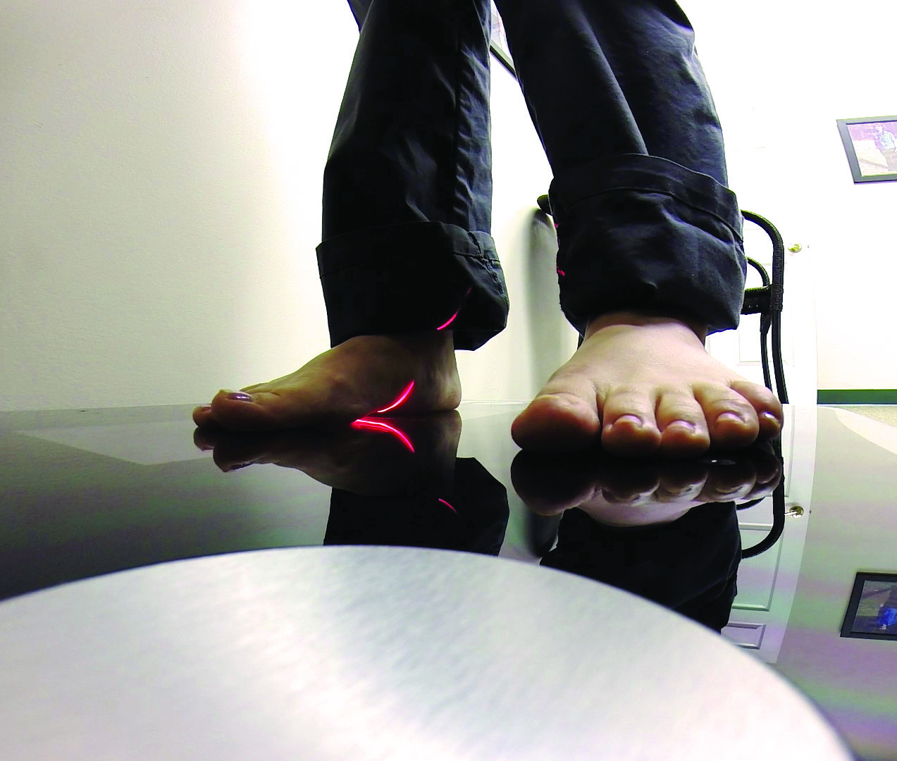
Take a look at the extensive wear patterns on your shoes and see if there are imbalances in the feet. If there are areas that are really worn/flattened out then there can be severe pronation taking place. This is one serious reason to look into foot orthotics,
Not to worry, as excessive foot pronation affects everyone.
While at school, work or conducting daily chores is when individuals pronate and while wearing shoes with no arch/spine support.
Pronation in the feet combined with shoes that don’t fit with no support has a negative impact on posture and alignment.
With no sufficient support, the body reverts to the imbalanced state. With continued use and not addressed will cause pain in the:
Knees
Hips
Back
Neck
And unfortunately, most of us ignore and even power through the pain caused by the poor fit and lack of support from these shoes.
Reasons to Use Custom Orthotics
The feet make up the body�s foundation. Just like a house, problems with the foundation will create problems throughout the structure over time. Therefore if the foundation is remedied properly then the other issues remedy themselves or are easier to treat because the feet are already taken care of.
Feet problems/issues create back problems. Fixing foot problems will make a difference with the back pain treatment plan.
Individuals think their feet are just fine. However, they don’t know what problems are in the background waiting for the worst moment to present. Imbalance/dysfunction is a silent issue that can ripple into many problems along with pain in the spine.
Feet are as unique as fingerprints. Custom orthotics will make a vast difference between the right and left foot and accommodate the body to achieve its full potential.
Custom orthotics are built just for you. Based on a high-resolution 3D scan of your feet, that is sent to a Foot Levelers� team of technicians, who create your 100% unique, tailored foot orthotic/s.
Don�t be fooled by over-the-counter knock offs. These are mass-produced insoles that can worsen pain. And while most cases of back pain respond well with Chiropractic care, adding custom foot orthotics will enhance the effectiveness of the treatment with no need for surgery or side effects from medications.
Your feet and your body will thank you and will improve health by maintaining proper posture, combined with full-body support to achieve optimal quality of life.
How to eliminate Back Pain naturally | (2020) Foot Levelers |El Paso, Tx
NCBI Resources
When there are problems with the feet, it can cause problems through the legs and all the way to the spine. This can cause the ankle to pronate, meaning it rolls inward. This alters the way the�bones of the foot�line up which extends through the tibia, or shin bone.
This can cause a condition called knock knees and it can change the way the entire body is aligned. This puts the body out of balance, destabilizing the spine, and can even cause the pelvis to tilt to one side or the other. When you are walking or standing, the stress caused by the misalignment can create a domino effect, causing or contributing to low back pain.
Our digestive health depends on the composition of our healthy gut microbiome or the bacteria in our gastrointestinal (GI) tract. This probiotic profile plays a fundamental role in our immune system and these can ultimately affect our inflammatory response. Also, the foods we eat, hormones, neurotransmitters, and even our adrenal and mitochondrial status can influence our digestive health. Abnormal or excess bacteria can cause many digestive health issues. Researchers and healthcare professionals have found that “fasting” can help promote a healthy gut microbiome and support overall digestive health. �
Several studies have shown that consuming enough fiber and foods that increase the amount of bacteria in the gastrointestinal (GI) tract is associated with improved insulin sensitivity as well as reduced immune reactions and inflammation, among many other health benefits. These same studies also demonstrated that fasting can have these same health benefits. Different types of fasting can be used as a treatment approach for a variety of digestive health issues. As a matter of fact, other studies have shown that fasting can help improve digestive health issues like SIBO, IBS, and leaky gut. �
An Experiment on Fasting and Digestive Health
Mike Hoaglin, former clinical director for the Dr. Oz show and current clinical lead for uBiome, a biotechnology company that helps healthcare professionals and patients understand how the gut microbiome affects overall health and wellness, demonstrated the importance of the bacteria in our gastrointestinal (GI) tract by sharing the outcome measures of an experiment he tried on himself. Biotechnology companies like uBiome can determine a patient’s probiotic profile, including “healthy” and pathogenic microorganisms which may be associated with digestive health issues like Crohn’s disease and ulcerative colitis. �
After learning how fasting can help improve your immune system, activate stem cells, and reduce your risk of developing many types of cancers, Mike became motivated to do his own five-day water fast to see how this strategic way of eating would affect his gut microbiome. He was also inspired to know how fasting could affect his energy levels as well as his mental acuity and brain fog. By submitting a stool sample, he determined the spectrum of bacteria in his gastrointestinal (GI) tract before starting the fasting process. Mike Hoaglin was under the supervision of his functional medicine practitioner. �
Understanding the Effects of Fasting
According to his uBiome probiotic profile test results, Mike had dysbiosis, an imbalance in the composition of his gut microbiome associated with decreased biodiversity of “healthy” bacteria and increased “harmful” bacteria known for causing inflammation. Mike Hoaglin scheduled five days in his schedule to start the fasting process after he talked to his functional medicine practitioner. As many people have described during the first several days of fasting, Mike had a very difficult time going without eating any food. He described feeling cranky and hungry, however, he was still able to sleep. �
Mike’s hunger had thankfully subsided by day three of the fasting process and, although he still had several days left of the treatment approach, the understood that the rest of the fasting process wasn’t going to be as challenging as it had been for the first two days, despite his blood glucose, or sugar, being low. Mike Hoaglin felt an increase in his energy levels by day four of the fasting process. He felt more mental clarity as his digestive system started using fat as energy instead of using sugar, or glucose. He immediately recognized that his stem cells had activated during day four of the fasting process. �
Mike ended the fasting process on day five at 5:00 pm by consuming a cup of bone broth. Bone broth is one of the most recommended type of foods to help people transition from fasting because it has essential amino acids, such as glutamine and glycine, that provide nutrition to the gastrointestinal (GI) tract as soon as it starts digesting food once again. Moreover, adding some Himalayan salt to your bone broth can also provide your cells with added minerals. Mike continued to transition from fasting by eating fiber-rich plant foods, healthy fats, and small amounts of lean protein, in easily digestible variations. �
Mike Hoaglin tested his gut microbiome following his fasting process and he was pleasantly surprised with the outcome measures of his probiotic profile. According to the uBiome test, fasting had practically “reset” Mike’s gut microbiome, or the bacteria in the gastrointestinal (GI) tract. The results demonstrated a balanced composition of his gut microbiome and he had increased the biodiversity of “healthy” bacteria and decreased “harmful” bacteria. After completing his experiment, Mike Hoaglin became more aware of how the type of foods we eat can ultimately affect our digestive health. �
Fasting is a well-known, strategical way of eating which can have a variety of digestive health benefits for many people. Many people can tremendously benefit from fasting. Fasting can activate autophagy, or the natural cellular detoxification process, to help sweep excess bacteria and undigested food debris away for elimination as waste, also activating anti-inflammatory processes to reduce inflammation and oxidative stress. During an experiment, fasting was shown to have tremendous benefits on overall digestive health. However, it’s important to keep in mind that fasting may not be for everyone. Make sure to talk to a qualified and experienced doctor before attempting any fasting approaches. – Dr. Alex Jimenez D.C., C.C.S.T. Insight
The following Neurotransmitter Assessment Form can be filled out and presented to Dr. Alex Jimenez. The following symptoms listed on this form are not intended to be utilized as a diagnosis of any type of disease, condition, or any other type of health issue. �
Our digestive health depends on the composition of our healthy gut microbiome or the bacteria in our gastrointestinal (GI) tract. This probiotic profile plays a fundamental role in our immune system and these can ultimately affect our inflammatory response. Also, the foods we eat, hormones, neurotransmitters, and even our adrenal and mitochondrial status can influence our digestive health. Abnormal or excess bacteria can cause many digestive health issues. Researchers and healthcare professionals have found that “fasting” can help promote a healthy gut microbiome and support overall digestive health. � Several studies have shown that consuming enough fiber and foods that increase the amount of bacteria in the gastrointestinal (GI) tract is associated with improved insulin sensitivity as well as reduced immune reactions and inflammation, among many other health benefits. These same studies also demonstrated that fasting can have these same health benefits. Different types of fasting can be used as a treatment approach for a variety of digestive health issues. As a matter of fact, other studies have shown that fasting can help improve digestive health issues like SIBO, IBS, and leaky gut. �
The scope of our information is limited to chiropractic, musculoskeletal, and nervous health issues or functional medicine articles, topics, and discussions. We use functional health protocols to treat injuries or disorders of the musculoskeletal system. Our office has made a reasonable attempt to provide supportive citations and has identified the relevant research study or studies supporting our posts. We also make copies of supporting research studies available to the board and or the public upon request. To further discuss the subject matter above, please feel free to ask Dr. Alex Jimenez or contact us at 915-850-0900.�
Curated by Dr. Alex Jimenez �
References:
�The Impact of Fasting on Your Microbiome.� Naomi Whittel, 12 Mar. 2019, www.naomiwhittel.com/the-impact-of-fasting-on-your-microbiome/.
Additional Topic Discussion: Chronic Pain
Sudden pain is a natural response of the nervous system which helps to demonstrate possible injury. By way of instance, pain signals travel from an injured region through the nerves and spinal cord to the brain. Pain is generally less severe as the injury heals, however, chronic pain is different than the average type of pain. With chronic pain, the human body will continue sending pain signals to the brain, regardless if the injury has healed. Chronic pain can last for several weeks to even several years. Chronic pain can tremendously affect a patient’s mobility and it can reduce flexibility, strength, and endurance. �
Neural Zoomer Plus for Neurological Disease
Dr. Alex Jimenez utilizes a series of tests to help evaluate neurological diseases. The Neural ZoomerTM Plus is an array of neurological autoantibodies which offers specific antibody-to-antigen recognition. The Vibrant Neural ZoomerTM Plus is designed to assess an individual�s reactivity to 48 neurological antigens with connections to a variety of neurologically related diseases. The Vibrant Neural ZoomerTM Plus aims to reduce neurological conditions by empowering patients and physicians with a vital resource for early risk detection and an enhanced focus on personalized primary prevention. �
Food Sensitivity for the IgG & IgA Immune Response
Dr. Alex Jimenez utilizes a series of tests to help evaluate health issues associated with food sensitivities. The Food Sensitivity ZoomerTM is an array of 180 commonly consumed food antigens that offers very specific antibody-to-antigen recognition. This panel measures an individual�s IgG and IgA sensitivity to food antigens. Being able to test IgA antibodies provides additional information to foods that may be causing mucosal damage. Additionally, this test is ideal for patients who might be suffering from delayed reactions to certain foods. Utilizing an antibody-based food sensitivity test can help prioritize the necessary foods to eliminate and create a customized diet plan around the patient�s specific needs. �
Gut Zoomer for Small Intestinal Bacterial Overgrowth (SIBO)
�
Dr. Alex Jimenez utilizes a series of tests to help evaluate gut health associated with small intestinal bacterial overgrowth (SIBO). The Vibrant Gut ZoomerTM offers a report that includes dietary recommendations and other natural supplementation like prebiotics, probiotics, and polyphenols. The gut microbiome is mainly found in the large intestine and it has more than 1000 species of bacteria that play a fundamental role in the human body, from shaping the immune system and affecting the metabolism of nutrients to strengthening the intestinal mucosal barrier (gut-barrier). It is essential to understand how the number of bacteria that symbiotically live in the human gastrointestinal (GI) tract influences gut health because imbalances in the gut microbiome may ultimately lead to gastrointestinal (GI) tract symptoms, skin conditions, autoimmune disorders, immune system imbalances, and multiple inflammatory disorders. �
Formulas for Methylation Support
XYMOGEN�s Exclusive Professional Formulas are available through select licensed health care professionals. The internet sale and discounting of XYMOGEN formulas are strictly prohibited.
Proudly,�Dr. Alexander Jimenez makes XYMOGEN formulas available only to patients under our care.
Please call our office in order for us to assign a doctor consultation for immediate access.
If you are a patient of Injury Medical & Chiropractic�Clinic, you may inquire about XYMOGEN by calling 915-850-0900.
�
For your convenience and review of the XYMOGEN products please review the following link. *XYMOGEN-Catalog-Download �
* All of the above XYMOGEN policies remain strictly in force. �
A pain specialist is a medical or osteopathic doctor that treats pain caused by disease, disorder, and trauma. Also known as interventional pain management specialists, these doctors are often anesthesiologists or physiatrists.
Pain medicine involves multi-disciplines and team effort with a patient’s primary physician, other doctors, and specialists in radiology, psychiatry, oncology, nursing, chiropractic/physical therapy, alternative medicine, etc.
Education and Training
Upon completion of medical school and a one-year internship, the specialist enters a residency program in anesthesiology, physical medicine or other fields like neurology. Upon residency completion, the doctor completes a one-year fellowship for advanced training in pain medicine.
Pain medicine specialists continue medical training and education throughout their careers.
Pain Management Goals
Pain medicine is aimed at managing acute or chronic pain through pain frequency and intensity reduction and ultimately alleviation.
Multidisciplinary treatment programs are utilized to address functional goals and normal day to day chores and activities.
A pain treatment plan’s goal is to regain well-being, increasing your level of activities like returning to work, school, etc and eliminate medication dependency.
Pain Types & Treatment
Specialists treat all types of pain.
Acute pain is severe/sharp and can signal something is wrong.
Touching a hot stove or slamming a finger in a door are examples of acute pain.
Chronic pain lasts 6 months or longer.
This type varies from mild to severe and is constant.
Spinal arthritis (spondylosis) is an example of chronic pain.
It can be difficult to manage, but combining a variety of treatments can show positive results. The pain types include:
Degenerative disease
Facet joint pain
Sciatica
Cervical and lumbar spinal stenosis
Spondylolisthesis
Vertebral compression fracture
Whiplash
An appointment consists of
These are like most medical visits. The focus is on the pain, the cause, what factors are contributing, and creating an optimal treatment management plan.
A physical examination
Neurological examination
Review of medical history particularly pain history.
Questions can include:
From zero to 10, 10 being the worst, where’s your pain at?
When did it start?
What were you doing when it started?
Does it spread or radiate out other areas of the body?
Is the intensity constant?
Is it worse at different times of the day or night?
What helps relieve the pain?
What worsens the pain?
What types of treatments have you tried?
Did it work and to what degree?
Do you take and what types of, over-the-counter medications, vitamins, and herbal supplements?
Do you take prescription medications?
A drawing of the body could be used to mark where the pain is felt, as well as indicate pain spread and type.
Diagnosis
Diagnosing the cause of pain is paramount to creating the proper management treatment plan. If the wrong diagnosis is made and the wrong treatment is initiated it might exacerbate the condition and/or create new injuries.
This can mean getting:
X-ray’s
CT scan
MRI
These imaging types help confirm the origin of the pain. When treating spine pain, arm, leg and foot symptoms may also be present. Therefore the correct diagnosis is essential to a successful treatment plan.
Spinal disorder pain treatment usually involves other specialists, like a primary care physician, neurosurgeon, orthopedic surgeon, radiologists, oncologists, nurses, chiropractors, physical therapists, and alternative medicine practitioners.
We focus on what works for you. We also strive to create fitness and better the body through researched methods and total wellness programs. These programs are natural and use the body�s own ability to achieve goals of improvement, rather than introducing harmful chemicals, controversial hormone replacement, surgery, or addictive drugs.
We want you to live a life that is fulfilled with more energy, a positive attitude, better sleep, less pain, proper body weight and educated on how to maintain this way of life. I have made a life of taking care of every one of my patients.
I assure you, I will only accept the best for you�
Back Pain Chiropractic Care El Paso, Texas
NCBI Resources
If you have low back pain�or have had it, you are not alone. Experts estimate that around�80% of people�will experience some type of back problem at some point in their lives.�The Global Burden of Disease 2010�lists low back pain as the number one cause of disability worldwide. The good news is the majority of back pain is mechanical in origin or is not organic.
If you are feeling any of these situations, then you might be experiencing low levels of glutathione, why not add some S-acetyl glutathione into your body.
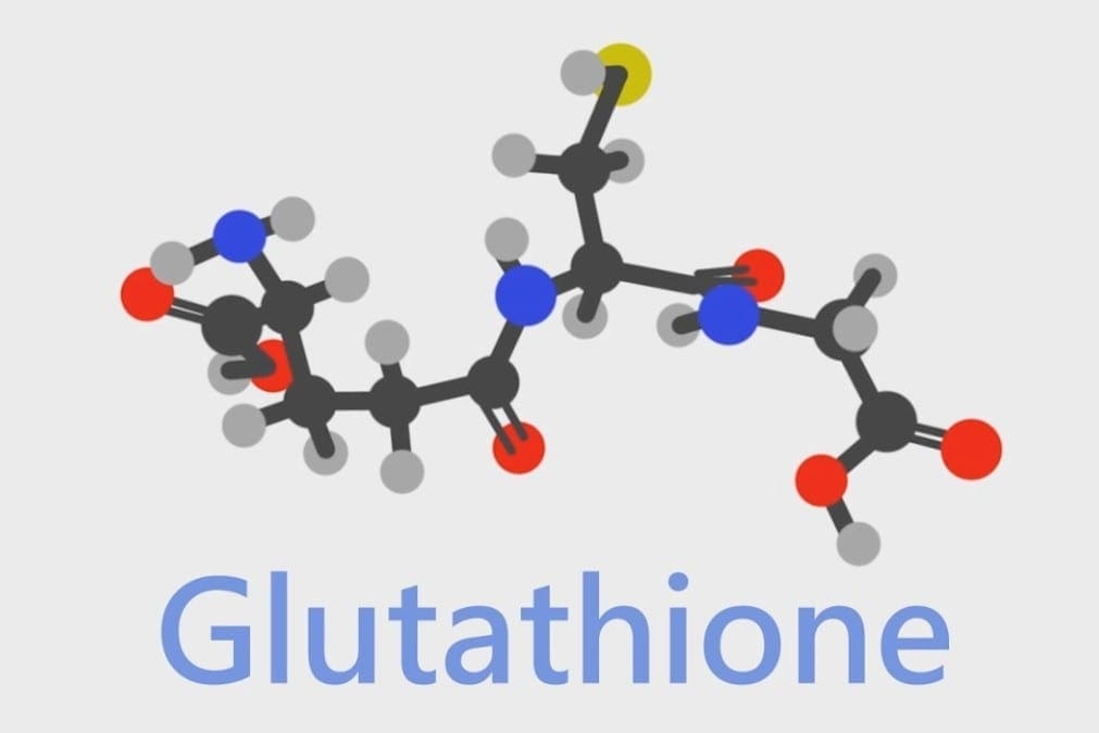
Glutathione
When glutathione is decreased in the body, it is known as glutathione deficiency or GSH. It is a tripeptide that is consist of L-glutamine, L-cysteine, and glycine while also being functional in the systems of the body. The biosynthesis of glutathione can be affected by some factors such as biochemical individuality or dietary factors. Another factor that can affect glutathione in the body is chronic oxidative stress. Chronic oxidative stress can deplete cellular glutathione in the body, causing it to develop inflammation and dysfunction to its organ systems.
There are ways to boost glutathione levels in the body since these nutrients and supplements are the precursors to glutathione. They consist of whey protein, vitamin C, and glutamine, and they can help raise the glutathione levels in the body to prevent inflammation and disruptive factors that can cause harm to the body; however, the results are inconsistent and need further research. Studies stated that biological individuality is different to every type of body since it is equivalent and can metabolize the precursor nutrients and supplements to the body.
Why Not Distribute Pure Glutathione?
Sadly though, when a person takes glutathione in an oral form, the results are unpleasant. When the person takes glutathione in the mouth, the oral dosage is oxidized instantly, before being absorbed into the body, thus leaving a foul smell. Taking glutathione in a vegetable capsule can help the individual receive glutathione without it being oxidized.
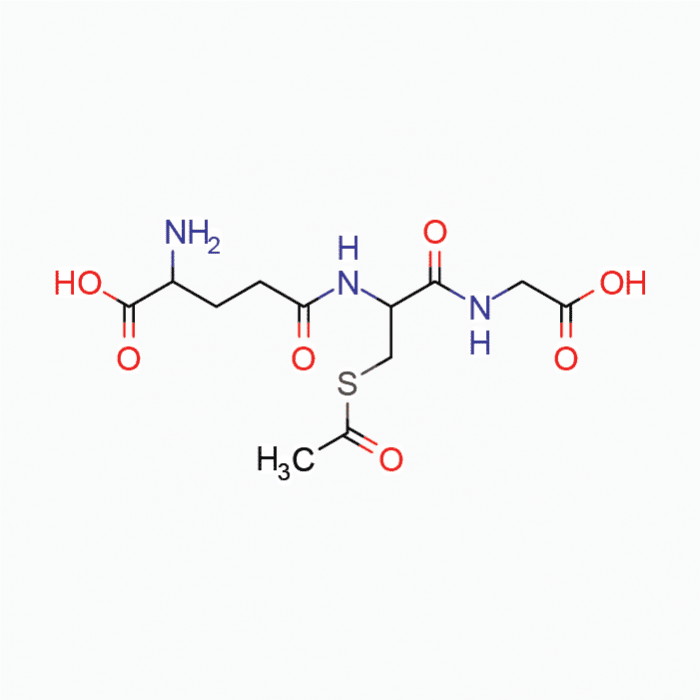
Many formulas can deliver a unique preparation to glutathione that can overcome the limitations it faces. Studies show that S-acetyl glutathione is stable enough to go through the intestinal walls and deposit the necessary nutrients into the body. With S-acetyl glutathione being consumed orally, it can increase the total glutathione and the percent-reduced glutathione so it can be beneficial for the body. With percent-reduced glutathione, it has a very significant biomarker for excellent status for a functional body.
The Mechanics of SAG Absorption
S-acetyl glutathione or SAG is a lipid-like compound that is taken by intact chylomicrons in the gut. The bond from this compound is placed into its thiol group that helps prevents oxidations in the body and allows molecules to pass into the cell walls after being absorbed into the gut. What happens is that the bond is cleaved by non-specific enzymes and helps prevent the breakdown of glutathione, while S-acetyl glutathione does not need expenditures to be cleaved once it crosses the cell walls in the body.
SAG Antioxidant Activity
Since glutathione helps tissue and organ functions throughout the body, it plays a critical role by protecting it from a variety of factors like, for example, oxidative stress, while also maintain cellular functions and supporting a healthy immune system. Studies show that many factors can increase oxidative stress exposure and adding insults to the body, therefore increasing the cellular consumption of the nutrients like glutathione, which provides antioxidant activity. When this happens, it leads to the result of a fiery cycle of oxidative stress and challenges detoxification to the body. Research states that complete biotransformation and protecting the body from oxidative stress is essential for the body to maintain cellular integrity and tissue health.
Benefits of Maintaining Healthy Glutathione Levels
There is plenty of information that is related to cellular health that has been surfaced. Research states that the mitochondria, which is the energy-producing powerhouse cell, has a role in being the primary functional cellular site for consuming oxygen and ROS (reactive oxygen species).� Studies show that S-acetyl glutathione can cross the membranes of the mitochondria by increasing the organ’s activity and minimizing ROS in the body. When ROS is reduced in the body, it can maintain the mitochondrial integrity and its function, while improving its health for the body to function correctly.
Studies show that S-acetyl glutathione can decrease TNF-alpha, NF-kappa beta, and F-2 isoprostane enzymes in the body. Additional studies show that there is a large amount of evidence that intracellular glutathione levels in macrophages can influence the Th1/Th2 cytokine pattern and can help promote a well-balanced immune reaction to the body.
Conclusion
Glutathione is an essential amino acid that is produced in the body. When there are low levels of glutathione in the body, S-acetyl glutathione can assist in maintaining those levels and by making sure that oxidative stress does not reach full capacity in the body to cause significant damage. Some products can provide more excellent stability, bioavailability, and digestive comfort for anyone who might be sensitive to N-acetyl L-cysteine.
The scope of our information is limited to chiropractic, musculoskeletal, and nervous health issues or functional medicine articles, topics, and discussions. We use functional health protocols to treat injuries or disorders of the musculoskeletal system. Our office has made a reasonable attempt to provide supportive citations and has identified the relevant research study or studies supporting our posts. We also make copies of supporting research studies available to the board and or the public upon request. To further discuss the subject matter above, please feel free to ask Dr. Alex Jimenez or contact us at 915-850-0900.
References:
Anderson, Michelle F, et al. �Glutathione Monoethylester Prevents Mitochondrial Glutathione Depletion during Focal Cerebral Ischemia.� Neurochemistry International, Pergamon, 20 June 2003, www.sciencedirect.com/science/article/abs/pii/S0197018603001335?via%3Dihub.
Ballatori, Nazzareno, et al. �Glutathione Dysregulation and the Etiology and Progression of Human Diseases.� Biological Chemistry, U.S. National Library of Medicine, Mar. 2009, www.ncbi.nlm.nih.gov/pmc/articles/PMC2756154/.
Fraternale, A., et al. �Antiviral and Immunomodulatory Properties of New Pro-Glutathione (GSH) Molecules.� Http://Www.eurekaselect.com, 31 May, 2006, www.eurekaselect.com/56205/article.
Kretzschmar, M. �Regulation of Hepatic Glutathione Metabolism and Its Role in Hepatotoxicity.� Experimental and Toxicologic Pathology, Urban & Fischer, 3 Nov. 2011, www.sciencedirect.com/science/article/abs/pii/S0940299396800546?via%3Dihub.
Locigno, Roberto, et al. �S-Acetyl-Glutathione Selectively Induces Apoptosis in Human Lymphoma Cells through a GSH-Independent Mechanism.� International Journal of Oncology, U.S. National Library of Medicine, Jan. 2002, www.ncbi.nlm.nih.gov/pubmed/11743644.
Lomaestro, Ben M, and Margaret Malone. �Glutathione in Health and Disease: Pharmacotherapeutic Issues – Ben M Lomaestro, Margaret Malone, 1995.� SAGE Journals, 1 Dec. 1995, journals.sagepub.com/doi/10.1177/106002809502901213.
Richman, PG, and A Meister. “Regulation of Gamma-Glutamyl-Cysteine Synthetase by Nonallosteric Feedback Inhibition by Glutathione.� The Journal of Biological Chemistry, U.S. National Library of Medicine, 25 Feb. 1975, www.ncbi.nlm.nih.gov/pubmed/1112810.
Vogel, Jens-Uwe, et al. �Effects of S-Acetylglutathione in Cell and Animal Model of Herpes Simplex Virus Type 1 Infection.� SpringerLink, Springer-Verlag, 18 Nov. 2003, link.springer.com/article/10.1007%2Fs00430-003-0212-z.
Scientists and healthcare professionals are starting to shine a light on the importance of the composition of our gut microbiome, or the population of “healthy” bacteria in our gastrointestinal (GI) tract. According to research studies, abnormal or excess amounts of gut bacteria can be one of the most common causes of a variety of digestive health issues, including SIBO and IBS. Our ancestors have included fermented foods like yogurt, kimchi, and sauerkraut as an important part of their traditional diet to regulate and manage the composition of their “healthy” bacteria: the gut microbiome. �
Finding ways to naturally improve our digestive health by maintaining a “healthy” probiotic profile has been a popular topic for many generations. As a result, eating fermented foods like those previously listed above, including other food groups with additional probiotics, and taking probiotic supplements has tremendously increased in popularity in recent years. Another way to naturally improve digestive health that has recently become more popular is fasting, strategic abstinence or reduction from several or all foods for a certain period of time. Fasting can ultimately help improve overall digestive health. �
Fasting can help support the healthy composition of our gut microbiome and it can be used as a treatment approach for a variety of conditions and diseases, such as headaches, migraines, eczema, metabolic syndrome, and obesity. Scientists and healthcare professionals have determined that fasting can stress the human body in a beneficial way. This stress benefits the healthy bacteria in the gastrointestinal (GI) tract because it helps activate autophagy or the natural cellular detoxification process. In the following article, we will discuss how fasting and autophagy can promote digestive health. �
Fasting and Autophagy Overview
Our gastrointestinal (GI) tract can often have a difficult job trying to repair our cells while sweeping undigested debris away to eliminate as waste because many people are constantly eating throughout the entire day. Many people are completely against the idea of fasting, or willingly skipping one or two meals per day, despite its benefits towards our digestive health. Because there are a variety of different methods and techniques for fasting, many people can follow this strategic way of eating and still take advantage of all its digestive health benefits. Fasting, however, may ultimately not be for everyone. �
Historically, many religious and spiritual practices used fasting as an important element in their culture to promote overall digestive health. There are currently a wide variety of fasting methods and techniques that are used to support natural well-being. Moreover, the treatment benefits of fasting are now being readily recognized in numerous research studies. The different types of fasting can ultimately vary from eating very little or nothing for a certain amount of time to drinking only water for a specific period of time, occasionally for up to five days, as a way to naturally improve digestive health. �
Intermittent fasting, a strategic way of eating that follows switching between unrestricted eating and restricted eating for a certain period of time, is one of the most common and practical fasting approaches for everyone. Scientists consider intermittent fasting to be safe and effective because you only go without eating any food for short periods of time. Research studies have demonstrated that using intermittent fasting for a total of 16 hours every day is enough to create the caloric restriction necessary to experience the benefits of fasting as well as to activate autophagy to help restore digestive health. �
The 5:2 diet is the strategic way of eating where a person consumes an average diet for five days and then greatly reduces their consumption of food to one-quarter of that of their normal diet for the other two days of the week. Every fasting approach is different but the purpose of abstinence or reduction from foods is to give our gut microbiome a break from digestion so they can focus on repairing our cells while sweeping undigested debris and excess bacteria away to eliminate as waste. Research studies suggest that the 16:8 diet may be the simplest fasting method or technique for people to follow. �
How Fasting and Autophagy Support Digestive Health
Our pancreas commonly triggers the release of glucagon when we have low blood glucose while the release of insulin is triggered to help reduce high blood glucose levels. Insulin decreases and glucagon increases during fasting which has been demonstrated to help promote improved metabolism as well as provide energy, mood changes, and weight loss. Fasting also helps promote the “healthy” composition of our gut microbiome or the population of “healthy” bacteria in our gastrointestinal (GI) tract. Scientists have associated fasting with the activation of the gene that supports overall digestive health. �
Optimal digestive health and “healthy” gut bacteria are important to help protect us from abnormal or excess bacteria, toxins, and other compounds that can trigger the immune system. Finally, fasting can help restore the integrity of the intestinal lining by managing inflammation that can ultimately help protect the human body against the variety of conditions and diseases associated with inflammation. The main benefit of fasting is that it can increase autophagy or the natural cellular detoxification process. With fasting, your gut health improves and you reduce your risk for a variety of digestive health issues. �
Fasting is a well-known, strategical way of eating which can have a variety of digestive health benefits for many people. Many people can tremendously benefit from fasting. Fasting can activate autophagy, or the natural cellular detoxification process, to help sweep excess bacteria and undigested food debris away for elimination as waste, also activating anti-inflammatory processes to reduce inflammation and oxidative stress. However, it’s important to keep in mind that fasting may not be for everyone. Make sure to talk to a qualified and experienced doctor before attempting any fasting approaches. – Dr. Alex Jimenez D.C., C.C.S.T. Insight
The following Neurotransmitter Assessment Form can be filled out and presented to Dr. Alex Jimenez. The following symptoms listed on this form are not intended to be utilized as a diagnosis of any type of disease, condition, or any other type of health issue. �
The scope of our information is limited to chiropractic, musculoskeletal, and nervous health issues or functional medicine articles, topics, and discussions. We use functional health protocols to treat injuries or disorders of the musculoskeletal system. Our office has made a reasonable attempt to provide supportive citations and has identified the relevant research study or studies supporting our posts. We also make copies of supporting research studies available to the board and or the public upon request. To further discuss the subject matter above, please feel free to ask Dr. Alex Jimenez or contact us at 915-850-0900.�
Curated by Dr. Alex Jimenez �
References:
�The Impact of Fasting on Your Microbiome.� Naomi Whittel, 12 Mar. 2019, www.naomiwhittel.com/the-impact-of-fasting-on-your-microbiome/.
Additional Topic Discussion: Chronic Pain
Sudden pain is a natural response of the nervous system which helps to demonstrate possible injury. By way of instance, pain signals travel from an injured region through the nerves and spinal cord to the brain. Pain is generally less severe as the injury heals, however, chronic pain is different than the average type of pain. With chronic pain, the human body will continue sending pain signals to the brain, regardless if the injury has healed. Chronic pain can last for several weeks to even several years. Chronic pain can tremendously affect a patient’s mobility and it can reduce flexibility, strength, and endurance. �
Neural Zoomer Plus for Neurological Disease
�
Dr. Alex Jimenez utilizes a series of tests to help evaluate neurological diseases. The Neural ZoomerTM Plus is an array of neurological autoantibodies which offers specific antibody-to-antigen recognition. The Vibrant Neural ZoomerTM Plus is designed to assess an individual�s reactivity to 48 neurological antigens with connections to a variety of neurologically related diseases. The Vibrant Neural ZoomerTM Plus aims to reduce neurological conditions by empowering patients and physicians with a vital resource for early risk detection and an enhanced focus on personalized primary prevention. �
Food Sensitivity for the IgG & IgA Immune Response
�
Dr. Alex Jimenez utilizes a series of tests to help evaluate health issues associated with food sensitivities. The Food Sensitivity ZoomerTM is an array of 180 commonly consumed food antigens that offers very specific antibody-to-antigen recognition. This panel measures an individual�s IgG and IgA sensitivity to food antigens. Being able to test IgA antibodies provides additional information to foods that may be causing mucosal damage. Additionally, this test is ideal for patients who might be suffering from delayed reactions to certain foods. Utilizing an antibody-based food sensitivity test can help prioritize the necessary foods to eliminate and create a customized diet plan around the patient�s specific needs. �
Gut Zoomer for Small Intestinal Bacterial Overgrowth (SIBO)
Dr. Alex Jimenez utilizes a series of tests to help evaluate gut health associated with small intestinal bacterial overgrowth (SIBO). The Vibrant Gut ZoomerTM offers a report that includes dietary recommendations and other natural supplementation like prebiotics, probiotics, and polyphenols. The gut microbiome is mainly found in the large intestine and it has more than 1000 species of bacteria that play a fundamental role in the human body, from shaping the immune system and affecting the metabolism of nutrients to strengthening the intestinal mucosal barrier (gut-barrier). It is essential to understand how the number of bacteria that symbiotically live in the human gastrointestinal (GI) tract influences gut health because imbalances in the gut microbiome may ultimately lead to gastrointestinal (GI) tract symptoms, skin conditions, autoimmune disorders, immune system imbalances, and multiple inflammatory disorders. �
Formulas for Methylation Support
�
XYMOGEN�s Exclusive Professional Formulas are available through select licensed health care professionals. The internet sale and discounting of XYMOGEN formulas are strictly prohibited.
Proudly,�Dr. Alexander Jimenez makes XYMOGEN formulas available only to patients under our care.
Please call our office in order for us to assign a doctor consultation for immediate access.
If you are a patient of Injury Medical & Chiropractic�Clinic, you may inquire about XYMOGEN by calling 915-850-0900.
�
For your convenience and review of the XYMOGEN products please review the following link. *XYMOGEN-Catalog-Download �
* All of the above XYMOGEN policies remain strictly in force. �
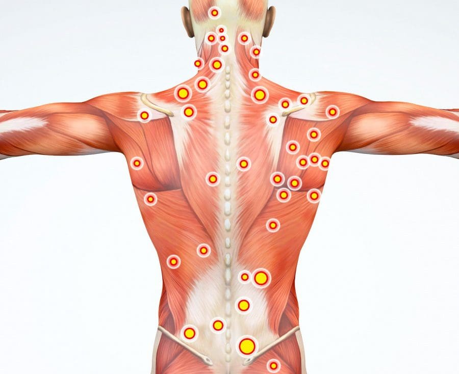
A trigger point is a knot or bundle of stiff spine muscle tissue that you can’t move or relax, and when touched pain spreads to the neck, shoulders, and upper back.
Pretty much everyone can relate to this type of pain in the neck or what is known as myofascial pain syndrome when several of these trigger points are grouped.
How to ease the pain and prevent it
Trigger points can form in muscles all over the body. Myofascial pain syndrome in the neck happens when trigger points develop in the muscles of the shoulders, upper back, and neck.
A Trigger Point
Trigger points have a unique connection to America. They were first identified in the 1940s by Janet Travell, MD, who was John F. Kennedy’s doctor. JFK had severe chronic back pain and had trigger point injections to ease the pain.
A trigger point is a sensitive area within the muscles. They are typically described as knots and feel like a bundle of tense, contracted muscles that twitch and spread pain when touched. The spreading pain is known as referred pain. Example: Trigger points in the shoulder send pain into the neck.
Trigger points cause muscles to stress and to contract. This results in:
Muscle weakness
Numbness
Limited muscle movement
Formation of Points in The Neck
They are usually caused by mechanical factors (factors that strain or stress the muscles).� Spinal trauma, like whiplash from an automobile accident or sports-related injury, can create trigger points.
They also develop through repetitive actions and routine everyday chores that can hurt the spine over time.
Straining the neck muscles from poor posture for extended times like craning the neck while working on a computer, carrying a heavy bag that stresses the muscles of the neck, upper back, and shoulders.
Trigger Points vs Fibromyalgia Tender Points
Trigger points do get confused with tender points of fibromyalgia. Trigger points and tender points are both defined as local areas of pain but are not the same.
Tender points do not cause referred pain the way trigger points do.
Tender points are symmetrical meaning that they are on both sides of the body. Whereas trigger points do not follow a symmetrical pattern.
But it can become complicated because individuals with fibromyalgia can have both tender points and trigger points. People with fibromyalgia can also have myofascial pain syndrome.
Trigger Point Diagnosis
Trigger points are a regular cause of different types of spine pain, that can range from neck pain to low back pain. However, doctors are still trying to understand how trigger points produce referred pain. This is why diagnosing trigger points can be difficult for doctors.
They are complex because they are easy to pinpoint but difficult to diagnose. As they can directly cause muscle pain but they can mimic other pain making conditions exactly the way Myofascial pain and fibromyalgia get confused.
Jaw pain
Earaches
Toothaches
These types of pain that do not go away could be caused by trigger point/s in the neck.
Individuals with chronic neck pain that don’t seem to have a cause, could be trigger points. A doctor will refer you to a physical therapist, chiropractor or another spine specialist to conduct an examination for trigger points.
Treatment
Treatment can range from home remedies, chiropractic care, physical therapy and if severe muscle injections. There is no one treatment that works, as everyone and their injuries are different, meaning that various treatment options need to be looked into.
Home
Before starting any home therapy, discuss it with a trained professional like a doctor, chiropractor, massage therapist, or physical therapist to identify the location of the trigger point to effectively treat it.
Treated with massaging the area but can be tough with hard-to-reach places in the upper back. If unable to reach the point slowly and gently roll over a foam roller, golf or tennis ball for quick relief.
Massage
Massage therapists are trained in relieving muscle pain. Deep tissue massage can relieve an irritated area. Regular massage sessions can reduce pain and prevent the points from reemerging.
Physical therapy
Physical therapy treats trigger points in different ways, this includes:
Massage
Heat
Electrical stimulation
Ultrasound
A cooling spray is applied followed with stretches to relax and relieve the contracted muscle/s
Medications
Muscle relaxants can be used to reduce the symptoms and relieve pain. However, these meds can have all kinds of side effects, and become habit-forming, so use should be limited and in conjunction with a proper chiropractic/physical therapy treatment plan.
Injections
If the pain continues despite the non-surgical treatments or worsens, then your doctor could recommend trigger point injections. Injections are late-stage therapy. Doctors want to avoid patients becoming dependant on injections and will prescribe injections with an exercise, chiropractic/physical therapy program for maximum relief and effectiveness.
Overall Health
The majority of people have felt tight muscles around the neck. Utilizing proper posture and healthy spinal mechanics can prevent trigger points and myofascial pain syndrome.
Our services are specialized and focused on injuries and the complete recovery process.�Our areas of practice include�Wellness & Nutrition, Chronic Pain,�Personal Injury,�Auto Accident Care, Work Injuries, Back Injury, Low�Back Pain, Neck Pain, Migraine Headaches, Sports Injuries,�Severe Sciatica, Scoliosis, Complex Herniated Discs,�Fibromyalgia, Chronic Pain, Complex Injuries, Stress Management, and Functional Medicine Treatments.
�Neck Pain and Chiropractic Treatment El Paso, Texas
NCIB Resources
Many people who have trigger points or myofascial pain syndrome in their spine have knots and tightness throughout their back and neck. To prevent myofascial pain syndrome one needs to practice a healthy lifestyle that promotes good spine health. Stretching and exercising regularly can help keep stress under control and prevent tension from building up, which makes it harder for trigger points to activate and cause pain.
If you are experiencing any of these situations, then you might have suffered from glutathione deficiency, why not trying some NAC supplements.
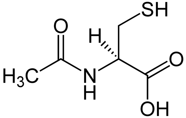
NAC and Its Benefits
NAC or N-Acetyl Cysteine is an amazing semi-essential amino acid. This amino acid can be produced from the body through other amino acids like methionine and serine. It can only become an essential amino acid when dietary intake of methionine and serine are low in the body. When a person is trying to incorporate NAC in their diet, it can be found in high protein foods like meats, dairies, and legumes. Studies show that consuming NAC is essential for a variety of health reasons, especially replenishing the most potent antioxidant, glutathione in the body.
Since NAC is a nutritional supplement that is exceedingly powerful, it can help glutathione be elevated in biosynthesis. NAC is recognized for supporting average mucous production, respiratory function, and eye health positively. Research shows that NAC can protect cell and tissue health from chronic illnesses and providing support for a healthy mental status in the body. There is even more research on NAC supplements, especially when someone increases their intake on the supplement. When there is an increase in NAC, and when it is consumed in the body, the effects are astounding. The NAC supplements can help the body boost the levels of some of the neurotransmitters and improving mental health.
In a 2011 study, researchers found that NAC is emerging to be a useful agent to help treat psychiatric disorders. The results of using NAC supplements to treat psychiatric disorders has helped alleviate some of these symptoms:
Addiction
Compulsive and grooming disorders
Schizophrenia
Bipolar disorders
Alzheimer�s disease
Since NAC can exert beneficial effects on the body, this supplement is useful to provide antioxidant, neuropathy, and anti-inflammatory properties to make sure that the body is functioning. Studies show that NAC can improve the outcomes of reducing lipopolysaccharides inflammation and preventing oxidative stress from being overexposed.
With NAC being a sulfur-containing derivative for the amino acid L-cysteine, this supplement provides supportive antioxidants and detoxification mechanisms for the body. Studies show that NAC can support the body�s antioxidant activity by neutralizing highly reactive hydroxyl radicals and serving as a source to sulfhydryl groups. They are thus enhancing the production or tripeptide glutathione in the body since it is a crucial component for antioxidant and detoxification enzymes.
Glutathione
Glutathione is a powerful antioxidant that has recently gained attention for its fantastic health benefits. This powerful antioxidant is found in every cell in the human body and can be absorbed in oral form. Research shows that even though the absorption of oral glutathione may be limited, the NAC supplementation can significantly increase the circulating levels of glutathione in the body. Studies stated that individuals who are infected with HIV, have glutathione deficiency in their system and have been associated with an impaired T-cell function and survival. So taking NAC orally can be used to replenish glutathione deficiency and is useful in the HIV infection.
Another study showed that taking NAC orally can help improve the responses of patients with chronic lung disease (CLD), chronic obstructive pulmonary disease (COPD), and cystic fibrosis (CF). The beneficial effects of taking NAC orally shows a decrease of inflammation in the lungs, improving the lung function, and reducing the neutrophil burden in cystic fibrosis airways.
Once this is done, though, NAC can help promote the production of glutathione and incorporate it into the crucial antioxidant enzymes and detoxification enzymes. With these enzyme activities being in play in the body, the glutathione is helping out by directly supporting their activities and the metabolism breakdown. Glutathione can also participate in fatty acid synthesis and can transport across the cell membrane.
Glutathione Factors
There are a variety of factors that can determine the requirements that glutathione can provide for the body. Glutathione can help control the toxin level exposure, increase the detoxification, and provide the overall needed support for antioxidants. Studies show that maintaining glutathione levels are essential to maintain the necessary health of the respiratory, hepatic, and the immune system from inflammation.
Research shows that since glutathione has multiple metabolic actions, they are essential for cellular homeostasis. Since it plays an important role, diseases like HIV, oxidative stress, chronic lung disease, and COPD can lower the body’s glutathione. The best way to make sure that individuals who have any chronic diseases, take NAC orally to prevent glutathione deficiency.
Glutathione can even help support antioxidant protection for lipids and proteins for the body as well as helping to maintain the standard response of inflammation due to injury. Studies show that elderly adults have altered their cellular redox levels and their dysregulated immune responses. Researchers also found out that the progression of chronic degenerative diseases of aging and that glutathione decreases with age naturally.
Conclusion
NAC is a semi-essential amino acid that has outstanding properties for the body. It helps replenishes the body�s glutathione and alleviate the symptoms caused by chronic illnesses. Taking NAC supplements is highly essential since it helps maintain adequate levels of glutathione to support overall health and well-being in the body. Some products help support glutathione levels as well as working well with NAC supplements by providing more excellent stability, bioavailability, and digestive comfort.
The scope of our information is limited to chiropractic, musculoskeletal, and nervous health issues or functional medicine articles, topics, and discussions. We use functional health protocols to treat injuries or disorders of the musculoskeletal system. Our office has made a reasonable attempt to provide supportive citations and has identified the relevant research study or studies supporting our posts. We also make copies of supporting research studies available to the board and or the public upon request. To further discuss the subject matter above, please feel free to ask Dr. Alex Jimenez or contact us at 915-850-0900.
References:
Atkuri, Kondala R, et al. �N-Acetylcysteine–a Safe Antidote for Cysteine/Glutathione Deficiency.� Current Opinion in Pharmacology, U.S. National Library of Medicine, Aug. 2007, www.ncbi.nlm.nih.gov/pmc/articles/PMC4540061/.
Dean, Olivia, et al. �N-Acetylcysteine in Psychiatry: Current Therapeutic Evidence and Potential Mechanisms of Action.� Journal of Psychiatry & Neuroscience : JPN, Canadian Medical Association, Mar. 2011, www.ncbi.nlm.nih.gov/pmc/articles/PMC3044191/.
Favier, A., et al. �Antioxidant Status and Lipid Peroxidation in Patients Infected with HIV.� Chemico-Biological Interactions, Elsevier, 23 Jan. 2003, www.sciencedirect.com/science/article/abs/pii/000927979490037X?via%3Dihub.
Grandjean, EM, et al. “Efficacy of Oral Long-Term N-Acetylcysteine in Chronic Bronchopulmonary Disease: a Meta-Analysis of Published Double-Blind, Placebo-Controlled Clinical Trials.” Clinical Therapeutics, Centre for Reviews and Dissemination (UK), Feb. 2000, www.ncbi.nlm.nih.gov/pubmed/10743980.
Hu, Heng-long, et al. �Antioxidants May Contribute in the Fight against Ageing: an in Vitro Model.� Mechanisms of Ageing and Development, Elsevier, 26 Jan. 2001, www.sciencedirect.com/science/article/abs/pii/S0047637400002128?via%3Dihub.
Keogh, Julian P., et al. �Cytotoxicity of Heavy Metals in the Human Small Intestinal Epithelial Cell Line I?407: The Role of Glutathione.� Taylor & Francis, 20 Oct. 2009, www.tandfonline.com/doi/abs/10.1080/15287399409531926.
Nakamura, Hajime, et al. �Redox Imbalance and Its Control in HIV Infection.� Mary Ann Liebert, Inc., Publishers, 5 July 2004, www.liebertpub.com/doi/10.1089/15230860260196245.
Nall, Rachel. �NAC: Use, Benefits, and Side Effects.� Medical News Today, MediLexicon International, 4 Dec. 2019, www.medicalnewstoday.com/articles/327219.php.
Ottenw�lder, H., and P. Simon. “Differential Effect of N-Acetylcysteine on Excretion of the Metals Hg, Cd, Pb, and Au.” SpringerLink, Springer-Verlag, July 1987, link.springer.com/article/10.1007/BF00295763.
Pace, Gary W., and Cynthia D. Leaf. �The Role of Oxidative Stress in HIV Disease.� Free Radical Biology and Medicine, Pergamon, 14 Jan. 2000, www.sciencedirect.com/science/article/abs/pii/0891584995000472?via%3Dihub.
Roberts, Robert L., et al. �N -Acetylcysteine Enhances Antibody-Dependent Cellular Cytotoxicity in Neutrophils and Mononuclear Cells from Healthy Adults and Human Immunodeficiency Virus-Infected Patients.� OUP Academic, Oxford University Press, 1 Dec. 1995, academic.oup.com/jid/article-abstract/172/6/1492/820544?redirectedFrom=fulltext.
Rosa, De, et al. �N?Acetylcysteine Replenishes Glutathione in HIV Infection.� Wiley Online Library, John Wiley & Sons, Ltd (10.1111), 24 Dec. 2001, onlinelibrary.wiley.com/doi/abs/10.1046/j.1365-2362.2000.00736.x.
White, Alexander C., et al. �Glutathione Deficiency in Human Disease.� The Journal of Nutritional Biochemistry, Elsevier, 17 Jan. 2003, www.sciencedirect.com/science/article/abs/pii/0955286394900396.
Witschi, A., et al. �The Systemic Availability of Oral Glutathione.� SpringerLink, Springer-Verlag, Dec. 1992, link.springer.com/article/10.1007%2FBF02284971.
Yal�in, Elvan, et al. �N-Acetylcysteine in Chronic Blepharitis.� Cornea, 1 Mar. 2002, insights.ovid.com/crossref?an=00003226-200203000-00007.
For many people, fasting, or the concept of willingly skipping meals for a specific period of time, may not seem like a very appealing way to improve digestive health. Because most people also eat about 3 meals a day, skipping one or two meals a day can ultimately cause them to feel moody, tired, and fatigued. However, for people with digestive health issues, such as SIBO, IBS, or leaky gut, they may already be feeling these symptoms, even after eating their 3 meals a day. In this article, we will discuss how fasting can be beneficial for some patients and how it can help improve their digestive health. �
Understanding the Digestive System
The digestive system starts the process of breaking down food from the moment we eat in order to absorb nutrients, such as vitamins and minerals. The digestive system will use approximately 25 percent of the calories we consume to even start the process of digestion. Digesting food requires tremendous effort from the human body because it alters many of its main functions and pulls many resources away from other structures to simply perform it. The immune system also activates every time we eat food in order to protect the gastrointestinal, or GI, tract from anything and everything that passes through. �
When fasting, however, the digestive system can start to heal and restore the human body. During a fast, the human body will utilize fat instead of sugar as the main source of energy fuel. An average person only has about 2,500 Kcal of glycogen to use as glucose for energy while the average person has about 100,000 Kcal of fat for energy. Moreover, it may take time for the human body to become adjusted to utilizing fat instead of sugar as the main source of energy fuel, which is why many people may not feel well until several days after they’ve started fasting. Fasting can also ultimately have other benefits. �
Inflammation
Inflammation is one of the main causes of a variety of chronic conditions and diseases, including digestive health issues. According to researchers and healthcare professionals, inflammation is the common cause of SIBO, small intestinal bacterial overgrowth, IBS, inflammatory bowel syndrome, and leaky gut. Environmental factors, such as toxins, processed foods, drugs and/or medications, alcohol, and food sensitivities or intolerances can all cause inflammation. Furthermore, stress can also cause inflammation and it can tremendously affect the process of digestion and overall digestive health. �
No food will ultimately pass through the gastrointestinal, or GI, tract during a fast. With the exception of water, fasting reduces the consumption of inflammatory compounds, further reducing inflammation in the human body. Anti-inflammatory cytokines become activated while pro-inflammatory cytokines become less active when fasting. The digestive system knows when we aren’t eating and it’ll ultimately trigger these structural and functional changes. Inflammation is also closely associated with oxidative stress. Oxidative stress and inflammation can affect our overall digestive health. �
Oxidative Stress
Fasting can help reduce inflammation and oxidative stress through our genes. Oxidative stress refers to the damage that happens to the cells and tissues of the human body when exposed to a variety of environmental factors, such as toxins. Proteins, lipids, and even the DNA of our cells can be affected by inflammation and oxidative stress, altering the structure and function of the cells. Eating antioxidants can help reduce inflammation and oxidative stress. It’s essential you make sure you consume enough antioxidants when you’re not fasting in order to prevent cell damage from inflammation and oxidative stress.
Fasting and the MMC for Digestive Health
Researchers and healthcare professionals have suggested that the development of several digestive health issues, including SIBO, IBS, and leaky gut, is associated with increased levels of oxidative enzymes as well as decreased amounts of antioxidant enzymes. However, the main source of these digestive health issues ultimately involves the gut microbiome or the bacteria in the gut. Small intestinal bacterial overgrowth, or SIBO, is a digestive health issue caused by the excess growth of the bacteria in the small intestine, eventually leading to leaky gut or intestinal permeability, among other problems. �
According to research studies and clinical trials, fasting can help change the population of the gut microbiome, encouraging the regulation of “healthy” bacteria. This digestive process is ultimately controlled by the migrating motor complex or the MMC. The MMC is a digestive process which regulates and maintains gastrointestinal, or GI, tract contractions throughout a period of time. The migrating motor complex helps sweep bacteria and undigested debris out for elimination as waste. Neurohormonal signals, such as somatostatin, serotonin, motilin, and ghrelin, control the MMC when eating and fasting. �
MMC activity triggers when we are fasting or in between meals. Once we consume food, however, nutrients like vitamins and minerals can affect the activation of the migrating motor complex, ultimately decreasing when MMC activity triggers, and essentially starting the digestive process once again. If we allow the MMC to complete its work during fasting, it can become much more difficult for food, undigested debris, and excess bacteria to stay in the gastrointestinal, or GI, tract. This is why fasting has been recommended as a treatment for SIBO. However, fasting may not be suitable for everyone. Although fasting can have a variety of digestive health benefits, make sure to contact a doctor before starting any fasting treatment plan or program. �
Fasting is a well-known, strategical way of eating which can have a variety of digestive health benefits for many people. Several digestive health issues, such as SIBO, IBS, and leaky gut, may tremendously benefit from fasting. Small intestinal bacterial overgrowth, or SIBO, is a severe health issue that causes excess bacteria to grow in the small intestine. Fasting can promote the migrating motor complex, or the MMC, to activate, sweeping excess bacteria and undigested debris away for elimination as waste, also triggering anti-inflammatory processes to reduce inflammation and oxidative stress. However, fasting may not be for everyone. Make sure to talk to a qualified and experienced healthcare professional before fasting. – Dr. Alex Jimenez D.C., C.C.S.T. Insight
Neurotransmitter Assessment Form
The following Neurotransmitter Assessment Form can be filled out and presented to Dr. Alex Jimenez. The following symptoms listed on this form are not intended to be utilized as a diagnosis of any type of disease, condition, or any other type of health issue. �
For many people, fasting, or the concept of willingly skipping meals for a specific period of time, may not seem like a very appealing way to improve digestive health. Because most people also eat about 3 meals a day, skipping one or two meals a day can ultimately cause them to feel moody, tired, and fatigued. However, for people with digestive health issues, such as SIBO, IBS, or leaky gut, they may already be feeling these symptoms, even after eating their 3 meals a day. In this article, we discussed how fasting can be beneficial for some patients and how it can help improve their digestive health. �
The scope of our information is limited to chiropractic, musculoskeletal, and nervous health issues or functional medicine articles, topics, and discussions. We use functional health protocols to treat injuries or disorders of the musculoskeletal system. Our office has made a reasonable attempt to provide supportive citations and has identified the relevant research study or studies supporting our posts. We also make copies of supporting research studies available to the board and or the public upon request. To further discuss the subject matter above, please feel free to ask Dr. Alex Jimenez or contact us at 915-850-0900.�
Curated by Dr. Alex Jimenez �
References:
Rory. �How To Heal Your Gut With Fasting.� Chewsomegood, MSc Personalised Nutrition, 9 Aug. 2018, www.chewsomegood.com/fasting-ibs/.
Additional Topic Discussion: Chronic Pain
Sudden pain is a natural response of the nervous system which helps to demonstrate possible injury. By way of instance, pain signals travel from an injured region through the nerves and spinal cord to the brain. Pain is generally less severe as the injury heals, however, chronic pain is different than the average type of pain. With chronic pain, the human body will continue sending pain signals to the brain, regardless if the injury has healed. Chronic pain can last for several weeks to even several years. Chronic pain can tremendously affect a patient’s mobility and it can reduce flexibility, strength, and endurance. �
Neural Zoomer Plus for Neurological Disease
Dr. Alex Jimenez utilizes a series of tests to help evaluate neurological diseases. The Neural ZoomerTM Plus is an array of neurological autoantibodies which offers specific antibody-to-antigen recognition. The Vibrant Neural ZoomerTM Plus is designed to assess an individual�s reactivity to 48 neurological antigens with connections to a variety of neurologically related diseases. The Vibrant Neural ZoomerTM Plus aims to reduce neurological conditions by empowering patients and physicians with a vital resource for early risk detection and an enhanced focus on personalized primary prevention. �
Food Sensitivity for the IgG & IgA Immune Response
Dr. Alex Jimenez utilizes a series of tests to help evaluate health issues associated with food sensitivities. The Food Sensitivity ZoomerTM is an array of 180 commonly consumed food antigens that offers very specific antibody-to-antigen recognition. This panel measures an individual�s IgG and IgA sensitivity to food antigens. Being able to test IgA antibodies provides additional information to foods that may be causing mucosal damage. Additionally, this test is ideal for patients who might be suffering from delayed reactions to certain foods. Utilizing an antibody-based food sensitivity test can help prioritize the necessary foods to eliminate and create a customized diet plan around the patient�s specific needs. �
Gut Zoomer for Small Intestinal Bacterial Overgrowth (SIBO)
�
Dr. Alex Jimenez utilizes a series of tests to help evaluate gut health associated with small intestinal bacterial overgrowth (SIBO). The Vibrant Gut ZoomerTM offers a report that includes dietary recommendations and other natural supplementation like prebiotics, probiotics, and polyphenols. The gut microbiome is mainly found in the large intestine and it has more than 1000 species of bacteria that play a fundamental role in the human body, from shaping the immune system and affecting the metabolism of nutrients to strengthening the intestinal mucosal barrier (gut-barrier). It is essential to understand how the number of bacteria that symbiotically live in the human gastrointestinal (GI) tract influences gut health because imbalances in the gut microbiome may ultimately lead to gastrointestinal (GI) tract symptoms, skin conditions, autoimmune disorders, immune system imbalances, and multiple inflammatory disorders. �
Formulas for Methylation Support
XYMOGEN�s Exclusive Professional Formulas are available through select licensed health care professionals. The internet sale and discounting of XYMOGEN formulas are strictly prohibited.
Proudly,�Dr. Alexander Jimenez makes XYMOGEN formulas available only to patients under our care.
Please call our office in order for us to assign a doctor consultation for immediate access.
If you are a patient of Injury Medical & Chiropractic�Clinic, you may inquire about XYMOGEN by calling 915-850-0900.
�
For your convenience and review of the XYMOGEN products please review the following link. *XYMOGEN-Catalog-Download �
* All of the above XYMOGEN policies remain strictly in force. �
If you are experiencing any of these situations, then your hippocampus might be lowered than usual.
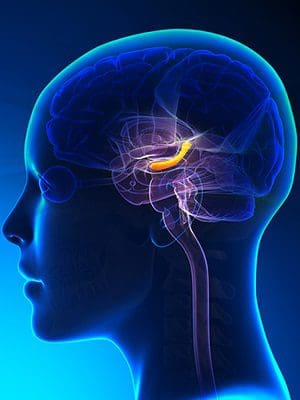
The Hippocampus
In the brain, there is an S-shaped structured located in the inner folds in the temporal lobe called the hippocampus. The hippocampus is a complex brain structure that has a layer of densely packed neurons, and its primary function involves how humans learn and how their memory works. The hippocampus is part of the limbic system as well since it works the feeling and reacting function in the body. The limbic system is situated at the edge of the cortex and includes the hypothalamus and the amygdala.
These structures help controls the body�s different functions like the endocrine system and the �fight or flight� reaction response. With the hippocampus helping humans process what information they are learning, this structure can retrieve two kinds of memories that are important; they are declarative memories and spatial relationship memories.
Declarative memories: These are memories that are related to facts and events a person experience. It includes examples like how to memorize speeches or line in a play that a person is doing.
Spatial relationship memories: These memories involve pathways or routes that a person must learn. An example of this is transportation drivers like cab drivers, bus drivers, and truckers who have to learn the routes in the places they are going to. So they use spatial memory and practice their routes many times until they have it in their memories. The spatial relationship memories are stored on the right side of the hippocampus.
Sadly though, the hippocampus can be damaged by neurological diseases like Alzheimer�s disease and PTSD (Post-Traumatic Stress Disorder). When it is damaged, a variety of conditions can affect the hippocampus�s ability to do its job for the brain, thus making the individual suffer from retaining information.
Hippocampus Conditions
Several conditions can cause problems to the body when the hippocampus is damaged. This is known as hippocampus atrophy, where the neurons and neuronal volume in the hippocampal that is a loss.
Alzheimer�s Disease
Alzheimer�s disease is when an individual begins to lose their memory. When the hippocampus is damaged, it can cause a dissociation between the cortexes and leads to information registration failure. Studies show that when Alzheimer�s disease is progressing, the hippocampus will lose its volume, and it will become harder for an individual to function in their daily lives.
Epilepsy
When a person has epilepsy, it might be due to a damaged hippocampus. Research shows that around 50 to 75% of patients with this disease may have hippocampal sclerosis, and in case they have died, they have medial temporal lobe epilepsy. More research states that the mechanics of hippocampal sclerosis in epilepsy can be related to the development of inflammation on the uncontrolled local hippocampus and blood-brain barrier damage.
Hypertension
When the hippocampus is damaged, hypertension can happen to a person. Hypertension is another name for high blood pressure, and it can lead to severe health complications to the body. Even though the causes of hypertension are still unknown, the risk factors from hypertension can include:
Environmental factors like stress or a lack of exercise
Hormone activity
Blood plasma
Studies show that hypertension and other risk factors are being increasingly viewed as a putative factor that is leading to hippocampal atrophy.
Cushing�s Disease
Cushing�s disease or Cushing syndrome is when the body is exposed to high levels of cortisol for a long time. Studies show that when there is a loss of cellular volume to the corticosteroid�s levels in the body and it could be responsible. When there is too much cortisol being produced in the body, it is one of the signs of Cushing syndrome. Some of the other signs include:
Weight gain
Fatty tissue deposits around the midsection, face, upper back and between the shoulders
Pink or purple stretch marks
Thinning, fragile skin that bruises easily
Slow healing cuts, insect bites and infections
Acne
Muscle weakness
Cognitive difficulties
Loss of emotional control
Since stress does play a role in the endocrine system and the neurological system, there are nearly 80 years of research on how much focus has been on the various levels of the HPA (hypothalamic-pituitary-adrenal) axis and the hormones it produces. It shows that glucocorticoids as the mediators for the stress effects on the hippocampus and being the contributing factor for stress-associated psychopathologies.
Conclusion
The hippocampus is located in the temporal lobe of the brain. This S-shaped structure can be easily damaged due to stress and other neurological factors that can affect the entire body and its systems. When harmful factors affect the hippocampus, it can lead the hormones that are producing to become imbalanced and cause dysfunction. Some products are here to make sure that the endocrine system is functioning properly and supporting the metabolic system, the gastrointestinal system, as well as making sure the hormones are balanced.
The scope of our information is limited to chiropractic, musculoskeletal, and nervous health issues or functional medicine articles, topics, and discussions. We use functional health protocols to treat injuries or disorders of the musculoskeletal system. Our office has made a reasonable attempt to provide supportive citations and has identified the relevant research study or studies supporting our posts. We also make copies of supporting research studies available to the board and or the public upon request. To further discuss the subject matter above, please feel free to ask Dr. Alex Jimenez or contact us at 915-850-0900.
References:
Anand, Kuljeet Singh, and Vikas Dhikav. �Hippocampus in Health and Disease: An Overview.� Annals of Indian Academy of Neurology, Medknow Publications & Media Pvt Ltd, Oct. 2012, www.ncbi.nlm.nih.gov/pmc/articles/PMC3548359/.
Dresden, Danielle. �Hippocampus: Function, Size, and Problems.� Medical News Today, MediLexicon International, 7 Dec. 2017, www.medicalnewstoday.com/articles/313295.php.
Felman, Adam. �Hypertension: Causes, Symptoms, and Treatments.� Medical News Today, MediLexicon International, 22 July 2019, www.medicalnewstoday.com/articles/150109.php.
Kim, Eun Joo, et al. �Stress Effects on the Hippocampus: a Critical Review.� Learning & Memory (Cold Spring Harbor, N.Y.), Cold Spring Harbor Laboratory Press, 18 Aug. 2015, www.ncbi.nlm.nih.gov/pmc/articles/PMC4561403/.
Team, Mayo Clinic. �Cushing Syndrome.� Mayo Clinic, Mayo Foundation for Medical Education and Research, 30 May 2019, www.mayoclinic.org/diseases-conditions/cushing-syndrome/symptoms-causes/syc-20351310.
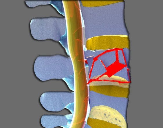
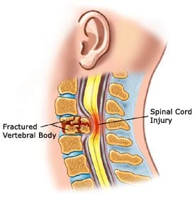
A burst fracture describes an injury to the spine where the vertebrae get compressed severely. These types of injuries occur from severe trauma, like an automobile accident or a serious fall, sports injury, work injury. These injuries entail a great deal of force into the spine, so much so that a vertebra can get crushed.
When crushed in the front of the spine, a wedge-shaped fracture occurs and is known as a compression fracture.
But if the vertebral body gets crushed in all directions this is known as a burst fracture.
The term burst means that the vertebral body spreads out in all directions.
Severe Injury
This is a much more severe injury than a compression fracture. With the bones crushed and possible rough jagged edges, if they spread out the spinal cord has a high probability of being injured. The fragments can bruise the spinal cord causing paralysis or partial neurologic injury. The spine becomes far less stable than from a compression fracture.
Nerve Injury
Neurologic injuries from a burst fracture can range from no injury to paralysis. This depends on the amount of force present at the time of the injury and how much the spinal canal is compromised.
A greater amount of force equals more bony fragments that can be forced into the spinal canal and cause higher loss of spinal cord function.
This can cause loss of:
Strength
Sensation
Reflexes below the injury
With an incomplete spinal cord injury, partial paralysis or partial reflex loss occurs.
With a mild burst fracture, only short-term symptoms could be present and no neurologic injury.
Intense Pain
Burst fractures can cause intense pain and the pain is right where the trauma took place.
But pain can also present in the legs and feet depending on how the spinal nerves were affected, shifted or pinched. Patients complain of an electric tingling or shooting type sensation in their legs with spinal cord compression. With a burst fracture, individuals are unable to walk right after the trauma. But the pain percentage present is severe enough that they know not to try and walk.
Diagnosis
If at the sight of the accident the patient says that they have severe back pain should not be in a seated flexed position. They need to be kept lying flat and transported in a flat position.
If they stand or sit with a burst fracture, it can increase the possibility of a neurologic injury.
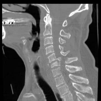
Burst fractures require immediate medical attention from an orthopedic or neurosurgeon. The patient is taken to an emergency room and x-rays, CT scans are gathered.
The diagnosis of a burst fracture is typically made with x-rays and a CT scan.
Sometimes, an MRI will be ordered to assess the amount of:
Soft tissue trauma
Bleeding
Ligament injury
The CT scan and x-rays allow the doctor to determine the level of the fracture, and if it is a:
Compression fracture
Burst fracture
Fracture-dislocation
This will determine how much the spinal canal has been compromised and if its angulation or angle has taken an abnormal bend or curve. These factors all contribute to the development of an optimal treatment plan.
The physical exam will document:
Spinal deformity and Angulation of the spine
Tenderness of the spine where the fracture is located
Neurologic exam
Neurologic exam should include testing:
Muscle strength
Sensation
Reflexes of the lower extremities
Testing of bowel and bladder control
Treatment & Recovery
A stable burst fracture can be treated without surgery.
A stable burst fracture falls into these parameters:
There is no neurologic injury
The angulation is less than 20 degrees
The amount of spinal canal compromise is less than 50%
With this type of treatment, a brace along with physical therapy/chiropractic can have excellent results.
A turtle or clamshell brace TLSO�(Thoracic Lumbar Sacral Orthosis) is a body cast used in the treatment of a burst fracture.
The brace is worn for eight to twelve weeks for adequate and optimal healing.
There are times when a fracture thought stable and treated can start to angulate. This may require surgery. However, all burst fractures require some form of treatment.
Recovery
Nonsurgical treatment patients stay in the hospital for one or two days while their brace is fit.
X-rays are done in the standing position to make sure the spine stays stable.
Pain medications are prescribed for three to four weeks
Non-narcotic medications can begin after the final week
When the brace is removed, physical therapy and chiropractic are instituted to help return strength to the core and lower extremities.
Surgical patients will remain in the hospital for three to five days.
They will be fitted with a brace after the incisions present less pain and are recovered from the surgery.
They can walk within one or two days with the help of a physical therapist.
X-rays are taken to follow the position of the spine and see how the healing is progressing.
Chiropractic/Physical therapy is implemented to help with core strength and lower extremity strength.
Recovery time depends on the severity of the neurologic injury.
Patients that don’t have a neurologic injury can make a full recovery with return to most activities.
Patients with partial neurological injuries can also expect to fully recover.
Unfortunately, with permanent neurologic injury, recovery can be limited.
But treatment for burst fractures today is superior to what they were years ago, especially with spine specialists and specific spinal procedures.
Chiropractic Rehab
Chiropractic is not a treatment for fractures but is a treatment for subluxations and rehabilitation with these types of fractures. Once a fracture has stabilized and healed properly, a chiropractic evaluation can rule out any lingering subluxation, herniation, and joint restriction. The adjustments are safe and effective in establishing optimal function to a subluxated joint.
Chiropractic Treatment For Car Accident Injuries El Paso, Texas
We focus on what works for you. We also strive to create fitness and better the body through researched methods and total wellness programs. These programs are natural and use the body�s own ability to achieve goals of improvement.
NCBI Resources
Chiropractors can help alleviate some of the long-term and immediate concerns associated with bone fractures. A chiropractor can help with compression techniques which are beneficial in maintaining the bone in place for healing. A chiropractor may also advocate wellness techniques, such as a healthy diet that will optimize the body�s ability to restore its original health and wellness. Chiropractors may also educate a patient on a variety of exercises and stretches to reduce the likelihood of complications and which, if done properly and at fixed intervals, will promote quicker recovery.
IFM's Find A Practitioner tool is the largest referral network in Functional Medicine, created to help patients locate Functional Medicine practitioners anywhere in the world. IFM Certified Practitioners are listed first in the search results, given their extensive education in Functional Medicine