We are familiar with neck stiffness or a crick. This can prevent us from comfortably moving the head all around. A crick can cause the spine, and shoulders to feel rigid and stressed from not being able to turn around and could cause an upper or low-back strain from having to turn the whole body to look back or even just to the side. Chiropractic treatment is available and will help, along with some self-care therapies that can be done at home.
Contents
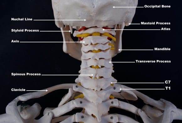
Crick in the Neck vs. Neck Stiffness
A crick in the neck is the same as a stiff neck. It develops when the neck muscles, tendons, and ligaments become strained/sprained. Most strains and sprains are minor but do cause inflammation/swelling of the neck�s soft tissues, which results in stiffness and, at times muscle spasms.
The symptoms
Cricks in the neck are uncomfortable, but not necessarily painful. If there is a pre-existing neck condition or injuries like whiplash the crick and stiffness could increase the uncomfortableness and generate pain.
The most common symptoms include:
Neck stiffness
Muscle stiffness
Reduced mobility affecting the neck�s range of motion
A popping sensation when trying to turn or tilt the head
Causes of a stiff neck or crick
There are different causes of neck stiffness. It can be a combination of things you can control and some you can�t.
Possible causes that you can control:
Poor posture working either sitting or standing for several hours without breaks or stretching.
Sleeping in a position that puts the neck in an awkward position or using a pillow that does not support the neck when sleeping.
Constantly looking down at a cell phone or tablet.
Stress and emotional tension can cause involuntarily tightening of the neck muscles and shoulders.
Heavy labor along with incorrect lifting techniques.
Reaching or having to look up/overhead for several hours like when painting a ceiling.
Possible causes that are out of your control:
Whiplash injury
Sports-injuries like a football stinger
Aging muscles and bones
Around 13% of cases the stiffness, and pain are caused by separate cervical spinal conditions, like:
Cold therapy reduces the swelling of soft tissues, like muscles and ligaments, while heat soothes the tightness by boosting blood circulation to the affected area. There are different products available that can deliver cold or heat to the neck and upper back.
Apply ice for 15 minutes each hour.
Apply heat therapy like a heating pad for 15 minutes every 2 or 3 hours.
Over-the-counter anti-inflammation medicines
Non-steroidal anti-inflammatory medications like ibuprofen and naproxen can help relieve inflammation and pain.
Because neck stiffness can be linked to lifestyle choices, individuals may find that they occur repeatedly. Simple neck stretches, chiropractic treatment, using a supportive pillow, and taking frequent breaks at your job can help prevent neck stiffness and keep you moving. These professionals have undergone extensive training in their field and are capable of treating neck pain effectively. So if you or a loved one are experiencing neck pain, give us a call. We�re ready to help!
In the following podcast video article, Dr. Alex Jimenez, a chiropractor in El Paso, TX, and Dr. Mario Ruja, another chiropractor in El Paso, TX, discuss chiropractic care and why it works. Chiropractic care is a safe and effective, alternative treatment option that focuses on the diagnosis, treatment, and prevention, of injuries and underlying conditions associated with the musculoskeletal and nervous system. Chiropractic care is a healthcare profession that has existed for many years throughout many civilizations and it focuses on the use of spinal adjustments and manual manipulations to carefully restore the original alignment of the spine and the human body as a whole. Dr. Alex Jimenez and Dr. Mario Ruja describe how they were first interested in becoming chiropractors, or doctors of chiropractic, as they also describe how they feel when they are able to provide pain relief to their patients. Dr. Jimenez and Dr. Ruja will focus on discussing why chiropractic care works and how it is different from other healthcare professions in the way it helps treat a variety of health issues associated with the spine, from neck pain to low back pain and sciatica. Chiropractic care can help promote overall health and wellness. � Podcast Insight
If you have enjoyed this video and/or we have helped you in any way
please feel free to subscribe and share us.
Thank You & God Bless.
Dr. Alex Jimenez RN, DC, MSACP, CCST
Metabolic syndrome is a collection of risk factors that can ultimately increase the risk of developing a variety of health issues, including heart disease, stroke, and diabetes, among other problems. Central obesity, high blood pressure, high blood sugar, high triglycerides, and low HDL are the 5 risk factors associated with metabolic syndrome. Having at least three of the five risk factors may suggest the presence of metabolic syndrome. Dr. Alex Jimenez and Dr. Mario Ruja explain the 5 risk factors associated with metabolic syndrome, in further detail, as they recommend diet and lifestyle modification advice and guidelines to help people with metabolic syndrome improve their overall health and wellness. From eating fiber and staying hydrated to exercise and better sleep, Dr. Alex Jimenez and Dr. Mario Ruja discuss how diet and lifestyle modifications can help improve the 5 risk factors associated with metabolic syndrome to ultimately prevent the risk of developing a variety of other health issues, including heart disease, stroke, and diabetes. � Podcast Insight
If you have enjoyed this video and/or we have helped you in any way
please feel free to subscribe and share us.
Thank You & God Bless.
Dr. Alex Jimenez RN, DC, MSACP, CCST
Everybody has a backstory. It is not easy. We got to PUSH Hard. It is not EASY…Dr. Alex Jimenez, a chiropractor in El Paso, TX, talks to Daniel (Danny) Alvarado, owner of the PUSH Fitness Center, about the importance of nutrition, diet, and fitness. Stress is the body’s natural response to any physical, mental, and emotional response. Although too much stress can be harmful, getting the right amount of stress is essential for survival. Dr. Alex Jimenez and Daniel Alvarado discuss how stress is the fundamental basis for inspiration and motivation. The PUSH Fitness Center was first created by Danny to help people achieve their optimal health and wellness goals. Hard-work and pushing towards your goal are essential for every individual. Danny discusses how he chooses to inspire and believe in his athletes in order to help them become the best person they can be. Nutrition, diet, and fitness can help prevent a variety of health issues, including metabolic syndrome, diabetes, stroke, and heart disease. Dr. Alex Jimenez, a chiropractor in El Paso, TX, and Daniel (Danny) Alvarado demonstrate how stress, inspiration, and motivation in people can support the hard work and the extra “push” they need to improve themselves, achieve their goals, and improve overall health and wellness. – Podcast Insight
If you have enjoyed this video and/or we have helped you in any way
please feel free to subscribe and share with us.
Thank You & God Bless.
Daniel Alvarado
Dr. Alex Jimenez
Facebook Fitness Center Page: https://www.facebook.com/PUSHftinessathletictraining/
Metabolic syndrome is a cluster of risk factors that can ultimately increase the risk of developing a variety of health issues, including heart disease, stroke, and diabetes, among other problems. Central obesity, high blood pressure, high blood sugar, high triglycerides, and low HDL or good cholesterol levels are the 5 risk factors associated with metabolic syndrome. Having at least three of the five risk factors may suggest the presence of metabolic syndrome. Dr. Alex Jimenez, Alexander Jimenez, Truide Torres, Kenna Vaughn, and Astrid Ornelas explain the 5 risk factors associated with metabolic syndrome, in further detail, as they recommend diet and lifestyle modification advice and guidelines, such as the ketogenic diet or the keto diet, as well as demonstrate the biochemical and chemical pathways that the body goes through during ketosis to help people with metabolic syndrome improve their overall health and wellness. From eating good fats and staying hydrated to exercise and better sleep, Dr. Alex Jimenez, Alexander Jimenez, Truide Torres, Kenna Vaughn, and Astrid Ornelas discuss how diet and lifestyle modifications, such as the ketogenic diet or keto diet, can help improve the 5 risk factors associated with metabolic syndrome to prevent the risk of developing a variety of other health issues, including heart disease, stroke, and diabetes. � Podcast Insight
If you have enjoyed this video and/or we have helped you in any way
please feel free to subscribe and share us.
Thank You & God Bless.
Dr. Alex Jimenez RN, DC, MSACP, CCST
In the following podcast video article, Dr. Alex Jimenez, a chiropractor in El Paso, TX, and Daniel (Danny) Alvarado, owner of PUSH Fitness Center in El Paso, TX, discuss the three points of weight loss. Excess weight and obesity are associated with metabolic syndrome and a variety of other health issues. Metabolic syndrome is a cluster of risk factors that can ultimately increase the risk of developing diabetes, stroke, and diabetes, among other complications. Dr. Alex Jimenez and Daniel Alvarado discuss how weight loss can be a safe and effective way to improve metabolic syndrome as well as overall health and wellness. Decreasing or eliminating sugar and carbohydrate consumption, increasing the consumption of proteins, �good� fats, and vegetables, as well as engaging and participating in exercise and physical activity can ultimately help promote weight loss to improve metabolic syndrome and a variety of other health issues. Furthermore, Dr. Alex Jimenez and Daniel Alvarado discuss how they can help people with excess weight and obesity achieve their weight loss goals by encouraging and motivating them through every step of the way. Weight loss is essential for people with metabolic syndrome to achieve overall health and wellness. � Podcast Insight
If you have enjoyed this video and/or we have helped you in any way
please feel free to subscribe and share us.
Thank You & God Bless.
Daniel Alvarado & Dr. Alex Jimenez
Subscribe: http://bit.ly/drjyt
Facebook Fitness Center Page: https://www.facebook.com/PUSHftinessathletictraining/
Yelp: El Paso Rehabilitation Center: http://goo.gl/pwY2n2
Yelp: El Paso Clinical Center: Treatment: https://goo.gl/r2QPuZ
Dr. Alex Jimenez, a chiropractor in El Paso, TX, and Dr. Mario Ruja, a chiropractor in El Paso, TX, discuss how chiropractic care can help with personal injuries, especially automobile accidents. Personal injuries can also include work injuries and slip-and-fall injuries. Auto accidents can cause a variety of injuries and underlying conditions, including neck pain, whiplash, back pain, low back pain, and sciatica. Sports injuries can also cause a variety of health issues. Chiropractic care is a safe and effective alternative treatment option that focuses on the diagnosis, treatment, and prevention of health issues associated with the musculoskeletal and nervous system. Dr. Alex Jimenez and Dr. Mario Ruja discuss how spinal adjustments and manual manipulations are commonly utilized to treat neck pain and back pain associated with personal injuries. Whiplash-associated-disorders are the most common types of health issues resulting after an automobile accident. Chiropractic care can carefully restore the original alignment of the spine, treating neck pain and whiplash caused by personal injuries, especially an auto accident. Dr. Alex Jimenez, a chiropractor in El Paso, TX, and Dr. Mario Ruja, a chiropractor in El Paso, TX further discuss how it’s fundamental for people who’ve been involved in a car crash to seek chiropractic care to treat soft tissue injuries that can cause neck pain and back pain. – Podcast Insight
If you have enjoyed this video and/or we have helped you in any way
please feel free to subscribe and share us.
Thank You & God Bless.
Dr. Alex Jimenez RN, DC, MSACP, CCST
Dr. Alex Jimenez, chiropractor in El Paso, TX, discusses BIA, BMI and basal metabolic rate with staff. Dr. Alex Jimenez, health coach Kenna Vaughn, Astrid Ornelas, Truide Torres, and biochemist Alexander Isaiah Jimenez all take part in a round table podcast discussion of the importance of measuring BMI, BIA, and basal metabolic rate. BMI or body mass index is frequently utilized to determine an individual’s relative weight according to their height. Healthcare professionals commonly utilize BMI, however, BMI may not be accurate for athletes because their body mass according to their height may demonstrate that they have excess weight or obesity utilizing BMI. BIA is the preferred analysis tool used to determine an athlete’s relative weight according to their height. Excess weight and obesity is a well-known risk factor that can ultimately increase the risk of metabolic syndrome, among other health issues, including diabetes, stroke, and heart disease. Dr. Alex Jimenez, health coach Kenna Vaughn, Astrid Ornelas, Truide Torres, and biochemist Alexander Isaiah Jimenez ultimately discuss in further detail how determining an individual’s BIA, BMI, and basal metabolic rate can help promote overall health and wellness as well as support weight loss for the general population and athletes alike. – Podcast Insight
If you have enjoyed this video and/or we have helped you in any way
please feel free to subscribe and share with us.
Thank You & God Bless.
Dr. Alex Jimenez RN, DC, MSACP, CCST
In the following podcast, Dr. Alex Jimenez, a chiropractor in El Paso, TX, and Dr. Mario Ruja, a chiropractor in El Paso, TX, discusses what metabolic syndrome is in more depth. Metabolic syndrome is a collection of conditions that can ultimately increase the risk of developing a variety of other health issues, including diabetes, stroke, and heart disease. According to the American Heart Association (AHA), a person may be diagnosed with metabolic syndrome if they have at least three of the following five risk factors, including abdominal obesity of more than 40 inches in men and more than 35 inches in women, fasting blood glucose levels of 100 mg/dL or above, blood pressure of 130/85 mm/Hg or above, blood triglycerides levels of 150 mg/dL or higher, and high-density lipoprotein (HDL) cholesterol levels of 40 mg/dL or less for men and 50 mg/dL or less for women. Having three or more of these risk factors can ultimately increase the risk of developing a variety of health issues, including diabetes, stroke, and heart disease. Dr. Alex Jimenez and Dr. Mario Ruja discuss in more detail how eating a lot of carbohydrates and sugar are ultimately associated with the risk factors of metabolic syndrome. � Podcast Insight
If you have enjoyed this video and/or we have helped you in any way
please feel free to subscribe and share us.
Thank You & God Bless.
Dr. Alex Jimenez RN, DC, MSACP, CCST
Dr. Alex Jimenez, a chiropractor in El Paso, TX, and Dr. Mario Ruja, a chiropractor in El Paso, TX, discuss chiropractic care and sciatica or sciatic nerve pain. Sciatica, or sciatic nerve pain, is a collection of symptoms, rather than a single type of injury or underlying condition, that includes several common symptoms, such as pain, discomfort, tingling and burning sensations, and numbness. Severe symptoms can also include muscle weakness. Sciatica, or sciatic nerve pain, can be caused by a variety of health issues, including a bulging or herniated disc, DDD, piriformis syndrome, spinal stenosis, and spondylolisthesis, among other health issues. Personal injuries like sports injuries, work-related injuries, automobile accident injuries, and slip-and-fall accidents can also cause low back pain and sciatica, or sciatic nerve pain. Dr. Alex Jimenez and Dr. Mario Ruja discuss the causes and symptoms of sciatica, or sciatic nerve pain, in further detail as well as the treatments. Chiropractic care is a safe and effective, alternative treatment option that utilizes spinal adjustments and manual manipulations to diagnose, treat, and prevent injuries and underlying conditions associated with the musculoskeletal and nervous system, including sciatica. Dr. Alex Jimenez, a chiropractor in El Paso, TX, and Dr. Mario Ruja, a chiropractor in El Paso, TX, demonstrate how chiropractic care can help relieve sciatica, or sciatic nerve pain, by carefully restoring any spinal misalignments, or subluxations, that may be affecting overall health and wellness. – Podcast Insight
If you have enjoyed this video and/or we have helped you in any way
please feel free to subscribe and share us.
Thank You & God Bless.
Dr. Alex Jimenez RN, DC, MSACP, CCST
IFM's Find A Practitioner tool is the largest referral network in Functional Medicine, created to help patients locate Functional Medicine practitioners anywhere in the world. IFM Certified Practitioners are listed first in the search results, given their extensive education in Functional Medicine