Popular hormone-based drugs for treating an enlarged prostate could increase men’s risk of type 2 diabetes, heart disease or stroke, a new study suggests.
A group of German men taking the drug Avodart (dutasteride) for three years wound up with higher blood sugar and cholesterol levels than men taking another class of prostate medication that does not affect male hormones, the researchers reported.
“Our small study suggests there are really adverse effects on metabolic function from these drugs that has not been reported previously,” said lead researcher Abdulmaged Traish. He is a professor of urology with the Boston University School of Medicine.
But Dr. Ashutosh Tewari, chair of urology for the Icahn School of Medicine at Mount Sinai in New York City, said the new findings run counter to prior clinical trials of the drug, and do not warrant any change in use at this time.
Still, Traish believes urologists should talk about these new results with patients before prescribing either Avodart or another hormone-based prostate drug called Proscar (finasteride). Both are in the class of drugs known as 5-alpha-reductase inhibitors.
“They should have a clear, open and honest discussion with their patients,” Traish said. “This drug might cause some of these problems.”
However, according to Tewari, “This is an interesting finding which is a little different than the large ‘controlled’ studies. It needs to be studied in a larger pool of patients in a prospective manner.”
The association seen in the study doesn’t prove a cause-and-effect relationship.
The prostate is a walnut-sized gland surrounding the urethra where it connects to the bladder. The prostate produces fluid that goes into semen, and is essential for male fertility. But as men age, their prostates tend to enlarge, pinching the urethra and making urination more difficult.
Avodart reduces production of dihydrotestosterone (DHT), a hormone linked to enlargement of the prostate gland. Treatment with Avodart can cause a man’s prostate to shrink by roughly 18 percent to 20 percent, Traish noted.
“The men urinate a little bit better,” Traish said. “They don’t have to stand an hour and a half in the bathroom at the airport.”
However, DHT also plays an important role in the function of other organs, particularly the liver, Traish said. He and his colleagues are concerned that reducing DHT could have other unknown health effects.
To examine the issue, Traish’s team reviewed records of 460 men treated at a single urologist’s office in Germany for enlarged prostate.
Half of the men had been prescribed Avodart to treat their problem, and the other half had been prescribed Flomax (tamsulosin). Flomax, in the class of drugs known as alpha-blockers, does not affect hormones, but works by causing the smooth muscle tissue of the prostate to relax, Traish said.
The researchers tracked all of the men for 36 to 42 months, performing blood tests and assessing prostate size and function.
Avodart was linked to an ongoing rise in blood sugar levels among men who received the drug, while men taking Flomax did not experience any such increase, the study authors said.
Further, long-term Avodart treatment was linked to increased “bad” LDL cholesterol levels in men, the investigators found. Men on Flomax experienced a smaller but yet significant increase in their LDL cholesterol levels, but also had an increase in their “good” HDL cholesterol levels, the findings showed.
Based on his findings, Traish said he would lean toward prescribing Flomax first rather than a hormone-based prostate drug.
“I would rather have my patient try something safer, and if it works for him, keep him on that,” Traish said.
Tewari noted that the clinical trials that found Avodart effective in treating enlarged prostate did not show any of these other metabolic problems.
Those clinical trials relied on men being randomly assigned Avodart, Tewari said. The men in this new study were not assigned medication randomly, but were allowed to choose their treatment following discussion with a doctor.
The new study also did not compare men taking Avodart to a control group taking a placebo, and relied on past data rather than an entirely new experiment, Tewari continued.
“This is interesting, yet needs to be verified in a controlled setting with a larger pool of patients,” Tewari explained. “At this time, I’m not too impressed with any clinical significance of this study.”
The study was published online recently in the journal Hormone Molecular Biology and Clinical Investigation.
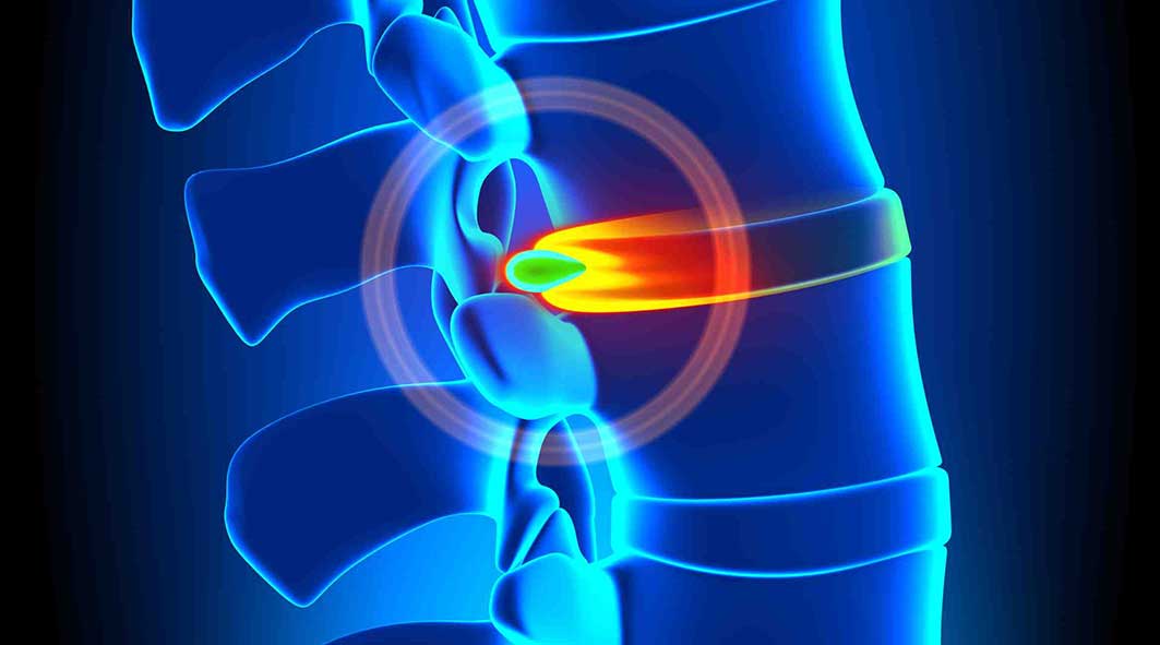
“The clinical diagnosis shows a disc bulge in their neck and some arthritis, so their neck symptoms are not associated with the crash. Lots of folks have those and do not have pain although it could be a minimal herniation. It’s our diagnosis that it was there before the crash.” This statement from an adjuster is an argument that has been made for many years, allowing insurance companies to inappropriately reduce settlements to their clients based on the client’s inability to prove when or how the damage or injury occurred. To factually counter this sort of statement, an individual must use imaging and age dating to discuss causality. Without medical experts utilizing the current medical and academic research available, it will continue to be difficult for any argument to be made explaining effects of these injuries and their mechanism based on fact vs. rhetoric.
Imaging of the spine is critically important in most cases of injured clients. In cases, imaging is necessary for proper diagnosis and future management of injuries. Imaging needs to be performed as per the academic and modern criteria to ensure an accurate diagnosis. The most common injuries in car accidents are spinal related, and the simple imaging available includes x-rays, CAT scans and magnetic resonance imaging (MRI), allowing medical providers to make an accurate diagnosis, when medically indicated.
Every medical provider has a permit to see and treat automobile related injuries. However a “license” is not the same as “specialization.” By way of example, though psychiatrists may have a license to do heart surgery and are MDs, it would not be in the patient’s best interest. Nor would I go to a spine surgeon for psychological concerns although they are licensed to treat medical conditions. In spinal trauma, certain suppliers specialize in connective tissue injuries of the spine, allowing us to go one step farther in diagnosis, prognosis and management, including “age-dating” these generally found disc and ligament injuries.
Understanding Age-Dating of Injuries
To understand age-dating, one wants to have a basic medical understanding of anatomy and physiology, and what tissue is commonly injured and the probable “pain generator”. Since neck injuries are the most common injuries cervical joints will be our focus. Related to anatomy, every set of two vertebrae in the neck is connected with three joints; two facet joints and a single disc. These joints allow for normal movement of the spine (mobility). There are multiple ligaments that are responsible for stability and hold together these joints. The correct balance of mobility and stability is critical when looking at the part of patient’s injuries, meaning that too little or too much movement in spinal joints can lead to pain, secondary to damaged tissue. The tissue most commonly hurt in a car crash is nerve, ligament, disc, facet and muscle/tendon. Spinal cord and bone injuries also happen although less frequently. To determine causality, the supplier should comment on what tissue is injured, and also use imaging to help determine if this injury occurred (age-dating).
There are two fundamental problems that must be addressed. Fardon and Milette (2001) reported, “The phrase ‘herniated disc’ does not infer knowledge of cause, relation to trauma or activity, concordance with symptoms, or need for treatment” (p. E108). Simply having a disc herniation’s presence, without a physical exam or without symptom documentation that is appropriate, does not allow one to comment on the cause of the injury. In a rear impact collision by way of example, even if the diagnosis is confirmed, additional criteria will need to be fulfilled to answer the question of “Was there sufficient force generated into the vehicle and the occupant to induce the cervical/lumbar herniation?” Fardon, in a follow-up study (2014) reported that disc injury “in the absence of significant imaging evidence of associated violent injury, should be classified as degeneration rather than trauma.” (p. 2531). Thus, we must more objectively define the subjective connotations of “violent injury” and address the issue of “degeneration as opposed to trauma”. Although this statement can frequently be misleading, it gives the trauma trained expert doctor a basis in going forward understanding that every patient’s physiology is unique and not subject to rhetoric, but clinical findings.
Violent injury to the occupant can occur when there are sudden acceleration and deceleration forces (g’s) generated to the neck and head which overwhelm connective tissue or pull them past their physiological limit. To determine the acceleration force, ?V (delta V) is utilized. ?V is the change in speed of the occupant vehicle when it is hit from behind (i.e., going from a stopped position to seven mph in 0.5 seconds because of forces moved from the “bullet” vehicle to the “target” vehicle). Utilizing these data, research allows us to make specific comments related to violent injury. Since the cervical spine is subjected to shearing forces, and compression, tension we are oversimplifying. Along with g-forces and the elastic nature of the majority of rear impact crashes makes it almost impossible to discover an actual minimum threshold for injury even though the literature has given us many examples of low-speed crashes which are dependent not simply on speed, but the mass (weight) of the subject vehicles. Each individual’s susceptibility to injury is unique. While g-forces alone are insufficient to predict injury, Krafft et al. (2002) reported that in low-speed collisions there’s an injury threshold of 4.2 g’s for males and 3.6 g’s for females. Krafft’s analysis is unique in that she has access to insurance data inaccessible to researchers. Panjabi (2004) revealed that forces as low as 3.5g impacts would lead to damage to the front of the disc, and 6.5g and 8g impacts would lead to disc damage posteriorly where the neurological components are.
Diagnosis for Disc and Ligament Injuries
A spinal biomechanical expert can look for evidence that is conclusive by disc and pathology, according to two phenomena. First, it is recognized that the body is electric. We’re measuring activity to diagnose when an EMG is done. Second, there are bioelectrical fields in all tissues. This typical field is disrupted when an injury occurs, and in the case of joints calcium is drawn to the damaged tissue. Issacson and Bloebaum (2010) reported “The particular loading pattern of bone has been documented as a significant piezoelectric parameter since potential gaps in bone have been known to be due to charge displacement during the deformation period” (p. 1271). For the patient, we have the ability to tell just how much of this process has occurred before or after their crash, especially if we take into consideration the tissue damage and signs of bone/calcium deposition.
In addition, the body begins a healing process that includes regeneration and remodeling of the soft and hard tissue as reported by Issacson and Bloebaum (2010). Spinal vertebrae have a unique structure of bone which allows it to adapt to abnormal mobility and stability (injury) by changing shape, which can be found on radiographs or MRI. Moreover, shape will change according to patterns based on the pressure or load it undergoes post-injury. Issacson and Bloebaum stated that “Physical forces exerted on a bone change bone structure and is a well-established principle…” (p. 1271). This is a further understanding of a scientific principle called Wolff’s law established in the 1800’s. Because we know what “normal” is, when we see “abnormal” findings as a result of mechanical stress we could broach the topic of an acute injury versus a degenerative process being the cause of the abnormality and create specific medical predictions accordingly.
He and Xinghua (2006) studied the predictability of the bone remodeling process and were able to make predictions of pathological changes that will occur in bone, specifically the osteophyte (bone spur) on the edge of a bone structure. Significantly, they noted their findings “confirmed that osteophyte formation was an adaptive process in response to this change of mechanical environment”. They noted that factors are crucial to the morphology of bones, particularly bones such as the femur and vertebrae.
For readers familiar with current academic and medical accepted nomenclature for disc injury, recognized from the combined task forces of the North American Spine Society (NASS), the American Society of Spine Radiology (ASSR) and the American Society of Neuroradiology (ASNR), disc herniations must have a directional component. When this occurs, the additional and abnormal pressure at the level of the disc damage matched with the direction of the herniation will cause that section of the vertebrae.
Thus, if there’s a C5/6 right sided herniation (protrusion/extrusion) secondary to a cervical acceleration/deceleration injury, then only that side of the vertebrae will change shape, creating an osteophyte. Facet arthritis is additionally caused by this compounded loading on the facet joint. This process is very similar to the formation of a callous on your hand or foot. The callous is a recognized and expected tissue response to increased load/friction exposure. Similarly, an osteophyte is a known and anticipated bone response to a rise in load/friction exposure.
At a basic level, the body has an electrical and mechanical response to injury leading to additional stress that leads to calcium (bone) to flow in the region of injury to further support the joint. The joint then abnormally grows, developing a called hypertrophy, degeneration, disc osteophyte complex, or arthritis/arthropathy, common terms seen in the reports of doctor and radiology.
Everybody is subject to these morphological (structural) changes, always and predictably determined by mechanical imbalances in the spine. He and Xinghua (2006) concluded that, “…it will actually take about over half a year to discover the bone morphological changes…” (p. 101). This indicates that it takes approximately six months to get an osteophyte (bone spur) to be demonstrable post-mechanical breakdown or failure. This again provides a time frame to better understand whether pathology of the intervertebral disc has been present for a long period of time (pre-existing) or has been produced as the direct result of the specific traumatic event by deficiency of the existence of an osteophyte, meaning the disc pathology is less than six months old, dependent on location and management of the pathology.
Conclusion
In conclusion, that by definition, a disc is a ligament connecting a bone to a bone and it has the structural responsibility to the vertebrae above and below to maintain the spinal system in equilibrium. Damage to the disc because of a tear (herniation or annular fissure) or a bulge will create abnormal load-bearing forces in the injury site. These present differently based on [1] if traumatic failure on the side of the disc lesion, or [2] if age related, as a general complex. Since other research and human subject crash testing have defined the term “violent trauma” as not being dependent upon the amount of damage done to the vehicle but rather to the forces to which the neck and head are exposed, we can now accurately predict in a demonstrable way the timing of causality of this disc lesion. This depends upon the symptomatology of the the morphology of the structure and is a subject that can be predicated upon speculation or rhetoric.
The scope of our information is limited to chiropractic and spinal injuries and conditions. To discuss options on the subject matter, please feel free to ask Dr. Jimenez or contact us at 915-850-0900 .�
References:
Fardon, D. F., & Milette, P. C. (2001). Nomenclature and classification of lumbar disc pathology: Recommendations of the combined task forces of the North American Spine Society, American Society of Spine Radiology, and American Society of Neuroradiology.�Spine, 26(5), E93�E113.
Fardon, D. F., Williams, A. L., Dohring, E. J., Murtagh, F. R., Rothman, S. L. G., & Sze, G. K. (2014). Lumbar Disc Nomenclature: Version 2.0:�Recommendations of the combined task forces of the North American Spine Society, American Society of Spine Radiology, and American Society of Neuroradiology.�Spine,�14(11), 2525-2545.
Krafft, M., Kullgren, A., Malm, S., and Ydenius, A. (2002). Influence of crash severity on various whiplash injury symptoms: A study based on real life rear end crashes with recorded crash pulses.� In�Proc. 19th�Int. Techn. Conf. on ESV, Paper�No. 05-0363, 1-7
Batterman, S.D., Batterman, S.C. (2002). Delta-V, Spinal Trauma, and the Myth of the Minimal Damage Accident.�Journal of Whiplash & Related Disorders, 1:1, 41-64.
Panjabi, M.M. et al. (2004). Injury Mechanisms of the Cervical Intervertebral Disc During Simulated Whiplash.�Spine 29 (11): 1217-25.
Issacson, B. M., & Bloebaum, R. D. (2010). Bone electricity: What have we learned in the past 160 years?�Journal of Biomedical Research, 95A(4), 1270-1279.
Studin, M., Peyster R., Owens W., Sundby P. (2016) Age dating disc injury: Herniations and bulges, Causally Relating Traumatic Discs.
Frost, H. M. (1994). Wolff’s Law and bone’s structural adaptations to mechanical usage: an overview for clinicians.�The Angle Orthodontist, 64(3), 175-188.
He, G., & Xinghua, Z. (2006). The numerical simulation of osteophyte formation on the edge of the vertebral body using quantitative bone remodeling theory.�Joint Bone Spine 73(1), 95-101.
Additional Topics: Weakened Ligaments After Whiplash
Whiplash is a commonly reported injury after an individual has been involved in an automobile accident. During an auto accident, the sheer force of the impact often causes the head and neck of the victim to jerk abruptly, back-and-forth, causing damage to the complex structures surrounding the cervical spine. Chiropractic care is a safe and effective, alternative treatment option utilized to help decrease the symptoms of whiplash.
In the prior writing we explored the criteria for vehicle integrity. In this writing we’ll expand on conservation of momentum. You’re encouraged to do so when you haven’t read the previous article.
Expanding on Conservation of Momentum
Remember we previously said, “The momentum moving into a collision could be accounted for at the outcome” when we discussed the concept of conservation of momentum. Here we will introduce the formula and walk through its parts; we have to comprehend this in order to explore each other influence.
The full formula:
Let�s walk through this, on the left side of the equation we have which is the weight of the first vehicle before the collision multiplied by which is the velocity (in feet per second) of the first vehicle before the collision. is the weight of the second vehicle before the collision times which is the velocity (in feet per second) of the second vehicle before the collision. On the right side of the equation we have which is the weight of the first vehicle after the collision multiplied by which is the velocity (in feet per second) of the first vehicle after the collision. is the weight of the second vehicle after the collision times which is the velocity (in feet per second) of the second vehicle after the collision.
Ok, I know this looks very intricate and the explanation is not jumping off the page so let’s write with a bit more ease of comprehension. Let us take the National Highway Transportation Safety Administration (NHTSA) standards for testing and place two of the identical mass vehicles in this. Let us use a 2012 Toyota Corolla, and we will say the other is blue and one is red because we need two of them.
Red Corolla * 5 mph + Blue Corolla * 0 mph = Red Corolla * 0 mph + Blue Corolla * 5 mph
The 2012 Toyota Corolla has a curb weight of 2,734 pounds, substituted in the formula it looks like this:
Now when we do the math to show the conservation of momentum we end up with the following:
20,094.9 + 0 = 0 + 20,094.9
20,094.9 = 20,094.9
Momentum conserved
Now we have proved the concept so we are going to apply it to a collision involving two different vehicles. We will substitute the 2012 red Toyota Corolla for a 2012 red Chevrolet Tahoe. The 2012 Chevrolet Tahoe weighs 5,448 lbs. Now the formula looks like this:
Red Tahoe * 5 mph + Blue Corolla * 0 mph = Red Tahoe * 0 mph + Blue Corolla * 9.96 mph
Now when we do the math to show the conservation of momentum we end up with the following:
40,042.8 + 0 = 0 + 40,042.8[1]
40,042.8 = 40,042.8
Momentum conserved
Three significant points can be observed in this protest.
First, when testing is done notice the change in rate at the Tahoe is 5 mph (5 to 0). This is less than the rates used by the Insurance Institute and we would expect the Tahoe to have minimal damage and no structural deformation.
The second point to note is the change in speed the Corolla experiences, 9.96 mph (0 to 9.96). This change in speed is four times the original.
Conclusion
Finally, neither vehicle exceeds the speed of 10 mph, which the automobile manufactures and insurance institute for highway safety often consider threshold for injury. This confirms that cars can easily deform and residents become injured in low speed crashes once you begin to check out the conservation of energy (momentum) and coefficient of forces moved to the target car.
The scope of our information is limited to chiropractic and spinal injuries and conditions. To discuss options on the subject matter, please feel free to ask Dr. Jimenez or contact us at 915-850-0900 .� References
Edmunds.com. (2012). 2012 Chevrolet Tahoe Specifications. Retrieved from Edmunds.com: www.edmunds.com
Edmunds.com. (2012). 2012 Toyota Corolla Sedan Specifications. Retrieved from Edmunds.com: www.edmunds.com
Brault J., Wheeler J., Gunter S., Brault E., (1998) Clinical Response of Human Subjects to Rear End Automobile Collisions. Archives of Physical Medicine and Rehabilitation, 72-80.
Additional Topics: Weakened Ligaments After Whiplash
Whiplash is a commonly reported injury after an individual has been involved in an automobile accident. During an auto accident, the sheer force of the impact often causes the head and neck of the victim to jerk abruptly, back-and-forth, causing damage to the complex structures surrounding the cervical spine. Chiropractic care is a safe and effective, alternative treatment option utilized to help decrease the symptoms of whiplash.
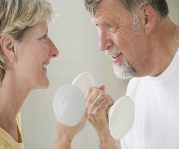
You may be familiar with osteoporosis, the so-called “brittle bone” disease, but there is another condition – sarcopenia – that can lead to disability as we age, a top expert says.
“Sarcopenia is an age-related condition that can make walking and daily functioning difficult. It can also set the stage for bone fractures but, unlike osteoporosis, few people are aware of it,” Dr. Neerav Padilya, Ph.D., tells Newsmax Health.
Sarcopenia is defined as condition resulting in age-related loss of muscle mass, strength, and function, the National Institutes of Health (NIH) says.
The condition was first described in 1997, and experts – including the NIH – say it is an independent risk factor for physical disability, unrelated to age or other health problems.
Our bones get larger and stronger until the age of 30, when muscle mass begins to diminish, and sarcopenia can begin to set in, says Padilya, vice president of research at Qurr, a New Jersey based company which makes a supplement that targets the condition.
“We rely upon our muscle mass for our mobility, as well as to maintain balance, so if you start to lose it due to sarcopenia, by time you’re in your late 60s or 70s, this will have a direct impact on your life,” adds Padilya, a researcher and patent-holding inventor.
“The condition also causes a lack of hand grip strength, so if this becomes weak, imagine trying to open a jar of pickles,” he adds.
In addition, a loss of muscle mass may increase the risk of diabetes, he says.
Research studies note that increased muscle mass can help the body handle glucose, reducing the risk of the disease, Padilya notes.
But the biggest danger that sarcopenia poses, says Padilya, is that it predisposes people to falling, and suffering a potentially life-threatening fracture.
“About 50 percent of the people over the age of 65 that die have suffered a fracture, so it is a very serious problem,” he adds.
Since muscle mass begins to diminish in your 30s, it’s never too early – or too late – to take steps to prevent sarcopenia, says Padilya.
Here are his recommendations:
Make sure you are eating enough protein. Eat foods with protein uniformly at meals throughout the day, not only at dinner. Consume 60 grams of protein daily, or 20 grams at each meal.
Get up and move. Even a small amount of activity on a daily basis really helps you reduce body fat and maintain muscle.
Introduce weight lifting and resistance training into your exercise program.
Make sure you’re getting enough vitamin D. Take a multi-vitamin with vitamin D in it, and also make sure you are getting calcium from milk or eggs. Bone health and muscle health are closely connected.
UK research has revealed that many middle-aged office workers are as sedentary as elderly pensioners.
Carried out by the University of Edinburgh’s Physical Activity for Health Research Centre, the team gathered data on 14,367 people in Scotland taken from the 2012-14 Scottish Health Survey, to look at how age and sex affected weekday and weekend sedentary time.
Defined as time spent in any waking activity done while sitting or reclined, sedentary time includes working, eating, reading, watching TV, or spending time on a computer.
Many recent studies have looked at the effects of sedentary time on health, with some experts warning that more than seven hours of inactivity a day can increase the risk of cardiovascular disease, type 2 diabetes, some cancers, and an early death, even if people are physically active at other times of the day.
The results of the new research found that men aged 45 to 54 spend on average 7.8 hours per weekday sitting down, compared with 7.4 hours for the over-75s.
Time spent sitting at work is the main reason for their sedentary time.
The team also found that only the youngest group of men — 16 to 24-year-olds — are significantly less sedentary than the over-75s on weekdays.
Most of the time spent sedentary in this age group is spent in front of a TV or screen.
At the weekend, those aged 25 to 54 were the least sedentary, sitting for between 5.2 and 5.7 hours a day, and in contrast the over 75s were the most sedentary, at 7.3 to 7.4 hours a day.
In addition, the researchers also found that men spend less time in front of a screen as they get older, with women peaking in middle-age.
The results now replace previous findings that older adults are the most sedentary age group in the UK and highlights the potential health risks of excessive sitting at work.
“Large parts of the population are dangerously sedentary, something we have underestimated. We need to tackle high levels of sedentary time in early and middle age, when patterns may develop. Our findings suggest that changing habits in the workplace could be an appropriate place to start, given how much time we spend sitting there every day,” commented one of the study’s authors Tessa Strain.
The findings were published in the Journal of Sports Sciences.
UTEP softball head coach Tobin Echo-Hawk announced the addition of pitcher Kira McKechnie on Wednesday. McKechnie played her first two years at Fresno State and will have two years of eligibility with the Miners.
McKechnie will join fellow transfer pitcher Julia Wright, and UTEP sophomore hurlers Devyn Cretz and Allie Johnson for the 2018 season.
�We are excited to have Kira on our roster for the upcoming season,� Echo-Hawk said. �It is always nice to add some depth to your pitching staff.�
McKechnie, a native of Sacramento, Calif., made a relief appearance in the circle during the 2017 campaign against San Diego and recorded a strikeout. In 2016, McKechnie made a pair of appearances in the circle, throwing 1.1 innings, while allowing two hits and no runs.
McKechnie attended Christian Brothers High School and was a dual-sport athlete. She was a four-year letterwinner in both softball and basketball. McKechnie was voted softball team captain in 2015 and capped her senior year with multiple accolades, which includes Sacramento Bee�s 2015 All-Metro first team, Cal-Hi first team All-State, CAL All-Optimist All-Star team, MaxPreps first team All-State and Cal-Hi D3 Athlete of Honor.
She was named Female Athlete of the Year by Character Combine, Bee Preps Show and MaxPreps Christian Brothers, while earning the Credit Union Athlete of the Week in 2015.
During her senior season, McKechnie hit .462 with 40 RBI, 11 doubles, a triple and eight home runs. She added 18 runs and was walked nine times. In the circle, McKechnie (12-7) fashioned a 1.95 ERA and recorded 195 Ks 140 innings (23 starts/25 appearances).
McKechnie has competed for the California Breeze (2005-09), Capital City Comets (2009-12), Nor Cal Patriots (2012-14) and Central Cal Dirt Dogs (2014-15). She also attended the 2012 OnDeck Elite Futures Camp and 2013 Colorado Sparkler All-Star/All-American game.
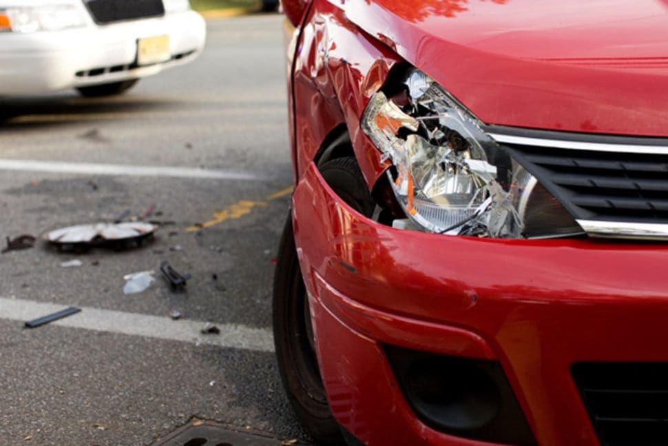
There are many factors that play a role in the dynamics of collisions. These include vehicle design and type, speeds, angles of approach, kinetic & potential energy, momentum, acceleration factor, friction… the list is quite long. There are a few constants in which we are curious. These constants are the building blocks of the planet and they make the world of collisions quantifiable and predictable.
Within this two-part series we will explore the factors which have the most influence in low speed collisions and how these factors are associated with injury. Note: nothing about these writings is inclusive, there is too much material to explore in depth. The objective of these writings is to present the concepts.
Conservation of Momentum & Auto Accidents
In this writing the subject of exploration is conservation of momentum and how it relates to low speed collisions and bodily injury of the occupant. Conservation of momentum is built on Sir Isaac Newton’s third law. Newton’s third law says “For every action there’s an equal and opposite reaction”.
In the interest of exploring conservation of momentum in a simple format, we aren’t likely to investigate and explain the history and physics of momentum; for this conversation, we’ll concentrate on the relationship to crash dynamics. It is momentum to speed collisions’ relationship that helps enlighten and is the causal factor of the injuries people who have held tight to the argument that is deceptive that no damage = no injuries.
While there is a formula and derivation, neither is needed just yet. For now, we’ll simply use the concept as follows: The momentum going into a collision can be accounted for in the outcome or the energy going in to the accident, must be accounted for at the end of the incident and that and what was exposed to and/or absorbed that energy.
Let’s apply some perspective to this notion with the following example.
Let us say we are standing at around a pool table and we are going to try the winning shot of the eight ball into a corner pocket. Following the cue ball is struck, we have and another. After the cue ball strikes the ball, then it stops moving and the eight ball begins moving. In this scenario the cue ball before the collision’s momentum is the same as the momentum of the eight ball after the collision[1]. The eight ball rolls to the corner pocket.
The transfer is extremely efficient due in part to the fact that neither pool balls can deform. Some of the energy would be used to perform this and less if either pool ball could deform. The National Highway Transportation Highway Safety Administration (NHTSA) mandates minimum performance standards for passenger vehicle bumpers. Vehicle bumpers are tested with 2.5 mph (3.7 fps)[2] impact equipment that has the same mass as the test vehicle. The test vehicle is struck with its brakes disengaged and the transmission in neutral. There’s no offset between the automobile and the barrier.
Performance Standards for Vehicle Safety
The NHTSA outlines acceptable damage to your vehicle’s various systems after the tests. Successful completion of these tests mandate operation of systems that are particular. The factory adjustment of the vehicle’s braking, steering, and suspension must be unaltered. In other terms, in order for a vehicle to pass these tests it canhave no change in its structure. If changes did occur the system that is braking, steering, and suspension would be out of factory adjustment.
The NHTSA isn’t alone in low rate bumper testing. The Insurance Institute for Highway Safety (IIHS) also conducts low rate bumper tests. The IIHS’s test rates are conducted at 6 mph (8.8 fps)[3] and the goal is to determine which vehicles have the least damage and therefore cost the least to repair. The vehicle ratings are proportional to the estimated cost of repair. The more costly the repair, the lower the rating.
While the vehicles used in the IIHS testing all show signs of contact with the barrier, none of the vehicles suffer harm which deforms the structure of the vehicle. Don’t have any change in its structure affecting the system, steering, and suspension, just as with the NHTSA the vehicles tested by the IIHS.
The lack of change in the structure (deformation) drives a test vehicle to accept the momentum transfer in the testing equipment. Further, the test vehicle is free to move after being destroyed. This testing scenario is like that of the cue ball and eight ball.
If a vehicle doesn’t deform during a low speed collision, then it will experience a change in speed (or velocity) very quickly; Consequently, the occupant(s) also experience this exact same change in speed. The key factor in these examples is that the mass of testing equipment and their vehicles involved, but what happens when the masses change?
Conclusion
When the mass of one vehicle changes the momentum also changes, the more mass the more momentum the vehicle can bring to the event and the greater the injury potential to the occupant. There are many complicating factors that now must be considered regarding injuries beyond the Laws of Momentum when determining trauma like the height, weight, muscle mass, occupant position, kind of seat belt used, etc.. However, the first step is to decide if there was enough energy as an initiating factor in low speed crashes to cause those injuries and to overcome no crash = no injury misconceptions and have a health expert in low speed injuries confirm relationship.
In the next installment, part II, we’ll discuss this in detail and it will necessary for the later subject of occupant injuries.
The scope of our information is limited to chiropractic and spinal injuries and conditions. To discuss options on the subject matter, please feel free to ask Dr. Jimenez or contact us at 915-850-0900 .�
REFERENCES:
Insurance Institute for Highway Safety. (2010, September). Bumper Test Protocol. Retrieved from Insurance Institute for Highway Safety: www.iihs.org
National Highway Transportation Safety Administration. (2011, October 1). 49 CFR 581 – BUMPER STANDARD. Retrieved from U.S. Government Publishing Office: www.gpo.gov
Additional Topics: Weakened Ligaments After Whiplash
Whiplash is a commonly reported injury after an individual has been involved in an automobile accident. During an auto accident, the sheer force of the impact often causes the head and neck of the victim to jerk abruptly, back-and-forth, causing damage to the complex structures surrounding the cervical spine. Chiropractic care is a safe and effective, alternative treatment option utilized to help decrease the symptoms of whiplash.
IFM's Find A Practitioner tool is the largest referral network in Functional Medicine, created to help patients locate Functional Medicine practitioners anywhere in the world. IFM Certified Practitioners are listed first in the search results, given their extensive education in Functional Medicine