Title: The chiropractic management of cervical Myelomalacia
Abstract: To examine the diagnosis and condition of a patient suffering from neck pain and radiation of pain into arms following a motor vehicle accident. Diagnostic studies include the chiropractic orthopedic and neurological examination, digital x-rays, range of motion and cervical MRI.
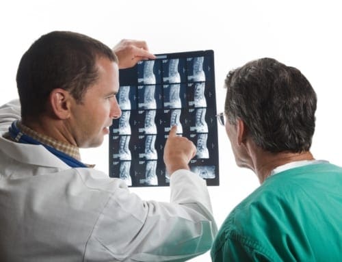
Introduction: On 10/10/2016, a 38-year-old male presented to our office for injuries he had sustained in an MVA on 10/01/2016. The patient stated that he was stopped at an intersection when the pickup behind him hit him at a fast speed, pushing him through the intersection. The patient stated that he had neck pain and stiffness the radiated into the trapezius area. He also complained about �tingling� into both hands. He also complained of lower back pain that he felt more than the neck. His review of systems was benign, other than the current symptoms of neck and back pain and tingling.
The patients Social/Family Medical History included his mother having high blood pressure and Diabetes.
Clinical Findings of Chiropractic and Myelomalacia
The patient is 6�0�. The patient weighs 211 pounds. The sitting blood pressure measured was 122/74.
An evaluation and management exam was performed. The exam consisted of a visual inspection of the spinal ranges of motion, digital palpation, manual testing of muscles, deep tendon reflexes and orthopedic and neurological findings. The Cervical exam showed the following decreased motion on visual exam in flexion, extension, left rotation, right rotation, right lateral flexion and left lateral flexion. All of the above motions produced pain.
When digital palpation was performed in the cervical and thoracic spinal areas, there was moderate spasm noted bilaterally in paraspinal areas with moderate tenderness noted.
In performing the cervical orthopedic and neurological testing, positive findings were present bilaterally with Foraminal Compression and Foraminal Decompression. Soto Hall test was positive when performed in the thoracic spine area. Manual, subjectively rated muscle testing was performed on certain muscles of the upper extremities. Based on the AMA Guides to the Evaluation of Permanent Impairment, 4th Ed., 1993/5th ed. 2001, differences were noted using the rating scale of five to zero. Five is full Range of Motion/Maximum Strength, Four is Full Range of motion with Moderate Resistance, Three is Full Range of Motion/Perceptible Weakness. The Deltoids and Triceps tested normally bilaterally at 5. The Biceps, forearm muscles and the intrinsic hand muscles all tested as a four on the right and a three on the left.
Grip Strength tests the strength of the hands which indicate nerve integrity from the cervical spine. In evaluation, the normal would be for a difference of strength in the preferred hand of 10% more. More than that would be a weakness in the opposite hand, less than that would be a weakness in the preferred hand. The preferred hand for this patient is the right hand. The testing below shows a definite decrease in strength in the left hand.
Hand tested
Rep one
Rep two
Rep three
Right
28
30
30
Left
18
18
20
Deep Tendon Reflexes were performed on the patient and were noted at a plus two bilaterally.
Using a Whartenburg pinwheel, dermatomes showed normal findings except for C8, which was hyposensitive on the left.
A Lumbar orthopedic and neurological exam was then performed. Upon visual examination, there was decreased motion in flexion, extension. right and left lateral flexion with pain present on all of the motions.
Lasegue�s Straight Leg Raising test was performed and was negative with 80 degree movement. Braggards test was performed and was negative bilaterally.
Kemps was done with the patient on both sides and was noted as negative. Ely test was noted as negative.
Digital palpation was performed and there was severe tenderness and spasm bilaterally in the lumbar paraspinal muscles.
Manual, subjectively rated muscle testing was performed on certain muscles of the lower extremities. Based on the AMA Guides to the Evaluation of Permanent Impairment, 4th Ed., 1993/5th ed. 2001, differences were noted using the rating scale of five to zero. Five is full Range of Motion/Maximum Strength, Four is Full Range of motion with Moderate Resistance, Three is Full Range of Motion/Perceptible Weakness. Muscle testing was done bilaterally in the Quadriceps, Hamstrings, Calf Muscles and Extensor Hallicus Longus and showed Full ROM and Strength.
Deep Tendon Reflexes were performed. They negative in the Achilles bilaterally, but +3 in the Patella bilaterally.
Based on the ortho/neuro findings and the history, the following x-rays were ordered:
AP/Lat/Flex/Ext/Bilateral Oblique�s/ APOM of the cervical spine, AP/Lat Thoracic
AP/Lat/Lateral Flexion/Oblique Lumbar�s. The x-rays were read and the Lumbar spine showed the discs were of a normal height and Georges line was un-interrupted. There the Lumbar curve appeared to be hypolordotic. On visual inspection, there was a decrease in the lateral bending bilaterally.
The Cervical spine showed that there was anterior spurring present in the C5/6 region of the cervical spine. In the lateral view, the normal curvature of the spine was no longer lordotic, but noted as a �Military Neck.� There was decreased range of motion noted in the flexion as well as the extension views. Also, noted on flexion and extension was paradoxical motion present at C1. Disc spaces were normal throughout the spine, except for narrowing of the disc space at C5/6, as well as spurring noted in the anterior part of the vertebral body.
Due to the injuries, orthopedic and neurological and x-ray findings, a cervical MRI was ordered. I recommended that the patient receive palliative therapy until a Cervical MRI could be obtained.
The MRI was obtained and personally reviewed. The Cervical MRI performed on 10/14/2016 revealed that C1/2 was unremarkable. There was a mild disc bulge at C2/3 and a moderate disc bulge which abuts the ventral cord and results in mild spinal canal stenosis at C3/4. There is also bilateral uncovertebral hypertrophy with moderate bilateral neural foraminal narrowing noted at C3/4. At C4/5, There is a mild disc bulge which abuts the ventral cord. There is a mild spinal canal stenosis. There is a bilateral uncovertebral hypertrophy with moderate bilateral neural foraminal narrowing. At C5/6, There is a moderate disc bulge which indents the ventral cord and results in severe spinal canal stenosis. There is a resultant T2 weighted hyperintense (high) signal abnormality in the spinal cord at this level. This may represent edema or myelomalacia. C6/7 shows that there is a mild disc bulge which abuts the ventral cord and results in mild spinal canal stenosis. There is bilateral uncovertebral hypertrophy with moderate bilateral neural foraminal narrowing. C7/T1 presents as unremarkable.
Test Study Treatment Impressions
At C5/6, there is a moderate disc bulge which indents the ventral cord and results in severe spine canal stenosis. There is resultant abnormal signal in the spinal cord at C5/6, which may represent myelomalacia or edema.
An alert was placed on this study.
Fig.1 (A) Sagittal T2 MRI of Cervical Spine
(B) Axial T2 MRI of the Cervical Spine.
A
B
The patient was notified of the MRI findings. The patient was informed that care would be discontinued until a consultation was done with a neurosurgeon. The patient stated that he was going to do that. He continued to try to get care, but we refused. The patient was instructed to go to the emergency room. The patient became angry stating that he wanted his records, that he was going to go to another chiropractor for them to �crack his neck�. The patient went to another chiropractor and based on our records, also refused to see the patient. The patient finally decided to go to the surgeon where disc surgery was performed to decompress the spinal cord.
The patient contacted our office and thanked us for being so adamant about his treatment.
Discussion of Results
There is much discussion in the MRI report concerning �bulges� and one must first have a handle on what is a bulge and herniation.
General radiologists often utilize various nomenclature such as bulge, protrusion, prolapse, herniation and a myriad of other descriptors. However, the nomenclature has been standardized and accepted by the North American Spine Society, the American Spine Society of Radiology and the American Society of Radiology by Fardone, Williams, Dohring, Murtagh, Rothman and Sze (2014):
�Degeneration may include any or all of the following: desiccation, fibrosis, narrowing of the disc space, diffuse bulging of the annulusbeyond the disc space, fissuring (i.e.., annular fissures), mucinous degeneration of the annulus, intradiscal gas, osteophytes of the vertebral apophyses, defects, inflammatory changes, and sclerosis of the endplates.� pg. 2528(1)
1. A disc in which the contour of the outer annulus extends, or appears to extend, in the horizontal (axial) plane beyond the edges of the disc space, usually greater than 25% (90�) of the circumference of the disc and usually less than 3 mm beyond the edges of the vertebral body apophysis.
2. (Nonstandard) A disc in which the outer margin extends over a broad base beyond the edges of the disc space.
3. (Nonstandard) Mild, diffuse, smooth displacement of disc.
4. (Nonstandard) Any disc displacement at the discal level.
Note: Bulging is an observation of the contour of the outer disc and is not a specific diagnosis. Bulging has been variously ascribed to redundancy of the annulus, secondary to the loss of disc space height, ligamentous laxity, response to loading or angular motion, remodeling in response to adjacent pathology, unrecognized and atypical herniation, and illusion from volume averaging on CT axial images. Mild, symmetric, posterior disc bulging may be a normal finding at L5�S1. Bulging may or may not represent pathological change, physiological variant, or normalcy. Bulging is not a form of herniation; discs known to be herniated should be diagnosed as herniation or, when appropriate, as specific types of herniation.� Pg. 2537(1)
Studin and Owens discuss this �nomenclature� in their article �Bulging Discs and Trauma: Causality and a Risk Factor�.
�There is now, based upon the literature and well respected experts, categories of disc bulges that can be deemed as direct sequella from trauma vs. those cases where there is pre-existing degeneration. It can also now be concluded, again based upon the literature that those patients can have an aggravation of the pre-existing condition that could persist a lifetime requiring perpetual care. To conclude these findings, a doctor trained in understanding the underlying pathology and sequella must be consulted to be able to render an accurate diagnosis that is demonstrable.�2 Pg. 26
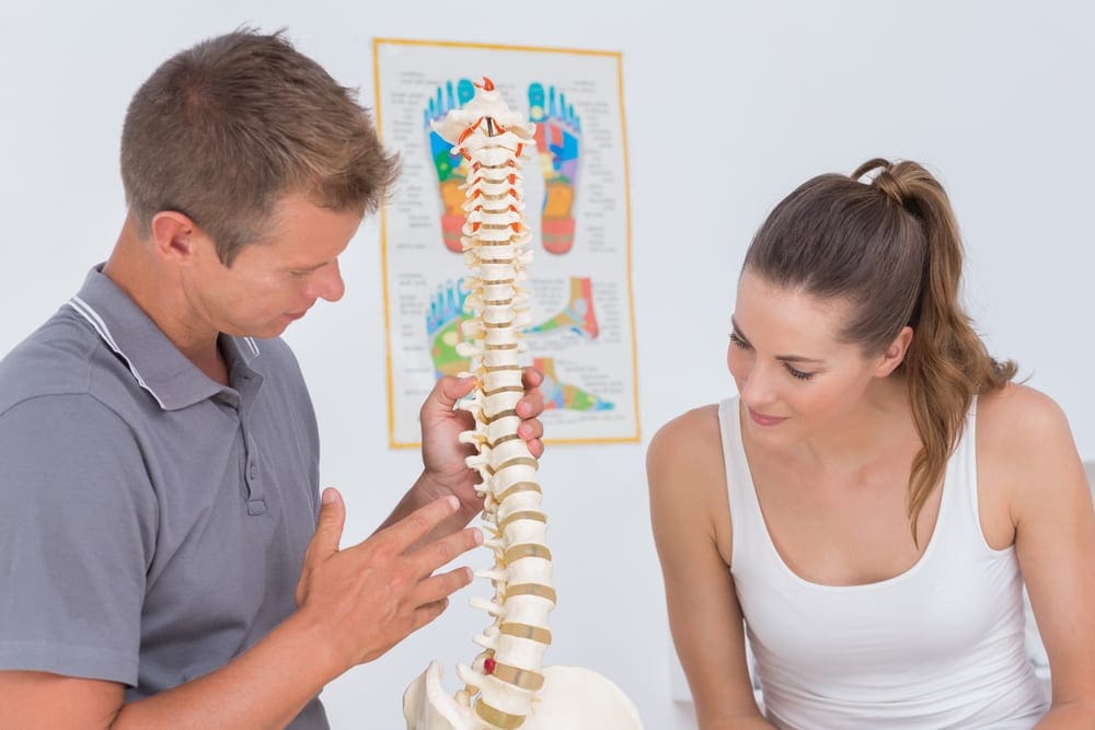
Understanding Cervical Myelomalacia
What is Myelomalacia? According to the MedicoLexicon, it is simply the �softening of the spinal cord�.3 Basically, it is ischemia that takes place in the spinal cord from abnormal pressure placed upon it. If left untreated, then that continues to spread and cause further damage to the cord. Once the cord has been damaged, there is no repair. Brandy Carrelli gives us a concise definition and the ramifications of it left untreated:
�The myelomalacia definition, strictly speaking is the �softening of the spinal cord�. After an acute injury, bleeding of the spinal cord may occur. As a result, there is �subsequent softening of normal tissues�. Myelomalacia can be caused by trauma or disease, but if it worsens, and if the bleeding reaches the cervical region of the body, it can be fatal. Bleeding can make the tissue necrotic. Fractured vertebrae can lead to bleeding in the spinal cord, as can some back surgery. Osteoporosis may also contribute to spinal instability and hemorrhaging. Sometimes circulatory problems can lead to a deterioration of tissues and bleeding. Myelomalacia can progress into impairment in the functioning of the lower extremities, below-normal or absent reflexes of the anus and pelvic limbs, loss of pain perception in the caudal region (near the coccyx), depression, respiratory problems due to �diaphragmatic paralysis�, and even neurological issues. Death could result from the respiratory paralysis. Damage occurs to the central nervous system. At first, the spinal cord damage may be minor. The most commonly injured areas are the lumbar spine (lower back) and cervical vertebrae (upper spine area).4
Disc degeneration, herniations (all variations) and bulging all describe what has happened to the disc itself. Once you have established a definitive diagnosis, then the question becomes, how is the disc affecting surrounding neurological components? Myelomalacia is the effect of that disc when the cord is affected by pressure. If there is bleeding into the cord, then the cord begins a degenerative spiral that can happen rather quickly. As you have read above, it can take what may simply appear as a minor issue to the patient that can lead to major neurological compromise and in extreme cases may lead to paralysis or death. Therefore, it is important carefully analyze the clinical indicators and image accordingly.
Myelomalacia is a relatively rare occurrence. According to Zhou, Kim, Vo and Riew,
�The overall prevalence of cervical myelomalacia was relatively low in the studied population, and it was affected by age, sex, and the specialties/subspecialties of referring providers. These results may help direct treatment guidelines and allow for informed discussions with patients in terms of the risk versus the benefit of surgery.�5 Pg. E252
It is a very common occurrence for the presence of disc bulging and herniations in chiropractic practices. It is of utmost importance for the chiropractor to not only order MRI when clinically indicated, it is important to be able to interpret those images as well. Once the clinical indicators begin to show a different story than presented by the patient symptomatically, it is the responsibility of the chiropractor to make the appropriate diagnosis, prognosis and treatment plan. In this case, that is an immediate neurosurgical referral. Although not a common finding in a chiropractic office, one must still be alert to the possibility of Myelomalacia. Managing the patient based upon an accurate diagnosis is your ultimate goal, and sometimes adjusting the patient isn�t the best first option as diagnosis and prognosis supersede treatment.
The scope of our information is limited to chiropractic and spinal injuries and conditions. To discuss options on the subject matter, please feel free to ask Dr. Jimenez or contact us at 915-850-0900 .
REFERENCES:
Fardon, D. F., Williams, A. L., Dohring, E. J., Murtagh, F. R., Gabriel Rothman, S. L., & Sze, G. K.
Studin M., Owens W. (2016) Bulging Discs and Trauma: Causality and a Risk Factor, American Chiropractor 34(6) 18, 20,22-24, 26, 28
http://www.medilexicon.com/dictionary/58294
Carrelli, B (2016) What is Myelomalacia? https://www.echiropractor.org/myelomalacia/
Zhou, Yihua; Kim, Sang D.; Vo, Katie; Riew, K. Daniel (2015) Prevalence of cervical myelomalacia in adult patients requiring a cervical magnetic resonance imagingSpine (Phila Pa 1976). 2015 Feb 15;40(4):E248-252.
Additional Topics: Recovering from Auto Injuries
After being involved in an automobile accident, many victims frequently report neck or back pain due to damage, injury or aggravated conditions resulting from the incident. There’s a variety of treatments available to treat some of the most common auto injuries, including alternative treatment options. Conservative care, for instance, is a treatment approach which doesn’t involve surgical interventions. Chiropractic care is a safe and effective treatment options which focuses on naturally restoring the original dignity of the spine after an individual suffered an automobile accident injury.
New studies in neuroscience suggest chiropractic care affects much more than back and neck pain. Celeste McGovern investigates an emerging body of evidence that spinal manipulation also improves your brain.
Imagine a convention that mixes cutting-edge natural health seminars with a surfer dude�s attitude, a revivalist�s enthusiasm and a good measure of live rock-�n-roll. That�s the California Jam, which took place in Costa Mesa in January. Billed as �the biggest health, wellness and chiropractic event on the planet�, it�s an annual meeting of thousands of unapologetically alternative practitioners who mill about three floors of exhibitions, sampling detox juices, protein snacks, �bulletproof� coffee and vitamins.
There�s a buzz about urine tests for metabolites; people are talking cellular detoxification and energy-balancing therapies, and they�re trading spinal adjustments on a row of tables. Inside the auditorium, a roster of headliner speakers takes the stage for two days, but one of the biggest ticket draws this year was a relatively unknown figure: neurophysiologist and chiropractor Heidi Haavik, who is pioneering a whole new field of research into what happens to a person�s brain when a chiropractor adjusts their spine.
�There is so much more to chiropractic care than back and neck pain, and headaches,� enthuses Haavik, who studied neuroscience after graduating from the New Zealand College of Chiropractic and is now focusing on research.
Up to now, there�s been a gulf between the available published research and the practice. A handful of studies have shown that chiropractic works only modestly�yet substantially better than drugs�at nipping neck and back pain,1 and may help with migraine,2 and even mysteriously lower blood pressure3 which, for 40 years, has been linked to joint dysfunction in the neck.
But the research is hardly enough to support its position as the most popular alternative medical treatment for more than a century, used by 30 million people in the US alone each year.
�Haavik�s research may finally explain scientifically the amazing results chiropractors have in clinical practice,� says Ross McDonald, a practising chiropractor and President of the Scottish Chiropractic Association.
The neuroscience studies explore the underlying mechanism of those results�how the spine and central nervous system (CNS) are interconnected and �talk� to each other, and how dysfunction in the spine can affect health and well-being.
One of Haavik�s studies, published this year in the journal Brain Sciences, looked at the effect of chiropractic adjustments in 28 patients with �subclinical� pain�those with a history of intermittent back or neck ache or stiffness for which they were never treated�but who were in pain the day of the experiment. On examination, all had tender spots and restricted joint movement in their spines.
Compared with �sham� adjustments, chiropractic spinal adjustments of these people induced significantly greater brain activity, or �cortical excitability� (which has to do with neuro-electrical signals produced when brain or peripheral muscles are stimulated), as measured by transcranial magnetic stimulation (TMS), which uses magnetic fields to stimulate nerve cells in the brain, as well as arm and leg muscle strength.
Increases in muscle strength have proved to be driven by brain activity resulting from spinal manipulation, and not by any changes made to the spinal cord itself. This offers a host of possibilities for, say, recovering muscle strength after nervous-system injuries. As the study concluded, �spinal manipulation may therefore be indicated� for patients who have lost muscle tone, or are recovering from a stroke or from orthopedic surgery that affects the muscles. It may even be of interest to athletes who participate in sports.4
These findings have confirmed a 2015 study which showed that, following a full-spine chiropractic adjustment session, voluntary leg muscle strength in study participants increased by 16 per cent, while electrical activity readings from the measured muscle increased by nearly 60 per cent. But most spectacularly, the researchers (from the Centre for Chiropractic Research in New Zealand) discovered a 45 per cent increase in the reflex pathway �drive� from the brain to muscle (an indicator of the ability of the brain to activate it). By contrast, the control participants who underwent the sham adjustment actually lost strength and brain drive to the measured muscle.5
This same Auckland-based team, led by Haavik and two colleagues are now embarking on some groundbreaking research involving brain-body communication in stroke patients.6 A preliminary study had tested the effect of a single chiropractic adjustment on 12 stroke patients, and found that it increased leg muscle strength by an average of 64 per cent and brain drive to the limb by more than 50 per cent. In contrast, both measurements fell after the sham adjustments in the controls.
Ordinarily, you wouldn�t expect to see muscles gain in strength after being asked to repeatedly resist something because muscles become fatigued. Now, that we have the technology to objectively measure an increase in muscle strength after an intervention, Haavik says, these results suggest that�chiropractic care is not only preventing fatigue, but making [muscles] even more efficient at producing force�.
The potential results of the new study could have a significant impact on the role of chiropractic care in people who have reduced muscle function as a result of stroke, she says.
Injury Risk
One interesting recent study by Haavik and her colleagues looked specifically at the impact of chiropractic on the risk of falls among older people.7
Falling is a significant cause of death, injury and health decline in the elderly, with about 30-40 per cent of older adults who still live independently suffering from at least one fall each year or more as they age.
In this randomized controlled trial, half of the group of 60 community-dwelling people, aged over 65 and living in Auckland, received 12 weeks of chiropractic care (two visits per week), while the other half received the �usual care�, which didn�t include seeing a chiropractor.
The patients were tested on their proprioception (in this case, their awareness of where their ankle joint was positioned), postural stability and ability to process �multi-sensory� information�a sound-induced flash illusion test, using flashing lights and beeps. This test is used to screen for fall risk, as it measures how well people can process two different kinds of stimuli at a time.8 The participants were also given a sensorimotor function test, which measured their ability to move their feet in response to a panel that suddenly lit up on the floor, plus a questionnaire based on their self-perceived health-related quality of life.
Over the 12 weeks of the study, the group receiving chiropractic care showed significant improvement in ankle joint position sense, meaning their brains may have become more accurately aware of what was going on in their feet; they were also able to react and move their foot onto the illuminated panel on the floor more quickly than before the chiropractic care. These improvements were not seen in the control group.
The chiropractic patients were 13 per cent better able to accurately report the correct number of flashes with the corresponding number of beeps�meaning they had lowered their risk for falls.
What�s more, at the end of the study, the participants who had received the chiropractic care reported a 2.4-fold increased improvement in the quality-of-life questionnaire compared with the controls.7
Your Plastic Brain
Haavik is now trying to explain how chiropractic achieves all this, and why restoration of proper movement is able to so profoundly affect the brain and overall health.
The CNS�the brain and spinal cord�and all the nerves beyond the CNS (the peripheral nervous system, or PNS) is a complex network comprising as many as 10 billion nerve cells (also called �neurons�) and 60 trillion synapses�tiny little junctions between neurons that mediate the �talk� across highly specialized neural circuits via chemicals called �neurotransmitters�. Indeed, nerves feed out of each segment of the spine like strands of spaghetti, and facilitate communication back and forth with various regions of the body.
Everything we do�from our basic motor reflexes to our capacity to experience abstract thoughts and feelings�relies on the precision of the computational processes performed by these CNS and PNS neural circuits. They, in turn, depend on having healthy excitatory and inhibitory systems.
A neuron gets �excited� when it�s �talked to� loudly enough, or stimulated, and this sends an electrical message down one of the neuron�s extensions (called �axons�), so allowing it to talk to another nerve cell by releasing more neurotransmitters at the synapses.
Such talk happens all the time as input comes in from our external senses (eyes, ears, mouth, nose and touch), as well as through an inner �map� of the location of our muscles and joints in three-dimensional space relative to each other (proprioception), as the brain carries out its decisions and functions.
Contrary to decades of scientific dogma, a recent wave of research has shown that the brain is actually highly adaptable to its ever-changing environment throughout life. It does this by keeping an up-to-date tab on its sensory inputs and its internal map of the self. This ability to adapt is known as �neural plasticity�.
Haavik likens the plasticity of the CNS to the subtle changes in the bed in a flowing stream. �You can never really step into the same river twice; the water, stones and silt of the riverbed are constantly changing,� she says. Likewise, your brain is changing with every thought and every execution, and is in a constant state of flux.
In fact, she believes her research demonstrates that vertebral subluxations (dysfunctional spinal segments; see box, page 33) lead to a breakdown in the way the brain perceives and controls the movement of the spine. And this spinal dysfunction doesn�t just affect how the brain then perceives and controls the spine, but also how it perceives and controls other parts of the body as well.
When the brain gets even slightly wrong information, it builds a faulty map that can impede neural signaling as effectively as damped sensory input�like wearing a blindfold or losing the sense of taste. And that translates to faulty functioning.
Chronic pain and neurodegenerative disorders have been linked to these faulty perceptions by the plastic brain.9 �Pain and conditions with other symptoms don�t necessarily happen all of a sudden for no reason. They can slowly develop without your awareness, a bit like a thousand straws on a camel�s back before it breaks,� says Haavik. �Only when the last straw is added do you feel the effect.�
Haavik�s team hypothesizes that spinal adjustments that restore normal movement also restore more normal data input from the spine to the brain. This, in turn, allows the spinal cord, brain stem and brain to process any incoming information more coherently.
�We believe this to be the mechanisms by which adjustments of vertebral subluxations can improve nervous system function, as observed daily in chiropractic practices all around the world.�
While the New Zealand researchers are reluctant to speculate on immunity, an emerging body of research is demonstrating the interconnectedness of both the nervous and immune systems too. An entirely new lymphatic system in the brain was only discovered in 2015 by a team of researchers at the University of Virginia,10 which highlights how limited our understanding of the brain, and the effect of the nervous system on global health, still is. It also raises further questions about how improving one system can lead to improvement in the other�and so perhaps why some people experience benefits to their immune-mediated disease with chiropractic manipulations.
�What is becoming clear is that chiropractic care seems to impact our brain�s inner reality by restoring the proper processing and integration of sensory information, which alters the way our brain controls our body,� says Haavik.
�It�s so exciting to see that there are other possible ways now to explain the effects of chiropractic that are actually congruent with current neuroscience,� she adds. �It�s actually more profound and powerful than we could have ever thought.�
The Many Faces Of Chiropractic
There are two schools of thought in chiropractic: the �mechanics�, who claim it should be absorbed into mainstream medicine as a standardized treatment for back and neck pain; and the �vitalists�, who believe that the treatment is much more far-reaching, as they�ve seen it help cases of fatigue, joint pain, migraines, allergies, asthma, bedwetting and even infertility.
The latter philosophy is radically different from the current medical paradigm. �The body has an innate ability to heal, provided there is no interference,� says Gilles LaMarche, vice president of professional relations at Life University in Atlanta, Georgia, the world�s largest chiropractic college. �It is self-developing, self-maintaining and self-healing.�
In this vitalistic view of chiropractic, when you get an infection or scrape your knee, the best practitioner merely assists the body in getting on with its own spontaneous and spectacular business of healing itself.
The chiropractor�s job, as vitalists see it, is to remove any interference in the body at the level of the spine, which they consider central.
�Conventional medicine doesn�t interpret symptoms as we interpret symptoms,� explains LaMarche from his end of chiropractic.
He sees fever, for instance, as one of the body�s natural mechanisms to fight infection: raising the body�s temperature kills bacteria and viruses, and facilitates other immune functions.
�Many doctors see fever as bad, as something to reduce,� he says, �and they give Tylenol [paracetamol], not considering it as a toxin that is actually going to stay in the liver and therefore interfere with healing and health.�
How Chiropractic Changes The Brain
So what�s going on in the brain after a chiropractic adjustment that could be increasing muscle strength in stroke patients? As a 2016 study from Aalborg University Hospital in Denmark demonstrated, a single chiropractic adjustment helps to improve something called �somatosensory integration� (when the brain receives accurate sensory input, so allowing it to properly organize and execute subsequent behaviors).1
Such a change mostly happens in the prefrontal cortex, that part of the brain known to be a key player in executive functions. It�s a sort of command control centre, integrating and coordinating the multiple neural inputs from a constantly changing environment to solve problems and achieve goals.
�Chiropractic care, by treating the joint dysfunction, appears to change processing by the prefrontal cortex,� the authors conclude.
So, while some chiropractors (and their patients) may have thought their adjustments were making changes locally and directly from the spine, in fact, the change is apparently effected indirectly by being sent to �central command� (the brain), then redirected back down neuronal chains to give the perception of reduced pain as well as other benefits.
�This suggests that chiropractic care may, as well, have benefits that exceed simply reducing pain or improving muscle function and may explain some claims regarding this made by chiropractors,� the study researchers say.
These claims include the ability of adjustments to increase muscle strength and core stability, improve reaction time and proprioception (your awareness of your body�s position in space), and so reduce the risk of injury.
What Is A Subluxation?
In 1895 in Iowa, the founder of chiropractic, Daniel David Palmer, claimed to have restored the hearing of deaf janitor Harvey Lillard by adjusting the part of his spine that Palmer could feel was �out of alignment�.
From this, he devised a theory that �misaligned� or �out-of-place� spinal segments interfere with proper nerve function, and that �adjusting� these segments back to their normal position relieves pressure on the nerves and restores neural function.
Chiropractors assess spines for areas where some of the small muscles that attach to the individual vertebrae have become tight due to injury, hunching over mobile phones and computers, or simply overuse. When these tight muscles cause the vertebrae�the small bones that make up the spine�to twist, certain parts of the bones can protrude and feel �misaligned� or �stuck�. Chiropractors call it a vertebral �subluxation� or �joint restriction�.
�It is more that a bone is functioning or moving in a less than ideal way�in a manner that is not �normal� for the body,� says Heidi Haavik.
And chiropractors counter this abnormality by �adjusting� it. �We don�t really put bones back in place when we adjust the spine,� she explains. The aim of the short, quick movements of chiropractic adjustments to the spine are to restore its natural range of movement.
How To Find A Good Chiropractor
All chiropractors must attend a licensed chiropractic college or university, and undergo at least four years of training in anatomy, neurology, physiology, radiology, pathology, clinical diagnostics and clinical nutrition, as well as physiotherapy and chiropractic techniques.
In the UK, chiropractors must pass a national exam to ensure competency. It is illegal to practice without first registering with the General Chiropractic Council.
Apart from these legal requirements, chiropractors have a broad range of approaches, specialities and techniques. Make sure to choose a chiropractor who:
��Meets your particular needs. Some chiropractors take a biomechanical approach, or treat a narrow range of conditions and only see people when they have a problem, like pain, while others take a �wellness� approach and treat people to prevent problems. Many chiropractors have special areas of focus: sports injuries, pregnancy, children, or even functional medicine, testing for metabolic deficiencies such as low vitamin D levels and prescribing supplements.
� Has a good reputation. It�s worth considering if other people have had good results.
� Talks with you at no cost to discuss your needs and their skills and services, and employs techniques that suit you. Some chiropractors use manual adjustments only, while others use devices like drop-tables�examination tables that move when the chiropractor adjusts so the impulse is delivered by the release-action of the table�and activators�hand-held tools that resemble a tire-pressure gauge and are spring-activated to deliver small and precisely controlled impulses to areas like the cervical (neck) spine. Some may also be trained in techniques like acupuncture, dry needling (acupuncture needles are inserted in muscle tissue to stimulate the release of �trigger points�, where muscles have gone into spasm) and active release technique (ART), which also targets contractions of muscles, ligaments and tendons to reduce joint stress.
� Carries out a thorough assessment before beginning treatment. A medical history and physical exam should be done to rule out conditions that need further referral or should not be treated by chiropractic. A chiropractor is trained to perform and read X-rays, which are sometimes required, but only if they meet standardized criteria.
� Gives you clear outcome measures to gauge improvement, such as less pain or an overall improved sense of wellbeing.
� Gives you enough time and attention. The best practitioner is also a coach or partner who can help you achieve your best state of health. Only choose someone who truly supports you.
Case Report: The Assessment of Traumatic Cervical Spine Injury and Utilization of Advanced Imaging in a Chiropractic Office.
Abstract: the objective is to explore the standard of care regarding the assessment of cervical spine injuries in a setting of a chiropractic office. Diagnostic studies include physical examination, range of motion studies, orthopedic testing and cervical spine. MRI.
Introduction: On January 30, 2017 a 49 year old female presented in my office to a second opinion examination at the request of her attorney. She had been involved in a rear-end collision on 12/12/2015. (2) She was transported to a local hospital and arrived with complaints of headaches, disorientation, right-sided neck pain and right arm pain. At the hospital emergency department CAT scan was taken of her brain, which proved to be negative. She received prescriptions of muscle relaxers and pain relievers and instructed to visit her primary care physician if her symptoms persisted.
Initial Examination
She consulted a local Chiropractor on December 15, 2015. The initial examination included the following from my review of the doctor�s notes: Presenting complaints were right-sided neck pain that radiates to the right arm. The doctor�s records show a positive cervical compression test and a positive maximum cervical compression test. Both produced pain bilaterally worse on the right. Facet provocation tests were positive for facet disease. Right side radicular pain pattern includes the trapezius and deltoid. No x-ray studies were included in the doctor�s orders. The patient received 23 chiropractic treatments from 12/15/2015 through 4/5/2016 for a diagnosis of cervical sprain/strain. The treatments consisted of spinal manipulation and a variety of soft tissue therapies.
Around January 15, 2017 I received a phone call from a local attorney regarding this patient and asking if I would do a second opinion examination on her due to persistent neck pain and right upper extremity pain. The patient presented on January 30, 2017 for my evaluation. My clinical findings are as follows:
Vitals: Age 49, weight 170 lbs. height 5� 8�, B.P 126/82, pulse 64, Resp. 16/min.
Appearance: in pain
Orthopedic/Range of motion: All cervical compression tests produced pain with radiation bilaterally worse on the right. Range of motion studies revealed: 40 degrees of left rotation and 32 degrees of right rotation with radiating pain produced by both motions.
Palpation: cervical spine palpation produced centralized spine pain that radiates to the right shoulder with numbness in the right arm and hand.
The patient informed me during the examination that her pain made it difficult to sleep through the night. If she was on her right side her right arm and hand would go numb immediately. A big part of this patient�s life was riding and caring for her horse and she could not do either because it resulted in severe neck and arm pain.
My recommendation to her and her attorney was to obtain a cervical spine MRI with a 1.5 Tesla machine due to the high quality images it can produce. MRI is a highly sensitive tool to evaluation of neurologic tissue including the spinal cord and nerve roots. (1) I bypassed the x-ray at this time due to the clinical presentation and 12% of spinal cord with injuries having no radiographic abnormality. (3)
Imaging
Figure 1: T2 Sagittal Cervical Spine MRI
Fig 2: T2 Axial Cervical Spine with Scout line through C3/4.
Radiology Report: The report and the images demonstrated a right paracentral disc extrusion measuring 9 mm and extending 8 mm cranial/caudal causing abutment of the spinal cord. (Fig 1)(2) Additionally the diameter of the central canal was reduced to 8.1mm and projected into the right lateral recess resulting in severe stenosis of the right neural canal. (Fig 2) Additional findings not pictured: C4/5 demonstrated a 2.5 mm bulging disc with facet hypertrophy with moderate stenosis of the left neural canal and severe stenosis of the right neural canal. C5/6 demonstrated a 1.5 mm posterior subluxation narrowing the central canal to 9.1 mm with unconvertebral joint hypertrophy resulting in moderate right and severe left neural canal stenosis. C6/7 revealed a broad based disc herniation worse on the left measuring 3.6 mm resulting in severe neural canal stenosis bilaterally complicated by unconvertebral joint hypertrophy. The MRI findings correlate with the patient�s clinical presentation. (4)
Discussion: When the patient returned to a consultation on the MRI findings my recommendation was to consult a neurosurgeon. (3) Her attorney asked me if the treating doctor acted incompetently. My only response was that I would have ordered the MRI immediately before treating the patient with manual manipulation. The case is likely to go to trial and there is a good chance that I will be called in as an expert witness. It is almost a guarantee that the defense attorney will ask me if I would have treated the patient for such a long period of time without an MRI or whether the treating doctor could have made the problem worse. The failure to accurately determine a diagnosis may result in malpractice action or a board hearing or both for this treating doctor and I would have ordered the MRI immediately considering the radicular findings and symptoms. After any myelopathic or significant radiculopathic symptoms a referral of advanced imaging needs to be performed in order to conclude and accurate diagnosis, prognosis and treatment plan prior to rendering care. Diagnostic appropriateness in the case of traumatic injury or with any etiology with neurologic symptoms or findings necessitates following triage protocols. In this case, an immediate 2-3mm MRI of the cervical spine is clinically indicated and proved integral to the safe care of this patient.
The scope of our information is limited to chiropractic and spinal injuries and conditions. To discuss options on the subject matter, please feel free to ask Dr. Jimenez or contact us at 915-850-0900 .
References:
Haris, A.M., Vasu, C., Kanthila, M., Ravichandra, G., Acharya, K. D., & Hussain, M. M. 2016. Assessment of MRI as a modality for evaluation of soft tissue injuries of the spine as compared to intraoperative assessment. Journal of Clinical and Diagnostic Research, 10(3), TC01-TC05
Schneider RC, Cherry G, Pantek H. The syndrome of acute central cervical spinal cord injury, with special reference to the mechanisms involved in hyperextension injuries of cervical spine. J Neurosurg 1954; 11: 546�577.
Tewari MK, Gifti DS, Singh P, Khosla VK, Mathuriya SN, Gupta SK et al. Diagnosis and prognostication of adult spinal cord injury without radiographic abnormality using magnetic resonance imaging: analysis of 40 patients. Surg Neurol 2005; 63: 204�209.
Miyanji F, Furian J, Aarabi B, Arnold PM, Fehlings MG. Acute cervical traumatic spinal cord injury: MR imaging Findings correlated with neurologic outcome-prospective study with 100 consecutive patients. Radiology 2007; 243: 820�827.
Additional Topics: Recovering from Auto Injuries
After being involved in an automobile accident, many victims frequently report neck or back pain due to damage, injury or aggravated conditions resulting from the incident. There’s a variety of treatments available to treat some of the most common auto injuries, including alternative treatment options. Conservative care, for instance, is a treatment approach which doesn’t involve surgical interventions. Chiropractic care is a safe and effective treatment options which focuses on naturally restoring the original dignity of the spine after an individual suffered an automobile accident injury.
Title: Conservative care and axial distraction therapy for the management of cervical and lumbar disc herniations and ligament laxity post motor vehicle collision.
Dr. Alex Jimenez, doctor of chiropractic, focuses on the diagnosis, treatment and prevention of a variety of injuries and conditions associated with the musculoskeletal and nervous systems, utilizing several chiropractic methods and techniques. The following procedures may be similar to his own but can differ according to the specific issue and complications by which the individual is diagnosed.
Abstract: This middle-aged female was injured in a vehicle collision causing her to sustain disc and additional ligament injuries in the cervical and lumbar spine. Diagnostic studies included physical examination, orthopedic and neurological testing, lumbar MRI, multiple cervical MRI�s, CRMA with motion cervical radiographs and EMG studies. Typically, conservative care is initiated prior to interventional procedures, and this case study seeks to explore the usage of passive therapy for mechanical spine pain and noted anatomic disc lesions after failure of interventional procedures. She reported both short term and long term success regarding pain reduction along with improvement in her activities of daily living after initiating conservative care, and continued to report further reductions in pain with periodic pain management using conservative care.
Introduction: The 49-year-old married female (Spanish speaking patient) reported that on March 4th, 2014 she was the seat-belted driver of a truck that was struck by a much larger fuel truck changing lines, hitting her vehicle at the front passenger side (far side, side impact). The force of the impact caused her truck to be lifted up and the right wheel popped off. Her head hit the window after impact and the spinal pain and complaints started approximately 24 hours later. Two days after the crash she went to the emergency department. Occupant pictures were taken describing an out of position occupant injury. She did not report any additional significant trauma after the collision.
Initial Diagnosis and Treatment for Disc Herniations
Prior to her evaluation at our clinic, she utilized multiple providers for diagnosis and treatment over the course of 11 months. She went to the emergency department, utilized 3 pain management medical doctors, neuropsychologist and a cognitive rehabilitation therapist. Imaging included radiographs and MRI of the right shoulder revealing rotator cuff tear; radiographs of the lumbar and thoracic spine, and left hand; CT of the head and cervical spine were performed; MRI cervical (3) and lumbar spine. Medications prescribed included Fentanyl, Percocet, Naprosyn, Cyclobenzaprine, Norco, Hydrocodone-acetaminophen, Soma, and Carisoprodol. Physical therapy was provided for spinal injuries and she did not respond to treatment. The neurosurgeon recommended epidural steroid injections and facet blocks. Cervical nerve blocks and cervical trigger point injections, cervical and lumbar epidural steroid injections (ESI), lateral epicondyle steroid injections were performed, none of which were palliative. Post-concussion disorder and PTSD with major depressive disorder were diagnosed.
On February 12th, 2015, she presented to our office with neck pain (average 6/10 VAS) that affected her vision, with paresthesia�s in both upper extremities radiating to the hands with numbness. She had low back pain (average 6/10 VAS), and she additionally reported paresthesia at the plantar surface of feet bilaterally. She had left elbow pain, right shoulder pain, knee pain, headaches and �anxiety� along with anterior sternal pain.
Her injuries were causing significant problems with her activities of daily living. Summarily she had increased pain with lifting, increased pain and restricted movement with bending, walking and carrying. She had been unable to perform any significant physical activity from the time of the crash in March 2014 until March 2015. Her right hand was always hurting and her forearms. She was not able to clean windows or do laundry, difficulty using stairs, problems with mopping, ironing and cleaning. She had to limit her walking and jogging primarily due to neck pain and right arm pain. She was not able to sit for long periods of time and sleeping was disrupted due to numbness in her hands. She was only able to walk on a treadmill for 10 minutes before having to stop due to pain, prior to the crash she would exercise for an hour.
Prior History: No significant prior musculoskeletal or contributory medical history was reported.
Research Study Conclusions
Clinical Findings (2/12/15): She had a height of 5�2�, measured weight of 127 lbs.
Visual analysis of the cervical spine revealed pain in multiple ranges of motion including flexion, extension, bilateral rotation and bilateral side bending. On extension pain was noted in the upper back, on rotation pain was noted in the posterior neck, and on lateral flexion pain was noted contralaterally.
Visual analysis of the lumbar spine revealed pain in the low back on all active ranges of motion, including flexion, extension and side bending, pain primarily at L5/S1.
Dual inclinometer testing was ordered based on visual active range of motion limitations with pain.
Sensory testing was performed of the extremities, C5-T1 and L4-S1. No neurological deficits other than right sided C5 hypoesthesia.
Foraminal compression test produced pain in the cervical spine. Foraminal distraction test caused an increase in pain in the neck. Jackson�s test on the right produced pain bilaterally in the neck. Straight leg raise bilaterally produced low back pain, double Straight leg raise produce pain at L5/S1 at 30 degrees.
Muscle testing of the upper extremities was tested at a 5/5 with the exception of deltoid bilaterally tested at a 4/5. The patient�s deep tendon reflexes of the upper and lower extremities were tested including Triceps, Biceps, Brachioradialis, Patella, Achilles: all were tested at 2+ bilaterally, equal and reactive. No evidence of clonus of the feet and Hoffman�s test was unremarkable.
C3-C5 right sided segmental dysfunction was noted on palpation. T5-T12 spinous process tenderness on palpation. Low back pain on palpation, particularly L5/S1.
Imaging Results
MRI Studies:
I reviewed the cervical MRI images taken May 2014 with the following conclusions (images attached):
Dramatic reversal of the normal cervical curvature, apex C5/6.
C5/6 herniation, indentation of the spinal cord anteriorly. High signal posterior on STIR.
Due to the angular kyphosis of the cervical spine and axial slices performed, C6/7 slices did not render a pure diagnostic image for disc disruption.
Fig. 1 (A) T2 Axial C5/6, 2 months post injury Fig. 1 (B) Sag T2 C5/6
I reviewed cervical MRI images taken September 17th, 2014 approximately 6-months post injury, and rendered the following conclusions:
Reversal of the normal cervical lordosis.
C5/C6 herniation (extrusion type) with indentation of spinal cord, appropriate CSF noted posteriorly.
I reviewed the cervical MRI dated October 24th, 2015 (images attached):
C4/5 herniation, extrusion type, left oriented into the lateral recess and neural canal causing moderate neural canal stenosis
Fig. 2 (A) 3D Axial C4/5, 19 months post injury Fig. 2 (B) Sag T2 C4/5
IMPRESSIONS: C4/5 herniation noted on 10/24/15 was not noted on prior images. The patient reported no additional injury or symptoms between MRI studies, so it is postulated that initial slices revealed a false negative; or due to the severity of abnormal cervical biomechanics, it is possible that the C4/5 disc herniated between the pre/post MRI�s with no significant increase in symptomatology. There was improvement at C5/6 related to disc abnormality and cord involvement (see below).
Fig. 3 (A) 3D Axial C5/6, 19 months post injuryFig. 3 (B) Sag T2 C5/6, 19 months post injury
The cervical flexion/extension images were digitized February 2016 and interpreted by myself and Robert Peyster MD, CAQ Neuroradiology, revealing a loss of Angular Motion Segment Integrity at intersegment C6/C7 measured at 19.7 degrees (maximum allowed 11 degrees), indicating a 25% whole person impairment according to the AMA Evaluation of Permanent Impairment Guidelines 5th edition1. CRMA provided from Spine Metrics, independent analysis.
Evidence of significant ligament injury causing functional subfailure was measured at C3/4 at 10.4 degrees and at C4/5 measuring 10.9 degrees regarding angular motion. Abnormal paradoxical translation motion measured at C6/7 and C7/T1.
Functional Testing:
EMG of the upper extremity revealed bilateral C6 radiculopathy, December 16th, 2015.
Range of Motion Cervical Dual Inclinometry:
Initial Max 4 months later % Improvement
Cervical Extension 44 42 -5%
Flexion 40 62 55%
Cervical Left 25 41 64%
Lateral flexion Right 12 26 117%
Cervical Left 46 59 28%
Rotation Right 43 73 70%
Conservative treatment rendered: A neurosurgical referral was made for assessment and surgical options. Conservative care was initiated despite failure of other medical procedures since there is �further evidence that chiropractic is an effective treatment for chronic whiplash symptoms�2-3. The patient was placed on an initial care plan of 2-3x/week for 5 months, with a gap in passive care for 1 month.
23 chiropractic visits. Instrument adjusting cervical spine was utilized with Arthrostim. Non-rotatory HVLA (high velocity low amplitude) spinal adjustments were performed thoracic and lumbar spine, applied A-P. No HVLA spinal adjustments to the cervical spine.
Prior to being placed at maximum medical improvement she had persistent low back symptoms, continued tingling in the fingertips and occasional neck pain at a 4/10, with her upper extremity paresthesia�s improved 50%. She continued with pain management chiropractic care after MMI, approximately 1 visit every 3-4 weeks with axial distraction to the cervical and lumbar spine, chiropractic adjustments as needed (PRN). 2 years/9 months post collision, and 1 year/9 months after initiating conservative care at our clinic, she reports only slight (1-2/10 VAS) spinal complaints with her primary concern being a torn rotator cuff injury from the crash that still requires surgical intervention. After initiating care at our clinic, no other interventional procedures were performed, although medication usage persisted. Due to improvement in symptoms and functional status, spinal surgery was not considered. She still utilizes Aleve PRN, 1-2 tablets. No significant active spinal rehabilitation was utilized. The patient was given at home active care consisting only of cervical and lumbar stretches, walking, and ice to affected areas.
Conclusion:While chiropractic care is safe even in the presence of herniations and radicular symptoms, �the likelihood of injury due to manipulation may be elevated in pathologically weakened tissues�4. Due to cord involvement, the provider decided to utilize low force procedures although HVLA spinal adjustments to the cervical spine could be considered safe due to lack of cord compression. HVLA spinal adjustments A-P were utilized in the lumbar and thoracic spine not only for short term pain relief but also as part of managing the chronic low back pain secondary to ligament/disc damage. While previously theorized to be only episodic, low back pain can be a lifelong condition requiring patients to seek ongoing care5. This care can be active, passive, pharmaceutical, interventional, or conservative in nature, but ongoing pain management therapy is often required for permanent ligament conditions. There is clear benefit to the patient population to be able to avoid surgical intervention due to risks, costs, ongoing prescription medication usage and adjacent level degeneration in the future6. Avoiding opioid usage is also a high priority in today�s environment.
Long term conservative care utilizing instrument spinal adjusting and targeted axial distraction therapy significantly reduced subjective reporting of pain, increased activities of daily living, and allowed the patient to avoid further spinal injections or surgical intervention. Considering that various interventional procedures failed prior to conservative care, it is important that providers work in an interdisciplinary environment such that the safest, and in this case the most effective, therapies are utilized first to reduce risk to the patient and maximize benefit and reduce costs.
In this case study, the patient utilized multiple pain management physicians, cervical nerve blocks and epidural steroid injections, and was not directed to conservative care for 11 months post injury. Utilizing chiropractic as conservative care would have enabled this patient to regain function and decrease pain while reducing costs and risks that are associated with medications and interventional procedures.
Competing Interest: There are no competing interests in the writing of this case report.
De-Identification: All of the patient�s data has been removed from this case.
The scope of our information is limited to chiropractic and spinal injuries and conditions. To discuss options on the subject matter, please feel free to ask Dr. Jimenez or contact us at 915-850-0900 .
Cocchiarella L., Anderson G. Guides to the Evaluation of Permanent Impairment, 5th Edition, Chicago IL, 2001 AMA Press.
Khan S, Cook J, Gargan M, Bannister G. A symptomatic classification of whiplash injury and the implications for treatment. Journal of Orthopaedic Medicine 1999; 21(1):22-25.
Whedon J, Mackenzie T, Phillips R, Lurie J. Risk of traumatic injury associated with chiropractic spinal manipulation in Medicare Part B beneficiaries aged 66-99 years. Spine, 2015; 40:264�270.
Hestbaek L, Munck A, Hartvigsen L, Jarbol DE, Sondergaard J, Kongsted A: Low back pain in primary care: a description of 1250 patients with low back pain in Danish general and chiropractic practices. Int J Family Med, 2014.
Faldini C., Leonetti D., Nanni M. et al: Cervical disc herniation and cervical spondylosis surgically treated by Cloward procedure: a 10-year-minimum follow-up study. Journal of Orthopaedics and Traumatology, June 2010.Volume 11, Issue 2,pp 99-103.
Additional Topics: Recovering from Auto Injuries
After being involved in an automobile accident, many victims frequently report neck or back pain due to damage, injury or aggravated conditions resulting from the incident. There’s a variety of treatments available to treat some of the most common auto injuries, including alternative treatment options. Conservative care, for instance, is a treatment approach which doesn’t involve surgical interventions. Chiropractic care is a safe and effective treatment options which focuses on naturally restoring the original dignity of the spine after an individual suffered an automobile accident injury.
It’s no surprise — overweight children who don’t properly learn self-regulating habits likely become obese adults. What is surprising is that one of the most common ways to help — restricting children’s diets — actually compounds the problem.
That’s the thrust of a new study by University of Illinois researchers who point to a disturbing pattern: Parents shame by withholding food due to weight gain, then children cope with the negative emotions by overeating.
Further exasperating the trend, overweight children are often rewarded with food by parents, and as they grow older, the children reward themselves with food.
Researchers who studied the pattern added a genetic component as well to better understand obesity. They reported that a child’s genetics, relating to cognition and emotion, likely play a key role. They found that when biological conditions were just right, a nudge by the social aspect sets kids on a path to obesity.
Kelly Bost, co-author of the study (published in Pediatric Obesity), and professor of child development at the University of Illinois, said: “When parents offer food to children whenever they are upset, children may learn to cope with their negative emotions by overeating, and they start to develop this relationship with food early in life; eating — especially comfort food — brings a temporary soothing. People intuitively understand that.”
The findings support the team’s hypothesis that a correlation exists between all factors: parenting approaches, combined with a child’s genetic make-up and restrictive feeding, and the child’s weight and the child’s propensity to be obese.
Bost said that children can effectively learn control for themselves: “Some of the things parents do, they may not think are related to how children are developing their eating habits. The ways parents respond or get stressed when children get upset are related in an indirect way. The way we respond to that emotion can help children to develop skills for themselves, to self-regulate, so that everyday challenges don’t become overwhelming things that they have to manage with respect to food.”
Bost and her team used data from the “Strong Kids” program, outreach developed by The Oregon Resiliency Project, an organization that is based on “research, training, and outreach effort aimed at social and emotional learning, mental health promotion, and social-emotional assessment intervention” of children, according to the organization’s website.
The team examined information about parents’ feeding styles, and how they typically reacted to their children’s (ages 2.5 to 3 years) negative emotions. The researchers examined these factors in combination with genetic data.
For the genetic factor, they looked at the COMT gene, a gene known for regulating cognition and emotion. This gene is the gatekeeper for dopamine, which controls the brain’s reward and pleasure centers.
Bost and her team studied minute variances in the gene pool to determine which children might be more susceptible to negative emotions or stress. They based their genetic research on the breakdown of amino acids in proteins that could lead to personality differences. One of them is the change produced by genetics in the form of a single part of our DNA: the nucleotide polymorphisms (SNPs). There are many types of SNPs; some affect the composition of protein and, depending on the change, affect the amount of dopamine in the brain, as presented by Psychology Today. Dopamine controls the brain’s reward and pleasure centers.
One type of SNP can change an amino acid from valine (Val) to methionine (Met). While largely academic, these two types of proteins influence emotion. Bost explained it best in the study: “We all carry two copies of genetic information — one from Mom, and one from Dad. In a person with Val/Val, the COMT system works three to four times faster than those with other combinations do, and therefore accumulates less dopamine in the front of the brain. Children who have at least one copy of Val tend to be more resilient emotionally. Those who are Met carriers have the propensity to be more reactive to negative emotion or stress.”
This genetic component was combined with the researchers’ studies. “We know that how parents respond to their children’s negative emotions influences the development of children’s response patterns over time,” Bost said in the study. “There is a whole body of literature linking emotion dysregulation to emotional overeating, dysregulation of metabolism, and risk for obesity, even starting at early ages. We wanted to begin to integrate information from these various fields to get a more holistic view of gene-environment interactions at this critical time in life for developing self-regulation.”
They began their research with a group of 126 children who were studied for the social aspect. For the genetic component, saliva samples were taken. Parents filled out questionnaires, rating how they typically respond to their common situations, including emotional outbursts.
Bost and colleagues found that parents most likely to use restrictive feeding were those who reported more frequent use of unresponsive stress-regulating strategies with their children — punishing or dismissive —and had children who were higher weight status and tested positive for the Met amino acid. But the same was not necessarily true for children who were Val carriers.
Bost and her team determined that breaking the cycle did not begin with blaming parents but instead by encouraging them to develop positive reinforcement and other techniques that could help their kids respond better and also help develop positive eating habits that likely would carry into adulthood.
While there exist myriad programs that focus on providing good nutrition or how to plan less stressful mealtimes, Bost explains that parents should also learn emotion regulation strategies in response to children who display emotional breakdowns and are eating to soothe — especially if the parents are restricting foods.
She added, “Sometimes the way parents respond is based on their own stress, belief systems, or the way they were raised. Educating parents from a developmental perspective can help them to respond to their children’s emotions in ways that will help their children learn to self-regulate their emotions and their food intake . . . responsive parenting involves an understanding of what stress-reducing approaches are most effective for a particular child.”
A new diet plan that dictates that you eat only one food for several weeks to lose weight fast is drawing warnings from many experts who say it is dangerous and only successful in the short term.
The Mono Diet (sometimes called the Banana Island or Monotrophic Diet) was popularized by a YouTube star nicknamed “Freelee the Banana Girl” (real name: Leanne Ratcliffe) who claimed it helped her lose weight, the New York Post reports.
Ratcliffe claimed to have lost 40 pounds eating close to 30 bananas a day.
A new version, the Sweet Potato Diet, promises the spud can help you lose 12 pounds in just two weeks. The hashtag #monomeal on Instagram, which highlights pictures of people’s meals containing a single food, has more than 38,000 posts, and the diet was one of the most searched in 2016, according to Google.
Frances Largeman-Roth, a registered dietitian and author of “Eating in Color,” tells the Post you can lose weight by eating only one food, but it’s likely to result from eating less.
“Yes, this diet can produce weight loss,” she says “But, the weight loss is a result of caloric restriction — not because any particular food is magically producing weight loss. It’s an incredibly restrictive and unbalanced diet and I do not recommend that anyone follow it.”
Experts warn such diets can also cause symptoms like dizziness and lead to some serious health problems, including dangerous metabolic changes and muscle loss.
Leg length discrepancy is a condition in which the legs are not of equal length. This might give an appearance that one leg is shorter compared to the other. The reasons for leg length discrepancy can be many, including defects that are congenital or may be acquired, which might include certain medical conditions, fractures, infections or injuries impacting the bone.
Leg length discrepancy might be a result of accurate discrepancy, which can be caused by real distinctions in the leg lengths. In other instances, the causes of leg length discrepancy might be due to circumstances that result in change in the angle of the hip or pelvic bone. In such cases, as the hip gets tilted to the other side and one side gets raised, the leg on that side seems to be shorter.
However, it is important to understand the foundation and causes of leg length discrepancy to handle the condition properly. It is also crucial to understand the impact of leg length discrepancy on an individual health and overall performance just as the the reasons are important. Mental and physical health can be affected by leg length discrepancy health insurance and will also be connected to spinal issues like scoliosis.
Can Limb Length Discrepancy Trigger Scoliosis?
Leg length discrepancy, due to uneven leg lengths, can impact the normal gait of the person. The main perform that is noticeable is the way a person walks or performs human anatomy actions. These can get afflicted or be difficult because of leg size discrepancy. Changes in normal movements can more lead to certain issues of the muscles like soreness, discomfort, weak imbalances or muscles on either side of the physique. Leg duration discrepancy can impact the hip, knees and ankle, can cause pain and dysfunction.
The muscles on both sides of the physique and those related to the hip can get pulled due to tilting of the hip-bone. This can be one the major effects of leg duration discrepancy, where the muscles get pulled to one side, creating changes in the curvature of the backbone. In to side ways pulling of the spinal curvature, which is termed as scoliosis, such adjustments can eventually result. There is much concern whether leg length discrepancy can cause scoliosis and it is important to understand correct therapy to be planned by this and a void further complications.
Limb Length Discrepancy and Scoliosis
Many studies have already been conducted, which revolve round the chance of leg duration discrepancy being an underlying cause of scoliosis. In the same time, leg length discrepancy can also result in pulling of the muscles that are back to one facet, which can contribute to some extent to or worsen existing scoliosis.
It might result in scoliosis, which might be useful in the beginning as the curvature gets tilted to one aspect. In scoliosis that is functional there might be slight tilting or pulling of the muscles to one side, without adjustments or damage to the structure of the spine. However, if functional scoliosis, which is caused or aggravated by leg-length discrepancy isn’t treated in time, it might worsen, causing changes in the structure of the curvature. This may result in structural scoliosis, which may not be disturbing and only more painful but also difficult to manage.
Some studies have revealed that scoliosis in certain persons is the result of mechanism, to make up for the leg length discrepancy. Simply stated, in leg length discrepancy, the legs are of unequal lengths, so to match the lengths the individual pulls the aspect down along with the hip starts to tilt. This, when continued for a longer period of time, can result in pulling to one aspect, making changes in the curvature. Scoliosis is one such change in spinal curvature, at which spine gets curved to one side, comprising alternative activities.
Symptoms of Scoliosis from Limb Length Discrepancy
A person that has developed scoliosis due to leg size discrepancy, usually presents with tilting of the hip. Along with the signs of leg length discrepancy, the individual may possibly also encounter pain in the muscles that are again, imbalances of muscle power and function of the muscles that are again. Bending, twisting movements might be difficult and it could also be painful to maintain or raise objects.
The appearance of the shoulders may possibly be different on account of scoliosis and one-shoulder can happen elevated in relation to the other. This could cause problems in neck, arm and shoulder movements and also hurt. It could sometimes result into serious degrees of scoliosis, if the status is left unattended.
Treatment of Scoliosis from Limb Length Discrepancy
It is importance to comprehend if leg-length discrepancy can trigger scoliosis. The treatment options might have to be planned appropriately if scoliosis has been resulted in by complications of leg-length discrepancy.
In some cases, leg size discrepancy can contribute to or worsen existing scoliosis, therefore, correcting leg duration discrepancy with heel raise have to be in the offing cautiously. It’s important to thoroughly examine any circumstance with leg-length discrepancy, as they can cause scoliosis in some instances. Prescribing a heel raise to appropriate leg length discrepancy can boost the chances of worsening the scoliosis due to tilting if scoliosis is obvious.
Hence, it really is essential to to examine the bio mechanics of the hi-P, evaluate the modifications in the spinal curvature in scoliosis as well as the tilting due to leg duration discrepancy. Depending on the the reasons, some cases of leg length discrepancy might require procedure for surgical correction of leg lengths. When the symptoms, scoliosis and causes like complications of leg length discrepancy, are correctly evaluated a multi disciplinary treatment approach may be planned.
Limb Length Discrepancy Explained (Video)
The scope of our information is limited to chiropractic and spinal injuries and conditions. To discuss options on the subject matter, please feel free to ask Dr. Jimenez or contact us at 915-850-0900 .
By Dr. Alex Jimenez
Additional Topics: Scoliosis Pain and Chiropractic
According to recent research studies, chiropractic care and exercise can substantially help correct scoliosis. Scoliosis is a well-known type of spinal misalignment, or subluxation, characterized by the abnormal, lateral curvature of the spine. While there are two different types of scoliosis, chiropractic treatment techniques, including spinal adjustments and manual manipulations, are safe and effective alternative treatment measures which have been demonstrated to help correct the curve of the spine, restoring the original function of the spine.
IFM's Find A Practitioner tool is the largest referral network in Functional Medicine, created to help patients locate Functional Medicine practitioners anywhere in the world. IFM Certified Practitioners are listed first in the search results, given their extensive education in Functional Medicine