Hypermobility Syndrome is a condition of the joints. Characterized by the ability of the joint to move beyond its normal range of motion and is sometimes called �loose joints� or �double jointed.� It is typically a genetic disorder and often identified in children. The gene passes from parent to child, so the condition tends to run in families. Estimated that 10 to 15 percent of children who are otherwise considered to be normal have joints that are hypermobile. However, it can be found in all ages and does not seem to be confined to a particular age group, ethnic group, or population although there are more cases of girls being hypermobile than boys.
Hypermobility Signs and Symptoms
The signs and symptoms of hypermobility can vary widely from person to person. Some people may not experience any symptoms while others have muscle and joint pain along with mild swelling. Usually noted in the evening or later afternoon as well as after moderate physical activity or exercise. The most common areas for pain and achiness are the elbows, knees, thigh muscle, and calf muscle. Often rest will provide relief.
A person who is hypermobile is usually more prone to soft tissue injuries and sprains. Additionally, the affected joints may be more inclined to become dislocated. It can also cause back pain, impaired joint position sense, and even flat feet, osteoarthritis, and nerve compression disorders. Other symptoms include increased bruising, chronic pain, loose skin, and thin scars. Children and young people who are hypermobile often experience growing pains more often than other children.
Most children will grow out of hypermobility; their joints will lose some of their flexibility as they get older along with the symptoms of rarely persist beyond childhood although some adults do find that they get dislocations and sprains much easier.
Causes of Hypermobility
The exact cause of hypermobility is not known, although it does seem to run in families. Genes play a large part in the process, particularly those involved in collagen production which is a vital protein for tendon, joint, and ligament development and function. There are also several�associated�conditions. Genetic disorders like Ehlers-Danlos and Marfan have hypermobility as a component as does Down Syndrome.
Hypermobility Treatment
Treatment for hypermobility depends on the patient. It depends on the symptoms that they are experiencing as well as the severity and how much of an impact the condition has on their quality of life. Mild symptoms may not require any treatment while more moderate to severe symptoms may warrant medication like naproxen, ibuprofen, or acetaminophen for pain. All of which,�can be bought over the counter.
Patients can ward off many of the symptoms or eliminate them by engaging in regular exercise, protecting the joints, practicing good posture, muscle strengthening exercises, and balancing techniques. Orthotics to correct flat feet can also be beneficial.
Chiropractic for Hypermobility
Many people use chiropractic for hypermobility pain and discomfort. The doctor will use adjustments to bring the joints into the appropriate movement pattern and the body into proper alignment, allowing the body to function as it should and relieves stress from joints that were compensating due to misalignment.
The patient may also be advised to do specific exercises at home, and get counseling on improving their posture. Because chiropractic treats the entire body, the patient will find that they learn how to best live with the condition without medication and manage pain naturally. Patients report dramatic improvement in their distress and mobility after regular, consistent chiropractic visits.
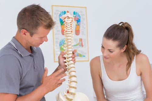
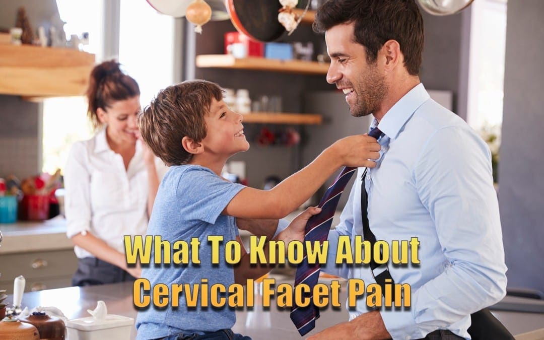
Cervical facet joint syndrome, or cervical facet osteoarthritis, is a degenerative condition marked by stiffness and pain in the cervical region (neck) of the spine.�Individuals can gain relief from various types of treatment, including chiropractic care.
Facet joint problems are among the most common sources of lower back and neck pain. They can cause debilitating, chronic problems with the neck and back and can lead to other more severe conditions and symptoms that can be disabling.
What is Cervical Facet Joint Pain?
The spine, comprised of a chain of bones known as vertebrae. Each one has two facet joints on the back side and a large disc on the front side, allowing the vertebrae to stack neatly, one on top of the other, providing stabilization for the entire body.
The facet joints are synovial joints, like other joints in the body and sometimes they can become inflamed or injured, causing pain and stiffness. Cervical facet joint pain is, quite literally, a pain in the neck. It means that the joints in the neck area have become injured or inflamed. Suffering from this condition can make it difficult for the patient to turn their head from side to side, or to move it up and down.
The cervical facet joints are almost always working. They undergo repetitive, constant motion and over time they can become torn or worn down. Problems within the joint can cause movement to be restricted, or it can have too much, both of which can cause pain.
Injury, such as whiplash, to the area, can also cause problems. If�not treated�appropriately�the condition�can be degenerative, and the patient can lose both flexibility and mobility, as well as suffer from chronic pain.
Symptoms of Cervical Facet Joint Pain
The symptoms of cervical facet joint pain tend to vary from patient to patient. A patient may experience one or several of these symptoms:
Tingling, weakness, or pain in the hand and arm
Neck pain
Upper back pain that can affect the shoulders
Pain between the shoulder blades
Headaches, typically located in the back of the head
Swelling and tenderness at the site of the inflamed facet joint
Decreased range of motion and flexibility in the neck
Treatment for Cervical Facet Joint Pain
When a patient diagnosed with cervical facet joint pain, the treatment is usually fairly conservative. Their doctor may recommend soft tissue massage, physical therapy, and posture correction. Combined with medications such as an anti-inflammatory like ibuprofen, or muscle relaxers to ease muscle spasms in the muscles that surround the affected joint.
If those methods do not give the patient relief, the doctor may take a more aggressive approach, prescribing facet joint injections that use steroid medications injected into the affected joint. This approach is intended to keep the pain localized while reducing it. The procedure can be performed in an outpatient setting and has a good record of being useful, but the results are temporary.
Chiropractic for Cervical Facet Joint Pain
Chiropractors have had much success in treating cervical facet joint pain. They can manipulate the areas that are affected, restoring painful, restricted facet joints to a point where they can move much more natural and without pain. Over time, with regular chiropractic treatments, they can help to reestablish range of motion in the neck area for their patients. All done without any medications or injections. It is a natural, gentle, effective method for relieving the pain and helping the patient enjoy a better quality of life.
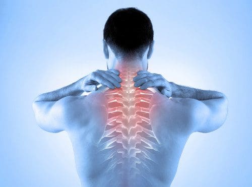
Sandra Rubio discusses the symptoms, causes, and treatments of neck pain. Headaches, migraines, dizziness, confusion, and weakness in the upper extremities are some of the most common symptoms associated with neck pain. Trauma from an injury, such as that from an automobile accident or a sports injury, or an aggravated condition due to improper posture can commonly cause neck pain and other symptoms. Dr. Alex Jimenez utilizes spinal adjustments and manual manipulations, among other chiropractic treatment methods like deep-tissue massage, to restore the alignment of the cervical spine and improve neck pain. Chiropractic care with Dr. Alex Jimenez is the non-surgical choice for restoring a patient’s overall well-being.
Neck Pain Symptoms & Chiropractic Treatment
Neck pain is a common health issue, with approximately two-thirds of the population being affected by neck pain at any time throughout their lives. Neck pain�that originates in the cervical spine, or upper spine, can be caused by numerous other spinal health issues. Neck pain can result due to the pinching of the nerves emanating from the vertebrae, or because of muscular tightness in both the upper spine and the neck. Joint disruption in the neck can generate a variety of other common symptoms, which�include�headaches, head pain, and migraines. There can also be a spinal joint discomfort. Neck pain affects about 5 percent of the global population as of 2010, according to statistics.
We are blessed to present to you�El Paso�s Premier Wellness & Injury Care Clinic.
As El Paso�s Chiropractic Rehabilitation Clinic & Integrated Medicine Center,�we passionately are focused on treating patients after frustrating injuries and chronic pain syndromes. We focus on improving your ability through flexibility, mobility and agility programs tailored for all age groups and disabilities.
If you have enjoyed this video and we have helped you in any way, please feel free to subscribe and recommend�us.
Dr. Alex Jimenez has great techniques to relieve the discomfort, the inflammation, the swelling, not only does he have a great technique to help with the horrible symptoms of sciatica, he also offers you great information when it comes to foods, anti-inflammatories, and we don’t go to prescription medications. So if you are looking for sciatica relief without the invasive procedures…you need to come see Dr. Jimenez.
Sandra Rubio
Are you currently suffering from debilitating sciatica symptoms? Chiropractic care may help you to find relief for your�sciatic nerve pain.�A doctor of chiropractic, or DC, regularly treats sciatica.
Sciatica is a collection of symptoms rather than a single condition, characterized by pain that originates from the lower back or buttock and travels down one or both legs into the feet. Sciatic nerve pain varies in frequency and intensity; minimum, moderate, severe and intermittent, constant, regular or irregular. Sciatica symptoms can happen when a spine illness, such as spinal stenosis or a bulging/ruptured disk, causes compression into the sciatic nerve or nearby nerves.
When this kind of compression occurs, it could lead to sensations of numbness or shooting pain. From the buttocks, back of the thighs, calves, and toes, sciatica pain may radiate down at times. Sciatic nerve pain is very similar to electrical shocks, and it may be dull, achy, sharp, toothache-like, and have pins�and needles feeling. Other symptoms include numbness, burning, and tingling sensations. Sciatica can be radiating or recognized as neuropathy pain, or neuralgia.
The misconception that sciatica is a disease�is common. However, sciatica is a symptom of a disease. Chiropractic care is a popular treatment which can help treat sciatica. The guide below discusses a comprehensive overview and a chiropractic treatment guide for sciatica.
Common Causes of Sciatica
Sciatica is commonly brought on by compression of the sciatic nerve in the lower back. Disorders known to activate sciatic nerve pain include lumbar spine subluxations, also known as misaligned vertebral bodies, herniated or bulging discs, also known as slipped disks, pregnancy and childbirth, tumors, and even non-spinal ailments such as diabetes, constipation, or sitting on an item�in the back pocket of your�pants.
One�frequent cause of sciatica is piriformis syndrome. Piriformis syndrome involves the piriformis muscle. The piriformis muscle and the thighbone located at the lower part of the backbone�connect and also assists in hip rotation. The sciatic nerve runs along these structures.
This muscle is vulnerable to injury from a difference in leg length, a slip and fall, or hip arthritis. Such circumstances can cause spasm and cramping to develop in the muscle, leading to inflammation and pain which can potentially end up pinching the sciatic nerve. Sciatic nerve wracking may lead to the loss of feeling,�called sensory loss, paralysis of a single limb or group of muscles, called monoplegia, and insomnia.�
Sciatic Nerve Pain Diagnosis
Before you discover you may need to see a healthcare professional for your sciatica symptoms, a chiropractor can be a good choice to start treatment for sciatic nerve pain. You may first want to visit your doctor to go over your symptoms and to find an accurate diagnosis of your condition. As soon as you’ve got a clear identification of the reason for sciatica, there are many conservative, or non-invasive treatment choices for sciatica which you can try, most of which may be used by a doctor of chiropractic, or chiropractor.
The physician’s first step when diagnosing sciatica is primarily to ascertain what is causing the individual’s relapse since there are lots of ailments that cause sciatica. Forming a diagnosis entails a review of the individual’s health history and a physical and neurological evaluation.
Diagnostic testing involves an x-ray, MRI, CT scan and/or electrodiagnostic tests,�including nerve conduction velocity and electromyography. These examinations and evaluations help to detect possible contraindications to other treatments and spinal adjustments. As described above sciatica may have many distinct causes, including the following:
Herniated discs
Spondylolisthesis
Tumors about the sciatic nerve
Pelvic injuries
Degenerative disc disease
If your healthcare professional says your condition can be treated with chiropractic care, then you may be able to find relief after proceeding with a couple of sessions, possibly more depending on the patient’s source of their symptoms. In the case that chiropractic care isn’t the ideal choice for the illness, your physician can research other treatment options.
Many research studies have demonstrated that chiropractic care is safe and effective for the treatment of lower back pain. Chiropractic is a healthcare profession which focuses on the non-surgical treatment of a variety of injuries and/or conditions associated with the musculoskeletal and nervous system, including sciatic nerve pain. Referred to as a collection of symptoms rather than a single health issue, sciatica can be treated by addressing the underlying problem with chiropractic care.
Dr. Alex Jimenez D.C., C.C.S.T.
Chiropractic Care for Sciatica
Chiropractic care is a form of complementary and alternative medicine, CAM, which relies on the idea that the body has an inherent intelligence that is interrupted by spinal ailments. The philosophy also teaches that these disruptions will be the foundation for all illness in the human body.
Chiropractic care�developed from the late 19th century as a means of adjusting spinal dislocations, referred to as subluxations by chiropractors, restoring the body’s natural integrity. Though several chiropractors still adhere to such beliefs, most chiropractors combine many different kinds of treatment modalities used in traditional medicine.
The objective of chiropractic treatment for sciatica is to assist your human body’s capacity to heal itself, without the need for�drugs and/or medications or surgical interventions. It’s based upon the scientific principle that motion contributes to pain,�structure, and function. Chiropractic care is well-known for being non-invasive, or non-surgical and prescription-free.
The treatment modalities utilized on a patient depends on the reason for their sciatica. A sciatica treatment program may include many distinct treatment�modalities, such as ice/cold therapies, ultrasound, TENS, and spinal adjustments as well as manual manipulations. Below, we will describe the treatment modalities used for sciatica.�
Treatment Modalities for Sciatica
Should you find that you need chiropractic care for sciatic nerve pain, your sciatica chiropractic treatment program plan may contain one or more of the following treatment modalities used by chiropractors, including:
Ultrasound is mild warmth created by sound waves which penetrate deep into tissues. Circulation increases and helps reduce cramping pain, swelling and muscle spasms.
TENS, or transcutaneous electrical nerve stimulation, is a small box-like, stainless-steel, mobile muscle stimulating machine. Variable intensities of electric stimuli control pain and reduce muscle spasms. Many healthcare professionals use versions of this TENS units.
Spinal adjustments and manual manipulations are the most common treatment modality used by chiropractors for sciatica. Manipulation helps to restore misaligned vertebral bodies back into their position in the spine and supports the restricted movement of the spinal column. Adjustment helps to decrease nerve-wracking responsible for causing pain, muscle soreness, other ailments, and inflammation. Adjustments should not be painful. Spinal adjustments and manual manipulations are�proven to be secure and effective.
A chiropractor may recommend the use of cold or heat therapies to relieve inflammation, stop spasms and loosen tight muscles associated with sciatic nerve pain. These can often be performed at home with proper guidance from a healthcare professional.
During training, students of chiropractic comprehend many modification methods enabling them to take care of various sorts of subluxations, injuries, and disorders. Techniques combine minimal strain and gentle pressure. Mastery of every treatment modality is an art which needs skill and accuracy. Spinal adjustments and manual manipulations are the treatments that distinguish chiropractic care.
Other disorders can lead to sciatica beyond the scope of chiropractic care. After diagnosis,� The person is referred to a different specialization if the doctor of chiropractic determines the patient’s disease requires additional treatment. Sometimes, co-manage is in the patient’s interest, and the chiropractor may continue to treat the patient with another doctor.
Pain relief for sciatica is possible. Seek sciatica chiropractic treatment for your symptoms. The scope of our information is limited to chiropractic as well as to spinal injuries and conditions. To discuss the subject matter, please feel free to ask Dr. Jimenez or contact us at�915-850-0900�.
Curated by Dr. Alex Jimenez
Additional Topics: Acute Back Pain
Back pain�is one of the most prevalent causes of disability and missed days at work worldwide. Back pain attributes to the second most common reason for doctor office visits, outnumbered only by upper-respiratory infections. Approximately 80 percent of the population will experience back pain at least once throughout their life. The spine is a complex structure made up of bones, joints, ligaments, and muscles, among other soft tissues. Because of this, injuries and/or aggravated conditions, such as�herniated discs, can eventually lead to symptoms of back pain. Sports injuries or automobile accident injuries are often the most frequent cause of back pain, however, sometimes the simplest of movements can have painful results. Fortunately, alternative treatment options, such as chiropractic care, can help ease back pain through the use of spinal adjustments and manual manipulations, ultimately improving pain relief.
David Garcia, maintenance facility worker, and a proud Dad in El Paso, TX at the Region 19 Education Services Center. However, Mr. Garcia’s everyday life has become influenced by his chronic lower back pain. After undergoing worsening symptoms for some time, David Garcia was recommended to seek chiropractic care with Dr. Alex Jimenez by his sister, a former patient of Dr. Jimenez. Mr. Garcia has since experienced enormous relief from his lower back pain, and he’s grateful to Dr. Alex Jimenez and his staff for supplying him with education regarding his health problems as well as adequately caring for him. David Garcia recommends Dr. Alex Jimenez as the non-invasive surgical selection for lower back pain.
Low Back Pain Relief
Low back pain is not a specific injury or condition but instead a group of symptoms which may be brought on by a wide variety of underlying health issues, all in varying levels of seriousness. The majority of low back pain does not have a definite cause but is believed to be the consequence of including strains or sprains, musculoskeletal issues. Obesity, smoking, weight gain during pregnancy, stress, poor physical condition, poor posture, and poor sleeping positions also have been attributed to developing low back pain. A complete list of potential causes comprises many less common ailments. Physical triggers might include osteoarthritis, degeneration of the disks between the vertebrae or a spinal disc herniation, broken vertebra(e) (like from osteoporosis) or, rarely, an infection or tumor of the spine. But there is relief with chiropractic care and therapy.
We are blessed to present to you�El Paso�s Premier Wellness & Injury Care Clinic.
As El Paso�s Chiropractic Rehabilitation Clinic & Integrated Medicine Center,�we passionately are focused on treating patients after frustrating injuries and chronic pain syndromes. We focus on improving your ability through flexibility, mobility and agility programs tailored for all age groups and disabilities.
If you have enjoyed this video and we have helped you in any way, please feel free to subscribe and recommend�us.
Damaris Foreman is a massage therapist at Dr. Alex Jimenez’s chiropractic massage therapy clinic. As an employee, Damaris has seen the healing procedure and the enormous improvement of several individuals receiving chiropractic care with Dr. Alex Jimenez. Damaris Foreman knows how chiropractic therapy procedures, like massage therapy, can help patients with a variety of health problems among others. Damaris clarifies how each patient is carefully cared for by Dr. Alex Jimenez and she adds that developing a strong bond with the patient through treatment is vital in the patient’s healing.
Chiropractic Massage Therapy
Massage treatment, clinically defined as the manipulation of the soft tissues of the human body to restore the health of those cells. Massage therapy consists of techniques that include holding and applying fixed or movable pressure, and causing movement to the body. Massage is often thought to impact the flow of blood vessels and the flow of blood and lymph, reduce muscular strain or flaccidity, affect the nervous system through stimulation or sedation, and enhance tissue healing. These effects can offer a variety of health benefits for people affected by musculoskeletal injuries and conditions, including those involving the nervous system.
We are blessed to present to you�El Paso�s Premier Wellness & Injury Care Clinic.
As El Paso�s Chiropractic Rehabilitation Clinic & Integrated Medicine Center,�we passionately are focused on treating patients after frustrating injuries and chronic pain syndromes. We focus on improving your ability through flexibility, mobility and agility programs tailored for all age groups and disabilities.
If you have enjoyed this video and we have helped you in any way, please feel free to subscribe and recommend�us.
Dr. Alex Jimenez has a great therapy for cervical sprains. He is great with his hands; he has been able to relieve a lot of headaches and a lot of cervical sprains with the special techniques that he has.
Sandra Rubio
Vertigo is the sensation of spinning or a rocking whenever you’re still. It tends to last for hours even days. Medically, it is distinct from dizziness since it involves the feeling of motion. Vertigo is a health issue affecting the internal ear, particularly in the semicircular canals. These structures line with cells within the inner ear that are responsible for providing feedback on our position, and they act like a gyroscope for your own body.
Causes for Vertigo
Various causes can cause vertigo. The reason may be central or peripheral. While peripheral problems�are due to a health issue in the inner ear, central problems can�occur in the brain or spinal cord. Small crystals within the ear, known as otoconia, can also become loose and lead to irritation in a health issue called benign paroxysmal positional vertigo or BPPV. A�buildup in the inner ear can also lead to vertigo. Headaches, head injuries, strokes, tumors, and multiple sclerosis may also cause vertigo.
Head injuries can increase the risk factor for developing vertigo. Additionally, drugs and/or medications like aspirin, blood pressure prescriptions, and even antidepressants have been found to cause vertigo. For some people, vertigo�is caused by alcohol consumption.
Diagnosis and Treatment for Vertigo
To diagnose vertigo, a health professional will need a full record of your signs and symptoms, including recent illnesses, previous medical problems, and�use of drugs and/or medications. Afterward, a physical exam is performed. For vertigo, these often feature a neurological examination to examine brain function and determine if it is peripheral or central.
The health issue may pinpoint signs or symptoms of abnormal eye movement. The Dix-Hallpike test or the roll test may be done to determine this diagnosis. The evaluation repositions the head and tracks symptoms. The head is quickly transferred from side to side. An MRI or a CT scan can also help exclude structural issues. Electronystagmography may additionally be carried out to diagnose the health issue. A vertigo diagnosis is essential before following up with the best treatment.
The most effective treatments in the event of peripheral vertigo include partial repositioning movements. It’s called the canalith repositioning procedure or the Epley maneuver. Particular head movements are performed to move the crystals back into place. Cawthorne head exercises may also be performed to achieve this in a series of eye and head movements. These improve vertigo and contribute to the decreased sensitivity of the nerves. However, this needs to be done on a regular basis for optimum results. A qualified and experienced healthcare professional, such as a chiropractor, can perform these types of treatments.
Furthermore,�chiropractic care can help correct any spinal misalignments, or subluxations, which may be contributing to vertigo. Chiropractic care is a safe and effective alternative treatment option which focuses on the treatment of a variety of injuries and conditions associated with the musculoskeletal and nervous system. A chiropractor may also offer lifestyle modifications to help speed up the recovery process. Although some drugs and/or medications, such as�Meclizine, can be used to manage vertigo, keep in mind that these may only provide temporary relief.
Vertigo may occur due to health issues in the inner ear as well as due to disturbances in the pathways of the nervous system. Regardless of the cause, the persistent sensation of dizziness, followed by other symptoms, can ultimately impact an individual’s quality of life. Many healthcare professionals like chiropractors can help treat symptoms of vertigo.
Dr. Alex Jimenez D.C., C.C.S.T.
Prognosis for Vertigo
Most patients with peripheral vertigo can find substantial relief with treatment. It’s been suggested that the Epley maneuver in cases of BPPV cures as many as 90 percent of affected patients. It is unlikely that vertigo will persist past a few days, although there is a 15 percent recurrence of BPPV in the first year after an episode. Tests for any structural�problems of the brain, spinal cord, or ear may be necessary if vertigo continues.
If you are feeling dizzy with an awareness of motion, you might have vertigo. Ensure that your world stops spinning with the help of a certified and experienced chiropractor. Many trained in the Epley maneuver and the Cawthorne head exercises for vertigo. A chiropractor can even offer you instruction on how to do these exercises. Contact a healthcare professional to complete an analysis of your symptoms and follow up with treatment.� The scope of our information is limited to chiropractic as well as to spinal injuries and conditions. To discuss the subject matter, please feel free to ask Dr. Jimenez or contact us at�915-850-0900�.
Curated by Dr. Alex Jimenez
Additional Topics: Acute Back Pain
Back pain�is one of the most prevalent causes of disability and missed days at work worldwide. Back pain attributes to the second most common reason for doctor office visits, outnumbered only by upper-respiratory infections. Approximately 80 percent of the population will experience back pain at least once throughout their life. The spine is a complex structure made up of bones, joints, ligaments, and muscles, among other soft tissues. Because of this, injuries and/or aggravated conditions, such as�herniated discs, can eventually lead to symptoms of back pain. Sports injuries or automobile accident injuries are often the most frequent cause of back pain, however, sometimes the simplest of movements can have painful results. Fortunately, alternative treatment options, such as chiropractic care, can help ease back pain through the use of spinal adjustments and manual manipulations, ultimately improving pain relief.
IFM's Find A Practitioner tool is the largest referral network in Functional Medicine, created to help patients locate Functional Medicine practitioners anywhere in the world. IFM Certified Practitioners are listed first in the search results, given their extensive education in Functional Medicine