A mattress topper for back pain can help by conforming closely to the body, correctly aligning the spine, and providing relief to the pressure points. The right quality mattress topper can provide relief when sleeping and can help keep the spine in proper alignment. Chiropractor Dr. Jimenez shares some top mattress toppers for back pain including:
Pros
What to consider when buying
Determine if a mattress topper is the best solution
Layla Memory Foam
Best Temperature Neutral
The Layla Memory Foam Topper is 2-inches thick and made from memory foam infused with copper. It’s enclosed in a polyester cover and becomes denser under deep compression points, like the hips and shoulders.
The Layla brand is known for coming with cooling copper-infusion technology improving airflow and blood circulation. The copper helps an individual sleep cooler than most memory foam and is available at a reasonable price. All sizes cost less than $400 and come with free shipping and returns, a 120-night trial, and a 5-year warranty.
Snuggle-Pedic Memory Foam
Best Memory Foam
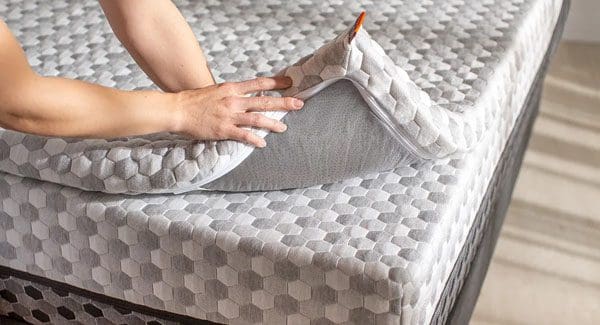
The Snuggle-Pedic is a two-layer structure, with softer memory foam on the outer layer and a firm channeled base layer for spinal support. It is a versatile design that makes it appropriate for back, side, and stomach sleepers. Memory foam mattress toppers can be too soft, providing no support, or too firm. This topper finds the balance. One side is soft-to-the-touch, with the other providing twice the support. This double layer system is soft but supportive. It is available in all sizes for less than $300 and comes with free shipping, returns, and customizations.
Saatva Latex Mattress Topper
Best Latex
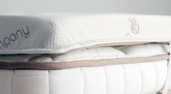
The Saatva is 1.5 inches of latex and is covered in organic cotton. The latex has ventilation to aid in breathability and the cotton cover adds to the overall temperature neutrality. For individuals that prefer all-natural materials, this latex topper is recommended. The latex used is Talalay latex and is natural and breathable. It also has ventilation spots and is hypoallergenic. For individuals that sleep hot, want a natural option, or have allergies, this could be an option. It comes with a 120-night trial period, free shipping and costs less than $350 in all sizes.
Zinus Memory Foam
Best For Side Sleepers
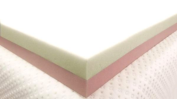
TheZinus topper measures four inches thick. It is a simple design with 2 inches of gel memory foam that is highly durable. Under the gel foam is a 2-inch-thick layer of support foam for added durability and support. This is an affordable option. The dual-layer provides cooling comfort, durable support and is designed to fit under any deep-pocket sheets. Side sleepers with hip and shoulder pain can appreciate the top layer�s softness and benefit from the durability of the bottom layer, preventing cradling and sinking.
Linenspa
Fastest Relief
The Linenspa is made with three inches of memory foam that conforms to the body’s curves while sleeping. This mattress topper is infused with temperature-regulating gel beads that evaporates any heat. It is available in two-inch, three-inch profiles and the foam is made to distribute weight evenly. With every size available for less than $105, this is a comfortable and affordable way to rejuvenate a worn mattress or soften a mattress that is too hard.
What to Look For
Mattress toppers can be beneficial for providing relief for aches and pains, but the best topper for back pain requires knowledge about materials, styles, and features that will provide relief. An overview of what to look for:
Materials
Memory foam and latex are ideal for pressure relief because they conform to the body�s curves without sagging. Look for high-quality materials, like American Talalay latex and open-cell foam.
Body Conformity Amount
The ideal amount of conformity provides pressure relief for painful areas. An ideal level depends on weight and sleeping positions. Look for details about the mattress weight, type, and sleeping style.
Sleeping Position
Sleeping on the back is a way to naturally align the spine, but a firm topper that does not sag is needed to provide the proper support.
Stomach sleepers need a topper that does not sag in the midsection, which can lead to neck pain.
Side sleepers need a topper that conforms to the contoured areas, like the shoulders and hips.
Price
Prices for mattress toppers vary. There are high-quality mattress toppers available for $150. For luxury, prices will be in the $300s and above.
Durability
Sagging is the main issue to avoid. Optimal spinal support is the objective. Do the research, read reviews, consult a chiropractor, and take plenty of time before investing.
Thickness
Toppers differ in thickness and can measure from one to seven inches thick. Thicker does not mean more comfortable and thicker toppers tend to be softer. Thinner toppers are usually firmer, which is better for back and stomach sleepers. What feels right depends on an individual’s sleeping position, weight, and size, however, most toppers fall in the two to three-inch thickness range.
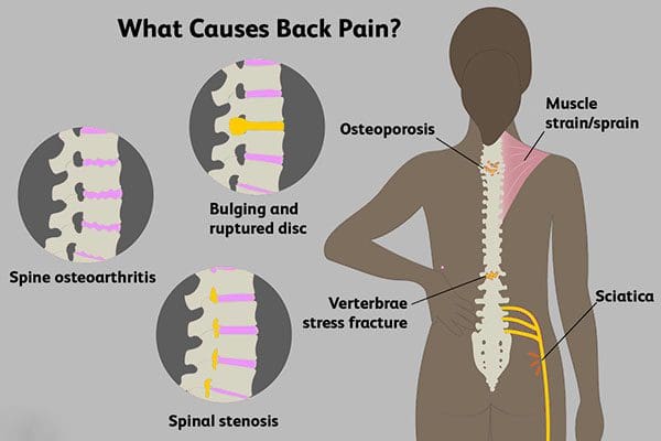
Back Pain Causes
Back pain can range from a dull ache to sharp sensations and can be the result of an auto accident, work injury, or personal injury that has developed over time. A sedentary lifestyle also contributes to low back pain. The following conditions are common sources of back pain:
Regardless of the cause or source, a mattress topper, which is more supportive than a mattress pad, can help alleviate back pain when sleeping, leading to a minimization of symptoms and improved quality of life.
Possible Option
Aching muscles, shooting/stabbing pain, reduced range of motion, and flexibility are detrimental to work, and home life. Whatever the case, there�s an option out there. An overview of mattress topper benefits:
The current mattress is old or not supportive enough, but don�t want to invest in a new mattress.
The current mattress is too firm, but a partner is comfortable on it. Mattress toppers are available in split sizes to soften/firm up one side.
A sedentary lifestyle has increased the frequency of dull low-back pain. A topper can help with back pain prevention.
Improve the comfort of a guest bed without spending too much. Similar to the first scenario, a mattress topper can be an upgrade for just several hundred dollars.
One of the most affordable sleep innovations today, but remember that durability, materials, and sleep factors, like weight, position, and height, should all be considered.
Composition of The Body
Sleep Stages
Sleep researchers are divided into two types of sleep: REM sleep is when dreams occur and NREM sleep which is non-REM sleep. NREM sleep is divided into stages:
Stage 1
Stage 1 makes up just 5-10% of a sleep cycle and is known as light sleeping. In this stage, the brain remains semi-conscious and is in between wakefulness and sleep. The brain waves start to elongate from alpha to theta waves.
Stage 2
Stage 2 is the bulk of the sleep cycle and makes up around 55%. In this stage, the body/brain is fully asleep and brain waves slow down even more.
Stage 3
Stage 3 is the deepest sleep state and is marked by elongated brain waves and slowed brain activity. It is also called short wave sleep. Slow-wave sleep makes up 15-25% of sleep but is the most important sleep state for body composition because most of the body’s restoration happens during this cycle.
REM/Rapid Eye Movement
This is the stage where dreams occur. It�s very distinct from the other three in that brain activity becomes more active. An individual passes through these sleep stages every 90 minutes. Getting 7-8 hours of sleep every night means going through 4-5 cycles. Going through a complete sleep cycle ensures an individual will go through Stage 3, which has a significant effect on body composition.
Dr. Alex Jimenez�s Blog Post Disclaimer
The scope of our information is limited to chiropractic, musculoskeletal, physical medicines, wellness, and sensitive health issues and/or functional medicine articles, topics, and discussions. We use functional health & wellness protocols to treat and support care for injuries or disorders of the musculoskeletal system. Our posts, topics, subjects, and insights cover clinical matters, issues, and topics that relate and support directly or indirectly our clinical scope of practice.*
Our office has made a reasonable attempt to provide supportive citations and has identified the relevant research study or studies supporting our posts. We also make copies of supporting research studies available to the board and or the public upon request. We understand that we cover matters that require an additional explanation as to how it may assist in a particular care plan or treatment protocol; therefore, to further discuss the subject matter above, please feel free to ask Dr. Alex Jimenez or contact us at 915-850-0900. The provider(s) Licensed in Texas& New Mexico*
References
Bolash R, Drerup M. How to Beat Insomnia When You Have Chronic Pain. Cleveland Clinic Web site. https://health.clevelandclinic.org/2015/12/managing-insomnia-for-those-with-chronic-pain/. Published December 18, 2015. Accessed April 18, 2017.
Improving Sleep: Special Health Report. Boston, MA: Harvard Medical School; 2015.
What is Sleep? American Sleep Association Web site. https://www.sleepassociation.org/patients-general-public/what-is-sleep/. Accessed April 18, 2017.
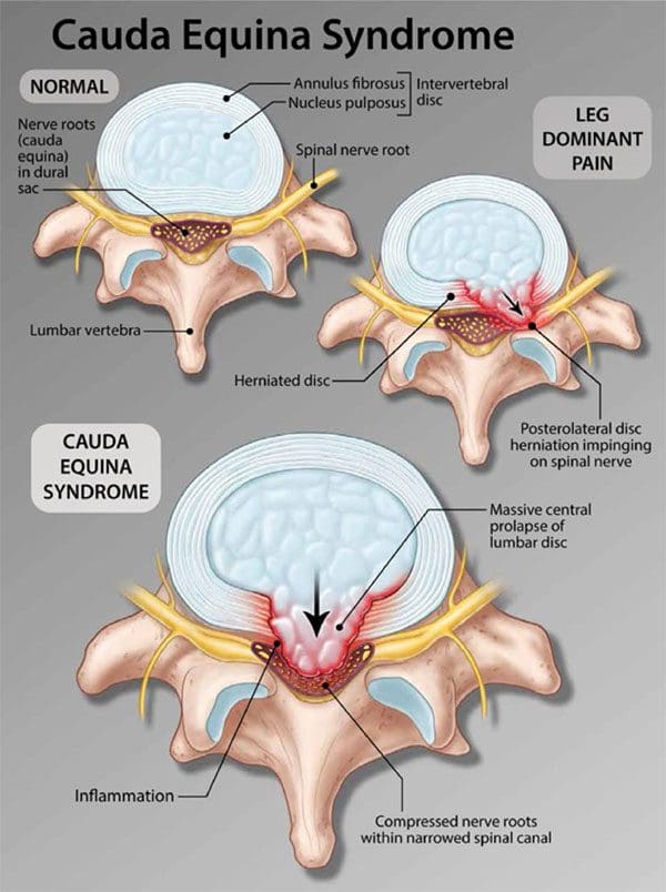
Cauda equina syndrome is an emergency that needs to be treated as soon as possible. It is a form of spinal nerve compression, but if left untreated, it can lead to permanent paralysis of one or both legs and permanent loss of bowel/bladder control. Lower back pain after sitting for too long or improperly lifting something heavy happens to most if not all of us.
However, sometimes pain in the lower back can be an indicator of something more serious. Especially, for individuals that are dealing with or managing back pain. One condition is cauda equina syndrome. It�s not like sciatica or arthritis, but it does have specific symptoms that individuals should be aware of.
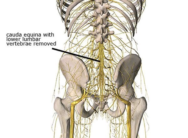
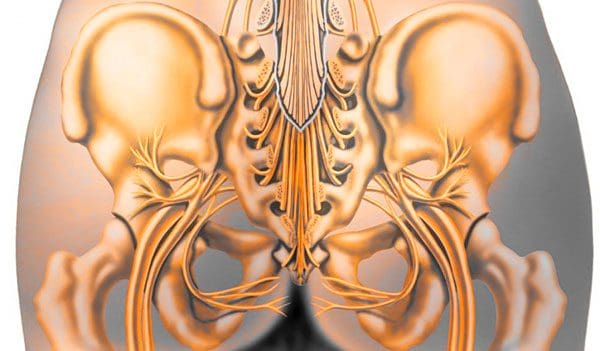
Cauda Equina Syndrome
The term comes from Latin that means horse�s tail. The cauda equina forms the group of nerves that run through the lumbar spinal canal. Generally, the condition means two things:
There is nerve compression of most of the lumbar spinal canal
Compression symptoms like numbness or weakness in the leg/s
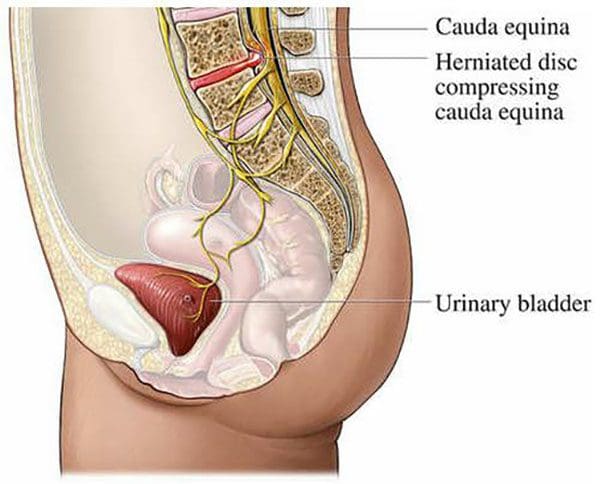
How cauda equina syndrome differs from typical compression of the lumbar spinal canal is that it can be caused by different issues, from fractures, tumors, and infections. More commonly, it is disc herniations that cause the problems. The key difference is the degree of nerve compression, and the number of nerves compressed.
For example, compression of a single nerve will not cause loss of bladder function. But compression of multiple nerves, especially the sacral nerves can cause loss of function. Nerve compression that leads to pain or numbness can be treated differently. Surgery is reserved for severe cases and for individuals that are not improving with non-invasive treatment.
Sneaky Presentation
One of the major factors is long-term compression that individuals do not realize they have. Individuals are more likely to be aware of symptoms from another spinal condition before cauda equina syndrome presents. However, the condition presents quickly but often other overlapping back problems mask cauda equina syndrome.
Causes
The syndrome can be brought on from anything that compresses the nerves. Most commonly, it is a root compression from degenerative processes, specifically lumbar disc herniations. Other causes include:
A doctor will examine any significant changes in bladder, bowel, or leg function that are considered red flags prompting an early and complete assessment. A physician will ask for a complete/detailed history of the onset and progression of symptoms.
The second is a close physical examination which includes testing sensation and strength along with a rectal exam to assess voluntary contraction. Also checking the body’s reflexes, assess walking gait and alignment. If most or all of the symptoms are presenting this will set in motion spinal imaging or an MRI. If the symptoms, exam, and imaging match, it will lead to an emergency admission to the hospital.
Body Composition Spotlight
Obesity and Osteoarthritis Connection
A variety of factors contribute to the development of osteoarthritis, including genetic factors and lifestyle choices. Research supports obesity is a significant risk factor in the development of osteoarthritis. It is pretty straightforward as body weight increases this equals increased load on the spine, and joints, especially the weight-bearing ones like the hips and knees. Increased pressure leads to early wearing, tearing, and eventual development of osteoarthritis. Added weight affects the body’s biomechanics and gait patterns.
However, obesity has also been shown to be a risk factor even on the non-weight-bearing joints. This is based on adipose tissue, which is more than just insulation. Adipose tissue is metabolically active and is involved in the secreting adipokines and cytokines which promote an inflammatory response. Pro-inflammatory adipokines and cytokines can have detrimental effects on joint tissue including damage to cartilage, synovial joints, and subchondral bone. The effect of inflammation on the joints in the body can contribute to the development of osteoarthritis.
Dr. Alex Jimenez�s Blog Post Disclaimer
The scope of our information is limited to chiropractic, musculoskeletal, physical medicines, wellness, and sensitive health issues and/or functional medicine articles, topics, and discussions. We use functional health & wellness protocols to treat and support care for injuries or disorders of the musculoskeletal system. Our posts, topics, subjects, and insights cover clinical matters, issues, and topics that relate and support directly or indirectly our clinical scope of practice.*
Our office has made a reasonable attempt to provide supportive citations and has identified the relevant research study or studies supporting our posts. We also make copies of supporting research studies available to the board and or the public upon request. We understand that we cover matters that require an additional explanation as to how it may assist in a particular care plan or treatment protocol; therefore, to further discuss the subject matter above, please feel free to ask Dr. Alex Jimenez or contact us at�915-850-0900. The provider(s) Licensed in Texas& New Mexico*
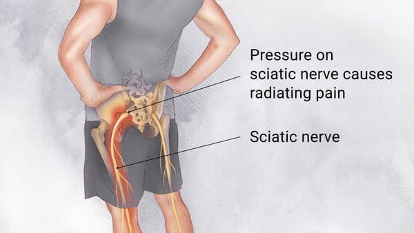
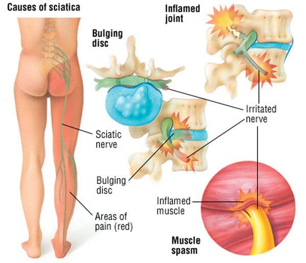
Trying to get a proper night’s rest and healthy sleep with sciatica can be difficult. Here we discuss how to combat sciatica discomfort for a better night�s rest. The sciatic nerves are the two largest nerves in the body. They run from the low back through the:
Hips
Buttocks
Down each leg into the feet
When the nerve gets:
Irritated
Inflamed
Pinched
Compressed
Sciatica can cause pain, tingling, numbness in the butt, lower back, leg, calf, and foot. It is a common condition that affects many individuals.
Is It Sciatica
Sciatica happens when the nerve becomes pinched from a bulging or herniated disc. In rare cases, the pain can result from a tumor putting pressure on the nerve or damage to the nerve caused by disease. The location and intensity of the pain depend on where the injury or damage occurred and how bad it is. Sciatica pain can be described as:
Dull
Sore
Numbing
Jolting
Throbbing
Hot
Stabbing
Radiating
For many sciatica usually resolves within a matter of weeks. However, once sciatica has presented future episodes are almost guaranteed to resurface and if not treated properly can lead to more serious problems.
Symptoms
The sciatic nerve/s can affect several areas of the body, making symptoms vary. The most common include:
Lower back pain starts at the low back, runs along the hips and buttocks down each leg.
Pain radiates/spreads down the butt/leg area sometimes described as a shooting pain and usually occurs on just one side.
Pain while sitting for long periods of time places pressure on the gluteal muscles, lower back, and nerves. This can cause or worsen the condition. When having to sit for a while, it is recommended to get up every hour or so and walk/move around. This gets the blood flowing and stretches out the tightened muscles.
Hip pain, as the sciatic nerves run through the hip joint and in some cases can cause pain to settle in the hip. Injuries in the hip can mimic the symptoms of sciatica. If there is hip pain that does not improve with time get checked by a doctor to rule out other causes like osteoarthritis, bursitis.
Numbness, some experience weakness in the legs and an altered sensation of numbness. This is caused by a herniated disc in the lower lumbar region.
Burning/tingling like a pins and needles sensation, especially in the feet and toes.
Conditions/Causes
There are several conditions that can cause sciatica:
Degenerative Disc Disease is where the discs of the spine deteriorate and become susceptible to painful herniation.
Spondylolisthesis is a painful condition where the lower vertebrae slip forward onto the bone directly below impinging the sciatic nerve.
Muscle Spasms and involuntary contractions of the muscles can cause sciatica if they compress the nerve.
Pregnancy sciatica is not uncommon. As the baby grows it places pressure on the nerve causing aches and pains.
Lumbar spinal stenosis is when the spaces in the low back begin to narrow compressing and irritating the nerve.
Risk factors include:
Age, as the spine gets older it becomes more susceptible to herniated discs and bone spurs, which are leading causes of sciatica.
Obesity and excess weight create added stress on the spine, which can inflame the nerves.
Occupation/work that requires standing/sitting for long periods or if a lot of heavy lifting is involved there is added pressure on the back increasing the risk for back problems.
Individuals with diabetes have an increased risk for nerve damage. When nerves are damaged, they can cause radiating pain.
Night Time
Sleeping at night can be a challenge, especially not being able to get into a comfortable position. Sleep deficits and insufficient sleep can reduce the body�s tolerance to pain and worsen inflammation. Many wake up with increased symptoms.
This is likely due to the fact that when the body is laying down the discs draw in and absorb fluid, which results in increased pressure within the disc, creating more pressure on the nerve. But there are some things to do to lessen pain and get a good night�s sleep. This includes changing sleep position, stretching, and practicing healthy sleep hygiene.
Sleeping Positions
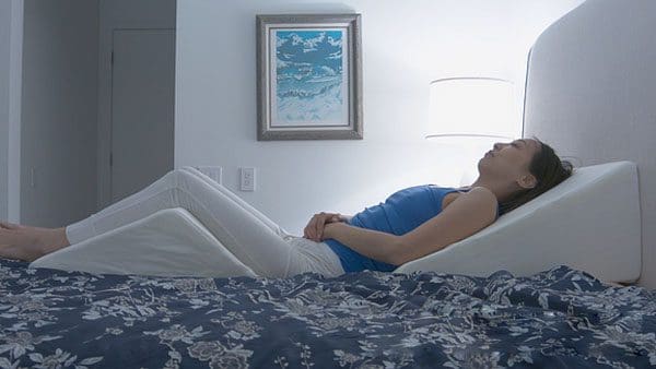
Sleeping on the back is considered the best sleeping position for sciatica because it eases pressure on the low back and discs where the nerves are located.
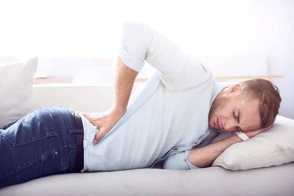
Sleeping on the side can be more comfortable and is a good position because it doesn�t place direct pressure on the muscles, discs, or sciatic nerve. But, it is important that the mattress is supportive enough to keep the spine aligned. If more support is needed place a pillow between the legs.
Sleeping with the knees elevated can help take the pressure off the low back. To achieve this place a pillow under the knees or, with an adjustable bed, use it to elevate the foot of the bed.
Sleeping with a body pillow provides extra comfort and helps the body remain in a certain position throughout the night. These pillows come in a variety of shapes, sizes.
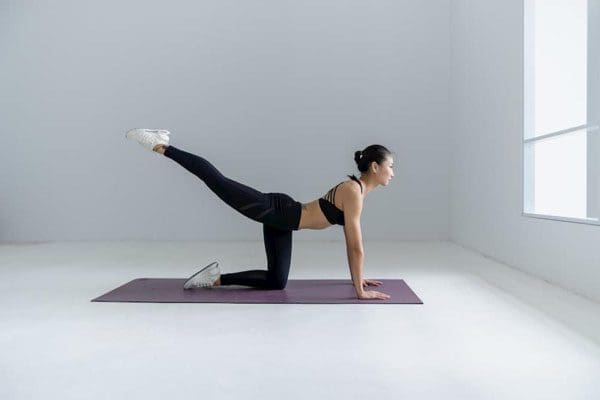
Stretches
Stretching can provide relief. It�s important to stretch to keep the body flexible and to prevent pain. Gentle stretches before bed, and after waking up will loosen the muscles and ligaments surrounding the spine and joints.
Stand and place one heel on an elevated surface, like a chair.
Fully extend the knee and flex the ankle by pointing the toes toward the ceiling.
Bend forward at the hips keeping the spine in a neutral position. Hold for 15 to 30 seconds.
Repeat with the other leg.
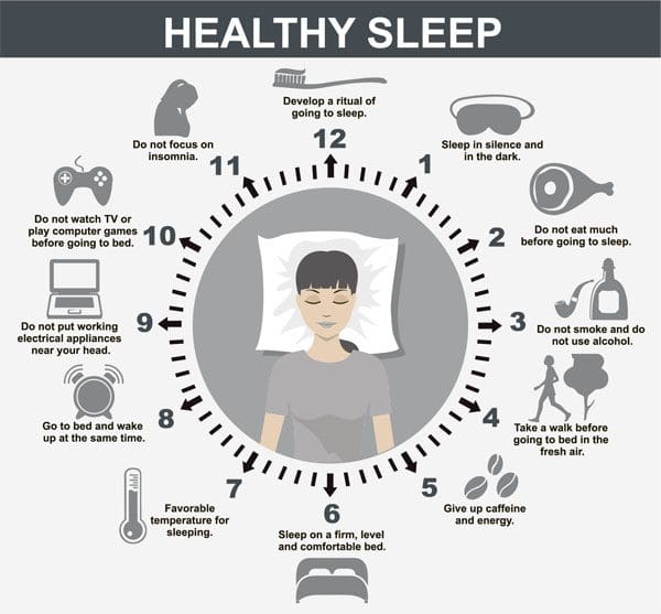
Sleep Hygiene
Proper sleep hygiene helps prepare for a good night�s sleep. Here are some tips to help improve sleep hygiene.
A nighttime routine will help unwind the body before bedtime. Start the routine 30 minutes before planning to go to sleep. Some things to do to help unwind:
Take a warm bath
Listen to relaxing/soothing music
Meditation
Reading
Get a new mattress. An old, sagging mattress can worsen sciatica and strain the back. The best mattresses for sciatica pain combines contour comfort to ease pressure points at the hips and shoulders with the proper support to keep the spine aligned.
Eye masks can help with artificial light which can fool the mind along with the circadian clock into thinking it�s daylight. Keeping out unwanted light all night can help.
Avoid blue light too close to bedtime like lamps and device screens. These are great for the day, as they help boost attention, reaction time, and mood. But at night it can be disruptive. Turn off electronics at least 30 minutes before bed to help the body adjust.
Room temperature control has found that most sleep better in a cool room. The optimal temperature is between 60 and 67 degrees.
Avoid exercise close to bedtime. Working out before bed can interfere with sleep. This is because exercise releases adrenaline keeping the mind and body alert.
Avoid stimulants before bedtime like caffeine, sugar, etc. which will keep the body up.
Medical Intervention
Sciatica pain can be mild or severe. Stretching or changing up sleeping position can help ease discomfort. But if the pain is severe or chronic, and if it prevents getting a good night�s sleep, consult a chiropractic professional.
InBody Spotlight
Sleep And Body Composition
A lack of sleep makes it harder to gain muscle and harder to lose fat.
Sleeping less means fewer opportunities to secrete growth hormone and develop muscle
Testosterone is negatively affected by lack of sleep
Sleeping less can increase cortisol levels, impairing muscle development
Irregular sleep throws off the body’s cycles, making the body feel hungrier
Sleeping less is linked to eating more snacks, increasing energy levels
Lack of sleep can cause reductions in Basal Metabolic Rate by 20%, reducing total energy output
Being tired reduces spontaneous movements, reducing total energy output
If trying to get into shape and change body composition, sufficient sleep is vital. Any positive changes to get more sleep are going to have positive changes in efforts to change body composition.
Dr. Alex Jimenez�s Blog Post Disclaimer
The scope of our information is limited to chiropractic, musculoskeletal, physical medicines, wellness, and sensitive health issues and/or functional medicine articles, topics, and discussions. We use functional health & wellness protocols to treat and support care for injuries or disorders of the musculoskeletal system. Our posts, topics, subjects, and insights cover clinical matters, issues, and topics that relate and support directly or indirectly our clinical scope of practice.*
Our office has made a reasonable attempt to provide supportive citations and has identified the relevant research study or studies supporting our posts. We also make copies of supporting research studies available to the board and or the public upon request. We understand that we cover matters that require an additional explanation as to how it may assist in a particular care plan or treatment protocol; therefore, to further discuss the subject matter above, please feel free to ask Dr. Alex Jimenez or contact us at 915-850-0900. The provider(s) Licensed in Texas& New Mexico*
References
Siengsukon, Catherine F et al. �Sleep Health Promotion: Practical Information for Physical Therapists.��Physical therapy�vol. 97,8 (2017): 826-836. doi:10.1093/ptj/pzx057
After all of these years, I am happy to announce that the Texas Supreme Court has finally made a decision regarding the Texas Board of Chiropractic Examiners et al v. Texas Medical Association case on January 29th, 2021. With great honor and gratitude, I’d like to continue to extend sincere thanks to everyone who worked hard on this case and whose tremendous efforts resulted in the decision. Thanks to the Supreme Court’s decision, chiropractors in Texas can now carry on their jobs accordingly. Below, I have provided a letter from Board President, Mark R. Bronson, D.C., F.I.A.N.M. on behalf of the Texas Board of Chiropractic Examiners stating the Texas Supreme Court’s decision in the Texas Board of Chiropractic Examiners et al v. Texas Medical Association case on January 29th, 2021. – Dr. Alex Jimenez D.C., C.C.S.T.
February 1, 2021
On behalf of the Texas Board of Chiropractic Examiners, I extend our sincere thanks and appreciation to everyone whose efforts resulted in the Texas Supreme Court’s decision in Texas Board of Chiropractic Examiners et al v. Texas Medical Association on January 29, 2021. Special thanks are due to all the attorneys at the Office of the Attorney General who worked on this case over these years.
The decision properly affirmed the validity of the Board’s scope of practice rule, which the court clearly said does not exceed our statutory scope of chiropractic practice. The court unequivocally held that the Board�s rules do not violate Occupations Code Chapter 201 or run counter to the chapter’s objectives set by the Texas Legislature, and in fact, carefully observe the statutory boundary between the medical and chiropractic professions. This decision, which recognizes the common sense and long-standing inclusion of associated nerves in chiropractic diagnosis and treatment, preserves and strengthens the essence of chiropractic.
Thanks to the court’s decision, our licensees can now fulfill their duties as vital portal-of-entry healthcare providers in Texas without fear. The court’s decision reaffirms the principles of economic freedom that have made Texas the best state in the nation to be a chiropractor.
Sincerely,
Mark R. Bronson, D.C., F.I.A.N.M. Board President
Texas Board of Chiropractic Examiners
The scope of our information is limited to chiropractic, musculoskeletal, physical medicines, wellness, and sensitive health issues and/or functional medicine articles, topics, and discussions. We use functional health & wellness protocols to treat and support care for injuries or disorders of the musculoskeletal system. Our posts, topics, subjects, and insights cover clinical matters, issues, and topics that relate and support directly or indirectly our clinical scope of practice.*
Our office has made a reasonable attempt to provide supportive citations and has identified the relevant research study or studies supporting our posts. We also make copies of supporting research studies available to the board and or the public upon request. We understand that we cover matters that require an additional explanation as to how it may assist in a particular care plan or treatment protocol; therefore, to further discuss the subject matter above, please feel free to ask Dr. Alex Jimenez or contact us at 915-850-0900. The provider(s) Licensed in Texas & New Mexico*
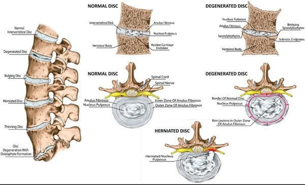
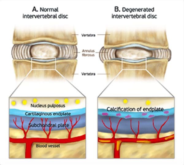
Chiropractic prevention for degenerative disc disease. In between the bones of the spine or the vertebrae are the protective shock-absorbing intervertebral discs. These protective cushions naturally begin wearing down or degenerating as the body ages. The common term for this is degenerative disc disease or DDD. It is not a disease, but the body’s natural aging process. The reason it is called degenerative disc disease is that it is a condition of the spine with deteriorating effects.
One of the things seen in degenerative disc disease and aging of the spine is that the soft disc, made up of a vast amount of water, slowly begins to dry out and dehydrate. The protein/sugar that is within the disc begins to dry out and that starts a degenerative domino effect along with tearing of the outside rings of the disc.
Causes and Risk Factors
The most common cause of disc degeneration is aging. With age comes the loss of fluids/water. This means the discs get thinner and provide less protection. The shock absorbers don�t absorb weight, impact, pressure like they used to. However, disc degeneration does not always occur later in life. Genetics can be a catalyst for development when young.
Individual environment vs. genetics
Stresses
Strains
Wear
Tear
Discs dry out
Rings crack or tear
If involved in an occupation that requires heavy lifting, twisting, and bending, this can generate disc damage, but physical stressors do not automatically enhance disc erosion. An individual can do this type of work their entire life and not experience problems. It depends on the environment and the overall health of the individual that can be assessed through chiropractic prevention and body composition analysis.
Chiropractic Prevention and Preserving The Discs
Individuals need training on proper body posture and body mechanics. A major contributing factor that increases the risk of nerve, spine/back pain due to disc damage. The back/spine should be moved like a crane. This means not bending over at the waist, but squatting down with the hips and knees when lifting an object. Not everything can be done this way, but if an individual can eliminate too much bending at the waist, it will definitely help.
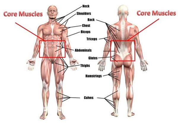
Do not neglect the core muscles. Core exercises will maintain the muscles around the waist, which also strengthens the rest of the body. All the muscles front, back, around the trunk, hips, and knees to the chest are all the core muscles.
Healthy Weight Less Spinal Stress
Beyond physical stressors, another important factor in preventing degenerative disc disease is maintaining a proper weight. Society has adapted more and more to a sedentary lifestyle. And home quarantine means being more sedentary. Emotional factors like:
Boredom
Anxiety
Depression
Fatigue
This can cause excessive junk food consumption and rapid weight gain.
Lifestyle Adjustments Health Coaching
Implementing a preventive lifestyle to prevent degenerative damage is challenging. Everyone really should try to live as healthy a lifestyle they can.
Smoking accelerates the aging process by drying out the tissue, especially the spinal discs. It is known as desiccation and is why smoking is a major risk factor for degenerative disc disease.
Disc stress
Anything that stresses the discs for any amount of time can generate pain. For example, standing too long even carefully standing straight can induce discomfort and pain. Sitting can help, but caution needs to be observed. Sitting too long places more pressure on the low back than when standing. There must be a balance between sitting and standing. Experts suggest getting up and moving every 20 to 30 minutes. This is a perfect time to get up and do in-home walking exercises, physical chores, anything that gets the body up and moving around.
Stretching
Regular bending and stretching need to become a natural reflex. When the body begins to feel sore, stretch out the area.
The good news is that everyone, even those with a genetic predisposition to greater disc loss or fusion, there are options for slowing down and minimizing disc damage. Maintaining a healthy weight, strengthening the core, and learning proper techniques for lifting for work, exercise, or sports will all help. If diagnosed with disc degeneration, talk to a doctor, physical therapist about developing a chiropractic prevention treatment plan. They�ll be able to develop a program specifically designed to help.
Bodily Composition
Human Immune System
The immune system is highly complex and essential in maintaining optimal health. The main function of this system is to neutralize pathogenic microorganisms like bacteria that get into the body and affect the body’s homeostasis, eliminate any harmful substances from the environment, and fight the body�s own cells that can cause illnesses like cancer. The defense system consists of innate and adaptive immune processes. The innate system includes exterior defenses like the:
Any organisms that get past the first line of defense, have to face the adaptive system, which is made up of T and B cells. The adaptive immune system is a learning defense that constantly adapts and evolves to be able to identify changes in pathogens that mutate over time. Together, these systems provide a fortified resistance to any long-term survival of infectious agents in the body. Healthy lifestyle adjustments can help strengthen and boost these systems working to keep the body safe.
Dr. Alex Jimenez�s Blog Post Disclaimer
The scope of our information is limited to chiropractic, musculoskeletal, physical medicines, wellness, and sensitive health issues and/or functional medicine articles, topics, and discussions. We use functional health & wellness protocols to treat and support care for injuries or disorders of the musculoskeletal system. Our posts, topics, subjects, and insights cover clinical matters, issues, and topics that relate and support directly or indirectly our clinical scope of practice.*
Our office has made a reasonable attempt to provide supportive citations and has identified the relevant research study or studies supporting our posts. We also make copies of supporting research studies available to the board and or the public upon request. We understand that we cover matters that require an additional explanation as to how it may assist in a particular care plan or treatment protocol; therefore, to further discuss the subject matter above, please feel free to ask Dr. Alex Jimenez or contact us at 915-850-0900. The provider(s) Licensed in Texas& New Mexico*
When lower back pain presents many want to retreat to the couch, bed and just lay down, but doctors, chiropractors, physical therapists, and spine specialists do not recommend this course of action. What they do recommend, other than treatment, is to engage in the easiest forms of exercise on the spine and back muscles. �
Staying sedentary is one of the worst things an individual can do to their back. When the back is aching exercise can usually help. This is because the muscles, ligaments, tendons are being stretched and not just staying still, which lets inflammation build up and swell. Moving keeps the blood flowing, allowing for broader healing and recovery.
However, back pain relief can be a challenge. Various treatment options exist because there are a variety of causes. The key is figuring out which type is best for each individual and their specific condition. An individual needs to know the cause of their type of back pain, as this determines which exercises should or should not be doing. The Pain and Therapy journal evaluated some of the best exercises for lower back pain. �
Physical Therapy Exercises
The McKenzie method can be very effective for acute disc herniation pain and sciatica. This type of exercise is to figure out if there is a specific position that helps the pain become centralized, correct any motion restrictions, and take the pressure off the region that is compressed or inflamed. Physical therapists incorporate McKenzie exercises as part of regular treatment. The strength-building moves are designed to help support the spine and consist of range-of-movement work and sustained positions. �
�
Home and Studio Workouts
Pilates is one of the easiest exercises for individuals with chronic low-back pain. Like McKenzie exercises, it utilizes sustained positions that strengthen the trunk/core muscles. The muscles are strengthened using small movements. Using the machine called a reformer, has built-in support for the spine. This is considered a low-key, muscle-toning workout that can ease chronic back pain. �
�
Water Exercise
Water exercises lessen the body’s weight, taking pressure/stress off the spine. Deep-water running with the water at shoulder-height can significantly improve low-back pain. In a study, a group of overweight/obese women worked out twice a week for an hour-long exercise session. After 12 weeks, improvements in pain intensity, personal care, sitting, standing, and sleeping were reported. �
�
Easiest Office Exercise
One of the easiest exercises is walking. It is great for the body. But the key is to walk more than usual around the office, or wherever work is. This is not about getting the heart rate up. It is about not staying in the same position for too long. When sitting and focused, an individual can stay in an uncomfortable position for some time and just push through it in an attempt to finish up the work.
Using a timer or an application that alerts every hour to get up and stretch is highly beneficial. Walk correctly to the bathroom, or just get up and walk around for a bit gets the blood pumping through the body and the muscles in motion stretching and contracting. �
�
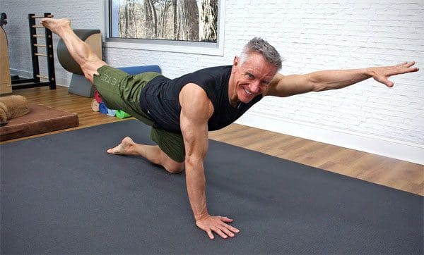
Stabilization Exercise
Strengthening workouts can be done at home.
Stretch while standing against the wall bringing the arms up and down.
Pull the elbows down into the back, which stops the hyperactive trapezius from tensing up.
Knee to the chest motion while lying on the back
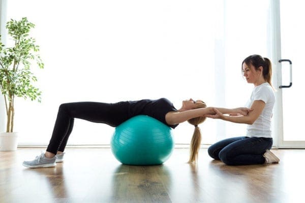
Abdominal crunches while balance on an exercise ball
Push the head back into the headrest while driving. This helps avoid the forward head posture.
Contact a doctor, chiropractor, or physical therapist that can recommend the best stabilization exercises for the specific pain/condition. �
�
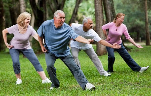
Tai Chi and Qigong
Tai Chi and Qigong are gentle exercises where an individual performs slow, controlled movements emphasizing balance and focus. Both can reduce pain, disability, and other symptoms associated with lower back pain. �
�
Body Composition Testimonial
�
Exercise After Childbirth
Physical activity for pregnant and post-birth, the American Congress of Obstetricians and Gynecologists recommends the following. The easiest exercise routines can be resumed gradually after pregnancy, once a doctor confirms it is medically safe, depending on the delivery, and the presence or absence of medical complications.
Regular aerobic exercise in lactating women has been shown to improve cardiovascular fitness without affecting milk production, composition, or infant growth.
Nursing women should consider feeding their infants before exercising in order to avoid exercise discomfort.
Nursing women also should ensure proper hydration before engaging in physical activity.
Take it slow.
�
Dr. Alex Jimenez�s Blog Post Disclaimer
The scope of our information is limited to chiropractic, musculoskeletal, physical medicines, wellness, and sensitive health issues and/or functional medicine articles, topics, and discussions. We use functional health & wellness protocols to treat and support care for injuries or disorders of the musculoskeletal system. Our posts, topics, subjects, and insights cover clinical matters, issues, and topics that relate and support directly or indirectly our clinical scope of practice.*
Our office has made a reasonable attempt to provide supportive citations and has identified the relevant research study or studies supporting our posts. We also make copies of supporting research studies available to the board and or the public upon request. We understand that we cover matters that require an additional explanation as to how it may assist in a particular care plan or treatment protocol; therefore, to further discuss the subject matter above, please feel free to ask Dr. Alex Jimenez or contact us at 915-850-0900. The provider(s) Licensed in Texas& New Mexico*
Chronic inflammation is quite common and can cause a variety of painful back conditions. Fortunately, chiropractic treatment can extinguish the inflammation at its core/root cause and bring long-lasting relief. Inflammation can be a significant factor in causing body and back pain. The proper and lasting treatment goes beyond anti-inflammatory medications. The objective is to extinguish the inflammation completely.
Inflammation
The number of diseases associated with chronic inflammation is increasing exponentially. An estimated 60% of Americans deal with at least one chronic condition caused completely or in part by inflammation. Inflammation is the body’s immune response to something that is not right in the body. Typically, there is:
Swelling
Redness
Warmth/heat signaling an inflammatory response from injury or infection
Endocrine, heart, lung, and neurologicalcomplications
Inflammation can generate inflammatory back pain as well when this happens. This is because the immune system is trying to protect the body from whatever is trying to infiltrate. The cells and chemicals fight the attack and try to heal whatever is going on. Sometimes, there is nothing going on but the immune system becomes hyper-reactive and attacks itself. This is the case of autoimmune disease, or what becomes chronic inflammation. Chronic inflammation lasts longer than 3 months and can lead to diseases like:
Autoimmune inflammation that presents as ankylosing spondylitis and rheumatoid arthritis is in a different category. This is when the body begins attacking itself. This can be triggered by a viral infection, which stimulates the immune system. This can cause:
Joint destruction
Ligament pain
Soft tissue swelling
Some conditions are hereditary
Inflammatory Spinal Conditions
Inflammation can travel into the body�s spinal and neurological systems. This results in painful conditions that can affect an individual’s quality of life.
Ankylosing Spondylitis
Ankylosing spondylitis usually starts in the lower back and can spread up. It is a form of arthritis that causes the vertebrae to fuse together. It can also cause inflammation in the urological and ophthalmological systems. It is believed to have a somewhat genetic connection. There is a marker called HLA B27 that is usually positive in patients and is more common in young males.
Rheumatoid Arthritis
Rheumatoid arthritis/RA causes inflammation in synovial joints. These produce a fluid that helps lubricate and nourish the joints. RA is found most commonly in the hands, wrists, and knees, but can also present in the spine�s facet joints that connect vertebrae. There is some genetic association but is common with smoking and obesity. It is diagnosed with lab work, like inflammation markers, rheumatoid factor, and physical examination. RA pain typically presents in the cervical spine or neck region.
Transverse Myelitis and Multiple Sclerosis
These conditions are closely tied and caused by inflammation in the central nervous system/CNS. The immune system attacks the nerve cells and removes the fatty substance that insulates the nerves and helps them transmit impulses to and from the central nervous system. This causes:
Pain
Weakness
Numbness
Bladder/bowel issues
Transverse myelitis affects the spinal cord whereas multiple sclerosis can affect the brain and the spinal cord. Transverse myelitis is typically acute, whereas multiple sclerosis is long-term and can have an increasing/decreasing course of progressive symptoms. And transverse myelitis can be a symptom of multiple sclerosis. Certain lifestyle choices/habits can cause or worsen inflammation. Obesity, smoking, and an unhealthy diet can have a significant impact on chronic inflammation.
Spinal Structures Affected
Inflammation can affect every area of the spine. From the lower back to inflammation of the vertebrae themselves. Injuries to the spine, that include the:
Bones
Discs
Ligaments
Joints
This can cause swelling and a build-up of fluid that can be found on an MRI. This is the preferred method of detecting inflammation. When the term itis is used this usually indicates a type of inflammation. For example, neuritis means inflammation of the nerve/s. This can be seen with nerve compression where the nerve is swollen on an MRI.
Extinguish Spinal Inflammation
Causes of inflammation can be traced back to lifestyle choices, but can also be alleviated through healthy lifestyle adjustments that turn into habits.
Nutritional Health Coaching
Recommended is to avoid or decrease processed foods, trans fats, and sugar. Consider supplementing vitamin D, magnesium, and omega 3s.
Quit Smoking
Quitting smoking will improve circulation and extinguish vascular inflammation.
Regular Physical Activity/Exercise
Aerobic exercises improve cardiac function and circulation as well as exercises that support the spine ergonomically. Core and pelvic stabilization are necessary for lower back pain.
Medication
For acute inflammation caused by injuries steroids and NSAIDs can help with severe pain and inflammation.
Chiropractic Extinguisher
When the spine and the body’s joints are in proper alignment, and the nerves are functioning at optimal levels the body’s biomechanics return to normal. This stops neuropeptides from producing and helps extinguish inflammation.
Surgery
With the spine, surgery is not recommended as a first-line treatment unless it is an emergency or there is potential for permanent neurological damage. If medication, chiropractic, physical therapy, supplements, lifestyle changes, and/or complementary treatments like acupuncture do not help and quality of life and function are significantly impacted, then elective surgery can be considered.
Body Composition Spotlight
All-around Exercise Regimen
As individuals continue to struggle with obesity and functional fitness with age, exercise becomes more important than ever. It is crucial to combine diet and regular physical activity/exercise to lose weight, have a favorable impact on body composition and lifespan. A well-rounded exercise regimen that incorporates all types of fitness. Aerobic exercise helps maintain an elevated heart rate and helps get rid of fat-free mass.
Resistance training helps to build lean muscle mass. Combining the two, with concurrent training, or utilizing a HIIT workout when there is not enough time will work wonders. Being better equipped and understanding why exercise is important for the body’s health, different types of exercise, and which ones are best suited for each individual’s needs will keep the body in top form for the long term.
Dr. Alex Jimenez�s Blog Post Disclaimer
The scope of our information is limited to chiropractic, musculoskeletal, physical medicines, wellness, and sensitive health issues and/or functional medicine articles, topics, and discussions. We use functional health & wellness protocols to treat and support care for injuries or disorders of the musculoskeletal system. Our posts, topics, subjects, and insights cover clinical matters, issues, and topics that relate and support directly or indirectly our clinical scope of practice.*
Our office has made a reasonable attempt to provide supportive citations and has identified the relevant research study or studies supporting our posts. We also make copies of supporting research studies available to the board and or the public upon request. We understand that we cover matters that require an additional explanation as to how it may assist in a particular care plan or treatment protocol; therefore, to further discuss the subject matter above, please feel free to ask Dr. Alex Jimenez or contact us at�915-850-0900. The provider(s) Licensed in Texas& New Mexico
IFM's Find A Practitioner tool is the largest referral network in Functional Medicine, created to help patients locate Functional Medicine practitioners anywhere in the world. IFM Certified Practitioners are listed first in the search results, given their extensive education in Functional Medicine