Discover the impact of traumatic brain injury on daily life and the best approaches to manage recovery effectively.
Introduction
Traumatic brain injury (TBI) happens when a sudden blow or jolt to the head damages the brain. It can change how a person thinks, moves, and feels. This article explains TBI in simple terms, including its causes, symptoms, and effects on the body. It also shows how chiropractors and nurse practitioners can work together to help people heal (Mayo Clinic, 2023; Cleveland Clinic, 2023).
What Is Traumatic Brain Injury?
A traumatic brain injury is harm to the brain from an outside force. The skull protects the brain, but a hard hit can still cause trouble inside. TBI can be mild, like a concussion, or severe, leading to long coma or disability. Every year, millions of people get a TBI from falls, car crashes, or sports (Mayo Clinic, 2023). The brain controls everything we do. When it gets hurt, problems can show up right away or weeks later. Early care matters a lot (Cleveland Clinic, 2023).
Common Causes of TBI
TBI starts with a strong force to the head or body. Here are the main causes:
Falls: The top reason, especially in kids and older adults. Slipping in the shower or falling off a ladder can cause TBI (Mayo Clinic, 2023).
Car accidents: High-speed crashes shake the brain inside the skull.
Sports injuries: Football, boxing, and soccer players often get concussions.
Violence: Gunshots, assaults, or shaken baby syndrome.
Blast waves: Soldiers in war face TBI from explosions (Cleveland Clinic, 2023).
Even a small bump can cause mild TBI if the brain moves rapidly within the skull (Hicks et al., 2020).
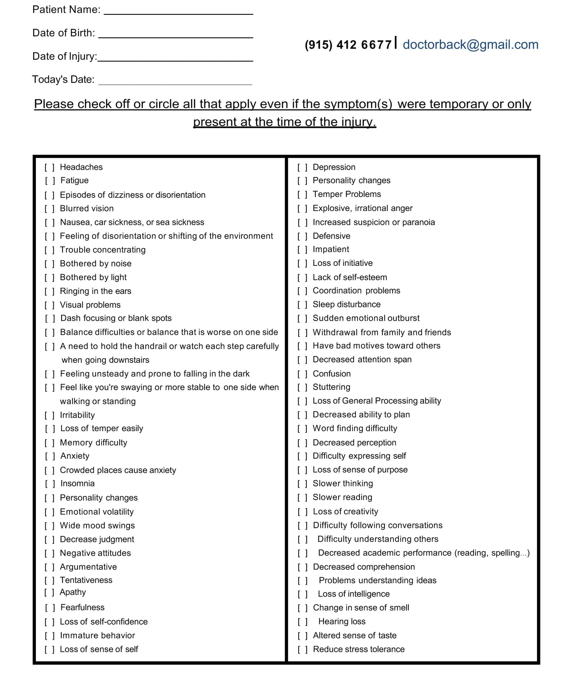
Symptoms of TBI
Symptoms depend on the severity of the injury. They can appear in the body, mind, or feelings.
Right-Away Signs
Losing consciousness for seconds or minutes.
Headache that will not stop.
Nausea or vomiting.
Feeling dizzy or losing balance.
Blurry vision or ringing in the ears (Mayo Clinic, 2023).
Later Signs
Trouble remembering new things.
Slow thinking or reading.
Hard time focusing.
Feeling sad, angry, or worried.
Sensitivity to light and noise.
Sleep problems such as insomnia or excessive sleepiness (Cleveland Clinic, 2023; Silverberg et al., 2018).
A chiropractor or nurse practitioner can find hidden signs by asking detailed questions about the accident and daily life (Jimenez, n.d.-a).
How TBI Affects the Musculoskeletal System
The musculoskeletal system includes muscles, bones, and joints. TBI often hurts this system because the force hits the whole body.
Neck pain and stiffness: Whiplash in car crashes strains neck muscles and spine.
Back pain: The spine can shift out of place, causing long-term pain.
Poor posture and balance: Brain signals to muscles get mixed up, making walking hard (Treleaven, 2017).
Muscle weakness: One side of the body may feel weak after severe TBI.
Spinal misalignment can press on nerves and slow healing. Chiropractors check the spine with gentle tests to spot these issues (Jimenez, n.d.-b).
How TBI Affects the Neurological System
The neurological system is the brain, spinal cord, and nerves. TBI directly damages this network.
Slow nerve signals: Thinking and moving feel delayed.
Seizures: Electrical storms in the brain.
Nerve pain: Tingling or burning in arms and legs.
Coordination loss: Hands shake or feet trip (Ellis et al., 2017).
Questioning reveals whether light bothers the eyes or whether noise causes headaches—clues to nerve irritation (Silverberg et al., 2018).
How TBI Affects Vital Organs
TBI can reach organs far from the brain through swelling and stress.
Lungs: Breathing problems if the brain stem is hurt.
Gut: Nausea, poor digestion, or constipation from nerve disruption.
Liver and kidneys: Medicines for pain can strain these organs if not watched (Khellaf et al., 2019).
A nurse practitioner orders blood tests to check organ health and adjust care (Jimenez, n.d.-c).
Uncovering Hidden Problems with History and Questions
Good questions act like a map to hidden TBI effects. A chiropractor or nurse practitioner asks:
“When did the injury happen?”
“Do bright lights hurt your eyes?”
“Do you feel sick after reading?”
“Any new pain in your neck or back?”
“How is your sleep?”
These answers guide exams. Gentle spine checks show tight muscles. Balance tests reveal wobbly steps. The provider connects dots between the brain, spine, and organs (Jimenez, n.d.-a; Haider et al., 2018).
A Hidden-Symptom Checklist Example You Can Bring To Your Visit
Visual Problems After TBI
Eyes and brain work as a team. TBI breaks the link.
– Double vision.
– Trouble tracking moving objects.
– Light sensitivity (photophobia).
– Dry eyes or blurry sight (Cleveland Clinic, 2023).
Simple eye tests in the office spot these issues early (Green et al., 2010).
Nausea and Digestive Signs
Nausea is common right after TBI. It can last if the vagus nerve is upset. Patients may feel full too fast or have reflux. A detailed diet history helps the nurse practitioner suggest gentle foods (Blyth & Bazarian, 2010).
Neurological Issues: Slow Thinking and Reading
Mild TBI slows the brain’s processing speed. Reading a page takes longer. Word-finding feels hard. Memory for new facts slips. Cognitive tests measure the gap and track improvement (McInnes et al., 2017).
Sensitivity to Light and Noise
Photophobia and phonophobia mean that normal lights or sounds feel painful. This comes from overactive brain circuits. Dark glasses and quiet rooms help in the short term, while therapy calms the nerves in the long term (Silverberg et al., 2018).
Sleep Issues Like Insomnia
Sleep heals the brain. TBI breaks the sleep cycle.
Hard to fall asleep.
Waking often.
Daytime fatigue.
Poor sleep slows recovery. A sleep diary guides the care plan (Wickwire et al., 2018).
Feeling Better Than Ever After a Semi-Truck Accident- Video
Why an Integrative Approach Works
Integrative care means a team effort. Chiropractic care fixes the body’s frame and nerves. Nurse practitioner care takes the whole health picture into account. Together, they speed healing and cut setbacks (Jimenez, n.d.-d; Gardner & Yaffe, 2015).
Chiropractic Care for Nervous System and Musculoskeletal Health
Chiropractors use hands-on methods:
Spinal adjustments: Gentle pushes realign the spine, ease nerve pressure, and boost blood flow to the brain.
Soft-tissue therapies: Massage relaxes tight neck and back muscles.
Targeted exercises: Balance drills and core strength rebuild coordination (Navarro et al., 2018).
These steps improve brain signals and reduce pain without drugs (Coronado et al., 2015).
Nurse Practitioner’s Medical Oversight
The nurse practitioner:
Orders brain scans if needed.
Manages pain, mood, or seizure medications.
Checks blood work for inflammation or hormone balance.
Guides nutrition to feed the brain (omega-3s, antioxidants).
Watches emotional health and refers to counseling (Haag et al., 2019).
Ongoing: Monthly check-ups, diet tweaks, and home exercise.
Patients track symptoms in a simple journal. The team reviews progress every two weeks (Jimenez, n.d.-e; Cnossen et al., 2017).
Real-Life Observations from Dr. Alexander Jimenez
Dr. Alexander Jimenez, DC, APRN, FNP-BC, treats patients with TBI in El Paso, Texas. He notices:
Neck misalignment often hides behind headaches.
Early spinal care cuts recovery time by weeks.
Teamwork with medical providers prevents medicine overload.
Simple home balance drills speed return to work (Jimenez, n.d.-f; Jimenez, n.d.-g).
His dual training lets him spot both spine and medical red flags fast.
Long-Term Outlook
Most mild TBI patients feel better in months with the right plan. Moderate to severe cases need longer care but still improve. Sticking to the integrative path raises the chance of full function (Maas et al., 2017).
Conclusion
Traumatic brain injury touches every part of life, from muscles to mood. Understanding causes and symptoms is the first step. Detailed history uncovers hidden effects on the musculoskeletal system, nerves, and organs. Chiropractic adjustments, soft-tissue work, and exercises rebuild the body’s foundation. Nurse practitioners guard overall health with medical insight. Together, this integrative, holistic plan guides patients back to daily joy.
References
Blyth, B. J., & Bazarian, J. J. (2010). Traumatic alterations in consciousness: Traumatic brain injury. Emergency Medicine Clinics of North America, 28(3), 571–594. https://pmc.ncbi.nlm.nih.gov/articles/PMC5657730/
Cnossen, M. C., van der Naalt, J., Spikman, J. M., Nieboer, D., Yue, J. K., Winkler, E. A., Manley, G. T., von Steinbuechel, N., Polinder, S., Steyerberg, E. W., & Lingsma, H. F. (2017). Prediction of persistent post-concussion symptoms after mild traumatic brain injury. Journal of Neurotrauma, 34(20), 2940–2947. https://pubmed.ncbi.nlm.nih.gov/29690799/
Coronado, V. G., Xu, L., Basavaraju, S. V., McGuire, L. C., Wald, M. M., Faul, M. D., Guzman, B. R., & Hemphill, J. D. (2015). Surveillance for traumatic brain injury-related deaths—United States, 1997–2007. MMWR Surveillance Summaries, 60(5), 1–32. https://pubmed.ncbi.nlm.nih.gov/21544045/
Ellis, M. J., Ritchie, L. J., Koltek, M., Hosain, S., Cordingley, D., Chu, S., Selci, E., Leiter, J., & Russell, K. (2017). Psychiatric outcomes after pediatric sports-related concussion. Journal of Neurosurgery: Pediatrics, 19(6), 698–707. https://pubmed.ncbi.nlm.nih.gov/26359916/
Gardner, R. C., & Yaffe, K. (2015). Epidemiology of mild traumatic brain injury and neurodegenerative disease. Molecular and Cellular Neuroscience, 66(Pt B), 75–80. https://pmc.ncbi.nlm.nih.gov/articles/PMC4461453/
Green, W., Ciuffreda, K. J., Thiagarajan, P., Szymanowicz, D., Ludlam, D. P., & Kapoor, N. (2010). Accommodation in mild traumatic brain injury. Journal of Rehabilitation Research and Development, 47(3), 183–199. https://pubmed.ncbi.nlm.nih.gov/20665345/
Haider, M. N., Leddy, J. J., Pavlesen, S., Clark, J., Wilber, C. G., & Willer, B. S. (2018). A systematic review of criteria used to define recovery from sport-related concussion in youth athletes. British Journal of Sports Medicine, 52(18), 1172–1179. https://pmc.ncbi.nlm.nih.gov/articles/PMC5818323/
Hicks, A. J., James, A. C., Spitz, G., & Ponsford, J. L. (2020). Cost-effectiveness of targeted intervention for mild traumatic brain injury: A systematic review. Brain Injury, 34(7), 845–856. https://pmc.ncbi.nlm.nih.gov/articles/PMC7248541/
Maas, A. I. R., Menon, D. K., Adelson, P. D., Andelic, N., Bell, M. J., Belli, A., Bragge, P., Brazinova, A., Büki, A., Chesnut, R. M., Citerio, G., Coburn, M., Cooper, D. J., Czeiter, E., Czosnyka, M., Dams-O’Connor, K., De Keyser, V., Diaz-Arrastia, R., Dreier, J. P., … Steyerberg, E. W. (2017). Traumatic brain injury: Integrated approaches to improve prevention, clinical care, and research. The Lancet Neurology, 16(12), 987–1048. https://pubmed.ncbi.nlm.nih.gov/29122524/
McInnes, K., Friesen, C. L., MacKenzie, D. E., Westwood, D. A., & Boe, S. G. (2017). Mild traumatic brain injury (mTBI) and chronic cognitive impairment: A scoping review. PLoS ONE, 12(4), e0174847. https://pmc.ncbi.nlm.nih.gov/articles/PMC5388340/
Navarro, R. R., Hernandez, A. M., & Smith, J. (2018). Chiropractic management of post-concussion syndrome. Journal of Chiropractic Medicine, 17(3), 189–196. https://pmc.ncbi.nlm.nih.gov/articles/PMC6359936/
Treleaven, J. (2017). Dizziness, unsteadiness, visual disturbances, and sensorimotor control in traumatic neck pain. Journal of Orthopaedic & Sports Physical Therapy, 47(7), 492–502. https://pubmed.ncbi.nlm.nih.gov/28622488/
Wickwire, E. M., Williams, S. G., Roth, T., Capaldi, V. F., & Lettieri, C. J. (2018). Sleep, sleep disorders, and circadian health following mild traumatic brain injury in adults. Clinics in Sports Medicine, 37(4), 565–579. https://pmc.ncbi.nlm.nih.gov/articles/PMC6239093/
What type of concussion tests are there to help establish the extent of head injuries and help assess improvement during recovery?
Concussion Tests
A concussion is a temporary change in brain function that occurs from a traumatic brain injury or TBI. It can cause problems with thinking and mood and can take weeks to years to heal. Concussion tests are done after a suspected head injury and are also used after diagnosis to assess healing progress. They are noninvasive tests that measure brain functions. Several tests vary in how they are given and what they measure.
Tests
A mild or moderate traumatic brain injury can cause damage to the brain that is not detectable with brain imaging tests. However, the damage can cause serious symptoms, including headaches, emotional changes, difficulty concentrating, and memory problems. (Haider M. N. et al., 2021) The effects of a concussion can be hard to describe, but concussion testing can help identify and quantify these changes. For individuals who don’t have time to heal or experience further brain injuries while recovering, the effects can be prolonged and worsen. This is one reason why concussion testing is vital to get a diagnosis and follow medical recommendations to avoid further injury to the brain. Diagnosis can help set goals, adjust, and assess how the effects improve over time. With improvement, individuals can participate in rehabilitation and follow their doctor’s instructions for gradually returning to work, school, and other activities.
Measurements
Concussion tests can measure subtle aspects of brain function, like visual or auditory perception and response speed (Joyce A. S. et al., 2015). The damage sustained can impair these abilities, like slow decision-making. A traumatic brain injury can be associated with serious injuries, like a skull fracture, swelling, bruise, or bleeding in the brain. These injuries can be detected with imaging tests and often require surgery or other interventions. Brain damage from bleeding or swelling would cause focal neurological symptoms and signs, including partial vision loss, numbness, and weakness. Individuals can have a concussion along with detectable brain injuries or in the absence of detectable brain injuries.
Types of Tests
There are several types of concussion tests. Individuals may have one or more of these, depending on the standard test that is used in their school, sports league, or by their doctor. These can include:
Online Checklists
Several different online checklists are available for concussion screening.
These tests may include questions about symptoms and are often used as self-tests but are not intended to replace an evaluation by a medical professional.
Baseline and Post-Injury Tests
Many schools and sports leagues conduct preseason skill measurements, including memory tests or tests of speed and accuracy, either in an interview form or with computer testing.
Individuals might be asked to retake the test that is used as a comparison if they have experienced a traumatic brain injury.
Standardized Assessment of Concussion – SAC
This five-minute test can be done on the sidelines after a sports injury or later.
It evaluates orientation, immediate memory, neurologic function, concentration, and delayed recall. (Kaufman M. W. et al., 2021)
King-Devick Concussion Test
This two-minute test can be performed on the sidelines after a sports injury or later to assess language, eye movement, and attention. (Krause D. A. et al., 2022)
Post-Concussion Symptom Scale
This test includes 22 questions involving neurocognitive factors, including difficulty concentrating or remembering, physical symptoms like headaches and dizziness, and emotional symptoms like sadness or irritability. (Langevin P. et al., 2022)
Sport Concussion Assessment Tool – SCAT
This test includes an on-field assessment noting concussion symptoms, memory assessment using Maddocks questions (a short list of specific questions), Glasgow Coma Scale (GCS), and cervical spine assessment.
An off-field assessment involves the evaluation of cognitive, neurological, balance, and delayed recall. (Kaufman M. W. et al., 2021)
Buffalo Concussion Physical Examination – BCPE
A modified physical examination that assesses neck tenderness and range of motion, head, jaw, and face abnormalities, eye movements examination, and coordination. (Haider M. N. et al., 2021)
After a concussion, individuals will also have a physical examination, including a full neurological examination, in a doctor’s office.
Results
A doctor will diagnose based on symptoms, physical examination, and concussion test results. For example, for individuals who have broken several bones and are taking powerful pain medications, concussion test results can be abnormal even if they did not experience a concussion. The results of concussion testing can be compared with results before the head injury. Often, baseline testing is required for participation in certain sports leagues at professional and amateur levels. A low score can indicate that head injury has impaired brain function. Sometimes, testing can be done within a few hours of the head trauma and then again a few days later. Responses of individuals who did not have measurements taken before a head injury can be compared with the average results of people their age.
Injury Medical Chiropractic and Functional Medicine Clinic
Injury Medical Chiropractic and Functional Medicine Clinic works with primary healthcare providers and specialists to develop an optimal health and wellness solution. We focus on what works for you to relieve pain, restore function, and prevent injury. Regarding musculoskeletal pain, specialists like chiropractors, acupuncturists, and massage therapists can help mitigate the pain through spinal adjustments that help the body realign itself. They can also work with other medical professionals to integrate a treatment plan to resolve musculoskeletal issues.
Lumbar Spine Injuries in Sports: Chiropractic Healing
References
Haider, M. N., Cunningham, A., Darling, S., Suffoletto, H. N., Freitas, M. S., Jain, R. K., Willer, B., & Leddy, J. J. (2021). Derivation of the Buffalo Concussion Physical Examination risk of delayed recovery (RDR) score to identify children at risk for persistent postconcussive symptoms. British journal of sports medicine, 55(24), 1427–1433. https://doi.org/10.1136/bjsports-2020-103690
Joyce, A. S., Labella, C. R., Carl, R. L., Lai, J. S., & Zelko, F. A. (2015). The Postconcussion Symptom Scale: utility of a three-factor structure. Medicine and science in sports and exercise, 47(6), 1119–1123. https://doi.org/10.1249/MSS.0000000000000534
Kaufman, M. W., Su, C. A., Trivedi, N. N., Lee, M. K., Nelson, G. B., Cupp, S. A., & Voos, J. E. (2021). The Current Status of Concussion Assessment Scales: A Critical Analysis Review. JBJS reviews, 9(6), e20.00108. https://doi.org/10.2106/JBJS.RVW.20.00108
Krause, D. A., Hollman, J. H., Breuer, L. T., & Stuart, M. J. (2022). Validity Indices of the King-Devick Concussion Test in Hockey Players. Clinical journal of sport medicine: official journal of the Canadian Academy of Sport Medicine, 32(3), e313–e315. https://doi.org/10.1097/JSM.0000000000000938
Langevin, P., Frémont, P., Fait, P., & Roy, J. S. (2022). Responsiveness of the Post-Concussion Symptom Scale to Monitor Clinical Recovery After Concussion or Mild Traumatic Brain Injury. Orthopaedic journal of sports medicine, 10(10), 23259671221127049. https://doi.org/10.1177/23259671221127049
The most common causes of TBI which result in ER visits include slip-and-fall accidents, blows to the head, and automobile accidents. Abrupt forces which jolt the brain violently within the skull, such as shock waves from explosions, which can also cause TBI. Traumatic brain injury can also result from bullet wounds or other injuries which penetrate the skull and brain. �
Doctors characterize traumatic brain injury as mild, moderate, or severe depending on whether the injury causes unconsciousness, how long it lasts, and other symptoms. Although most traumatic brain injuries are characterized as mild because they’re not considered life-threatening, even a mild TBI can have serious and long-lasting effects if left untreated. � Resulting from an impact to the head which interrupts brain function, TBI is a threat to cognitive health in two ways: �
The effects of traumatic brain injury, which may be long-lasting or even permanent, can include unconsciousness, inability to recall the event, confusion, difficulty learning new information, trouble speaking, unsteadiness, lack of coordination, and health issues associated with vision or hearing, among other common symptoms.
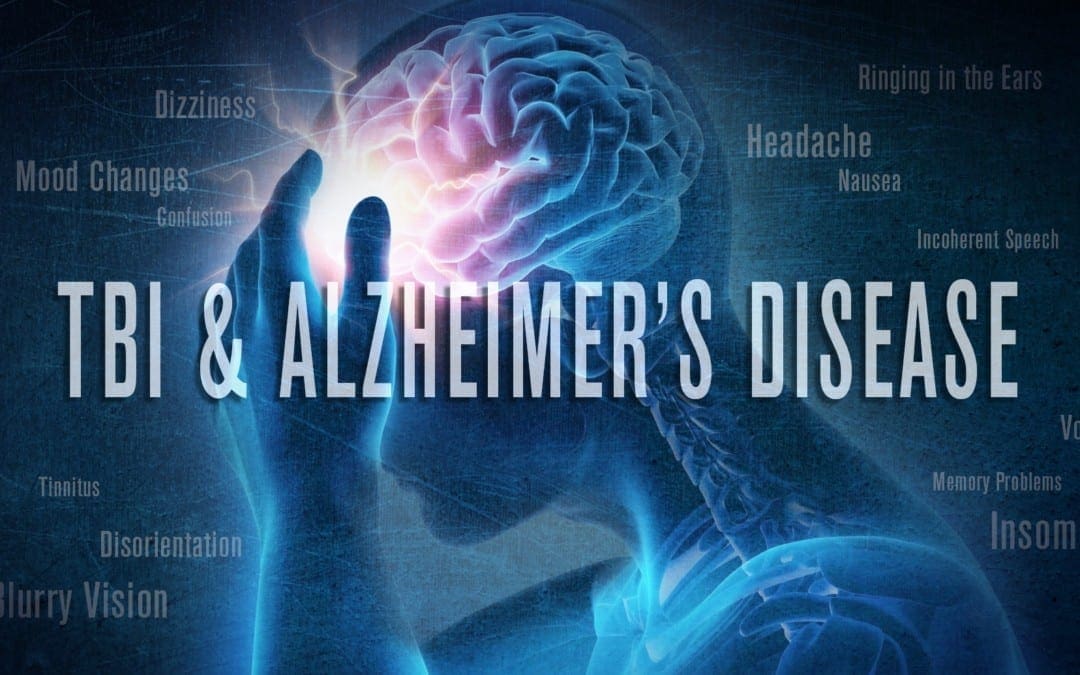
TBI may increase the risk of developing Alzheimer’s disease or dementia, years after the injury takes place.
According to the Centers for Disease Control and Prevention (CDC), approximately 2.8 million TBI-associated ER visits, hospitalizations, and deaths occurred in 2013, the latest year for which information is available. The purpose of the following article is to discuss traumatic brain injury (TBI) and its connection with Alzheimer’s disease and other health issues. �
Traumatic Brain Injury Causes
Slip-and-fall accidents are the most common cause of traumatic brain injury, where falls pose a potentially serious risk factor for older adults. According to a CDC special report evaluating data from several federal agencies, approximately 56,000 seniors are hospitalized every year as a result of head injuries sustained in falls. A serious TBI from a slip-and-fall accident may ultimately result in long-term cognitive changes and reduced ability to function as well as overall mood changes. �
About 775,000 older adults have traumatic brain injury-related disability. Measures to reduce the risk of falls include: �
Using a walker or other assistive device to compensate for mobility problems, muscle weakness or poor balance.
Having your vision checked regularly and using glasses or contact lenses that correct for changes.
Working with your doctor to watch for medication side effects or interactions among drugs you�re taking.
Avoiding household hazards, such as clutter, loose rugs or poor lighting.
Automobile accidents are another common cause of traumatic brain injury (TBI). People can reduce the risk of being involved in an auto accident by keeping their vehicle in good condition, following the rules of the road, and buckling their seat belt. Wearing a helmet and when biking, inline skating, or playing contact sports can also help protect the head from TBI. �
TBI Symptoms
The severity of symptoms for traumatic brain injuries largely depends on whether the injury is mild, moderate, or severe. Mild traumatic brain injury (TBI), also known as a concussion, can either not cause unconsciousness or can cause unconsciousness which lasts for 30 minutes or less. Mild traumatic brain injury (TBI) symptoms may include: �
Inability to remember the traumatic event immediately before or up to 24 hours after
Confusion and disorientation
Difficulty learning new information
Headache
Dizziness
Blurry vision
Nausea and vomiting
Ringing in the ears
Trouble speaking coherently
Mood changes or changes in sleeping patterns
These symptoms will commonly manifest at the time of the TBI or soon after, however, these may sometimes not develop till several days or even weeks following the traumatic event. Mild TBI symptoms are generally temporary and these will clear up within hours, days, or weeks following the traumatic even, however, they can occasionally last several months or longer. �
Moderate traumatic brain injury can cause unconsciousness which lasts more than 30 minutes but less than 24 hours and severe traumatic brain injury can cause unconsciousness for more than 24 hours. Symptoms of moderate and severe traumatic brain injury are similar to those of mild traumatic brain injury but these are more serious and longer-lasting. �
In all types of TBI, cognitive changes are the most common symptoms. The ability to learn and remember new information is also frequently affected. Other commonly affected cognitive skills include the ability to pay attention, organize thoughts, plan effective strategies for completing tasks and activities, and/or make sound judgments. More severe changes in cognitive skills may develop years after the traumatic event where the person may appear to have recovered from the previous TBI. �
TBI Diagnosis
Evaluations performed by healthcare professionals to help diagnose traumatic brain injury (TBI) generally include: �
Questions about the traumatic event
Analysis of the person’s level of consciousness and confusion
Neurological tests to analyze memory and thinking, vision, hearing, touch, balance, and reflexes
Let your doctor know if you are taking any drugs and/or medications, especially blood thinners, because they can increase the chance of complications. Also, inform your healthcare professional if you drink alcohol or take illicit drugs. �
Depending on the cause of the TBI and the severity of symptoms, brain imaging with computed tomography (CT) may be necessary to determine if there�s swelling or bleeding in the brain. If you experience a traumatic brain injury, it should be noted in your permanent medical record and mentioned whenever familiarizing a new doctor with your medical history. �
Traumatic Brain Injury Treatment
The most serious traumatic brain injuries commonly require specialized hospital care and can also need several months of rehabilitation. Most traumatic brain injuries are mild and can be treated with either a short hospital stay for observation or at-home monitoring followed by outpatient rehabilitation, if necessary. Treatment of dementia in a person with a history of traumatic brain injuries varies depending on the type of dementia diagnosed. Treatment strategies for Alzheimer’s disease or another type of dementia are ultimately the same for people with and without a history of traumatic brain injury. �
Alzheimer’s disease and other types of dementia which may occur as a long-term result of traumatic brain injury (TBI) are progressive health issues which worsen over time. As with all types of dementia, they can affect a person’s quality of life, shorten lifespan, and complicate the effort to manage other health issues effectively. However, because other types of dementia, such as CTE, are considerably new for researchers and healthcare professionals, clinical guidelines for diagnosis and treatment do not exist. Several research studies are underway to gain further insight into the patterns of TBI and Alzheimer’s disease which may be implicated in CTE and to develop strategies for prevention, diagnosis, and treatment. �
As previously mentioned in the article above, Alzheimer�s disease and other types of dementia which may occur as a long-term result of traumatic brain injury (TBI) are progressive health issues which may ultimately worsen over time. As with all types of dementia, these can affect quality of life, shorten life span, and complicate the effort to manage other health issues effectively. It’s essential for patients and healthcare professionals to diagnose and treat a traumatic brain injury to prevent further health issues in the future, including Alzheimer’s disease and dementia. – Dr. Alex Jimenez D.C., C.C.S.T. Insight
According to research studies, TBI is ultimately associated with Alzheimer�s disease and other types of dementia. Doctors commonly characterize traumatic brain injury as mild, moderate, or severe depending on whether the previous traumatic event causes unconsciousness, how long it lasts, and other well-known symptoms. The scope of our information is limited to chiropractic, musculoskeletal and nervous health issues as well as functional medicine articles, topics, and discussions. To further discuss the subject matter above, please feel free to ask Dr. Alex Jimenez or contact us at 915-850-0900 . �
Curated by Dr. Alex Jimenez �
Additional Topic Discussion: Chronic Pain
Sudden pain is a natural response of the nervous system which helps to demonstrate possible injury. By way of instance, pain signals travel from an injured region through the nerves and spinal cord to the brain. Pain is generally less severe as the injury heals, however, chronic pain is different than the average type of pain. With chronic pain, the human body will continue sending pain signals to the brain, regardless if the injury has healed. Chronic pain can last for several weeks to even several years. Chronic pain can tremendously affect a patient’s mobility and it can reduce flexibility, strength, and endurance.
Neural Zoomer Plus for Neurological Disease
Dr. Alex Jimenez utilizes a series of tests to help evaluate neurological diseases. The Neural ZoomerTM Plus is an array of neurological autoantibodies which offers specific antibody-to-antigen recognition. The Vibrant Neural ZoomerTM Plus is designed to assess an individual�s reactivity to 48 neurological antigens with connections to a variety of neurologically related diseases. The Vibrant Neural ZoomerTM Plus aims to reduce neurological conditions by empowering patients and physicians with a vital resource for early risk detection and an enhanced focus on personalized primary prevention. �
Formulas for Methylation Support
XYMOGEN�s Exclusive Professional Formulas are available through select licensed health care professionals. The internet sale and discounting of XYMOGEN formulas are strictly prohibited.
Proudly,�Dr. Alexander Jimenez makes XYMOGEN formulas available only to patients under our care.
Please call our office in order for us to assign a doctor consultation for immediate access.
If you are a patient of Injury Medical & Chiropractic�Clinic, you may inquire about XYMOGEN by calling 915-850-0900.
�
For your convenience and review of the XYMOGEN products please review the following link.*XYMOGEN-Catalog-Download �
* All of the above XYMOGEN policies remain strictly in force.
Traumatic brain injury (TBI) is one of the most common causes of disability and death among the general population, especially in young adults. Additionally, TBI is associated with a variety of neurodegenerative diseases, such as Alzheimer�s disease (AD) and Parkinson�s disease (PD). It is essential for patients and healthcare professionals to understand the pathophysiological mechanisms of traumatic brain injury and neurodegenerative diseases to diagnose factors which may ultimately cause neurodegeneration associated with TBI as well as determine possible treatment approaches. �
Oxidative stress, neuroinflammation, and glutamatergic excitotoxicity have previously been associated with TBI and neurodegenerative diseases. As a matter of fact, oxidative stress is believed to be an essential pathological mechanism which connects TBI to neurodegenerative diseases. Research studies have demonstrated that reactive oxygen species and their subsequent byproducts may play a role as novel fluid markers for the identification and monitoring of cellular damage. These reactive oxygen species can also serve as a suitable treatment approach to ultimately help reduce the risk of neurodegenerative diseases and promote quality of life for people suffering from TBI and other health issues. �
Pathogenesis of TBI and Neurodegenerative Diseases
Several research studies have demonstrated the development of neurodegenerative diseases following TBI. Previous research studies have also shown a three times higher prevalence of PD following TBI. Likewise, the prevalence of AD has also been shown to be higher following TBI. Moreover, traumatic brain injury has been demonstrated to be a risk factor for ALS with several research studies demonstrating an increased risk of neurological diseases in professional Italian soccer players. A case-control research study of ALS patients in the United States also found an increased risk of ALS with repeated TBI. However, it currently appears unlikely that a single occurrence of TBI could considerably affect the risk of ALS. Additionally, chronic traumatic encephalitis (CTE), a tau pathology, has been demonstrated in NFL players and professional athletes which suffer from repeated TBI. Because of the prevalence of neurodegenerative diseases and other health issues appears to increase after TBI, it is relevant to discuss the pathogenesis of TBI and neurodegenerative diseases. �
In several research studies, TBI patients and TBI animal models have been shown to demonstrate characteristic pathological mechanisms in key proteins, indicating the disruption of axonal transport due to axonal injury. The accumulated proteins which result in protein neuropathy include A?, ?-synuclein, and tau protein. These abnormal proteins are specifically interesting because it is well-known that A? protein aggregation is an essential pathological factor of AD, ?-synuclein protein aggregation is an important characteristic of PD, and tau protein aggregation is fundamental in the pathogenesis of CTE and AD. Surprisingly, these protein neuropathological changes occur in all three proteins through oxidative stress-associated free radicals and reactive aldehydes which are commonly increased following TBI. Additionally, the reactive aldehyde byproducts of lipid peroxidation have been demonstrated to result in further lipid peroxidation. Provided that these pathological proteins can also cause the development of free radicals through excitotoxicity or changes in mitochondrial ion balance. Because reactive aldehydes can cause further lipid peroxidation and protein carbonylation, it is possible that oxidative stress also plays a key role in a self-propagating cycle of lipid peroxidation, protein carbonylation, and neurodegenerative protein aggregation. Further research studies are still necessary to determine these outcome measures. �
TBI patients and TBI animal models have also demonstrated behavioral signs and symptoms, such as post-TBI dementia which resembles AD, post-TBI motor deficits which offer evidence of post-TBI brain tissue damage in the region of the hippocampus thus, resembling brain tissue damage in AD, and damage in the basal ganglia thus, resembling the brain tissue damage which occurs in PD. Functional magnetic resonance imaging (fMRI) research studies have also shown transient and persistent neuropathological functional changes in the brain of TBI patients which may contribute to the development of chronic neurodegenerative diseases. These changes observed in post-injury patients suggest that TBI could cause the initial tissue damage which resembles or results in processes in the pathophysiology of neurodegenerative diseases. �
Based on the essential role which oxidative stress plays in post-TBI secondary injury and in the pathophysiology of neurodegenerative diseases, it is possible that oxidative stress is a key process in connecting TBI to the increased prevalence of neurodegenerative diseases. Furthermore, oxidative stress may serve as a therapeutic, diagnostic, or prognostic marker in evaluating the risks of long term neurological diseases following TBI which can help determine a proper treatment approach. �
Treatment of TBI and Neurological Diseases
Considering the considerable risks caused by TBI, it is clear that there is a need for effective methods and techniques for early diagnosis and treatment of TBI patients to ultimately reduce the prevalence of post-TBI neurological sequelae. Currently, the diagnosis of TBI is primarily based on the patient’s provided history and clinical observations. Several clinical systems have been developed for the evaluation of mTBI, which is the most common type of clinical TBI, including the Sports Concussion Assessment Tool and Military Acute Concussion Evaluation. However, these assessments are made to be utilized immediately after injury and, as such, quickly decreasing in sensitivity with delayed evaluation. Moreover, the Glasgow Coma Scale has been utilized for decades and allows for both quick and constant communication of the patient’s condition nevertheless, the currently accepted threshold score of 13 may not be adequate to exclude visible abnormalities on computed tomography imaging which require neurosurgical intervention. Due to these outcome measures in current diagnostic methods and techniques, civilian and military work-groups have recommended the development of fluid or imaging-based biomarkers for the diagnosis of mTBI to ultimately determine the most appropriate treatment approach. �
Several substances and proteins have been suggested to play an essential role as fluid biomarkers, including glial fibrillary acidic protein (GFAP), calcium-binding protein S100B, and tau protein. In most cases, the presence of these biomarkers demonstrates a blood-brain barrier disruption within the central nervous system. These proteins have been demonstrated to be acutely increased following TBI in human participants, however, these currently face challenges of low specificity, poor correlation with the development of post-concussive symptoms, and poor correlation with imaging abnormalities. �
Provided the key role of oxidative stress and neuroinflammation in secondary neuronal injury and neurodegeneration, it is possible that the results of these processes may also serve as suitable biomarkers. As previously mentioned, plasma levels of several oxidative stress and inflammation-associated markers have been demonstrated to be increased in serum up to 42 days following multiple blast injuries and as early as one day following a single injury. Furthermore, lipid peroxidation products, such as acrolein and 4-hydroxynonenal, have also been demonstrated to be associated not only in TBI secondary injury but also in other types of neuronal health issues, such as spinal cord injury and ischemia-reperfusion injury. Provided that these peroxidation products are not only a cause of damage but also able to cause the modification of biomacromolecules where it is possible that measured increases may be able to demonstrate not only present damage but also continued secondary injury. Treatment of oxidative stress could help as a possible prophylactic treatment to decrease the risk of post-TBI neurodegeneration. Direct supplementation with endogenous antioxidants, such as glutathione and superoxide dismutase, has not demonstrated considerable benefits because these do not easily cross the blood-brain barrier. However, the glutathione precursor N-acetylcysteine has demonstrated several acute benefits in both animal and human research studies. Additionally, focusing on substances of the oxidative cascade, such as reactive aldehydes, has been demonstrated as a possible treatment due to the more lengthened half-lives of these substances when compared to ROS. However, despite the lengthened increase of inflammatory and oxidative byproducts, trials of antioxidant therapies have generally favored acute treatment, often within hours of the TBI, suggesting that acute treatment is appropriate. �
Considering the essential role of post-TBI oxidative stress in the development and progression of chronic neurodegenerative diseases, diagnosis and treatment of this process seem to be promising for the management and regulation of neurodegenerative diseases following TBI. Provided their connection to oxidative stress, inflammatory markers, and lipid peroxidation byproducts could serve as surrogate biofluid markers. Finally, antioxidant treatment strategies can help neutralize perpetuation of cellular and molecular damage and decrease risks of long-term neurological sequelae. �
As previously mentioned in the article above, oxidative stress seems to be the key pathological mechanism connecting neuroinflammation and glutamatergic excitotoxicity in both TBI and neurodegenerative diseases. Due to the increased prevalence of TBI and neurodegenerative diseases, the development of new safe and effective, early diagnosis and treatment approaches is fundamental for overall health and wellness. Many healthcare professionals can improve symptoms and health issues associated with TBI and neurodegenerative diseases. – Dr. Alex Jimenez D.C., C.C.S.T. Insight
TBI is associated with a variety of neurodegenerative diseases, such as Alzheimer�s disease (AD) and Parkinson�s disease (PD). It is essential for patients and healthcare professionals to understand the pathophysiological mechanisms of traumatic brain injury and neurodegenerative diseases to diagnose factors which may ultimately cause neurodegeneration associated with TBI as well as determine possible treatment approaches. The scope of our information is limited to chiropractic, musculoskeletal and nervous health issues as well as functional medicine articles, topics, and discussions. To further discuss the subject matter above, please feel free to ask Dr. Alex Jimenez or contact us at 915-850-0900 . �
Curated by Dr. Alex Jimenez �
Additional Topic Discussion: Chronic Pain
Sudden pain is a natural response of the nervous system which helps to demonstrate possible injury. By way of instance, pain signals travel from an injured region through the nerves and spinal cord to the brain. Pain is generally less severe as the injury heals, however, chronic pain is different than the average type of pain. With chronic pain, the human body will continue sending pain signals to the brain, regardless if the injury has healed. Chronic pain can last for several weeks to even several years. Chronic pain can tremendously affect a patient’s mobility and it can reduce flexibility, strength, and endurance.
Neural Zoomer Plus for Neurological Disease
�
Dr. Alex Jimenez utilizes a series of tests to help evaluate neurological diseases. The Neural ZoomerTM Plus is an array of neurological autoantibodies which offers specific antibody-to-antigen recognition. The Vibrant Neural ZoomerTM Plus is designed to assess an individual�s reactivity to 48 neurological antigens with connections to a variety of neurologically related diseases. The Vibrant Neural ZoomerTM Plus aims to reduce neurological conditions by empowering patients and physicians with a vital resource for early risk detection and an enhanced focus on personalized primary prevention. �
Formulas for Methylation Support
XYMOGEN�s Exclusive Professional Formulas are available through select licensed health care professionals. The internet sale and discounting of XYMOGEN formulas are strictly prohibited.
Proudly,�Dr. Alexander Jimenez makes XYMOGEN formulas available only to patients under our care.
Please call our office in order for us to assign a doctor consultation for immediate access.
If you are a patient of Injury Medical & Chiropractic�Clinic, you may inquire about XYMOGEN by calling 915-850-0900.
�
For your convenience and review of the XYMOGEN products please review the following link.*XYMOGEN-Catalog-Download
* All of the above XYMOGEN policies remain strictly in force.
Traumatic brain injury (TBI) is one of the most common causes of disability and death in people. About 1.6 million individuals suffer traumatic brain injuries in the United States every year. TBI can cause a process of injury which may ultimately cause a variety of neurodegenerative diseases and other health issues. Many of the neurodegenerative diseases following TBI include health issues such as Alzheimer’s disease (AD), Parkinson’s disease (PD), and amyotrophic lateral sclerosis (ALS). �
The mechanisms underlying the pathogenesis which result in these type of neurodegenerative diseases, however, are still completely misunderstood. Where many of the health issues following TBI have a high incidence, there are currently only several treatment approaches which can help prevent the pathological development of chronic neurological diseases. �
An understanding of the mechanisms underlying TBI and neurodegenerative diseases is fundamental to determine the possible connection between these health issues, to allow the safe and effective diagnosis and treatment. In the following article, we discuss the pathological mechanisms of neurodegenerative diseases and how they’re associated with traumatic brain injury (TBI), including Alzheimer’s disease (AD), Parkinson’s disease (PD), and amyotrophic lateral sclerosis (ALS). �
Pathological Mechanisms of Neurodegenerative Diseases
Although many neurological diseases may have different symptoms, AD, PD, and ALS have several common characteristics. Each neurodegenerative disease is caused by genetic risk factors, however, most cases are idiopathic or unknown. The pathological mechanisms of these health issues are ultimately characterized by the degeneration of brain cells or neurons together with several common symptoms. Moreover, abnormal clusters or dysfunction of the substances amyloid-? (A?), ?-synuclein, and superoxide dismutase (SOD1) are generally found in AD, PD. Although the exact pathological mechanisms of neurodegenerative diseases have not been fully determined, it has been suggested that oxidative stress, glutamatergic excitotoxicity, and neuroinflammation play fundamental roles in neurological diseases such as AD, PD, and ALS. �
AD has a tremendous prevalence among older adults which can greatly decrease their rate of survival and their overall quality of life. In 2008, as many as 24 million people worldwide had dementia, where most had AD, a number which is expected to double every 20 years as the population ages. The pathological mechanisms of AD include the presence of neuritic plaques and the loss of cholinergic neurons or brain cells in the human brain, however, the underlying risk factors leading to these events are still unclear. Neurodegeneration in AD is believed to happen due to the accumulation of amyloid ?-peptide (A?) in plaques in the brain tissue however its aggregation and toxicity are still completely misunderstood. �
Research studies have demonstrated that oxidative stress may play a fundamental role in the pathogenesis of AD because of increased neurotoxic markers of lipid peroxidation, such as 4-hydroxynonenal, in human participants, increased brain protein oxidation in AD, increased nuclear DNA oxidation in the brain of AD patients, 30 percent increased activity of the free radical scavenging enzyme SOD-1 in cell lines of AD patients, and considerable evidence that beta amyloid creates free radical peptides. In addition, it has been demonstrated that free radicals and lipid peroxidation caused by A? can ultimately result in neuronal death in AD. In vitro and animal research studies have demonstrated that the antioxidant effect of cannabinoids was able to prevent neurodegeneration in the neurological disease, suggesting the role of oxidative stress in AD. �
Neuroinflammation has also been associated wit A? toxicity which has likewise been connected to oxidative stress by inflammatory cytokine activity. The purpose of inflammation is to restore cellular homeostasis and balance redox equilibrium, however, inflammation changes with co-localized A? deposits, inflammatory-related proteins, and activated microglial cells in AD. Microglia and astroglia recognize misfolded proteins which can trigger an immune response that may be responsible for the progression and severity of the neurodegenerative disease. The microglial cells promote A? clearance and support neuroprotective properties in early stages of AD, but as the health issue progresses, inflammatory cytokines downregulate A? clearance genes and promote A? accumulation, ultimately causing neurodegeneration. Moreover, cytokines can trigger the creation of arachidonic acid which aggravates neurodegeneration by increasing extracellular levels of glutamate, known to cause excitotoxicity in AD as well as causing the creation of superoxide free radicals which are responsible for cellular death. Furthermore, research studies suggest that non-enzymatically glycated tau causes oxidative stress which results in cytokine gene expression and release of A?-peptide in AD, demonstrating pathological mechanisms between cytokines and oxidative stress which causes the progression and severity of AD. In addition, oxidative damage from reactive oxygen species and lipid peroxidation products, such as 4-hydroxy-2-nonenal (HNE), can restrict glutamate transporters, causing a decreased glutamate uptake that is fundamental for neuronal survival, an increased glutamate concentration in the synaptic cleft, and subsequent excitotoxicity which ultimately causes neurodegeneration in AD. �
Neurodegenerative Diseases in Functional Neurology
Chronic traumatic encephalopathy (CTE) is a neurodegenerative disease associated with repeated blunt force impacts to the head with the transfer of acceleration and deceleration forces to the brain or repetitive mild traumatic brain injuries, although the central pathological mechanisms for the development of neurodegeneration in CTE has not been discovered. CTE has been associated with behavioral and personality changes, parkinsonism, and dementia. Research studies demonstrated similarities between CTE and Alzheimer�s disease but these were different in the predominance of tau protein deposition over amyloid. The tau protein deposition in CTE has been previously demonstrated to restrict kinesin-dependent transport of peroxisomes and the loss of peroxisomes makes the cells vulnerable to oxidative stress, ultimately causing neurodegeneration. This tau protein deposition, which occurs in AD, also restricts the transport of amyloid precursor protein (APP) in axons or dendrites, causing its accumulation in the cell body. Along with tau proteins, portions of TDP43, a nuclear RNA/DNA binding protein which controls the transcription of thousands of genes, have been demonstrated in AD, PD, ALS, and CTE, which cause the misfolding of SOD1, affecting the surrounding cells with free-radical damage. The research studies have also demonstrated the purpose of oxidative stress in CTE neurodegeneration and in other neurological diseases. �
Chronic inflammation has also been demonstrated in CTE and AD, which is believed to aggravate neurodegeneration and, as previously mentioned, it is ultimately associated with oxidative stress though inflammatory cytokines. Moreover, it has been demonstrated that after the initial head trauma in CTE, microglia activate and release toxic levels of cytokines and excitotoxins, such as glutamate, where the excitotoxins restrict phosphatases, resulting in hyperphosphorylated tau, neurotubule dysfunction, and neurofibrillary tangle deposition, all of which are fundamental factors of CTE. Research studies have also demonstrated a synergy between proinflammatory cytokines and glutamate receptors which increase reactive oxygen species and worsens neurodegeneration in the injured brain associated with TBI and neurological diseases. �
Parkinson�s disease is the second most prevalent neurodegenerative disease with a prevalence of approximately 0.3 percent of the older adult population. PD is characterized by the development of ?-synuclein rich Lewy bodies and subsequent death of the dopaminergic neurons of the substantia nigra. Several genetic risk factors have also been demonstrated, including mutations to the ubiquitin-proteasome system. Although the pathological mechanisms which trigger dopaminergic degeneration in non-hereditary PD are still unclear, it has been suggested that oxidative modification or carbonylation of the lysine-rich N-terminus and the non-amyloid factor of ?-synuclein may ultimately cause an ?-synuclein aggregation. �
The reactive carbonyls created as secondary products in oxidative stress have been demonstrated to develop lysine adducts and promote ?-synuclein aggregation in vitro. Additionally, animal models of PD utilizing agents, such as 1-methyl-4-phenyl-1,2,3,6-tetrahydropyridine, have demonstrated the increased development of superoxide in dopaminergic cells associated with the cortex. Furthermore, mitochondrial localization of ?-synuclein has been demonstrated to promote oxidative stress in vitro. Neuroinflammation is believed to be a partial cause for the oxidative stress in PD with activated microglial cells demonstrated in the substantia nigra and striatum of deceased PD patients. Activated microglia were also demonstrated in rhesus monkeys up to 14 years after model induction. In addition, glutamatergic excitotoxicity is believed to play a fundamental role in PD. Rotigotine, an FDA approved dopamine receptor agonist, has been suggested to improve the efficiency of glutamate transporter 1 (GLT-1) and has been demonstrated to support neuroprotection against glutamatergic excitotoxicity in dopaminergic cell culture as well as a variety of other functions in the human brain in Parkinson’s disease. �
ALS is a fatal neurodegenerative disease characterized by the death of motor neurons in the central nervous system (CNS) and it is the most common motor neuron disease. Approximately 10 percent of all ALS cases have been associated with genetic causes while the majority are idiopathic or of unknown cause. Mutations affecting superoxide dismutase (SOD1) are responsible for almost 20 percent of all familial cases, however, this is responsible for only 2 percent of all overall cases. Despite the characterized mutations, the exact pathological mechanisms of ALS have yet to be fully determined. �
Research studies utilizing SOD1 mutant mouse models have demonstrated the development of SOD1 aggregates. Given the fundamental role of SOD1 in detoxification of superoxide radicals, it has been previously mentioned that loss of function could cause increased cellular exposure to reactive oxygen species, however, this hypothesis has been challenged by outcome measures in the normal development of SOD1 deficient mice in the absence of considerable traumatic injuries. Furthermore, research studies demonstrated that SOD1 mutant animals ultimately demonstrated no considerable improvement in symptomatic progression with knockout or coexpression of wild type SOD1 which suggests that the mutation results not in the loss of function but rather in the gain of toxic properties. Research studies in rats and human patients suggest that, similar to ?-synuclein and A?, SOD1 mutation cause the development of potentially cytotoxic protein aggregates even in patients without SOD1 mutations. Additionally, the catalysis changes achieved by several mutant variants causes decreased astroglial reuptake of glutamate through restriction of GLT-1. Riluzole, an FDA approved treatment for ALS, has been suggested to help improve glutamatergic excitotoxicity with increased glutamate uptake through GLT-1 and blockade of sensitive channels. Oxidative stress is also involved in neuronal death and in the progression of ALS. �
Given its fundamental role in maintaining and regulating damage from neuroinflammation and excitotoxicity, it is possible that oxidative stress also plays a fundamental role in the pathophysiology of AD, PD, and ALS in a similar fashion to TBI. As such, addressing oxidative stress in neurodegeneration could serve as an effective treatment strategy in neuroprotection. �
Conclusion
Despite the prevalence of TBI the significant neurological sequelae associated with such injuries, diagnosis, and treatment of TBI remains greatly misunderstood. In addition, the causing factors connected to TBI and neurodegenerative diseases, such as AD, PD, ALS, and CTE, have not been fully determined. Several processes, including oxidative stress and neuroinflammation, have also been found to be common between secondary TBI and several neurodegenerative diseases. In particular, oxidative stress appears to be the key mechanism connecting neuroinflammation and glutamatergic excitotoxicity in both TBI and neurological diseases. It is possible that the oxidative cascade caused by TBI ultimately causes and results in the characteristic pathologies of neurodegenerative diseases through oxidation or carbonylation of essential proteins. �
Due to the high prevalence of TBI and neurodegenerative diseases, the development of new safe and effective treatment approaches for TBI is fundamental. Given the essential role that oxidative stress plays in connecting secondary injury and neurodegeneration, detection of ROS and key byproducts could serve as a method or technique for the diagnosis and treatment of potential cellular damage. Finally, these reactive species may serve as a viable therapeutic target for reducing long-term neurodegenerative disease risk following TBI, helping to reduce the disability and death as well as improve the quality of life of individuals in the United States that suffer from traumatic brain injury (TBI) and other health issues. �
TBI is among one of the most common causes of disability and death among the general population in the United States. According to a variety of research studies, mild, moderate, and severe traumatic brain injury has been associated with neurodegenerative diseases, such as Alzheimer’s disease and Parkinson’s disease, as well as a variety of other neurodegenerative diseases. It is fundamental to understand the pathophysiological mechanisms of neurodegenerative diseases while further research studies are still required to determine the association between TBI and neurological diseases. – Dr. Alex Jimenez D.C., C.C.S.T. Insight
Traumatic brain injury (TBI) is one of the most common causes of disability and death in people. About 1.6 million individuals suffer traumatic brain injuries in the United States every year. TBI can cause a process of injury which may cause a variety of neurodegenerative diseases and health issues, such as Alzheimer’s disease (AD). The scope of our information is limited to chiropractic, musculoskeletal and nervous health issues as well as functional medicine articles, topics, and discussions. To further discuss the subject matter above, please feel free to ask Dr. Alex Jimenez or contact us at 915-850-0900 . �
Curated by Dr. Alex Jimenez �
Additional Topic Discussion: Chronic Pain
Sudden pain is a natural response of the nervous system which helps to demonstrate possible injury. By way of instance, pain signals travel from an injured region through the nerves and spinal cord to the brain. Pain is generally less severe as the injury heals, however, chronic pain is different than the average type of pain. With chronic pain, the human body will continue sending pain signals to the brain, regardless if the injury has healed. Chronic pain can last for several weeks to even several years. Chronic pain can tremendously affect a patient’s mobility and it can reduce flexibility, strength, and endurance.
Neural Zoomer Plus for Neurological Disease
Dr. Alex Jimenez utilizes a series of tests to help evaluate neurological diseases. The Neural ZoomerTM Plus is an array of neurological autoantibodies which offers specific antibody-to-antigen recognition. The Vibrant Neural ZoomerTM Plus is designed to assess an individual�s reactivity to 48 neurological antigens with connections to a variety of neurologically related diseases. The Vibrant Neural ZoomerTM Plus aims to reduce neurological conditions by empowering patients and physicians with a vital resource for early risk detection and an enhanced focus on personalized primary prevention. �
Formulas for Methylation Support
XYMOGEN�s Exclusive Professional Formulas are available through select licensed health care professionals. The internet sale and discounting of XYMOGEN formulas are strictly prohibited.
Proudly,�Dr. Alexander Jimenez makes XYMOGEN formulas available only to patients under our care.
Please call our office in order for us to assign a doctor consultation for immediate access.
If you are a patient of Injury Medical & Chiropractic�Clinic, you may inquire about XYMOGEN by calling 915-850-0900.
�
For your convenience and review of the XYMOGEN products please review the following link.*XYMOGEN-Catalog-Download �
* All of the above XYMOGEN policies remain strictly in force.
IFM's Find A Practitioner tool is the largest referral network in Functional Medicine, created to help patients locate Functional Medicine practitioners anywhere in the world. IFM Certified Practitioners are listed first in the search results, given their extensive education in Functional Medicine