Self-myofascial release, also known as “foam rolling,” has changed from a once mysterious technique used solely by professional athletes, athletes, and therapists to a familiar everyday method for people at all levels of fitness.
Products, technology, and data have introduced an increasing array of training and recovery methods to the individual.
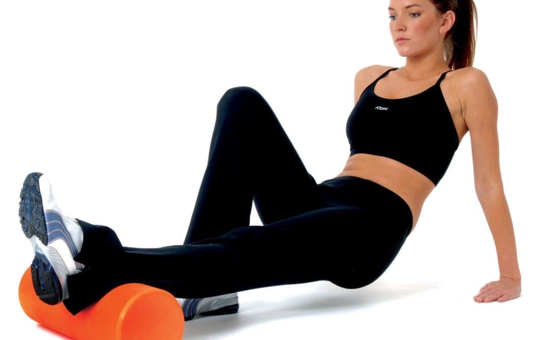
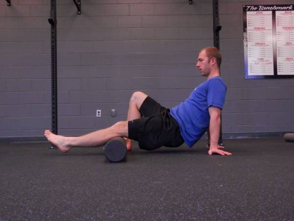
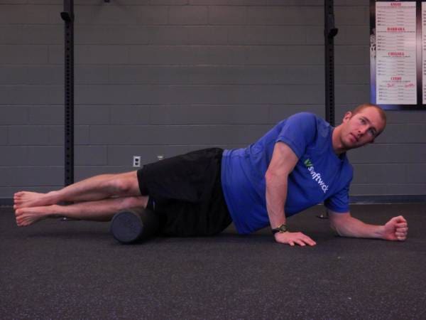
Self-myofascial release is a fancy word for self-massage, utilized to release muscle stiffness or trigger points. This technique can be performed using a foam roller, lacrosse ball, Theracane, or your own hands. By applying pressure to these painful areas, you are able to assist in the recovery of muscles and helping to restore them. Proper function means your muscles are healthy, elastic, and ready to perform at a moment’s notice.
Determining Tight Muscles & Trigger Points
Trigger points are referred to as “knots” which form in muscles. They’re unique and may be identified once they begin to refer pain. Pain referral, for our purposes, may be described as the pain felt when pressure is applied to a single area of their body, but the pain is felt or radiated in a different area.
A common case of a trigger point is felt while foam rolling your iliotibial (IT) band as it causes pain to radiate up to the hip or all the way down the leg to the ankle. When rolling on tight/sore muscles you may experience pain or discomfort. It should be uncomfortable, but not unbearable, and it must relieve the symptoms, when you are done.
For many, deep tissue massage is simple to understand. Somebody is able to exercise the knots in your muscles, and it is commonly known that this process may be uncomfortable and occasionally painful. Because only you can feel what is happening, self-myofascial discharge provides the consumer the capability to control the recovery and healing procedure by applying pressure in precise places.
It is always suggested to consult with your physician or physical therapist to get therapeutic/sharp pain and receive approval prior to beginning self-myofascial release. You will be cleared immediately and your doctor will encourage the practice. Releasing trigger points helps reestablish appropriate movement patterns and pain free movement, and finally, to boost functionality. Utilizing stretching alone isn’t always enough to discharge muscles. Imagine a bungee cord with a knot tied into it and then envision stretching the cord. This creates tension, stretching the part of the muscle and the attachment points. The knot, however, has remained unaltered.
Foam rolling can assist in dividing these muscle knots, resuming normal blood flow and function. The aim to any recovery or corrective technique is to get you back to normal functioning’s point, as if nothing was ever wrong.
Causes of Trigger Points & Tight Muscles
Both have exactly the same contributing factors such as training, flexibility, movement patterns, posture, nutrition, hydration, rest, anxiety, and other lifestyle factors. Our bodies learn to compensate for what we throw at them daily, but we can transcend our ability to recover via intense workouts, bad posture, and other lifestyle factors.
Deep compression can help to break up or relax tight muscles and adhesions formed between muscular layers and their environment. Imagine you are currently tenderizing your muscles. They should be soft and supple as a baby’s muscles. If our muscles are not taken care of properly we can experience loss of motion that is debilitating.
The deep compression of self-myofascial release enables normal blood flow to return and the recovery of healthy tissue. The body wants to be healthy and strong, but an extra boost is required to attain optimum tissue and muscle health.
How Do I Know What to Foam Roll and How to Do It?
Areas to concentrate on can be identified in two different ways. The first is through screenings. When you have followed the two posts – screening and stylish hinge screening – and also have had struggles with either movement, foam rolling should be included by you into retrieval program and your workout. You may target you are currently focusing on.
If after using the foam roller your motion enhances, you’ve got a more specific plan to follow. Second, muscles and trigger points are discovered utilizing techniques’ listing below and researching every one.
To foam roll correctly, apply moderate pressure to a particular muscle or muscle group using the roller and your own leg. You should roll slowly, no longer than one inch. Pause for several moments when you find areas that are painful or tight and relax as far as you can. You should begin to feel that the muscle releasing, and pain or the distress should reduce.
If a place is too painful to use direct pressure, then change the roller and then apply pressure on the surrounding area and gradually work to loosen the entire area. The purpose is to restore muscles – it isn’t a pain tolerance evaluation. You could also use different objects to operate on muscles such as lacrosse ball, a tennis ball, Theracane, or Trigger Point Therapy Kit.
Never roll a joint or bone. Avoid your back. To target these muscles I advise using lacrosse or tennis balls. If you’re experiencing difficulties with your neck, refer these problems to an appropriate medical practitioner and need attention that is advanced.
What Happens After Foam Rolling?
You might be sore the next day. It should feel like your muscles are worked/released, but you shouldn’t push yourself to the purpose of excessive soreness. Drink lots of water, get enough sleep , and eat clean. Fuel your muscles and this can help flush your system. Before focusing on precisely the same place give it 24-48 hours.
The scope of our information is limited to chiropractic and spinal injuries and conditions. To discuss options on the subject matter, please feel free to ask Dr. Jimenez or contact us at 915-850-0900 .�
Additional Topics: Sports Care
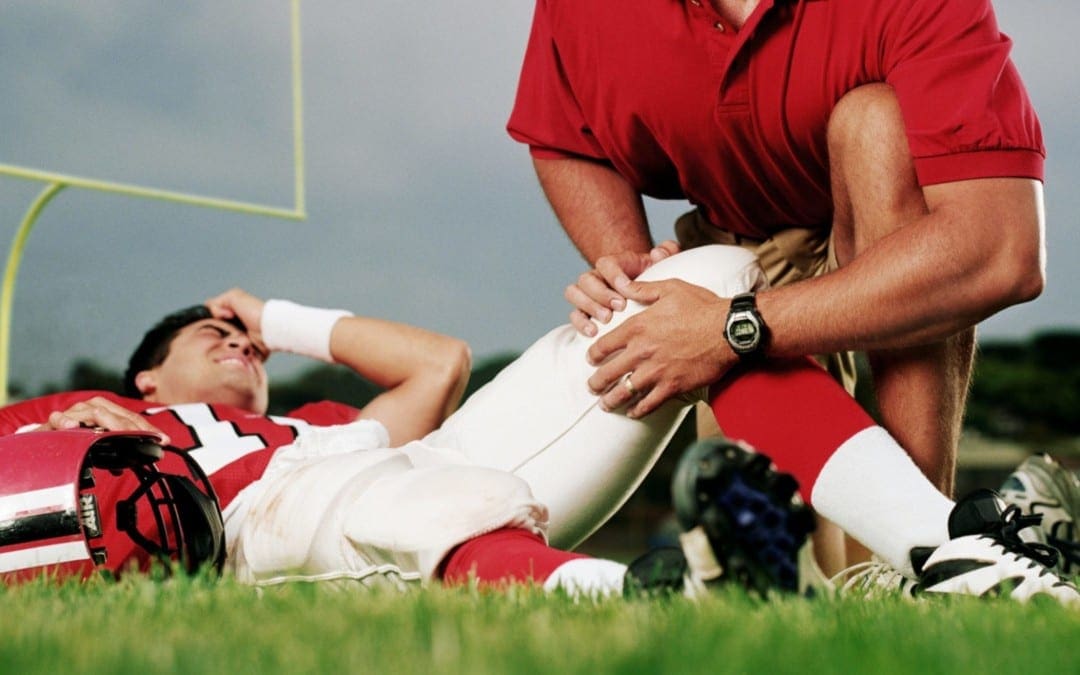
Many athletes engage in frequent warm-up stretches and exercises before participating in their specific sport of physical activity, in order to avoid experiencing sports injuries. Although these can help prevent a variety of sports injuries, athletes may still suffer an injury as a result of an accident. From chiropractic care to surgery, in severe cases, sports care is important for athletes to continue participating in their specific sport of physical activity.
Concussion, also known as mild traumatic brain injury (MTBI), has been a poorly understood condition known to the majority of healthcare providers as difficult to objectify and manage.
Historically, there has been no testing available to conclude an accurate diagnosis. In the absence of objective imaging findings of bleeding in the brain, a diagnosis of “mild traumatic brain injury” has been affixed to the condition, whereas if there’s evidence of traumatic bleeding then the diagnosis “traumatic brain injury” is applied.
Although Hartvigsen, Boyle, Cassidy and Carroll (2014) reported that 600 out of 100,000 Americans are affected every year by concussion, Jeter et al, (2012) reported that close to 40 percent of people experiencing a mild brain injury do not report it to their doctor, making accurate statistics very tricky to conclude. Despite potential under reporting in the people, we realize concussion is an issue that has consequences that are important from the perspective of a clinical result and we cannot afford to ignore this condition.
Mechanism of Injury: Mild Traumatic Brain Injury
Mild traumatic brain injury or concussion results from transfer of mechanical energy from the outside environment to the brain due to traumatic events where there’s a sudden acceleration and then a sudden deceleration of the mind and brain, such as in a Coup/Contrecoup injury during a whiplash scenario. As the brain is freely moving to a degree because it’s only surrounded by cerebral spinal fluid, it continues moving in the original direction and as the head “whips” rapidly in the opposite direction, the brain bounces off parts of the inner skull, which in turn rebounds shortly after the head changes direction. This is one easily defined mechanism of MTBI that doesn’t cause gross bleeding, yet leaves the brain injured through direct compression or overstretching (axonal shearing) of central nervous system components.
Although this has been examined extensively in the military, it’s been recently investigated in professional sports, where after several lawsuits and lives at risk, there are now definitive “concussion protocols” in place. Part of the protocols as reported from the British Journal of Sports Medicine (2016) is the Sports Concussion Assessment Tool 2 or SCAT2 that’s been adopted by numerous professional sports leagues. However, the majority of concussion victims are not active participants in the military or a professional sports team and many find their way into chiropractic practices as a consequence of sports injuries, car accidents, slip and falls and every other sort of head trauma etiology. Even though the mechanisms might vary, the induced end results are the same.
For generalized patient intake protocols, according to both Medicare and academia standards, a questionnaire outlining a summary of body systems is mandated, and part of those questions center on brain function. As reported by Jeter et al behavioral and cognitive symptoms, signs and symptoms are reported on standard patient intake questionnaires and require consideration of a diagnosis of concussion.
Prominent symptoms of concussion include: balance issues, vomiting, nausea, headache, drowsiness, dizziness, fatigue, vision, light or noise sensitivity and sleep disturbances. Cognitive symptoms include deficits in attention, concentration, memory, mental processing speed, and working memory or decision making. Behavioral symptoms include anxiety, depression, irritability, depression and aggression. The researchers went on to report that approximately 25 percent of the cases can have these symptoms persist.
Diagnosis and Treatment for MTBI
As a profession, chiropractic is a important part of the rehabilitation for the concussion population as the post-traumatic patient typically presents to the average chiropractic practice. As chiropractors (along with all healthcare providers), even if you mix the history with the above symptoms inclusive of neurological, behavioral and cognitive traits, you then have the direction or “triage road map” of the way to conclusively differentially diagnose your individual, including what tests to consider conducting in order to do so. The first line of testing is to consider imaging to rule out bleeding and ensure the patient does not require an immediate consultation. Treating blindly can place your patient in risk that is possible.
Imaging of the brain requires either MRI or CAT scans, MRI being the more sensitive, and in the absence of bleeding, the diagnosis is limited to MTBI or concussion (used interchangeably). More recently, diffusion tensor imaging (DTI) has been a tool available to picture mTBI victims that uses tissue water diffusion speeds to determine bleeding at a very small level giving demonstrable evidence to brain injury. As reported by Soares, Marques, Alves, and Sousa, (2013), DTI has several issues to overcome to certify accuracy including, but not limited to, tissue type, integrity, barriers and quantitative diffusion rates that are required to infer molecular diffusion prices. DTI is a model based upon assumption with a outlook as a tool.
Historically, MTBI was exclusively diagnosed by an omission of advanced imaging findings and the presence and persistence of the neurology, cognitive and behavioral signs and symptoms. Today, brain-derived neurotrophic factors (BDNF) offer responses about carpal brain pathology that is both conclusive and reproducible. Based on Korley et al. (2015), brain-derived neurotrophic factors is a secreted autocrine (compound hormone or messenger in blood) which promotes the development, maintenance, survival, differentiation and regeneration of neurons. BDNF also is important for synaptic plasticity (strengthening of synapses over time) and memory processing. Germane to MTBI and concussion, BDNF has been implicated in decreasing brain injury, with elevations and restoring traumatic brain injury.
Korley went on to report that BDNF levels were the highest in the normal group with lower values in mTBI and even lower in traumatic brain injury (TBI) subjects. In addition BDNF values were associated with incomplete recovery of patients that were MTBI compared to moderate or severe TBI patients. Because of this, it has been ascertained that BDNF has for identifying associated sequelae at 6 23, a prognostic value.
Korley stated that BDNF is the most abundantly secreted brain neurotrophin and as a secreted protein and can be readily measured using well-established immune-assay methods, identifying it as a non-necrosis brain injury biomarker. This distinguishes BDNF from other biomarkers which are components of neurons and myelin based proteins among other structures. In order for structural fibers to be found in high abundance in circulation, adequate cellular necrosis and damage to the blood barrier membrane must be observed, however BDNF does not require cellular damage or necrosis to be observed in circulation enabling DDNF to be more plentiful in flow than structural proteins.
Following a traumatic brain event, BDNF supports synaptic reorganization and recovery during the brain circuitry “reconnection” phase. Therefore, a better prognosis is indicated by lowered values. In patients with a co-morbidity of BDNF of anxiety, depressive disorders and schizophrenia BDNF values on the day of injury predispose this population to incomplete recovery as a risk element. Korley et al.. Concluded that serum BDNF discriminates between MTBI and TBI cases. Also, diminished BDNF values are associated with recovery in identifying and useful symptoms 6-months post-trauma.
Conclusion
Simply put, a blood test could assist providers in concluding the existence and/or severity of traumatic brain injury or mild traumatic brain injury. An early diagnosis is afforded by the results so you can devise a treatment plan inclusive of changing activities of everyday living to prevent additional damage and optimize the repair procedure with minimizing further chemical, physical or emotional stressors.
Based upon interviews with leading neurologists and neurosurgeons who understand and have first-hand expertise of both receiving chiropractic care and handling and treating MTBI patients, it is strongly recommended that until the signs and symptoms of the neurologic, cognitive and behavioral abate that high-velocity rotational cervical adjustments be avoided to enable the brain to “repair and rewire” the connections without additional possibilities of and Coup/ Contrecoup energy to the mind. This is a recommendation which we agree while recognizing that chiropractic care should not be avoided adapted to allow the brain to heal.
The scope of our information is limited to chiropractic and spinal injuries and conditions. To discuss options on the subject matter, please feel free to ask Dr. Jimenez or contact us at 915-850-0900 .�
References:
1. Hartvigsen, J., Boyle, E., Cassidy, J. D., & Carroll, L. J. (2014). Mild traumatic brain injury after motor vehicle collision: What are the symptoms and who treats them? A population-based 1-year inception cohort study. Archives of Physical Medicine and Rehabilitation, 95(Suppl. 3), S286-S294.
2. Jeter, C. B., Hergenroeder, G. W., Hylin, M. J., Redell, J. B., Moore, A. N., & Dash, P. K. (2013). Biomarkers for the diagnosis and prognosis of mild traumatic brain injury/concussion. Journal of Neurotrauma, 30(8), 657-670.
3. British Journal of Sports Medicine. (2016). Sport concussion assessment tool 2. Retrieved from http://bjsm.bmj.com/content/43/Suppl_1/i85.full.pdf
4. Soares, J. M., Marques, P., Alves, V., & Sousa, N. (2013). A hitchhiker�s guide to diffusion tensor imaging. Frontiers in Neuroscience, 7(31), 1-14.
5. Korley, F. K., Diaz-Arrastia, R., Wu, A. H. B., Yue, J. K., Manley, G. T., Sair, H. I., Van Eyk, J., Everett, A. D., Okonkwo, D. O., Valadka, A. B., Gordon, W. A., Maas, A. I., Mukherjee, P., Yuh, E. L., Lingsma, H. F., Puccio, A. M., & Schnyer, D. M., (2015). Circulating brain-derived neurotrophic factor has diagnostic and prognostic value in traumatic brain injury. Journal of Neurotrauma, 32, 1-11.
Additional Topics: Weakened Ligaments After Whiplash
Whiplash is a commonly reported injury after an individual has been involved in an automobile accident. During an auto accident, the sheer force of the impact often causes the head and neck of the victim to jerk abruptly, back-and-forth, causing damage to the complex structures surrounding the cervical spine. Chiropractic care is a safe and effective, alternative treatment option utilized to help decrease the symptoms of whiplash.
According to the American Academy of Orthopedic Surgery �The most common soft tissues injured are muscles, tendons, and ligaments.
Acute injuries are caused by a sudden trauma, such as a fall, twist, or blow to the body. Examples of an acute injury include sprains, strains, and contusions.�� (http://orthoinfo.aaos.org/topic.cfm?topic=A00111) We must also not forget that there are other soft tissues that can get injured and the true definition of soft tissue, which is anything not bone is soft tissue.
This includes the brain, lungs, heart and any other organ in the body. However, in medicine soft tissue injuries are commonly known to be limited to the muscles, ligaments and tendons.
Soft Tissue Injury Classification
When we look at the type of structures that muscles, tendons and ligament are composed of, we will realize that they are connective tissue. According to the National Institute of Health �Connective tissue is the material inside your body that supports many of its parts. It is the “cellular glue” that gives your tissues their shape and helps keep them strong. It also helps some of your tissues do their work (http://www.nlm.nih.gov/medlineplus/connectivetissuedisorders.html). Unlike fracture repair where the bone is replaced and usually heals properly if aligned and rested, connective tissue disorders undergo a different type of wound repair that has aberrant tissue replacement as sequella to bodily injury and has subsequent abnormal permanent function.
If we focus on sprains or ligamentous injuries, according to the American Academy of Orthopedic Surgery there are three types of sprains:
Sprains are classified by severity:1
Grade 1 sprain (mild):�Slight stretching and some damage to the fibers (fibrils) of the ligament.
Grade 2 sprain (moderate):�Partial tearing of the ligament. There is abnormal looseness (laxity) in the joint when it is moved in certain ways.
Grade 3 sprain (severe):�Complete tear of the ligament. This causes significant instability and makes the joint nonfunctional.
Regardless of the severity of the sprain, there is tissue damage or bodily injury and the next step is to determine if there is healing or wound repair. According to Woo, Hildebrand, Watanabe, Fenwick, Papageorgiou and Wang (1999) ��as a result the combination of cell therapy with growth factor therapy may offer new avenues to improve the healing of ligament and tendon. Of course, specific recommendations regarding growth factor selection, and timing and method of application cannot be made at this time.
Previous attempts at determining optimal doses of growth factors have provided contradictory results. Although growth factor treatment has been shown to improve the properties of healing ligaments and tendons, these properties do not reach the level of the uninjured tissue.� (p. s320)
�No treatment currently exists to restore an injured tendon or ligament to its normal condition.�, stated Dozer and Dupree (2005). (pg. 231).
Soft Tissue Recovery Process
According to Hauser, Dolan, Phillips, Newlin, Moore and Woldin (2013) �injured ligament structure is replaced with tissue that is grossly, histologically, biochemically and biomechanically similar to scar tissue. Fully remodeled scar tissue remains grossly, microscopically and functionally different from normal tissues� (p. 6) �the persisting abnormalities present in the remodeled ligament matrix can have profound implications on joint biomechanics, depending on the functional demands placed on the tissue.
Since remodel ligament tissue is morphologically and mechanically inferior to normal ligament tissue, ligament laxity results, causing functional disability of the affected joints and predisposing other soft tissues in and around the joints further damage.� (p.7) �studies of healing ligaments have consistently shown that certain ligaments do not heal independently following rupture, and those that didn�t feel, do so characteristically inferior compositional properties compared with normal tissue. It is not uncommon for more than one ligament undergo injury during a single traumatic event.� (p.8) �osteoarthritis for joint degeneration is one of the most common consequences of ligament laxity.
Traditionally, the pathophysiology of osteoarthritis was thought to be due of aging and wear and tear on the joint, but more recent studies have shown that ligaments play a critical role in the development of osteoarthritis. Osteoarthritis begins when one or more of ligaments become unstable or lax, and the bones began to track improperly and put pressure on different areas, resulting in the rubbing the bone on cartilage. This causes breakdown of cartilage and ultimately leads to deterioration, whereby the joint is reduced to bone on bone, a mechanical problem of the joint that leads to abnormality of the joints mechanics. Hypomobility and ligament laxity have become clear risk factors for the prevalence of osteoarthritis.� (p.9)
Looking globally at the research over the last 16 years, in 1999 it was concluded that the most current treatments to repair or heal the injured ligament do not reach the level of the uninjured tissue. In in 2005 it was concluded that no treatment currently exists to restore an injured tendons or ligaments to its normal condition. In addition the current standard of ligament research in 2013 concluded that that ligaments do not feel independently, but damage ligaments are a direct cause of osteoarthritis and biomechanical dysfunction (abnormality of joint mechanics). The latest research has also concluded that ligament damage or sprains is the key element in osteoarthritis and not simply aging or wear and tear on the joint.
As a result it is now clear based upon the scientific evidence that a soft tissue injury is a connective tissue disorder that has permanent negative sequela and is the cause of future arthritis. This is no longer a debatable issue and those in the medical legal forum who are still arguing �transient soft tissue injuries� are simply rendering rhetoric out of ignorance and a possible ulterior motive because the facts clearly delineate the negative sequella based upon decades of multiple scientific conclusions.
The caveat to this argument is that although there is irrefutable bodily injury with clear permanent sequella, does it also cause permanent functional loss in every scenario? Those are two separate issues and as a result of the function of ligaments, which is to connect bones to bones the arbiter for normal vs. abnormal function is ranges of motion of the joint. That can be accomplished by either a two-piece inclinometer for the spine, which according to the American Medical Association Guides to the Evaluation of Permanent Impairment, 5th Edition (p. 400) is the standard (and is still the medical standard as the 6th Edition refers to the 5th for Ranges of motion).
The other diagnostic demonstrable evidence to conclude aberrant function is to conclude laxity of ligaments through x-ray digitizing. Both diagnostic tools confirm demonstrably loss of function of the spinal joints. ��
The scope of our information is limited to chiropractic and spinal injuries and conditions. To discuss options on the subject matter, please feel free to ask Dr. Jimenez or contact us at 915-850-0900 .�
Woo S, Hildebrand K., Watanabe N., Fenwick J., Papageorgiou C., Wang J. (1999) Tissue Engineering of Ligament and Tendon Healing, Clinical Orthopedics and Related Research 367S pgs. S312-S323
Tozer S., Duprez D. (2005) Tendon and Ligament: Development, Repair and Disease, Birth Defects Research (part C) 75:226-236
Hauser R., Dolan E., Phillips H., Newlin A., Moore R. and B. Woldin (2013) �Ligament Injury and Healing: A Review of Current Clinical Diagnostics and Therapeutics, The Open Rehabilitation Journal (6) 1-20
Cocchiarella L., Anderson G., (2001) Guides to the Evaluation of Permanent Impairment, 5th Edition, Chicago IL, AMA Press
Additional Topics: Preventing Spinal Degeneration
Spinal degeneration can occur naturally over time as a result of age and the constant wear-and-tear of the vertebrae and other complex structures of the spine, generally developing in people over the ages of 40. On occasion, spinal degeneration can also occur due to spinal damage or injury, which may result in further complications if left untreated. Chiropractic care can help strengthen the structures of the spine, helping to prevent spinal degeneration.
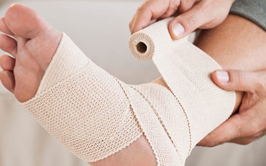
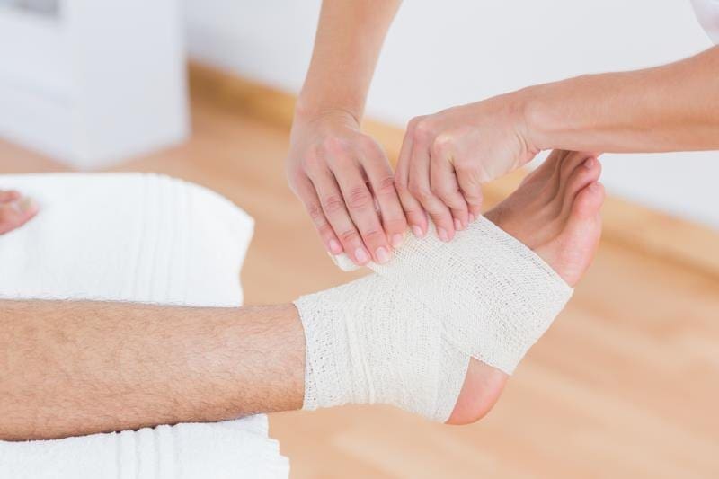
Sprains, strains and tears are different types of injuries, and it’s important to know how they differ, a sports massage therapist says.
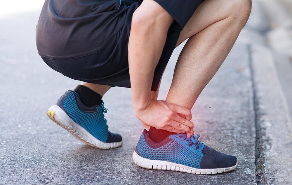
A sprain is the overstretching or tearing of ligaments, which are the tissues that connect bones to each other and stabilize them.
“Sprains occur when the joint is forced into an unnatural position. They happen most often in the ankle but can occur at any joint, such as the wrist or knee,” said Martin Mufich. He is also a clinical assistant professor at Texas A&M College of Nursing. Symptoms of a sprain include joint or muscle pain, inflammation, hampered movement, tenderness and bruising. “A mild sprain should take approximately seven to 10 days to heal,” Mufich said in a university news release.
“A torn ligament is considered a severe sprain that will cause pain, inflammation, bruising and result in ankle instability, often making it difficult and painful to walk. Recovery from a torn ligament may take several weeks, and should be done under the supervision of a health-care provider,” he explained.
A strain is the overstretching or tearing of a muscle or a tendon, which connects the muscles to the bones. It can occur from a single incident or over time. “An acute strain is an instantaneous stretch or tear of the muscle or tendon, whereas, a chronic strain stems from repetitive motions over time that place stress on the muscle or tendon,” Mufich said. Symptoms of a strain include muscle spasms, weakness, cramping, immobility, pain, bruising and swelling. It can take a few weeks for symptoms of a mild-to-moderate strain to ease, he explained.
A tear is the ripping of tissue in ligaments, muscles or tendons.
“Typically, the worse a tear, the more inflammation and pain a person will experience, and the longer it will take for the injury to heal,” Mufich said. In general, the treatment for sprains, strains and tears involves a plan called “RICES” — Rest, Ice, Compression, Elevation and Stabilization. However, for some severe tears, such as those of the anterior cruciate ligament (ACL) in the knee, surgery may be needed.
Mufich said that it is normal to experience some discomfort during the healing process from any of these injuries, but there should not be any sharp pain. “If you are not seeing improvements within 24 hours or it is getting worse, contact a health-care provider,” he advised.
SOURCE: Texas A&M, news release, Feb. 23, 2017�
For more information, please feel free to ask Dr. Jimenez or contact us at 915-850-0900 .
Preventing Sports Injuries
Many athletes largely depend on chiropractic care to enhance their physical performance. New research studies have determined that aside from maintaining overall health and wellness, chiropractic can also help prevent sports injuries. Chiropractic is an alternative treatment option utilized by athletes to improve their strength, mobility and flexibility. Spinal adjustments and manual manipulations performed by a chiropractor can also help correct spinal issues, speeding up an athlete’s recovery process to help them return-to-play as soon as possible.
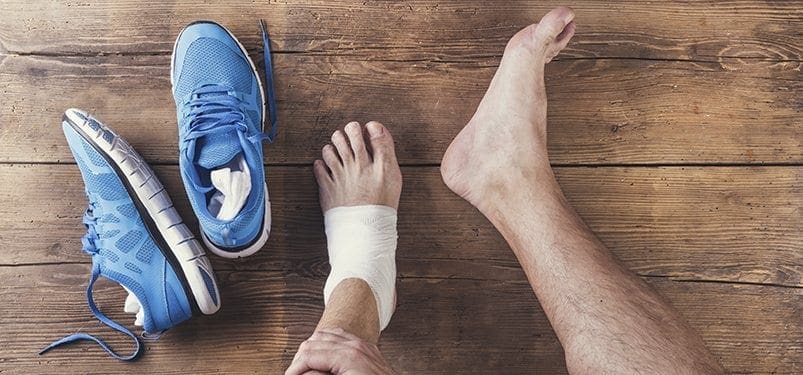
Whether your doctor recommends surgery for a ruptured Achilles tendon may depend partly on your age and activity level, foot experts say.
The Achilles tendon is a band of tissue that runs down the back of the lower leg and connects the calf muscle to the heel bone. A rupture is a complete or partial tear of the tendon that leaves the heel bone separated or partially separated from the knee.
Length of recovery from this type of injury varies depending on whether a patient undergoes surgical or nonsurgical treatment.
“Treatment processes are dependent upon a patient’s overall health, activity level and ability to follow a functional rehabilitation protocol,” said Dr. Jeffrey McAlister, a foot and ankle surgeon in Sun City West, Ariz. Advances in treating Achilles tendon rupture were discussed by McAlister and other specialists at a recent meeting of the American College of Foot and Ankle Surgeons, in Las Vegas.
Typically, less active and unhealthy patients receive nonsurgical treatment, since they are not trying to return to active sports, McAlister said in a college news release. But this approach usually involves a long rehabilitation/recovery period (9-12 months). Also, these patients may be at increased risk of potentially dangerous blood clots due to inactivity during this period.
“For more athletic and younger patients, the surgical option may be best,” said Dr. Michael VanPelt, a Dallas foot and ankle surgeon. “We anticipate these patients have shorter healing times.”
But because there is low blood flow to the Achilles tendon, healing after surgery can be tricky.
“Advances in surgical techniques to repair Achilles tendon ruptures include limited incision, or smaller incision, surgical approaches to help patients have smaller scars, and less of a chance of wound complications,” said Dr. Jason Kayce, a Phoenix foot and ankle surgeon.
For more information, please feel free to ask Dr. Jimenez or contact us at 915-850-0900 .
Preventing Sports Injuries
Many athletes largely depend on chiropractic care to enhance their physical performance. New research studies have determined that aside from maintaining overall health and wellness, chiropractic can also help prevent sports injuries. Chiropractic is an alternative treatment option utilized by athletes to improve their strength, mobility and flexibility. Spinal adjustments and manual manipulations performed by a chiropractor can also help correct spinal issues, speeding up an athlete’s recovery process to help them return-to-play as soon as possible.
What home remedies are effective for sprains and strains?
Initial treatment for sprains and strains should occur as soon as possible. Remember RICE!
Rest the injured part. Pain is the body’s signal to not move an injury.
Ice the injury. This will limit the swelling and help with the spasm.
Compress the injured area. This again, limits the swelling. Be careful not to apply a wrap so tightly that it might act as a tourniquet and cut off the blood supply.
Elevate the injured part. This lets gravity help reduce the swelling by allowing fluid and blood to drain downhill to the heart.
Over-the-counter pain medication is an option. Acetaminophen (Tylenol) is helpful for pain, but ibuprofen (Motrin, Advil) or naproxen (Aleve) might be better because these medications relieve both pain and inflammation. Remember to follow the guidelines on the bottle for appropriate dose of the medicine, especially for children and teens. Underlying medical conditions or use of other prescription medicines may limit the use of over the counter pain medications.
What is the treatment for sprains and strains?
Sprains and strains can usually be treated with home therapy using the RICE interventions. However, if the injury is more severe, your care provider may suggest splinting or casting to rest the injured joint. In some cases, operations are required to fix complete tears of muscles or tendons to allow complete return of function and to allow those muscles to do their job of moving the body. Significant tears of ligaments that stabilize joints also may need repair, but again, most are treated with short-term immobilization and early return to activity. Sometimes, resting the injury requires some help. Slings for arm injuries or crutches for leg injuries can be used, in addition to a variety of removable splints to protect the injured area from further damage and movement. Resting also helps relieve some of the muscle spasm associated with the injury.
Occasionally, if the injury is especially severe, the physician may want to use a nonremovable splint made of plaster or fiberglass. Although the splint may look like a cast, it doesn’t have plaster or fiberglass completely encircling the injured area. Instead, by only going partially around an injury, there is some room to allow for swelling that may occur during the next few days.
For more information, please feel free to ask Dr. Jimenez or contact us at 915-850-0900 .
Preventing Sports Injuries
Many athletes largely depend on chiropractic care to enhance their physical performance. New research studies have determined that aside from maintaining overall health and wellness, chiropractic can also help prevent sports injuries. Chiropractic is an alternative treatment option utilized by athletes to improve their strength, mobility and flexibility. Spinal adjustments and manual manipulations performed by a chiropractor can also help correct spinal issues, speeding up an athlete’s recovery process to help them return-to-play as soon as possible.
The body is meant to move. Muscles allow that movement to happen by contracting and making joints flex, extend and rotate. Muscles attach on each side of the joint to bone by thick bands of fibrous tissue called tendons. When a muscle contracts, it shortens and pulls on the tendon, which allows the joint to go through a range of motion.
A strain occurs when the muscle tendon unit is stretched or torn. The most common reason is the overuse and stretching of the muscle. The damage may occur in three areas:
The muscle itself may tear.
The area where the muscle and tendon blend can tear.
The tendon may tear partially or completely (rupture).
Joints are stabilized by thick bands of tissue called ligaments which surround them. These ligaments allow the joint to move only in specific directions. Some joints move in multiple planes; therefore, they need more than one group of ligaments to hold the joint in proper alignment. The ligaments are anchored to bone on each side of the joint. If a ligament is stretched or torn, the injury is called a sprain.
Causes of Sprains and Strains
Sprains and strains occur when the body is put under stress. In these situations, muscles and joints are forced to perform movements for which they are not prepared or designed to perform. An injury can occur from a single stressful incident, or it may gradually arise after many repetitions of a motion.
Signs and Symptoms of Sprains and Strains
The first symptom of a sprain or strain injury is pain. Other symptoms, such as swelling and spasm, can take time (from minutes to hours) to develop.
Pain is always a symptom that indicates that there is something wrong with the body. It is the message to the brain that warns that a muscle or joint should be protected from further harm. In work, exercise, or sport, the pain may come on after a specific incident or it may gradually progress after many repetitions of a motion.
Swelling almost always occurs with injury, but it may take from minutes to hours to be noticed. Any time fibers of a ligament, muscle, or tendon are damaged, some bleeding occurs. The bleeding (such as bruising on the surface of the skin) may take time to be noticed.
Because of pain and swelling, the body starts to favor the injured part. This may cause the muscles that surround the injured area to go into spasm. Hard knots of muscle might be felt near the site of the injury.
The combination of pain, swelling, and spasm causes the body to further protect the injured part, which results in difficulty with use. Limping is a good example of the body trying to protect an injured leg.
REFERENCE:
Young, Craig C. “Ankle Sprain.” Medscape.com. Dec. 16, 2014. <http://emedicine.medscape.com/article/1907229-overview>.
For more information, please feel free to ask Dr. Jimenez or contact us at 915-850-0900 .
Preventing Sports Injuries
Many athletes largely depend on chiropractic care to enhance their physical performance. New research studies have determined that aside from maintaining overall health and wellness, chiropractic can also help prevent sports injuries. Chiropractic is an alternative treatment option utilized by athletes to improve their strength, mobility and flexibility. Spinal adjustments and manual manipulations performed by a chiropractor can also help correct spinal issues, speeding up an athlete�s recovery process to help them return-to-play as soon as possible.
IFM's Find A Practitioner tool is the largest referral network in Functional Medicine, created to help patients locate Functional Medicine practitioners anywhere in the world. IFM Certified Practitioners are listed first in the search results, given their extensive education in Functional Medicine