
Why Do I Need an X-ray or MRI for Lower Back Pain El Paso, TX?
Low back pain is one of the most common ailments for people visiting a doctor or an urgent care clinic. When the back pain becomes intense, it can get you thinking something is seriously wrong with your back. The doctor might offer an x-ray or MRI scan to put your concerns at ease.
Fortunately, most cases of low back pain, even acute pain, improve within days or a few weeks. Most cases are remedied with chiropractic, physical therapy, heat/ice therapy, and rest. And a lot of these cases do not require any form of spinal imaging. However, those are why X-ray, MRI, and CT scans are necessary to figure out what’s happening.
- Strained muscle
- Sprained ligament
- Poor posture
These typical causes of low back pain can be painful and limit activities.
Back Pain Lasting Longer Than 2/3 Weeks
Subacute pain lasts between 4 and 12 weeks, while chronic back pain lasts three months or longer. These are not indications of a severe lower back spinal condition.
Less than 1% of people with low back pain are diagnosed with the condition that may require spine surgery:
- Cauda equina syndrome
- Spinal infection
- Metastatic spinal cancer
X-rays or MRIs for Diagnosing Low Back Pain
Doctors may recommend an x-ray or MRI if the low back pain is from a traumatic injury, like a:
- Slip
- Fall
- Automobile accident
Other potential causes of low back pain may warrant medical imaging immediately or later.
The diagnostic process starts with the evaluation of the low back symptoms and how they relate to what was found during the:
- Physical exam
- Neurological exam
- Medical history
A doctor utilizes these results to determine whether spinal imaging is necessary, along with the type of imaging test, x-ray, or MRI and the timing to confirm a diagnosis.
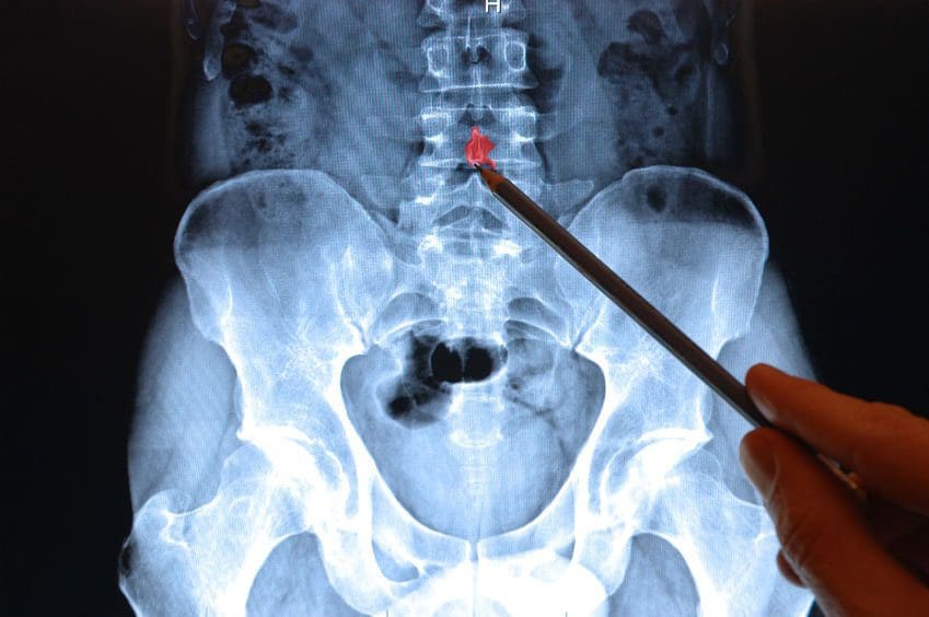
A Low Back X-Ray/MRI
X-ray spinal imaging best detects bony structural problems but is not so great with soft tissue injuries. X-ray series may be performed to diagnose vertebral compression fractures.
- Anterior
- Posterior
- Lateral views
MRI is a radiation-free test. MRIs create 3-D anatomical views of the spinal bones and soft tissues. A contrast dye like gadolinium is used to enhance and improve the quality of the images. The contrast is injected through an intravenous line in your hand or arm before or during the test. An MRI can evaluate neurological symptoms, like radiating pain or pain that develops after a cancer diagnosis.
Symptoms, Co-existing Medical Diagnoses, and Conditions that may Require Spine Imaging
Neurological symptoms
- Low back pain that radiates, fans out, or downward into the buttocks, legs, and feet
- Abnormal reflexes in the lower body can indicate nerve disruption
- Numbness, tingling, and possibly weakness develop
- Inability to lift your foot, aka foot drop
Co-existing medical diagnoses and conditions
- Cancer
- Diabetes
- Fever
- Osteoporosis
- Previous spinal fracture
- Spine surgery
- Recent infection
- Immunosuppressant medication use
- Corticosteroid medication
- Weight loss
X-ray Radiation Exposure
Radiation to your entire body is measured through the millisievert (mSv), also known as the effective dose. The radiation dose is the same amount every time you experience an x-ray. When undergoing an x-ray, the radiation not absorbed by the body creates the image.
The effective dose helps a doctor measure the risk for possible side effects of radiographic imaging:
- CT scans use radiation as well
- Specific body tissues and organs in the lower back are sensitive to radiation exposure, like the reproductive organs.
MRI Radiation-Free Why Not Just Use This Test All The Time
MRI cannot be used on all patients because of its powerful magnet technology. Pregnant women or individuals with metal inside their body, like a spinal cord stimulator, heart pacemaker, etc., cannot be scanned with an MRI.
MRI testing is also expensive; doctors do not want to prescribe unnecessary tests that increase costs. Or because of the fine detail that MRIs provide, sometimes a spinal issue can look severe but is not.
Example: An MRI of the lower back reveals a herniated disc in a patient with no back/leg pain or other symptoms.
This is why doctors bring all their findings like the symptoms, physical exam, and medical history to confirm a diagnosis and create a custom treatment plan.
Imaging Test Takeaways
If low back pain takes its toll, listen to what the doctor recommends. They might not order a lumbar x-ray or MRI immediately but remember the issues mentioned above, like neurological symptoms and co-existing medical conditions. But these tests help discover the cause or causes of the pain. Remember this is to help get patients to their optimal health and pain-free.
How to eliminate Back Pain naturally | (2020) Foot Levelers |El Paso, Tx
NCBI Resources
Imaging diagnostics is an essential element in the evaluation of spine trauma. The rapid evolution of imaging technology has tremendously changed the assessment and treatment of spine injuries. Imaging diagnostics utilizing CT and MRI, among others, are helpful in acute and chronic settings. Spinal cord and soft-tissue injuries are best evaluated by magnetic resonance imaging, or MRI, whereas computed tomography scanning or CT scans best evaluate spinal trauma or spine fracture.


