According to chiropractic physiological concepts, the body is in a state of relaxation when the body’s natural intelligence can properly function. With regular movement, work, school, chores, tasks, the body takes a beating getting pushed, pulled, and twisted. This results in loss of function, disharmony, chronic conditions, and disease. Chiropractic assistance restores the communication flow with various types of adjustments.
Like a vehicle that has something loose or is out of place, but can still move, so do individuals continue to work despite their body telling them that something is wrong or out of place. As they continue the body can start to break down. Even with a minor vertebral subluxation. A subluxation is when a vertebra/spine’s bones interfere with the transmission of information along the spine and throughout the central nervous system. The messages/transmissions to and from the brain disrupt the body’s ability to function properly resulting in health issues. A chiropractor adjusts the body to correct the subluxation. This facilitates the body’s natural ability to:
Adapt
Regulate
Heal itself through its own natural intelligence.
Chiropractic is based on principles of physiology. These principles have a significant impact on individuals:
Overall health
Vitality
Well-being
Self-healing and Regulating
When individuals cut themselves, or when a bone gets broken, the body will heal itself. No thought or practice is needed. The body breathes, digests food, the heart continues to beat and the kidneys filter out waste products.
Brain, Nervous System, and Body Health
The brain and nervous system are what controls and coordinate the body’s ability to heal and self-regulate. The brain sends out information through the nervous system to the organs of the body. The information tells the organs what to do and when to do it. The organs reply to the brain through the nervous system letting the brain know everything is operating correctly.
To function properly and maintain optimal function, the body depends on the proper circulation of nerve transmissions/information traveling through the nervous system.
Spinal State
The condition or state of the spine if out of place even just a little could interfere with the transmission of information via the nerve pathways. Misalignments and subluxations don’t always hurt so individuals don’t know this is happening. The nervous system is protected by the skull and spinal bones, known as the vertebrae. The spinal bones allow for free movement. Subluxations and misalignments:
Stretch
Pinch
Irritate
Choke
Rub the nerve fibers.
This alters the information/messages traveling and interferes with the body’s ability to function as it should.
Chiropractic Assistance
Chiropractic assistance will diagnose and correct misalignments and subluxations, restoring proper nerve circulation. These spinal adjustments unlock and realign the bones allowing for optimal movement and the communication pathways are fixed. A healthy spine and nervous system are vital to healthy body function. This is the body’s natural state. How to know if you have a subluxation, have your spine checked by a chiropractor.
Body Health Assistance
Body Composition Goals
For individuals that fall within the extremely overweight and obese body fat percentage category, an effective strategy is to lose fat while preserving the existing Lean Body Mass. Individuals with high body fat percentages often have existing muscle developed from carrying the added weight when moving. Preserving this muscle is important. Having more muscle means increased metabolism and more strength.
To start losing body fat it is recommended to cut down/cut out processed foods and reduce caloric intake.
Consult with a health coach and nutritionist to find out about different types of diets and types of physical activity.
One study showed that obese individuals could quickly improve body composition by:
Maltese, Paolo Enrico et al. “Molecular foundations of chiropractic therapy.” Acta bio-medica : Atenei Parmensis vol. 90,10-S 93-102. 30 Sep. 2019, doi:10.23750/abm.v90i10-S.8768
Pickar, Joel G. “Neurophysiological effects of spinal manipulation.” The spine journal: official journal of the North American Spine Society vol. 2,5 (2002): 357-71. doi:10.1016/s1529-9430(02)00400-x
Rosner, Anthony L. “Chiropractic Identity: A Neurological, Professional, and Political Assessment.” Journal of chiropractic humanities vol. 23,1 35-45. 20 Jul. 2016, doi:10.1016/j.echu.2016.05.001
Vining, Robert et al. “Assessment of chiropractic care on strength, balance, and endurance in active-duty U.S. military personnel with low back pain: a protocol for a randomized controlled trial.” Trials vol. 19,1 671. 5 Dec. 2018, doi:10.1186/s13063-018-3041-5
Neurophysiology: There are two ways that nociceptive information reaches the central nervous system. One is the neospinothalamic tract for quick pain and two is the paleospinothalamic tract for slow pain that increases.
Neurophysiology Of Pain Part II
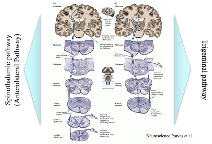
Intensity, Location & Quality of Pain…
… involve Spinothalamic and Trigeminal Pathways
The trigeminal pathway brings information from the face area.
The spinothalamic pathway brings information from the rest of the body.
Both these pathways project to the sensory cortex, which also receives information on innocuous stimuli such as touch, pressure and warmth via a separate pathway.
2 Pain Transmission Pathways For Location Intensity Quality
There is difference between the objective and subjective aspects of injury and pain.
Despite similar injury, people can differ in how much pain they feel.
Depending on the context, pain may not be felt despite injury, e.g. battlefield injury, during intense sports.
This suggests that there is a physiological mechanism that controls the transmission of nociceptive signals to the brain or modifies the interpretation of pain.
The pain control system can also explain the placebo effect.
Pain Modulation Pathway
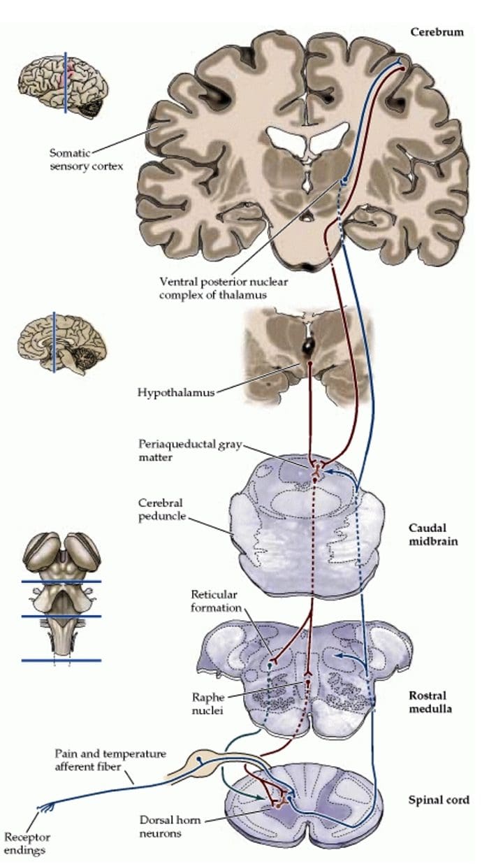
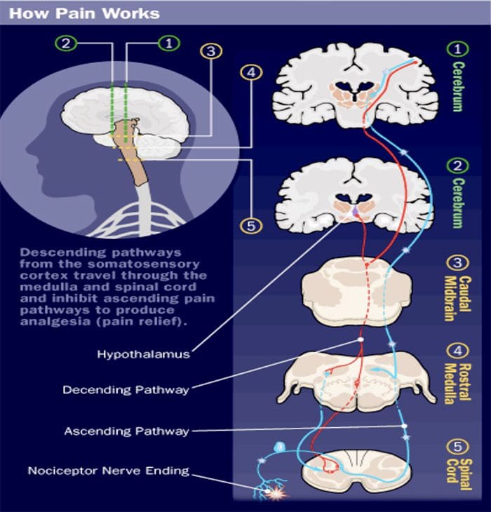
Nerve signals are sent form the somatic sensory cortex and hypothalamus to the periaqueductal gray matter (PAG).
PAG sends signals to the parabrachial nucleus, medullary reticular formation, locus coeruleus, and Raphe neulei.
These in turn can control the in the transmission of nociceptive signals from the spinal cord to the brain.
This involves different involves different neurotransmitters.
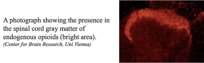
Endogenous Opioids
Internally produced molecules with opioid-like action which regulate transmission of nociceptive signals.
Three classes of these molecules have been identified. All are peptide molecules
Enkephalins
Endorphins
Dynorphins
Despite these being powerful, endogenous modifiers of nociceptive signals, it has been difficult to produce and administer them in a way than can used in clinical practice.
Location Of Nerve Cells With Endogenous Opioid Receptors
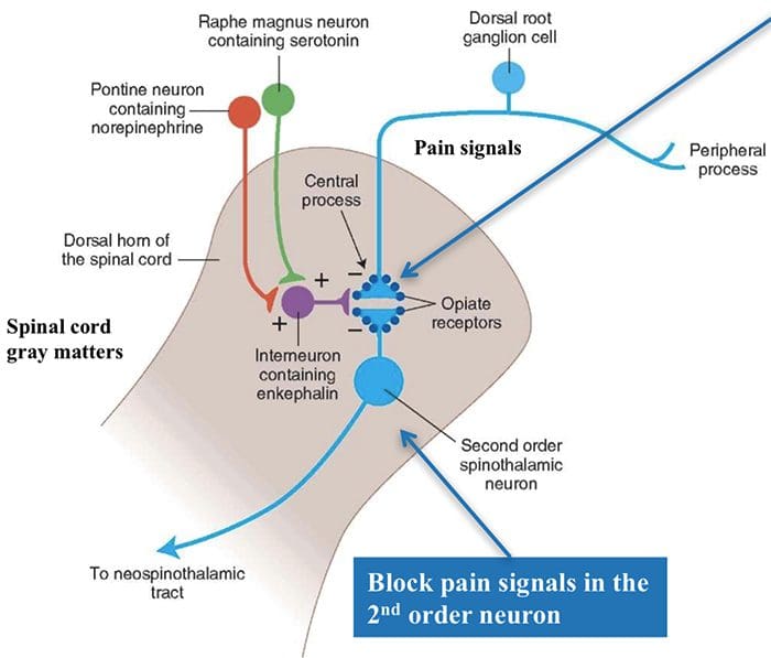
In the spinal cord, endogenous opioids can prevent transmission between 1st order nerve cells (bringing signals from the periphery) and 2nd order spinal nerve cells that transmit the signals to the brain.
Also can prevent the increased synaptic efficiency, which plays a role in hyperalgesia.
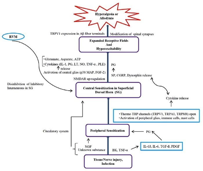
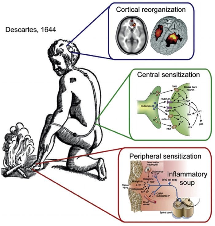
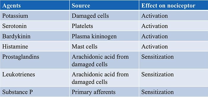
Knowing the molecules involved in the �inflammatory soup� and how they are synthesized provides possible targets for pain reduction.
e.g. prostaglandins are produced by the COX enzyme. The activity of this enzyme is blocked by non-steroidal anti- inflammatory drugs (NSAIDs) such as ibuprofen, diclofenac.
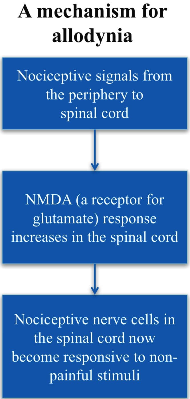
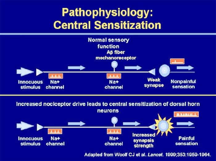
Allodynia
A condition when normally non- painful stimuli cause pain, e.g., touch, light pressure, cold.
Involves changes in the synaptic sensitivity of the nociceptive neurons in the spinal cord (central sensitization).
Drugs such as ketamine, block NMDA receptors and so reduce transmisison of the nociceptive stimuli.
Gate Control Theory of Pain
Mother says to child, �Come I will rub the area which is painful and this will make it feel better.�
After stubbing a toe, we instinctively rub the area; this reduces the sensation of pain.
Ronald Melzack and Patrick Wall in 1962 provided an possible explanation for this effect.
Ascending Tracts | Pain Modulation: Gate Control Theory
Gate Theory
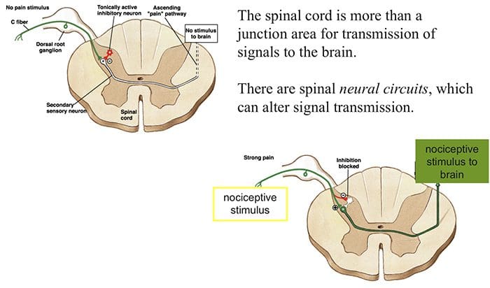
Rubbing the area that hurts stimulates receptors of innocuous stimuli like touch, pressure and vibration.
These mechano-receptors send signals along the A? nerve fibers that:
(1) stimulate spinal nerves (inhibitory inter-neurons) that in turn inhibit signaling in the 2nd order neurons (projection neuron) and (2) directly inhibit the 2nd order neuron to reduce or stop pain signal from being sent to the brain
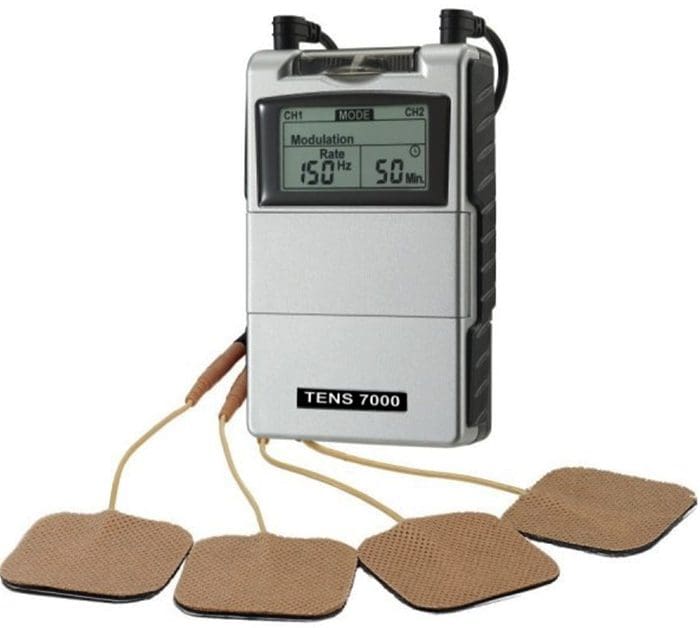
Transcutaneous Nerve Stimulation (TENS) is based on the Gate Control Theory. Nerves of the innocuous sensory system are stimulated and they in turn, inhibit transmission of nociceptive stimuli in the spinal cord.
Abnormalities Of Pain System
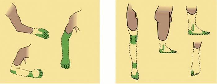
Phantom Pain
Patients with amputation often have burning or tingling pain in the body part removed.
One possible cause is that nerve fibers at the stump are stimulated and the brain interprets the signals as originating in the amputated portion.
The other is the rearrangement within the cortical areas so that area say for the hand now responds to signals from other parts of the body but still interprets them as coming for the amputated hand.
Peripheral Sensitization
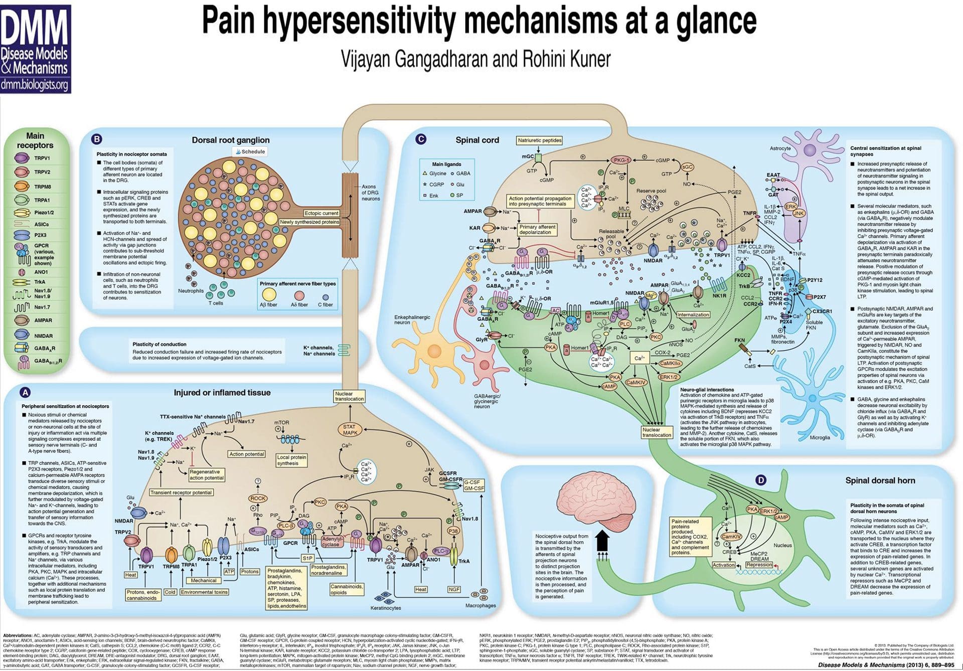
Peripheral sensitization represents a reduction in the threshold and/or an increase in magnitude of responsiveness at the peripheral ends of sensory nerve fibers.
This occurs in response to chemical mediators released by nociceptors and non-neuronal cells (e.g. mast cells, basophils, platelets, macrophages, neutrophils, endothelial cells, keratinocytes and fibroblasts) at the site of tissue injury or inflammation.
Basically, it is an increased sensitivity to an afferent nerve stimuli.
Central Sensitization
Peripheral & Central Sensitisation
A condition of the nervous sytem that is associated with the development and maintenance of chronic pain.
Known as �wind-up� or persistent high reactivity.
�Plastiticity in pain pathways� or the persistence of pain even after an injury has healed.
Is this negative or positive plasticity?
Central Sensitization & C Fibers
Two Main Characteristics Of Central Sensitization:
� Allodynia � occurs when a person experiences pain with things that are normally not painful, ie, soft touch causes pain.
� Hyperalgesia � occurs when a stimulus that is typically painful is perceived as more painful that it should be, ie, a simple bump.
Both are due to hyperreactivity of the nervous system.
Neurophysiology of pain: Pain�defined is the unpleasant sensation that accompanies injury or near injury to tissues, though it can also occur in the absence of such damage if the nociception system is not functioning. Nociception means the system that carries pain signals of injury from the tissues. This is the physiological incident that comes with pain.
Neurophysiology Of Pain
Objectives
Basics of the nervous system
Synaptic function
Nerve impulses
Transduction of peripheral painful stimuli
Central pathways
Central Sensitization
PeripheralSensitization
Control or modulation of pain signals
Pathophysiology of pain signaling pathway
Definition Of Pain
“Pain is an unpleasant sensory and emotional experience associated with actual or potential tissue damage, or described in terms of such damage”.
It is important to know the basic structure of the nervous system.
This will help in:
� Understanding the mechanism by which nociceptive signals are produced.
� Know the different regions of the nervous system involved in processing these signals.
� Learn how the different medications and treatment for pain management work.
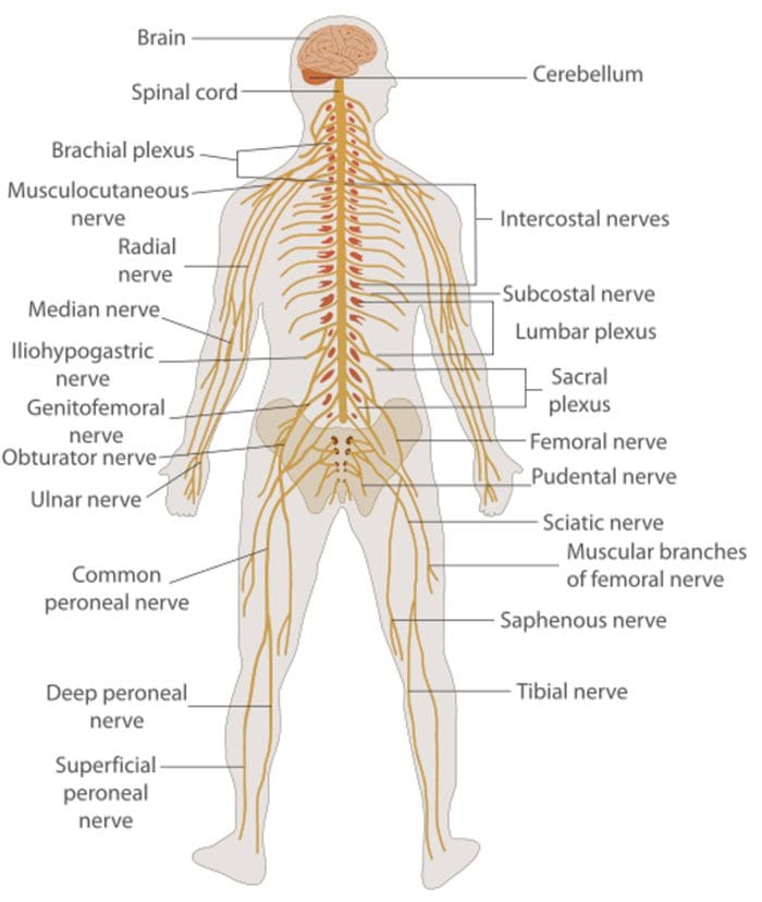
Nervous System
Central nervous system (CNS)
Brain and Spinal Cord
Peripheral Nervous System (PNS)
Nerve fibers go to all parts of the body.
Send signals to the different tissues and send signals back to the CNS.
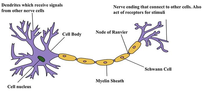
Nerve Cells
The nervous system is made up of nerve cells which send long processes (axons) to make contact with other cells.
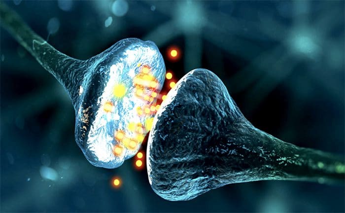
Nerve Cell-To-Nerve Cell Communication
Nerve cells communicate with other cells by releasing a chemical from the nerve endings � Neurotransmitters
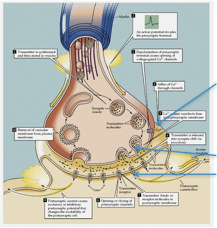
Basic Steps In Synaptic Transmission
Synaptic Transmission
Steps in the passage of signal from one nerve cell to other.
Drugs are used to block the transmission of signals from one nerve cell to other.
These drugs can effect:
Ca2+ ion channel to prevent Ca2+ inflow which is essential for neurotransmitter (NT) release, e.g., the action of gabapentin.
Release of NT.
Prevent NT from binding to its receptor so stop further transmission of the signal.
Electrical Impulse
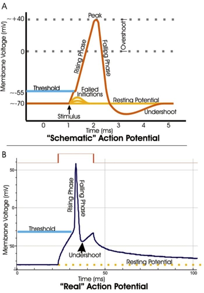
Signals move along a nerve process (axon) as a wave of membrane depolarization called the Action Potential.
The inside of all nerve cells has a negative electrical potential of around � 60 mV.
When stimulated this negative electrical potential becomes positive and then negative again in milliseconds.
The action potential moves along the nerve process (axon) to the nerve ending where it cause release of NT.
Action Potential
When there is no stimulation the membrane potential is at its Resting Potential.
When stimulated, channels in the nerve membrane open allowing the flow of sodium ions (Na+) or calcium ions (Ca2+) into the nerve or cell. This makes the inside less negative and in fact positive -the peak of the action potential (+40 mV).
These channels than close and by the opening of K+ channels the membrane potential returns to its resting level.
Stopping Action Potentials To Stop Nociceptive Stimuli
Nociceptive stimuli are those that will create a sensation of pain after they are processed in the CNS.
Nociceptive signals can be prevented from reaching the CNS by blocking the action of the channels that control the movement of ions across the nerve membrane.
A number of anesthetic agents stop Na+ channel from working and hence stop the generation of actions potentials and transmission of signals to the CNS.
Sensory Systems
The sensory system that can be divided into two divisions:
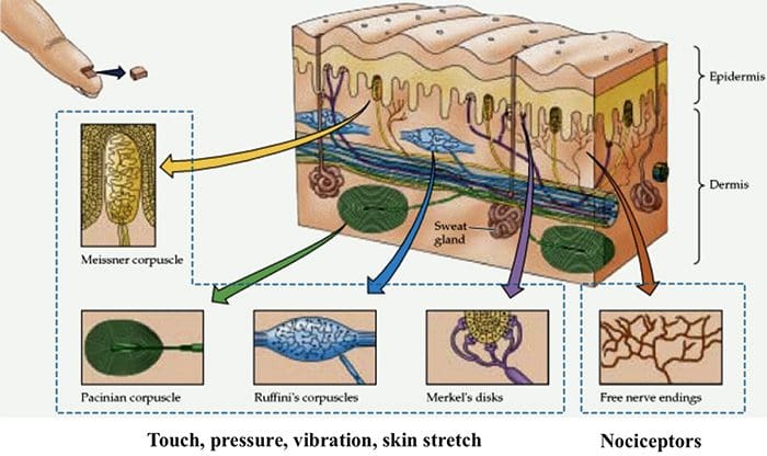
A Sensory System that transmits innocuous stimuli such as touch, pressure, warmth.
A System that transmits stimuli that indicate that tissues have been damaged = nociceptive .
These two systems have different receptors and pathways in the PNS & CNS
Nociceptors are free nerve endings that respond to stimuli that can cause tissue damage or when tissue damage has taken place.
Present in membrane of free nerve endings are receptors (protein molecules) whose activity changes in the presence of painful stimuli.
(Note the use of the same term receptor is used for cell or organs or molecules that involved in transduction of a stimuli.)
Transduction
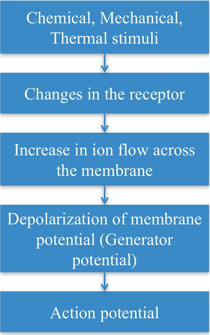
Transduction is the process of converting the stimuli into a nerve impulse.
For this to occur the flow of ions across the nerve membrane has to change to allow entry of either Na+ or Ca2+ ions to cause depolarization of the membrane potential.
This involves a receptor molecule that either directly or indirectly opens the ion channels.
Chemical Agents…
… which can cause the membrane potential at the free nerve ending (nociceptor) to produce an action potential.
Many stimuli � mechanical, chemical and thermal � give rise to painful sensation making transduction a complex process.
Recently receptor molecules have been identified�� Transient Receptor Potential (TRP) channels � that respond to a number of strong stimuli.
TRP receptors are also involved in transmitting the burning sensation of chili pepper.
In time, drugs that act on these receptors will be developed to control pain.
Different TRP Channels
Capsasin, the active ingredient in chili pepper, is used in patches for relief of pain.
Menthol and peppermint gels are used to relieve muscle pain.
Motor Output & Sensory Input To Spinal Cord
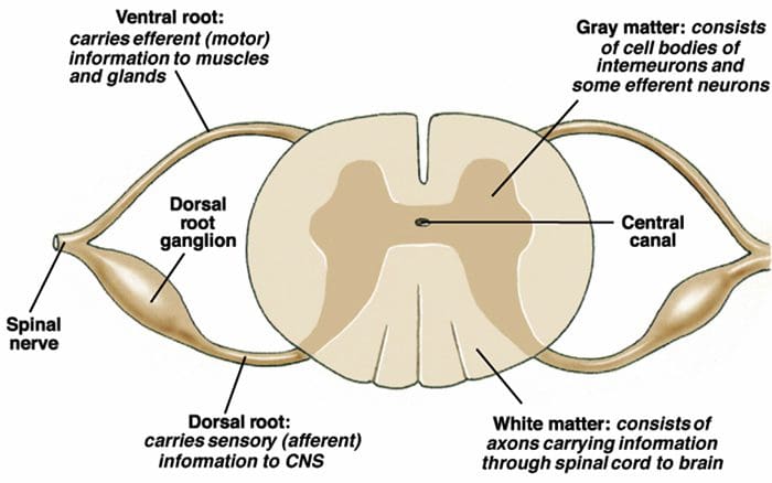
Sensory nerves have their cell body outside the spinal cord in the dorsal root ganglia ( = 1st order neurons).
One process goes to the periphery, the other goes to the spinal cord where it makes synaptic contact with nerve cells in the spinal cord ( = 2nd order neurons).
The 2nd order neuron sends processes to other nerve cells in the spinal cord and to the brain.
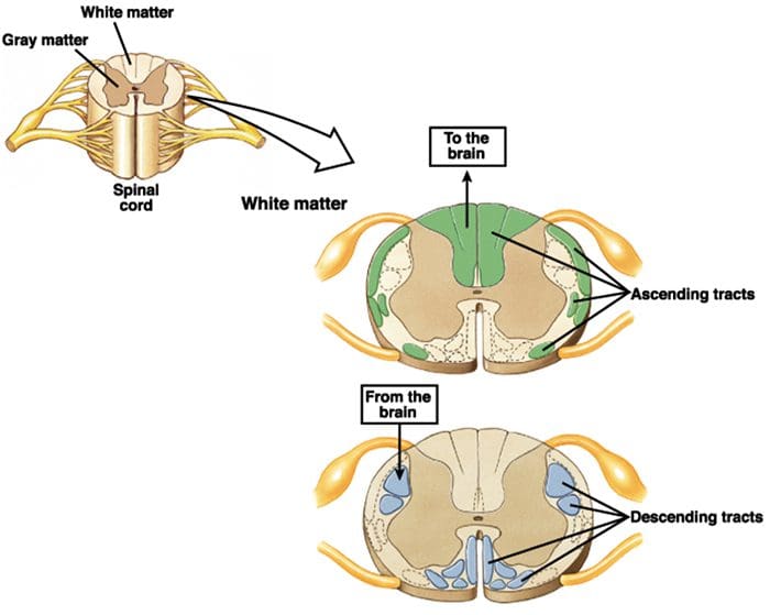
2nd Order Nerve Cells Send Nerve Fibers In The Spinal Cord White Matter
Transmission Of Nociceptive Signals From The Periphery To The Brain
Silverthorn
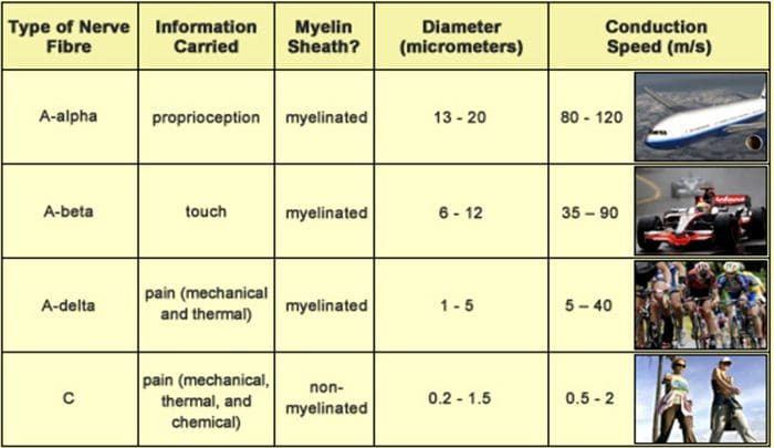
A Delta (?) & C Nerve Fibers
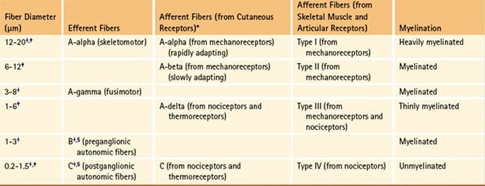
Nerve fibers are classified according to the:
� (1) diameter of the nerve fiber and
� (2) whether myelinated or not.
A? and C nerve fiber endings respond to strong stimuli.
A? are myelinated and C are not.
Action potentials are transmitted 10 times faster in the A?
(20 m/sec) fibers than in C fibers (2 m/sec).
A? & C fibers
A? fibers respond mainly to mechanical and mechno-thermal stimuli.
C fibers are polymodal, i.e. the nerve ending responds to several modalities � thermal, mechanical and chemical
This polymodal ability is due to the presence of different receptor molecules in a single nerve ending.
Fast & Slow Pain
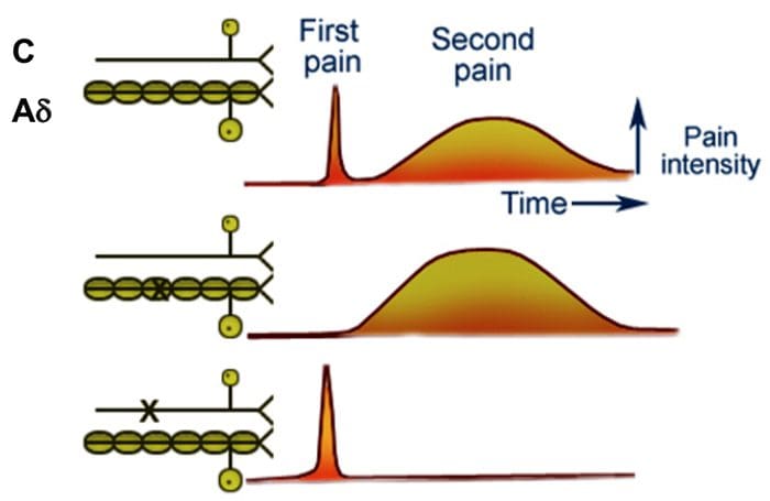
Most people when they are hit by an object or scrape their skin, feel a sharp first pain (epicritic) followed by a second dull, aching, longer lasting pain (protopathic).
The first fast pain is transmitted by the myelinated A? fibers and the second pain by the unmyelinated C fibers.
Central Pain Pathways
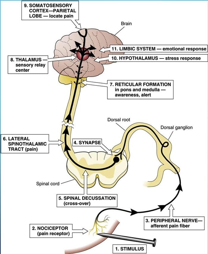
Nociceptive signals are sent to the spinal cord and then to different parts of the brain where sensation of pain is processed.
There are a pathways/regions for assessing the:
Location, intensity, and quality of the noxious stimuli
Unpleasantness and autonomic activation (fight-or-flight response, depression, anxiety).
Dr. Sletten Discussing Central Sensitization Syndrome (CSS)
El Paso, TX. Chiropractor, Dr. Alexander Jimenez continues the discussion on the anatomy of nerve fibers, receptors, spinal tracts and brain pathway/s. As the spinal nerve nears the spinal cord, it splits into the dorsal and ventral roots. The dorsal root only contains the axons of sensory neurons. While the ventral roots contain only the axons of motor neurons. Some of the branches synapse with local neurons in the dorsal root ganglion, posterior (dorsal) horn, and even the anterior (ventral) horn, at the spine where they enter.
Other branches travel short distances up or down the spine to interact with neurons at other levels of the spinal cord. A branch can also turn into the posterior (dorsal) column white matter to connect with the brain. Spinal nerve systems that connect to the brain are contralateral, in that the right side of the body is connected to the left side of the brain and the left side of the body is connected to the right side of the brain.
Cranial nerves convey specific sense information from the head and neck directly to the brain. Whereas spinal information is contralateral, cranial nerve systems are for the most part�ipsilateral, meaning that a cranial nerve on the right side of the head is connected to the right side of the brain. Some cranial nerves contain only sensory axons. Other cranial nerves have both sensory and motor axons, including the trigeminal, facial and glossopharyngeal. General senses of somatosensation for the face travel through the trigeminal system.
PATHWAYS
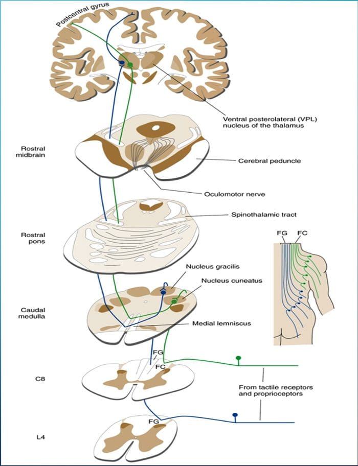
THE POSTERIOR COLUMN� MEDIAL LEMNISCUS SYSTEM CONVEYS INFORMATION ABOUT TOUCH AND LIMB POSITION
POSTERIOR COLUMN MEDIAL LEMNISCAL PATHWAY
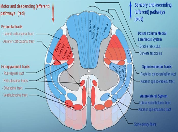
The term posterior column refers to the entire contents of a posterior funiculus, exclusive of its share of the propriospinal tract. The posterior columns consist mainly of ascending collaterals of large myelinated primary afferents carrying impulses from various kinds of mechanoreceptors (although substantial numbers of second-order fibers and unmyelinated fibers are also included). This has traditionally been considered the major pathway by which information from low-threshold cutaneous, joint, and muscle receptors reaches the cerebral cortex.
2-Minute Neuroscience: Touch & The Dorsal Columns-Medial Lemniscus
DAMAGE TO THE POSTERIOR COLUMN�MEDIAL LEMNISCUS SYSTEM CAUSES IMPAIRMENT OF PROPRIOCEPTION AND DISCRIMINATIVE TACTILE FUNCTIONS
�As might be expected from the types of afferents contained in the posterior columns, this pathway carries information important for the conscious appreciation of touch, pressure, and vibration and of joint position and movement. However, because input from cutaneous receptors also reaches the cortex by other routes, damage to the posterior columns causes impairment, but not abolition, of tactile perception. Complex discrimination tasks are more severely affected than is the simple detection of stimuli. Other functions, such as proprioception and kinesthesia, are classically considered to be totally lost after posterior column destruction. The result is a distinctive type of ataxia (incoordination of movement); the brain is unable to direct motor activity properly without sensory feedback about the current position of parts of the body. This ataxia is particularly pronounced when the patient�s eyes are closed, preventing visual compensation.�
Given the role of the posterior column, the patient should be screened for any abnormalities regarding their sense of fine touch, vibration, barognosis, graphesthesia, stereognosis, kinaesthesia, two-point discrimination and conscious proprioception:
A common way of testing for fine touch is to ask the patient to recognize common objects placed within a cloth using their touch.
Vibration sense can be tested using a low pitched C128 tuning fork placed along a bony prominence of the desired corresponding spinal level(s) to be tested.
Barognosis refers to the ability to determine the approximate weight of an object.
Graphesthesia refers to the ability to recognize writing on the skin by touch. The practitioner can draw out a letter on the patients skin as a way of testing.
Kinaesthesia refers to ones own sense of body motion (excluding equilibrium which is controlled in part by the inner ear) and is commonly tested using the subject�s ability to detect an externally imposed passive movement, or the ability to reposition a joint to a predetermined position.
Proprioception is often assessed using the Rombergs test. This examination is based on the notion that a person requires at least two of the three following senses to maintain balance while standing: proprioception; vestibular function and vision. A patient who has a defect within their proprioceptive mechanism can still maintain balance by using vestibular function and vision. In the Romberg test, the patient is stood up and asked to close their eyes. A loss of balance is interpreted as a positive Romberg sign.
THE SPINOTHALAMIC TRACT CONVEYS INFORMATION ABOUT PAIN AND TEMPERATURE
A GOOD BRAIN CAN MODULATE PAIN
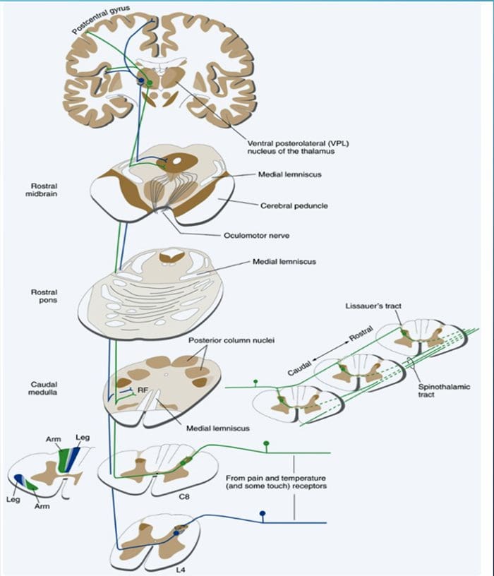
SPINOTHALAMIC TRACT
Pain is a complex sensation, in that a noxious stimulus leads not only to the perception of where it occurred but also to things such as a rapid increase in level of attention, emotional reactions, autonomic responses, and a greater likelihood that the event and its circumstances will be remembered. Corresponding to this complexity, multiple pathways convey nociceptive information rostrally from the spinal cord. One of them (the spinothalamic tract) is analogous to the posterior column�medial lemniscus pathway.
SPINOTHALAMIC TRACTS
Two main parts of the Spinothalamic Tract (STT)
Lateral Spinothalamic Tract
Transmission of pain and temperature
Anterior Spinothalamic Tract
Transmission of crude touch and firm pressure
DAMAGE TO THE ANTEROLATERAL SYSTEM CAUSES DIMINUTION OF PAIN AND TEMPERATURE SENSATIONS
Examination:
Given the role of the spinothalamic tract, the patient should be screened for any abnormalities regarding their sense of touch, pain, temperature, and pressure sensation.
Screening for such abnormalities is commonly done using gentle pin pricks and cotton wool, to contrast between sharp and soft, following cutaneous sensory nerve root distributions. Hot and cold discrimination can be ascertained using the cold metal arm of a tuning fork, and a warm palm or heated object.
2 Minute Neuroscience: Pain & The Anterolateral System
HAUSER ET AL. FIBROMYALGIA, 2015
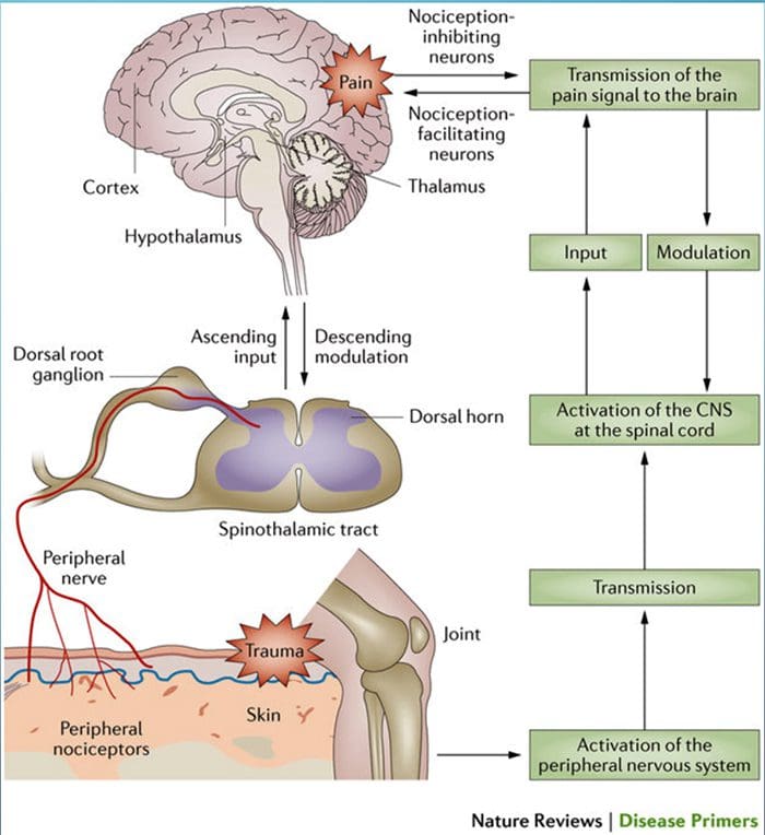
�Pain processing and its modulation: Activation of peripheral pain receptors (also called nociceptors) by noxious stimuli generates signals that travel to the dorsal horn of the spinal cord via the dorsal root ganglion. From the dorsal horn, the signals are carried along the ascending pain pathway or the spinothalamic tract to the thalamus and the cortex. Pain can be controlled by nociception- inhibiting and nociception-facilitating neurons. Descending signals originating in the supraspinal centers can modulate activity in the dorsal horn by controlling spinal pain transmission. CNS, central nervous system.�
SPINAL INFORMATION REACHES THE CEREBELLUM BOTH DIRECTLY AND INDIRECTLY
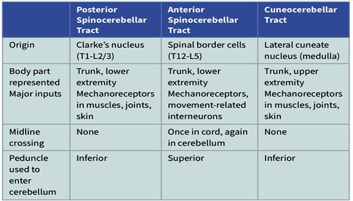
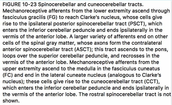
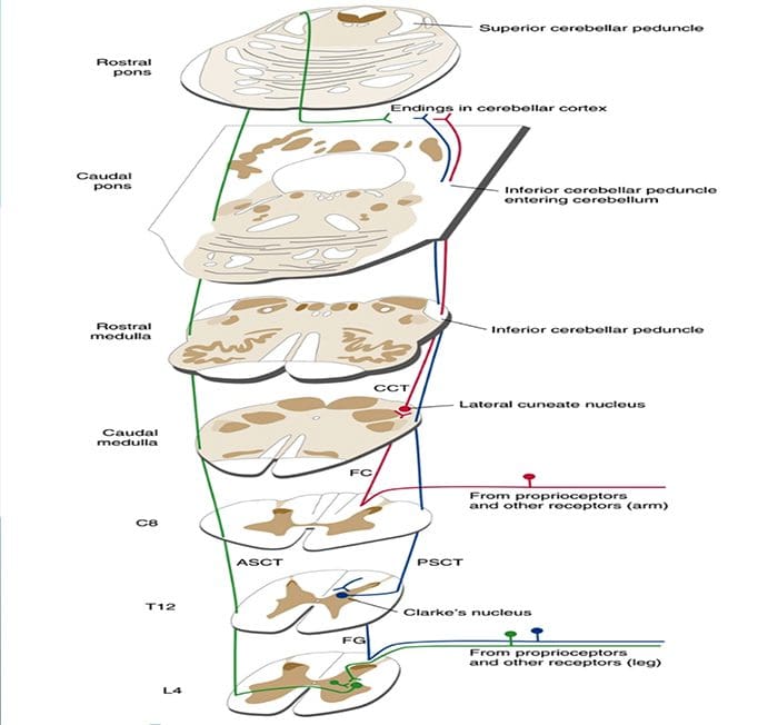
The spinal cord is an important source of information used by the cerebellum in the coordination of movement. This information reaches the cerebellar cortex and nuclei both directly, by way of spinocerebellar tracts, and indirectly, by way of relays in brainstem nuclei. A number of spinocerebellar tracts have been described, some representing the upper extremity and others the lower extremity. Only three have been well characterized.
Ascending Tracts | Spinocerebellar Tract
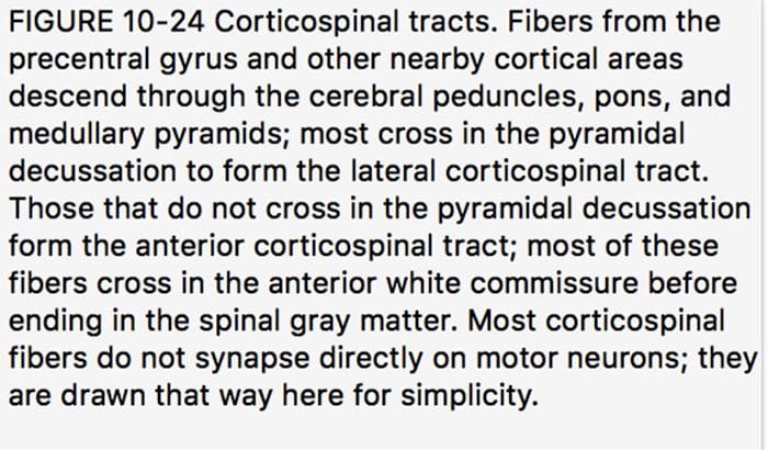
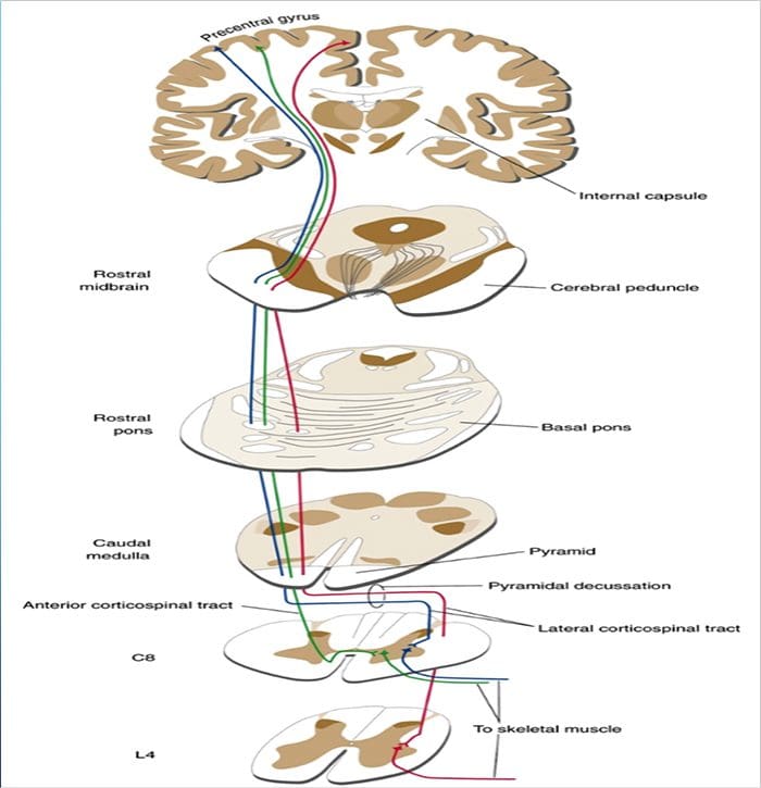
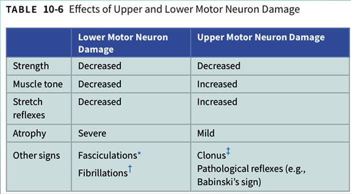
DESCENDING PATHWAYS INFLUENCE THE ACTIVITY OF LOWER MOTOR NEURONS
El Paso, TX. Chiropractor, Dr. Alexander Jimenez discusses the anatomy of nerve fibers, receptors, spinal tracts and brain pathways. Regions of the Central Nervous System (CNS) coordinate various somatic processes using sensory inputs and motor outputs of peripheral nerves. Important areas of the CNS that play a role in somatic processes are separated in the spinal cord brain stem. Sensory pathways that carry peripheral sensations to the brain are referred to an ascending pathway, or tract. Various sensory modalities follow specific pathways through the CNS. Somatosensory stimuli activate receptors in the skin, muscles, tendons, and joints throughout the entire body. The somatosensory pathways are divided into two separate systems based on the location of the receptor neurons. Somatosensory stimuli from below the neck run along the sensory pathways of the spinal cord, and the somatosensory stimuli from the head and neck travel through cranial nerves.
ANATOMY OF RECEPTORS, NERVE FIBERS, SPINAL CORD TRACTS AND BRAINSTEM PATHWAYS
RECEPTORS AND RECEPTOR BASED THERAPY
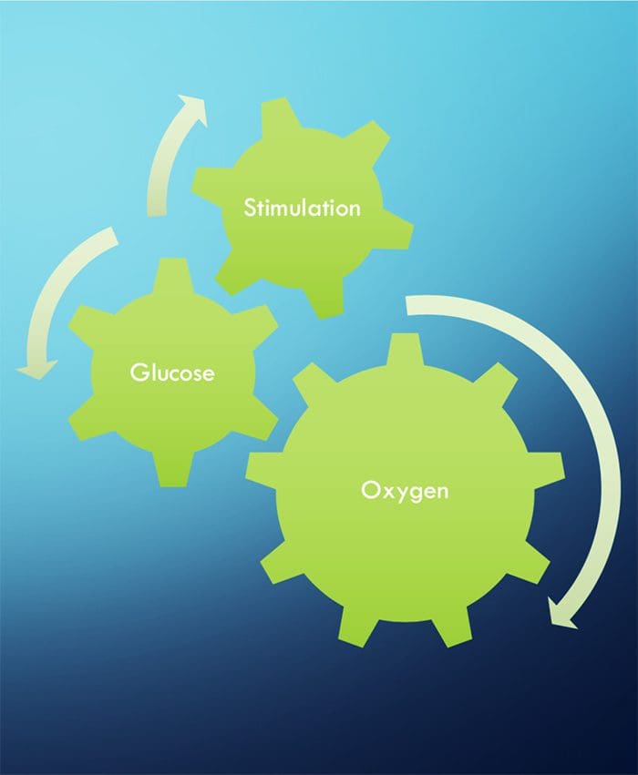
NEURONS NEED THREE THINGS TO SURVIVE!
FUNCTIONAL NEUROLOGY KEY CONCEPTS
The cell needs three things to survive.
Oxygen, glucose and stimulation.
Stimulation = Chiropractic, exercise, etc.
Stimulation leads to neuronal growth
Neuronal growth leads to plasticity
Subluxations alter the frequency of firing of neurons
Activation of one side will stimulate ipsilateral cerebellum and contralateral cortex (usually)
Proper stimulation CAN reduce pain.
CHIROPRACTIC IS RECEPTOR-BASED THERAPY
INTRODUCTION
The ongoing activity and output of the CNS are greatly influenced, and sometimes more or less determined, by incoming sensory information.
The basis of this incoming sensory information is an array of sensory receptors, cells that detect various stimuli and produce receptor potentials in response, often with astonishing effectiveness.
The health of the neuron, however, plays a huge role in how neurons can produce receptor potentials, the endurance of the neuron and the ability to create plasticity.
�Neurons that fire together, wire together.� Hebbian Theory
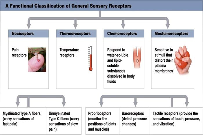
TYPES OF RECEPTORS
Chemoreceptors
Smell, taste, interoceptors
Thermoreceptors
Temperature
Mechanoreceptors
Cutaneous receptors for touch, auditory, vestibular, proprioceptors
Nociceptors
Pain
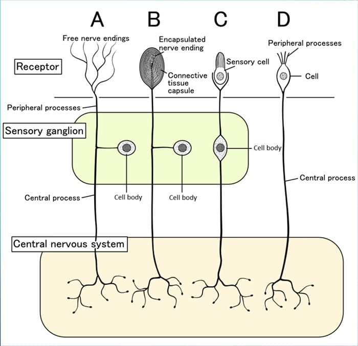
PARTS OF RECEPTORS
Although their morphologies vary widely, all receptors have three general parts:
1. Receptive Area 2. Area Rich In Mitochondria
Health of the neurons within the receptors will determine its response to stimulation
3. Synaptic Area To Pass Messages To The CNS
RECEPTIVE FIELDS
These are particular areas in the periphery where application of an adequate stimulus causes the receptors to respond.
Neurons in successive levels of sensory pathways (second- order neurons, thalamic and cortical neurons-also have receptive fields, although they may be considerably more elaborate than those of the receptors.
TRANSDUCTION
Sensory receptors use ionotropic and metabotropic mechanisms to produce receptor potentials
Sensory receptors transduce some physical stimulus into an electrical signal � a receptor potential � that the nervous system can understand.
Sensory receptors are similar to postsynaptic membranes as their adequate stimuli are analogous to neurotransmitters.
THE DIAMETER OF A NERVE FIBER IS CORRELATED WITH ITS FUNCTION
BIGGER = FASTER
Larger fibers conduct action potentials faster than do smaller fibers.
A? fibers are the largest and most rapidly conducting myelinated fibers.
The slowest conducting fibers of the body are the C fibers
RECEPTORS IN MUSCLES AND JOINTS DETECT MUSCLE STATUS AND LIMB POSITION
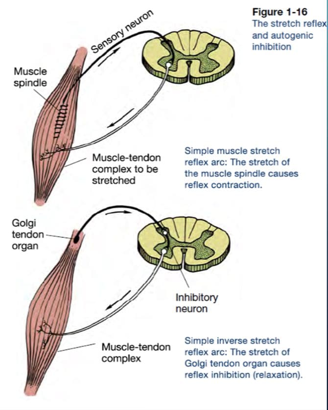
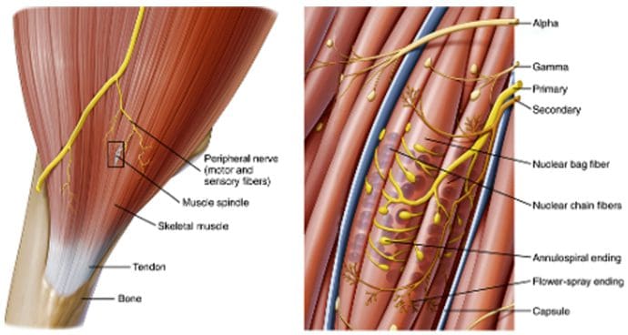
MUSCLE SPINDLES
Muscle spindles (Fig. 9-14) are long, thin stretch receptors scattered throughout virtually every striated muscle in the body.
These muscle spindles sense muscle length and proprioception (�one�s own� perception).
They are quite simple in principle, consisting of a few small muscle fibers with a capsule surrounding the middle third of the fibers.
These fibers are called intrafusal muscle fibers (fusus is Latin for �spindle,� so intrafusal means �inside the spindle�), incontrast to the ordinary extrafusal muscle fibers (�outside the spindle�).
The ends of the intrafusal fibers are attached to extrafusal fibers, so whenever the muscle is stretched, the intrafusal fibers are also stretched.
The central region of each intrafusal fiber has few myofilaments and is noncontractile, but it does have one or more sensory endings applied to it.
When the muscle is stretched, the central part of the intrafusal fiber is stretched, mechanically sensitive channels are distorted, the resulting receptor potential spreads to a nearby trigger zone, and a train of impulses ensues at each sensory ending.
GOLGI TENDON ORGANS
Golgi tendon organs are spindle-shaped receptors found at the�junctions between muscles and tendons. They are similar to Ruffini endings in their basic organization, consisting of interwoven collagen bundles surrounded by a thin capsule (Fig. 9-16).
Large sensory fibers enter the capsule and branch into fine processes that are inserted among the collagen bundles. Tension on the capsule along its long axis squeezes these fine processes, and the resulting distortion stimulates them.
If tension is generated in a tendon by making its attached muscle contract, tendon organs are found to be much more�sensitive and can actually respond to the contraction of just a few muscle fibers.
Thus Golgi tendon organs very specifically monitor the tension generated by muscle contraction and come into play whe
n fine adjustments in muscle tension need to be made (e.g., when handling a raw egg).
�
Thus the mode of action of Golgi tendon organs is quite different from that of muscle spindles (Fig. 9-17). If a muscle�contracts isometrically, tension is generated across its tendons, and the tendon organs signal this; however, the muscle spindles signal nothing because muscle length has not changed (assuming that the activity of the gamma motor neurons remains unchanged).
In contrast, a relaxed muscle can be stretched easily, and the muscle spindles fire; the tendon organs, however, experience little tension and remain silent. A muscle, by virtue of these two types of receptors, can have its length and tension monitored simultaneously.
IFM's Find A Practitioner tool is the largest referral network in Functional Medicine, created to help patients locate Functional Medicine practitioners anywhere in the world. IFM Certified Practitioners are listed first in the search results, given their extensive education in Functional Medicine



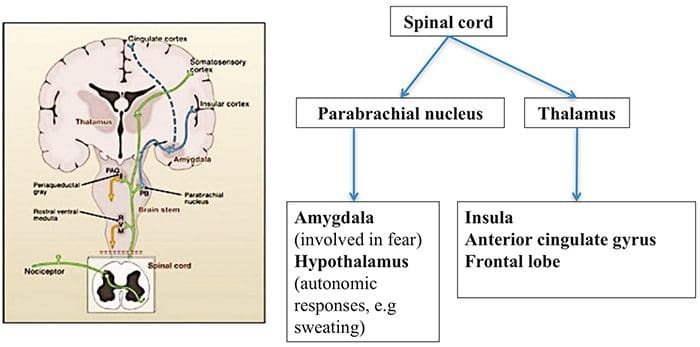 Brain Areas Involved In Processing Of Nociceptive Signals
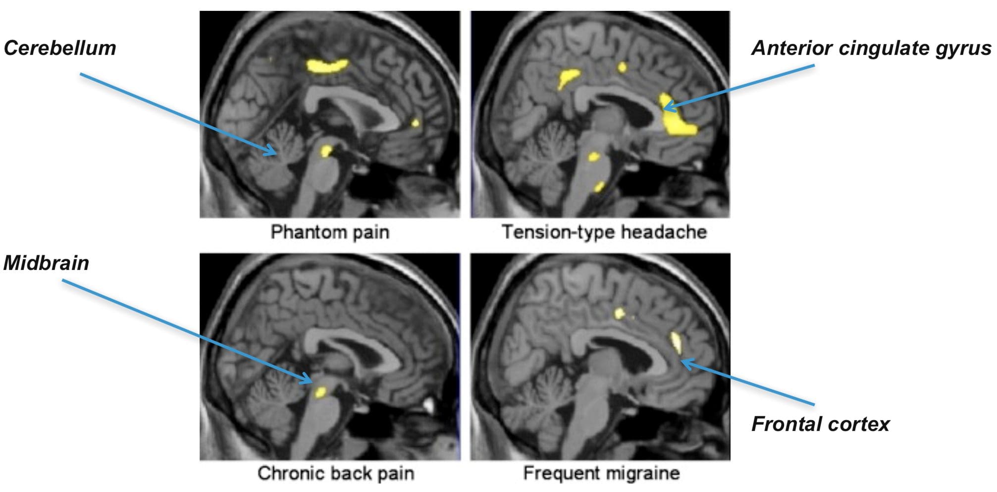 The Anterior Cingulate & Insula Cortex Are Activated In Human Subjects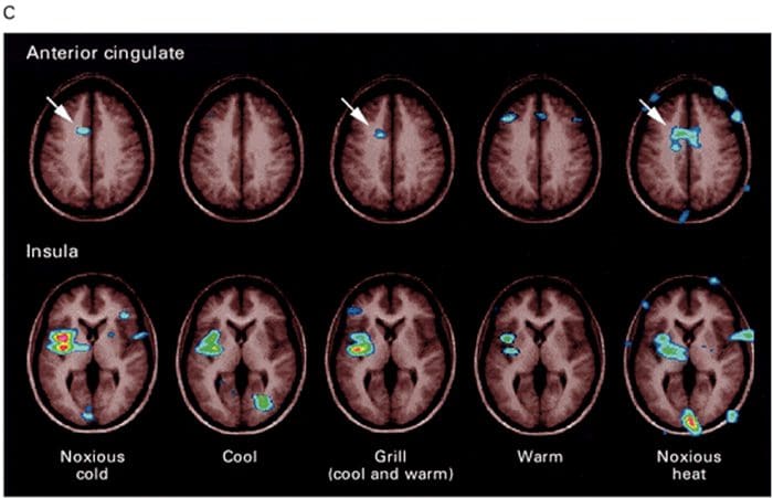
 Inflammatory Soup – Hyperalgesia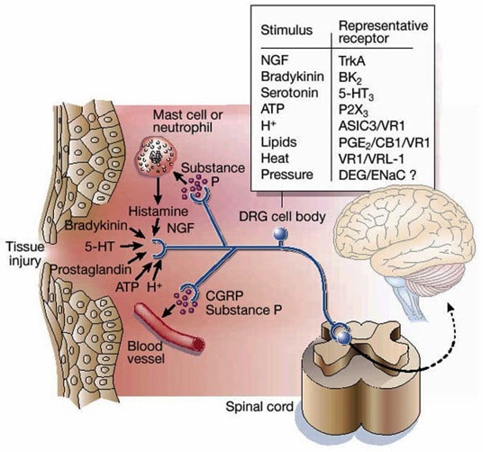
 Gate Control Theory of Pain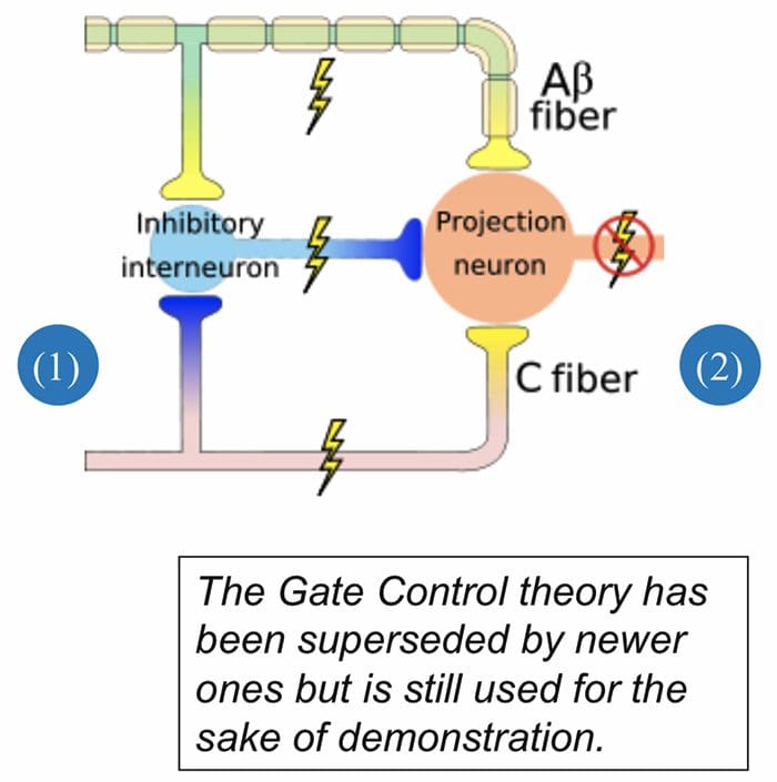
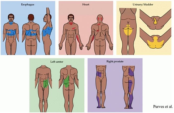
 Abnormalities Of Pain System
 Peripheral Sensitization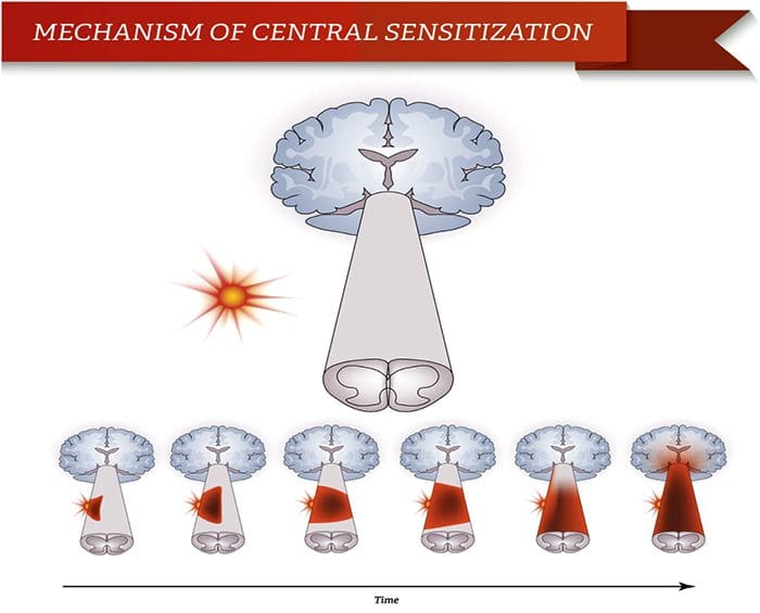
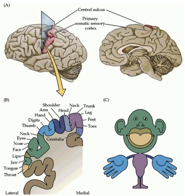
 Somatosensory Cortex Organization
 Cortical Reorganization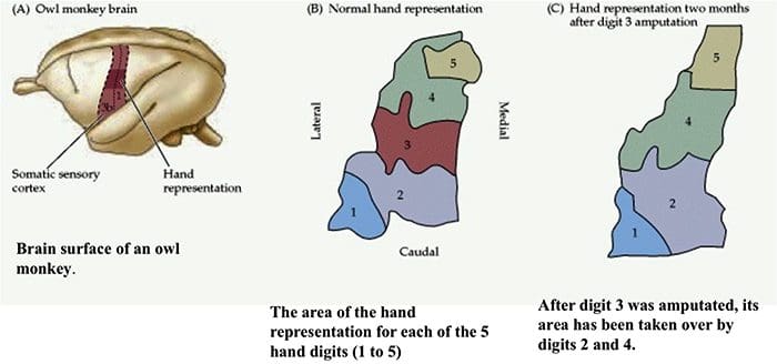
 Nerve Cell-To-Nerve Cell Communication
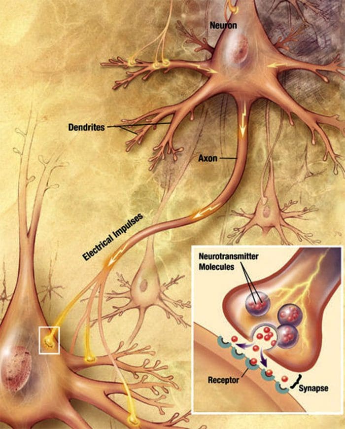 Nerve cells communicate with other cells by releasing a chemical from the nerve endings � Neurotransmitters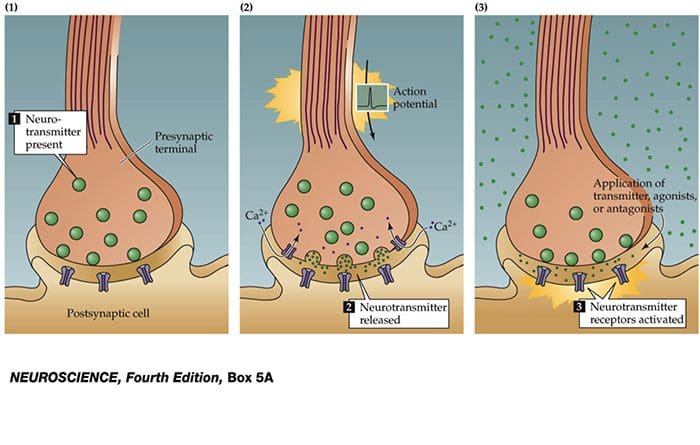
 TRP Channels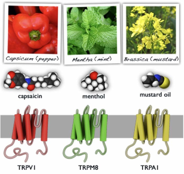
 The term posterior column refers to the entire contents of a posterior funiculus, exclusive of its share of the propriospinal tract. The posterior columns consist mainly of ascending collaterals of large myelinated primary afferents carrying impulses from various kinds of mechanoreceptors (although substantial numbers of second-order fibers and unmyelinated fibers are also included). This has traditionally been considered the major pathway by which information from low-threshold cutaneous, joint, and muscle receptors reaches the cerebral cortex.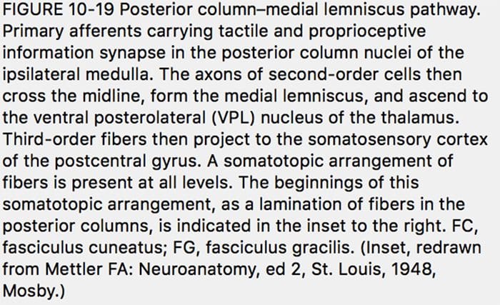
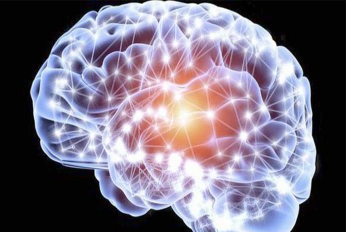 The cell needs three things to survive.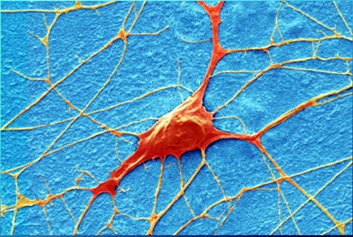
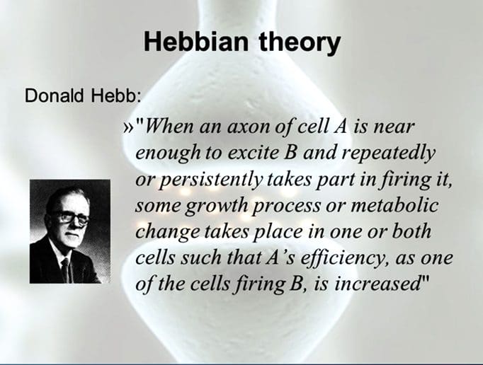 The health of the neuron, however, plays a huge role in how neurons can produce receptor potentials, the endurance of the neuron and the ability to create plasticity.
 Chemoreceptors
 Although their morphologies vary widely, all receptors have three general parts: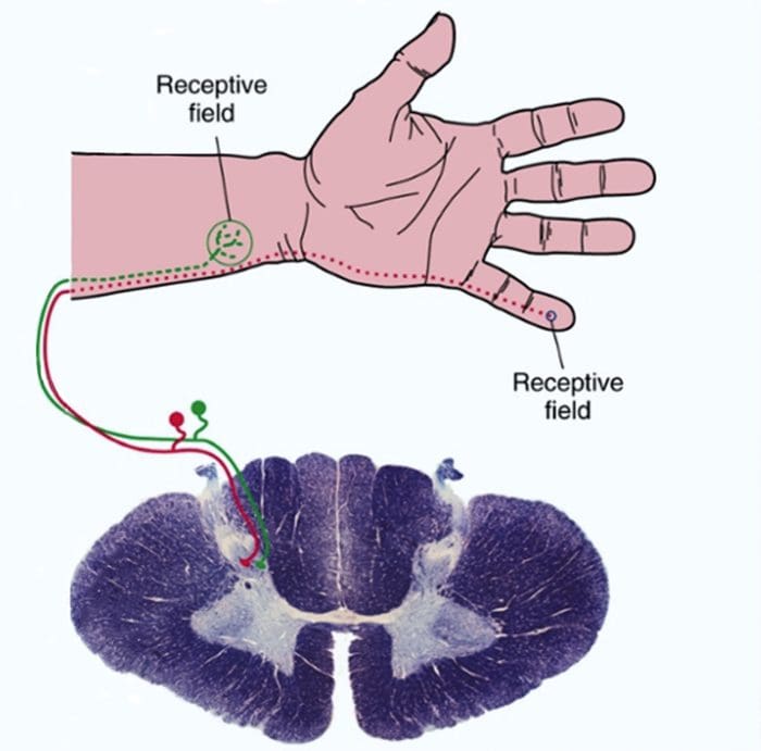
 Sensory receptors use ionotropic and metabotropic mechanisms to produce receptor potentials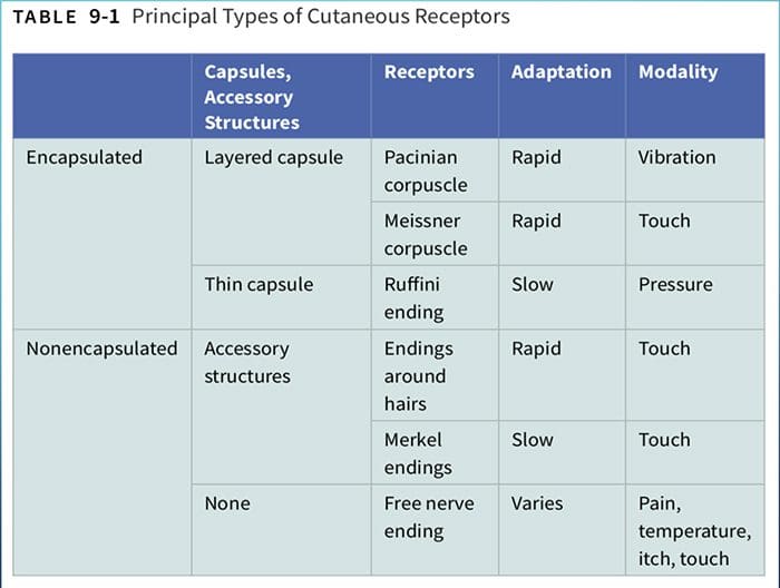
 Larger fibers conduct action potentials faster than do smaller fibers.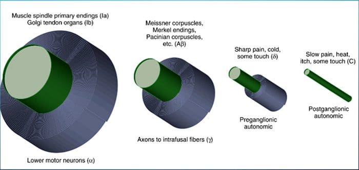

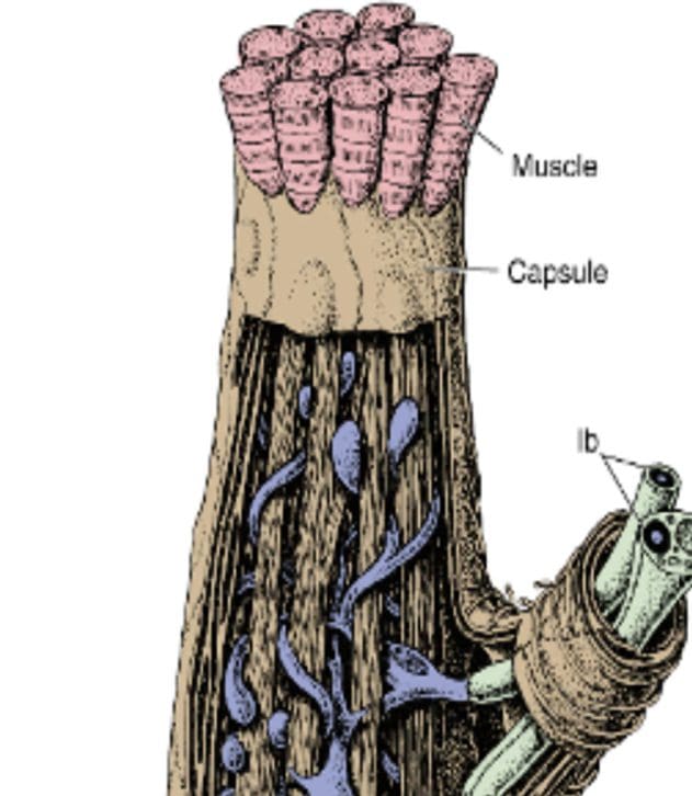 junctions between muscles and tendons. They are similar to Ruffini endings in their basic organization, consisting of interwoven collagen bundles surrounded by a thin capsule (Fig. 9-16).
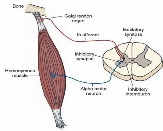 sensitive and can actually respond to the contraction of just a few muscle fibers.