Neurophysiology: There are two ways that nociceptive information reaches the central nervous system. One is the neospinothalamic tract for quick pain and two is the paleospinothalamic tract for slow pain that increases.
Neurophysiology Of Pain Part II
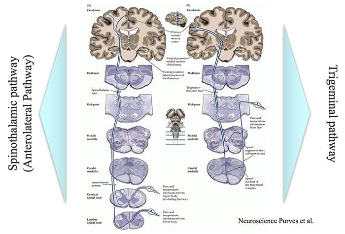
Intensity, Location & Quality of Pain…
… involve Spinothalamic and Trigeminal Pathways
The trigeminal pathway brings information from the face area.
The spinothalamic pathway brings information from the rest of the body.
Both these pathways project to the sensory cortex, which also receives information on innocuous stimuli such as touch, pressure and warmth via a separate pathway.
2 Pain Transmission Pathways For Location Intensity Quality
There is difference between the objective and subjective aspects of injury and pain.
Despite similar injury, people can differ in how much pain they feel.
Depending on the context, pain may not be felt despite injury, e.g. battlefield injury, during intense sports.
This suggests that there is a physiological mechanism that controls the transmission of nociceptive signals to the brain or modifies the interpretation of pain.
The pain control system can also explain the placebo effect.
Pain Modulation Pathway
Nerve signals are sent form the somatic sensory cortex and hypothalamus to the periaqueductal gray matter (PAG).
PAG sends signals to the parabrachial nucleus, medullary reticular formation, locus coeruleus, and Raphe neulei.
These in turn can control the in the transmission of nociceptive signals from the spinal cord to the brain.
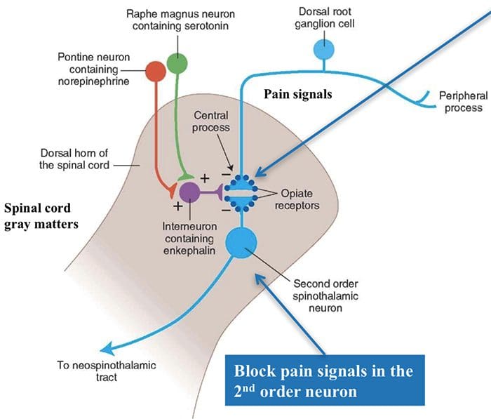
This involves different involves different neurotransmitters.
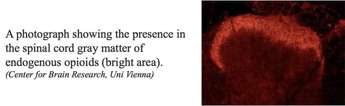
Endogenous Opioids
Internally produced molecules with opioid-like action which regulate transmission of nociceptive signals.
Three classes of these molecules have been identified. All are peptide molecules
Enkephalins
Endorphins
Dynorphins
Despite these being powerful, endogenous modifiers of nociceptive signals, it has been difficult to produce and administer them in a way than can used in clinical practice.
Location Of Nerve Cells With Endogenous Opioid Receptors
In the spinal cord, endogenous opioids can prevent transmission between 1st order nerve cells (bringing signals from the periphery) and 2nd order spinal nerve cells that transmit the signals to the brain.
Also can prevent the increased synaptic efficiency, which plays a role in hyperalgesia.
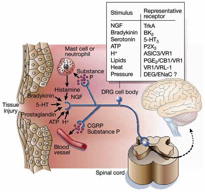
Knowing the molecules involved in the �inflammatory soup� and how they are synthesized provides possible targets for pain reduction.
e.g. prostaglandins are produced by the COX enzyme. The activity of this enzyme is blocked by non-steroidal anti- inflammatory drugs (NSAIDs) such as ibuprofen, diclofenac.
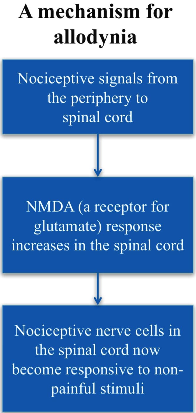
Allodynia
A condition when normally non- painful stimuli cause pain, e.g., touch, light pressure, cold.
Involves changes in the synaptic sensitivity of the nociceptive neurons in the spinal cord (central sensitization).
Drugs such as ketamine, block NMDA receptors and so reduce transmisison of the nociceptive stimuli.
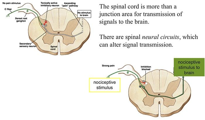
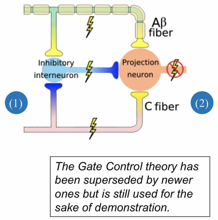
Gate Control Theory of Pain
Mother says to child, �Come I will rub the area which is painful and this will make it feel better.�
After stubbing a toe, we instinctively rub the area; this reduces the sensation of pain.
Ronald Melzack and Patrick Wall in 1962 provided an possible explanation for this effect.
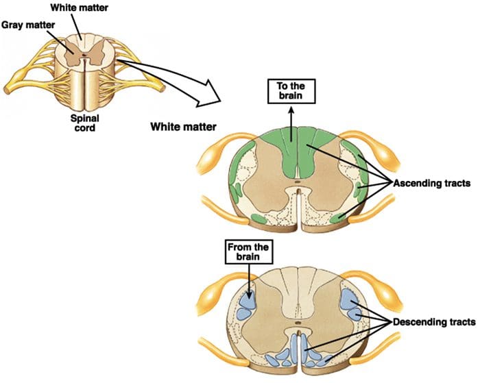
Ascending Tracts | Pain Modulation: Gate Control Theory
Gate Theory
Rubbing the area that hurts stimulates receptors of innocuous stimuli like touch, pressure and vibration.
These mechano-receptors send signals along the A? nerve fibers that:
(1) stimulate spinal nerves (inhibitory inter-neurons) that in turn inhibit signaling in the 2nd order neurons (projection neuron) and (2) directly inhibit the 2nd order neuron to reduce or stop pain signal from being sent to the brain
Transcutaneous Nerve Stimulation (TENS) is based on the Gate Control Theory. Nerves of the innocuous sensory system are stimulated and they in turn, inhibit transmission of nociceptive stimuli in the spinal cord.
Abnormalities Of Pain System
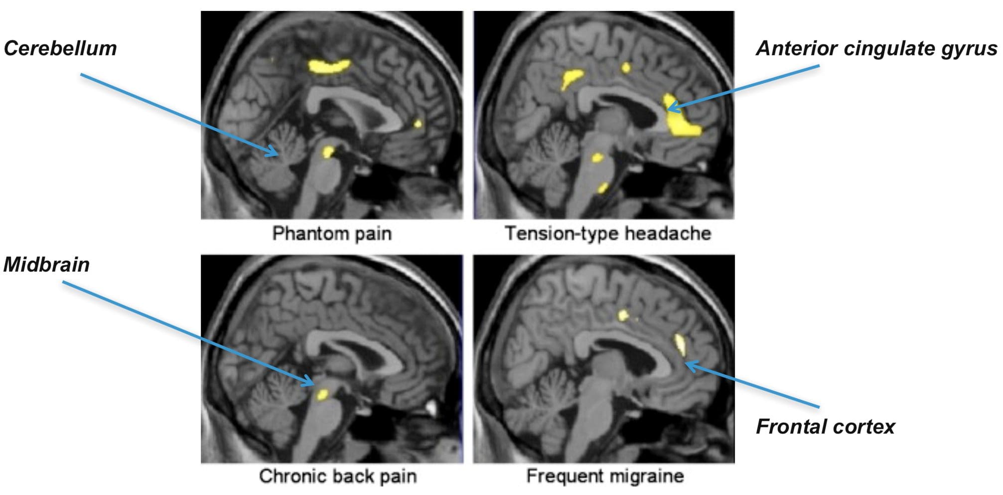
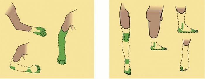
Phantom Pain
Patients with amputation often have burning or tingling pain in the body part removed.
One possible cause is that nerve fibers at the stump are stimulated and the brain interprets the signals as originating in the amputated portion.
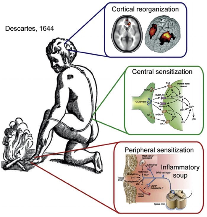
The other is the rearrangement within the cortical areas so that area say for the hand now responds to signals from other parts of the body but still interprets them as coming for the amputated hand.
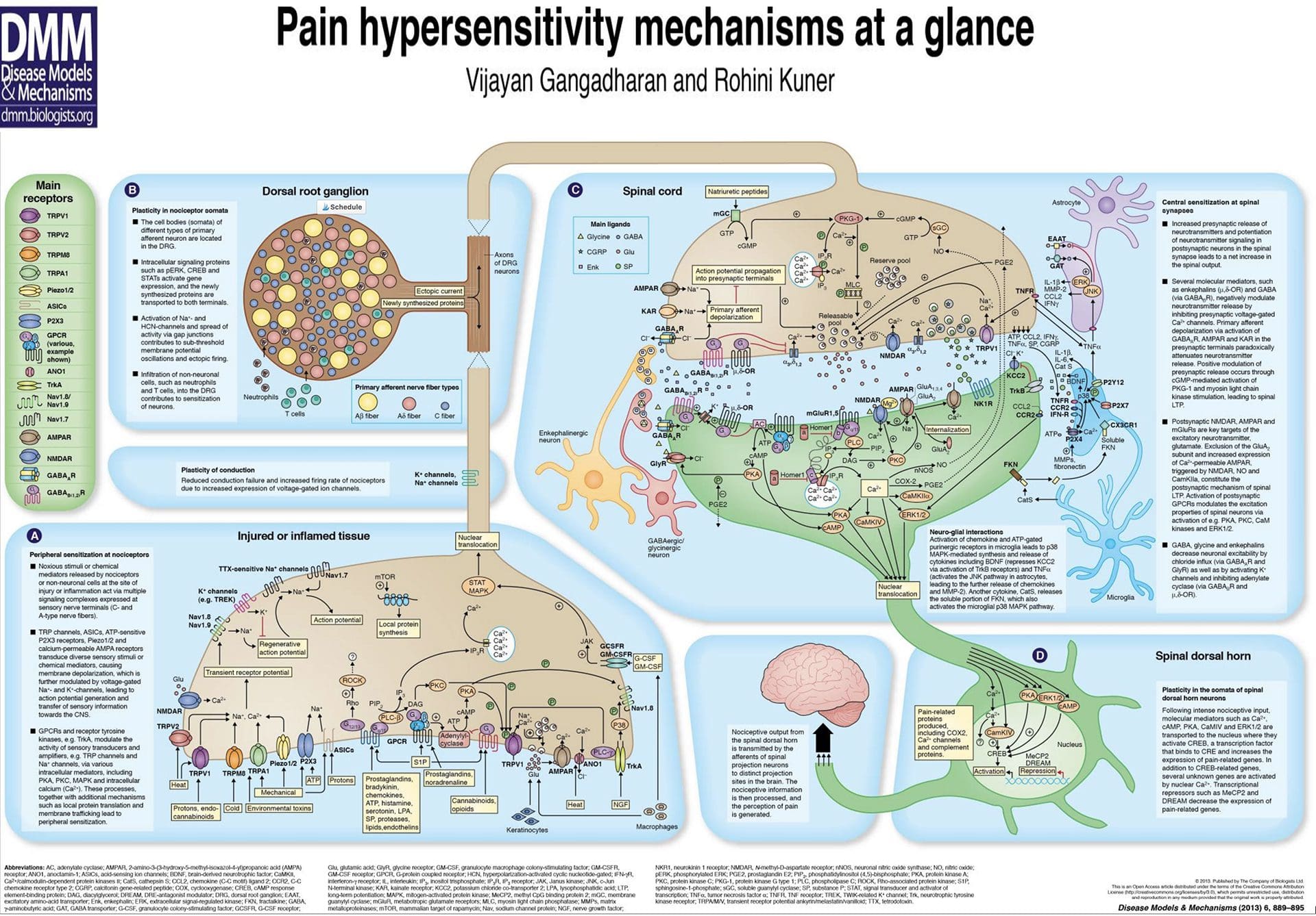
Peripheral Sensitization
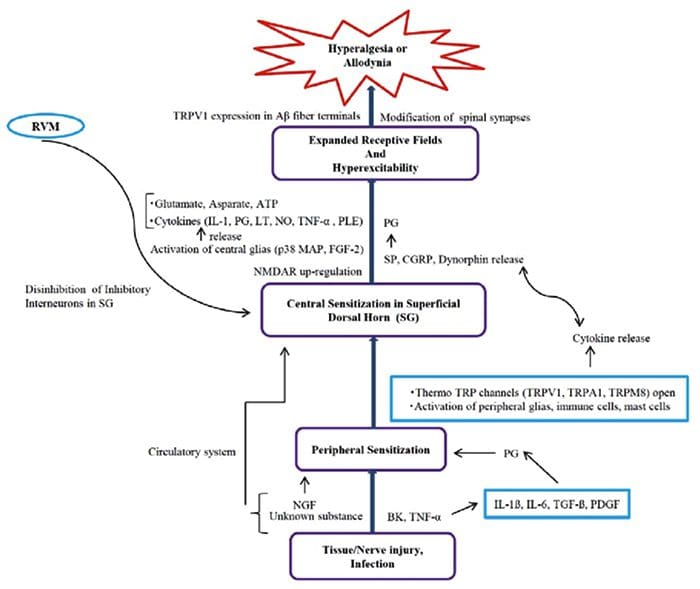
Peripheral sensitization represents a reduction in the threshold and/or an increase in magnitude of responsiveness at the peripheral ends of sensory nerve fibers.
This occurs in response to chemical mediators released by nociceptors and non-neuronal cells (e.g. mast cells, basophils, platelets, macrophages, neutrophils, endothelial cells, keratinocytes and fibroblasts) at the site of tissue injury or inflammation.
Basically, it is an increased sensitivity to an afferent nerve stimuli.
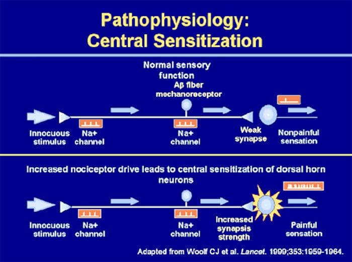
Central Sensitization
Peripheral & Central Sensitisation
A condition of the nervous sytem that is associated with the development and maintenance of chronic pain.
Known as �wind-up� or persistent high reactivity.
�Plastiticity in pain pathways� or the persistence of pain even after an injury has healed.
Is this negative or positive plasticity?
Central Sensitization & C Fibers
Two Main Characteristics Of Central Sensitization:
� Allodynia � occurs when a person experiences pain with things that are normally not painful, ie, soft touch causes pain.
� Hyperalgesia � occurs when a stimulus that is typically painful is perceived as more painful that it should be, ie, a simple bump.
Both are due to hyperreactivity of the nervous system.
Neurophysiology of pain: Pain�defined is the unpleasant sensation that accompanies injury or near injury to tissues, though it can also occur in the absence of such damage if the nociception system is not functioning. Nociception means the system that carries pain signals of injury from the tissues. This is the physiological incident that comes with pain.
Neurophysiology Of Pain
Objectives
Basics of the nervous system
Synaptic function
Nerve impulses
Transduction of peripheral painful stimuli
Central pathways
Central Sensitization
PeripheralSensitization
Control or modulation of pain signals
Pathophysiology of pain signaling pathway
Definition Of Pain
“Pain is an unpleasant sensory and emotional experience associated with actual or potential tissue damage, or described in terms of such damage”.
It is important to know the basic structure of the nervous system.
This will help in:
� Understanding the mechanism by which nociceptive signals are produced.
� Know the different regions of the nervous system involved in processing these signals.
� Learn how the different medications and treatment for pain management work.
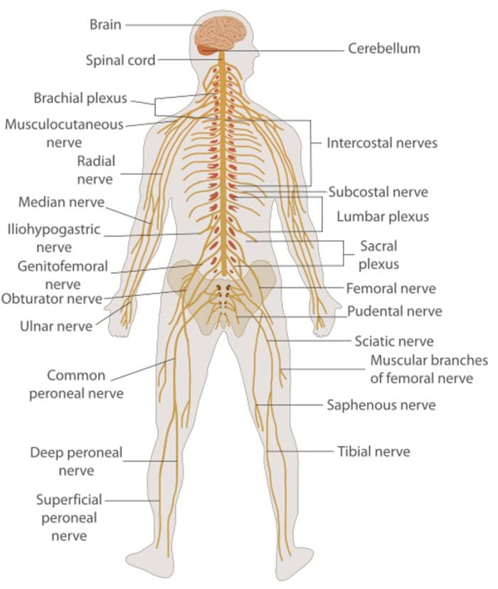
Nervous System
Central nervous system (CNS)
Brain and Spinal Cord
Peripheral Nervous System (PNS)
Nerve fibers go to all parts of the body.
Send signals to the different tissues and send signals back to the CNS.
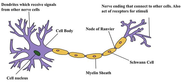
Nerve Cells
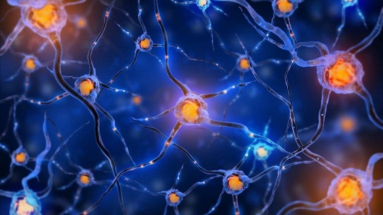
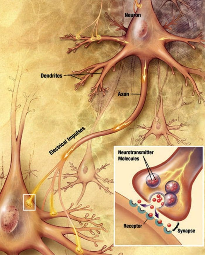
The nervous system is made up of nerve cells which send long processes (axons) to make contact with other cells.
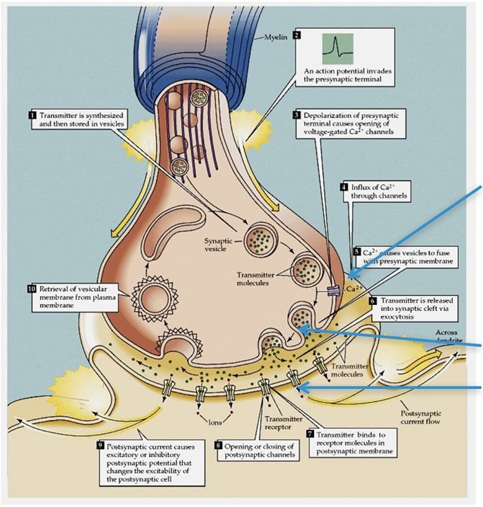
Nerve Cell-To-Nerve Cell Communication
Nerve cells communicate with other cells by releasing a chemical from the nerve endings � Neurotransmitters
Basic Steps In Synaptic Transmission
Synaptic Transmission
Steps in the passage of signal from one nerve cell to other.
Drugs are used to block the transmission of signals from one nerve cell to other.
These drugs can effect:
Ca2+ ion channel to prevent Ca2+ inflow which is essential for neurotransmitter (NT) release, e.g., the action of gabapentin.
Release of NT.
Prevent NT from binding to its receptor so stop further transmission of the signal.
Electrical Impulse
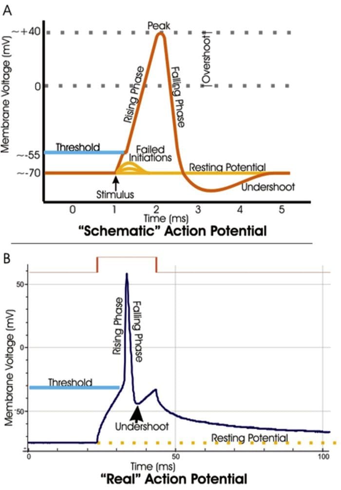
Signals move along a nerve process (axon) as a wave of membrane depolarization called the Action Potential.
The inside of all nerve cells has a negative electrical potential of around � 60 mV.
When stimulated this negative electrical potential becomes positive and then negative again in milliseconds.
The action potential moves along the nerve process (axon) to the nerve ending where it cause release of NT.
Action Potential
When there is no stimulation the membrane potential is at its Resting Potential.
When stimulated, channels in the nerve membrane open allowing the flow of sodium ions (Na+) or calcium ions (Ca2+) into the nerve or cell. This makes the inside less negative and in fact positive -the peak of the action potential (+40 mV).
These channels than close and by the opening of K+ channels the membrane potential returns to its resting level.
Stopping Action Potentials To Stop Nociceptive Stimuli
Nociceptive stimuli are those that will create a sensation of pain after they are processed in the CNS.
Nociceptive signals can be prevented from reaching the CNS by blocking the action of the channels that control the movement of ions across the nerve membrane.
A number of anesthetic agents stop Na+ channel from working and hence stop the generation of actions potentials and transmission of signals to the CNS.
Sensory Systems
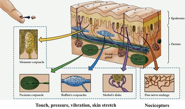
The sensory system that can be divided into two divisions:
A Sensory System that transmits innocuous stimuli such as touch, pressure, warmth.
A System that transmits stimuli that indicate that tissues have been damaged = nociceptive .
These two systems have different receptors and pathways in the PNS & CNS
Nociceptors are free nerve endings that respond to stimuli that can cause tissue damage or when tissue damage has taken place.
Present in membrane of free nerve endings are receptors (protein molecules) whose activity changes in the presence of painful stimuli.
(Note the use of the same term receptor is used for cell or organs or molecules that involved in transduction of a stimuli.)
Transduction
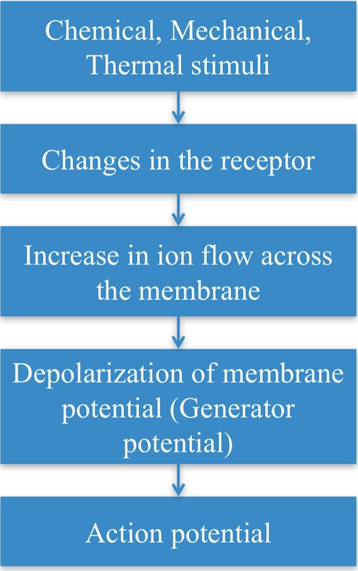
Transduction is the process of converting the stimuli into a nerve impulse.
For this to occur the flow of ions across the nerve membrane has to change to allow entry of either Na+ or Ca2+ ions to cause depolarization of the membrane potential.
This involves a receptor molecule that either directly or indirectly opens the ion channels.
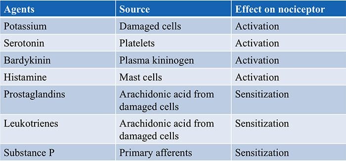
Chemical Agents…
… which can cause the membrane potential at the free nerve ending (nociceptor) to produce an action potential.
Many stimuli � mechanical, chemical and thermal � give rise to painful sensation making transduction a complex process.
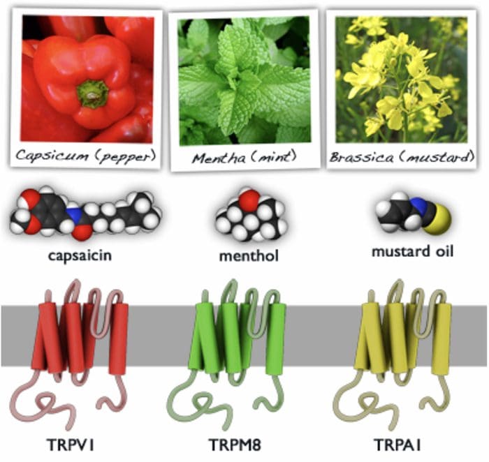
Recently receptor molecules have been identified�� Transient Receptor Potential (TRP) channels � that respond to a number of strong stimuli.
TRP receptors are also involved in transmitting the burning sensation of chili pepper.
In time, drugs that act on these receptors will be developed to control pain.
Different TRP Channels
Capsasin, the active ingredient in chili pepper, is used in patches for relief of pain.
Menthol and peppermint gels are used to relieve muscle pain.
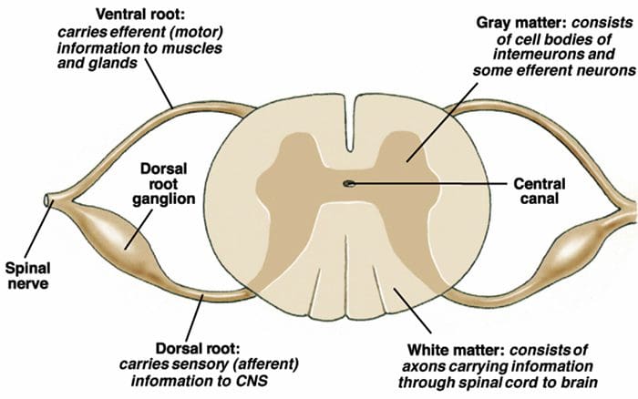
Motor Output & Sensory Input To Spinal Cord
Sensory nerves have their cell body outside the spinal cord in the dorsal root ganglia ( = 1st order neurons).
One process goes to the periphery, the other goes to the spinal cord where it makes synaptic contact with nerve cells in the spinal cord ( = 2nd order neurons).
The 2nd order neuron sends processes to other nerve cells in the spinal cord and to the brain.
2nd Order Nerve Cells Send Nerve Fibers In The Spinal Cord White Matter
Transmission Of Nociceptive Signals From The Periphery To The Brain
Silverthorn
A Delta (?) & C Nerve Fibers
Nerve fibers are classified according to the:
� (1) diameter of the nerve fiber and
� (2) whether myelinated or not.
A? and C nerve fiber endings respond to strong stimuli.
A? are myelinated and C are not.
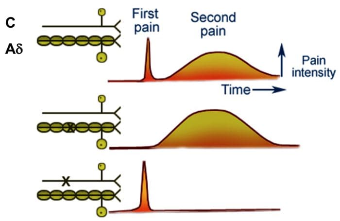
Action potentials are transmitted 10 times faster in the A?
(20 m/sec) fibers than in C fibers (2 m/sec).
A? & C fibers
A? fibers respond mainly to mechanical and mechno-thermal stimuli.
C fibers are polymodal, i.e. the nerve ending responds to several modalities � thermal, mechanical and chemical
This polymodal ability is due to the presence of different receptor molecules in a single nerve ending.
Fast & Slow Pain
Most people when they are hit by an object or scrape their skin, feel a sharp first pain (epicritic) followed by a second dull, aching, longer lasting pain (protopathic).
The first fast pain is transmitted by the myelinated A? fibers and the second pain by the unmyelinated C fibers.
Central Pain Pathways
Nociceptive signals are sent to the spinal cord and then to different parts of the brain where sensation of pain is processed.
There are a pathways/regions for assessing the:
Location, intensity, and quality of the noxious stimuli
Unpleasantness and autonomic activation (fight-or-flight response, depression, anxiety).
Dr. Sletten Discussing Central Sensitization Syndrome (CSS)
Neurons are believed to establish neuronal connections through innate predetermined programs during the developmental process of the brain. It’s also believed that neurons gravitate to areas of attraction and move away from areas of repulsion in a theory known as the chemoaffinity hypothesis. The Chemoaffinity hypothesis claims that neurons first make connections with their targets based on interactions with specific molecular markers and, therefore, that the first wiring diagram of an organism is indirectly determined by its genotype.
These markers are created during cellular differentiation and aid not just with synaptogenesis, but also act as guidance cues for their individual axon. The development of the mature nervous system formations demands axons to navigate to their correct targets in order to establish neuronal connections or synaptic connections. Growing axons create highly motile structures, known as growth cones, which direct the axon to its goal. They do it by responding to specific guidance molecules that either attract or repel the growth cone.
The Theory of Neuronal Connections
The concept that axons are directed principally by molecular determinants, rather than mechanical determinants, such as cells, extracellular material and other neurons, was established by Roger Wolcott Sperry, a neuropsychologist and neurobiologist, in 1963. However, it was not until the discovery of guidance molecules including netrins, semaphorins, ephrins and Slits, that Sperry’s chemoaffinity hypothesis became widely recognized as a prevalent mechanism for guidance of not only axons, but of all cells.
In 1981, Roger Sperry received the Nobel Prize for Physiology or Medicine for his discoveries concerning the functional specialization of the cerebral hemispheres. He performed studies on patients with epilepsy in whom the corpus callosum, or the bundle of axons fibers which connects the two brain hemispheres, was severed to stop seizures. A number of tests and evaluations revealed the way both brain hemispheres hold independent streams of conscious awareness, perceptions, thoughts and memories, and most fundamentally, that neuronal connections are formed and preserved with a high degree of precision.
Having demonstrated that the institution of specific neuronal connections is fundamental to the overall function of the brain, Sperry turned to look at how these connections are created, and used his chemoaffinity theory to describe how axons find the right target during the development of the brain. Others had raised the possibility that compound determinants might function in axon guidance, but it was Sperry that supplied the direct histological evidence and proposed the chemoaffinity hypothesis for axon guidance.
Roger Sperry and his Chemoaffinity Hypothesis
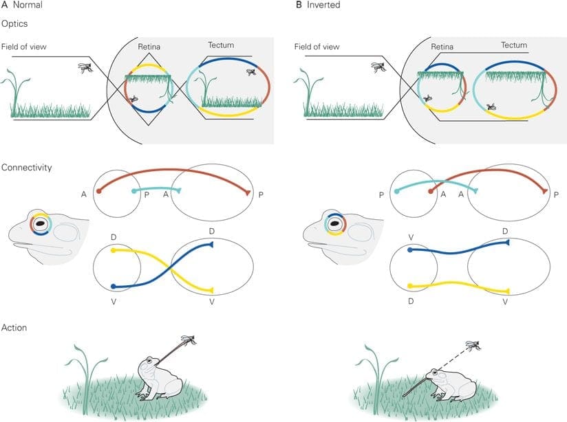
Roger Wolcott Sperry pioneered the inception of the Chemoaffinity Hypothesis after the 1960’s in a series of elegant experiments employing the retinotectal system of the African Clawed Frog, he sectioned the optic nerves and steered the eyes 180 degrees. He wished to know whether vision would be ordinary following regeneration or if the animal would eternally see the world ‘upside down’. If the latter held true, this could reveal that the nerves were somehow guided back to their original sites of termination; however, restoration of normal eyesight would mean that the nerves had resumed at new sites. Sperry showed that these creatures did indeed view the world ‘upside down’.
According to the experiment, initial�eye orientation gives that the top of the eye is Dorsal, and the underside is Ventral. Post-operation, the “top” of the eye is now Ventral, and the base is Dorsal. After a food source was supplied, the frog extended its tongue, meaning that the Dorsal-Ventral orientation of the eye still remained. In follow up experiments, the eye was dispersed and rotated 180� and the optic nerve was also cut to determine if this could affect the Dorsal-Ventral orientation. The results were identical. It was those studies which directed Sperry to suggest that complicated chemical codes, under genetic control, direct axons to their targets, his chemoaffinity hypothesis.
In his first theory, Sperry proposed that distinct cells bear different cell-surface proteins that serve as markers, a notion that demanded an unsatisfactorily high variety of proteins. He revised his model suggesting that double gradients of guidance cues in the afferent and target areas would enable proper axon targeting. There is now extensive experimental data to support the chemoaffinity hypothesis, as well as the requirement for gradients of receptor and/or ligand, such as ephrins and Eph receptors, in the projection and target regions is well established.
Roger Wolcott Sperry concluded that every individual optic nerve and tectal neuron used some kind of chemical markers that dictated their connectivity through development. He concluded that if the eye had been rotated, each optic fiber and every tectal neuron possessed cytochemical labels that uniquely denoted their neuronal kind and place and that optic fibers may use these labels to navigate to their own matching target cell, hence the visuomotor impairment.�Although certain aspects and details about Sperry’s model are unproven or incorrect, the basic notion of this chemoaffinity hypothesis has become dogma in developmental neurobiology.
Dr. Alex Jimenez’s Insight
Over the years, the principle to understand the establishment of neuronal connections has continued throughout the field of neurophysiology as well as prenatal development of the brain. Neuronal connections are believed to be established during the migration of growth cones guided by extracellular guidance cue gradients. Although this theory has been revisited countless of times, Roger Sperry was the first to explain how axons navigate to their correct targets in his chemoaffinity hypothesis. Countless experimental and clinical data now exists to support the chemoaffinity hypothesis.
The scope of our information is limited to chiropractic as well as to spinal injuries and conditions. To discuss the subject matter, please feel free to ask Dr. Jimenez or contact us at 915-850-0900 .
Curated by Dr. Alex Jimenez
Additional Topics: Sciatica
Sciatica is medically referred to as a collection of symptoms, rather than a single injury and/or condition. Symptoms of sciatic nerve pain, or sciatica, can vary in frequency and intensity, however, it is most commonly described as a sudden, sharp (knife-like) or electrical pain that radiates from the low back down the buttocks, hips, thighs and legs into the foot. Other symptoms of sciatica may include, tingling or burning sensations, numbness and weakness along the length of the sciatic nerve. Sciatica most frequently affects individuals between the ages of 30 and 50 years. It may often develop as a result of the degeneration of the spine due to age, however, the compression and irritation of the sciatic nerve caused by a bulging or herniated disc, among other spinal health issues, may also cause sciatic nerve pain.
IFM's Find A Practitioner tool is the largest referral network in Functional Medicine, created to help patients locate Functional Medicine practitioners anywhere in the world. IFM Certified Practitioners are listed first in the search results, given their extensive education in Functional Medicine

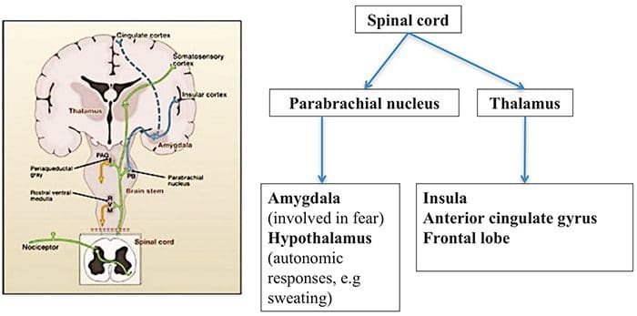 Brain Areas Involved In Processing Of Nociceptive Signals
 The Anterior Cingulate & Insula Cortex Are Activated In Human Subjects
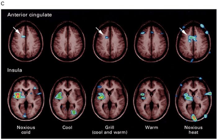 Adapted from Craig et al. 1994, 1996. From Principles of Neural Science, Kandell et al.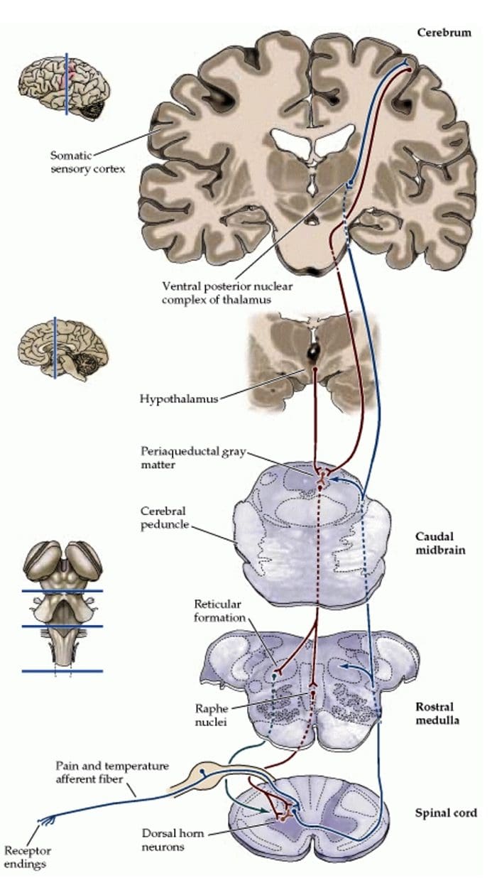
 Inflammatory Soup – Hyperalgesia
 Julius-D & Basbaum-AI, Nature 2001;413:203
 Gate Control Theory of Pain
 http://wikidoc.org/images/f/fe/Gate_control_A_firing.png
 Abnormalities Of Pain System
 Peripheral Sensitization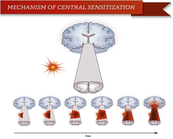
 Somatosensory Cortex Organization
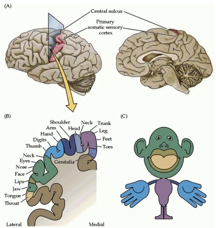 Cortical Reorganization
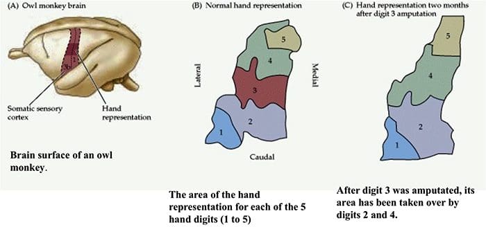 Neuroscience. 2nd edition. Purves D, Augustine GJ, Fitzpatrick D, et al., editors. Fig 25.14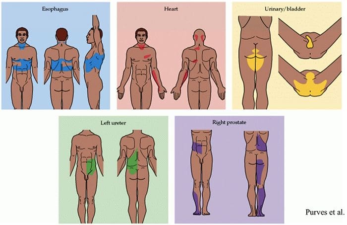
 Nerve Cell-To-Nerve Cell Communication
 Nerve cells communicate with other cells by releasing a chemical from the nerve endings � Neurotransmitters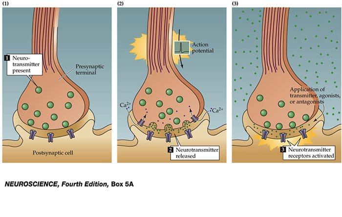
 TRP Channels