Bone neoplasms and tumor-like conditions affecting the knee can be benign or malignant. Age at Dx is crucial for DDx
In patients <40: Benign bone neoplasms: Osteochondroma, Enchondroma are relatively frequent
Fibrous cortical defect (FCD) & Non-ossifying fibroma (NOF) are particularly frequent in children
Giant cell tumor (GCT) is the m/c benign neoplasm of the knee in patients between 20-40 years of age
Malignant bone neoplasms in <40: m/c Osteosarcoma and 2nd m/c Ewing sarcoma
In patients >40: malignant neoplasms: m/c are secondaries d/t bone metastasis. Primary bone malignancy:�the m/c
Multiple Myeloma (MM). Less frequently:�a 2nd�peak of Osteosarcoma (post-radiation or Paget�s), Fibrosarcoma or Malignant�Fibrous�Histiocytoma�(MFH) of bone.
Clinically: knee pain, pathological fracture
Some tumor-like conditions like FCD/Non-ossifying fibroma are asymptomatic and may regress spontaneously. Occasionally NOF may present with pathologic fracture. N.B. any knee/bone pain in a child/adolescents should be�treated with clinical suspicion and adequately investigated.
Imaging: 1st step: radiography
MRI with T1+C is crucial for lesion characterization/regional extent, staging and pre-operative planning. CT may�help with pathologic Fxs detection. If malignant bone neoplasms considered, CXR/CT, PET-CT to investigate�metastatic spread and staging are important
Imaging Approach Bone Neoplasms
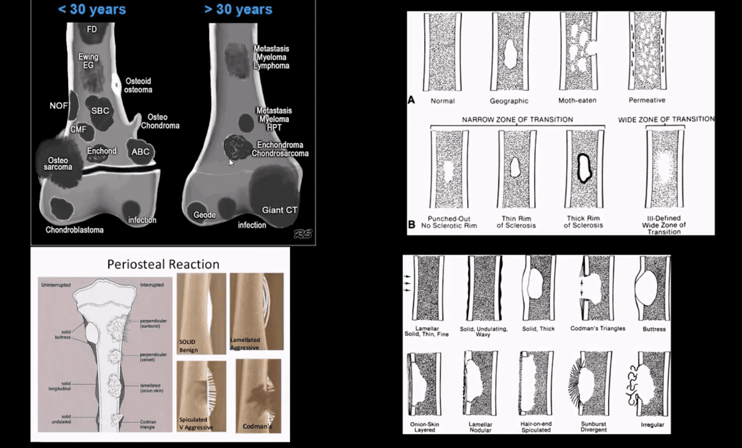
Approach to imaging Dx of bone neoplasms includes age, bone location (epiphysis vs. metaphysis vs. diaphysis), zone of transition surrounding the lesion, periosteal response, type of matrix, permeating or moth-eaten destruction vs. sclerotic, ground-glass, osteoid, cartilaginous matrix, soft tissue invasion, etc.
Key x-radiography features to DDx benign vs. malignant bone neoplasm:
Zone of transition: lesion is geographic with a narrow zone of transition vs. ill-defined wide zone of transition suggesting aggressive bone resorption
What type of bone destruction occurred: soap-bubbly appearance vs. osteolytic vs. osteosclerotic changes
Is there a round-glass matrix? Is there a well-defined rim of the sclerotic border with septations potentially suggesting slow growth and encapsulation like most benign processes.
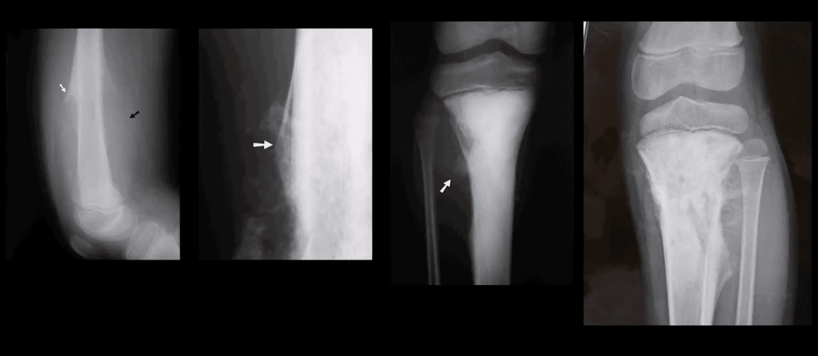
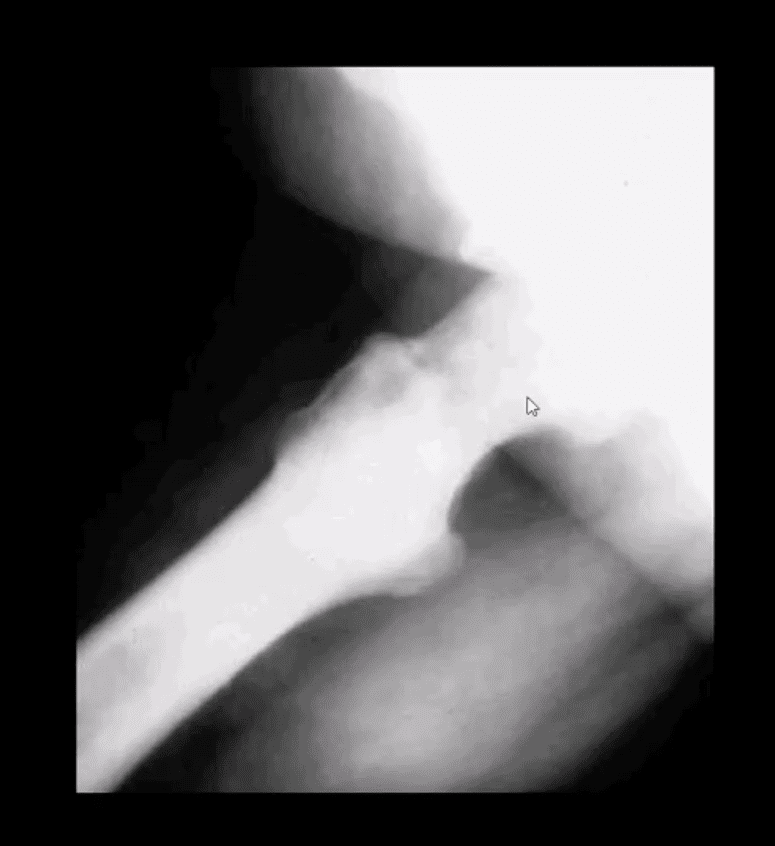
Periosteal proliferation: solid vs. aggressive spiculated/sunburst/hair-on-end with local soft tissue invasion and Codman triangle (study next slide)
FCD & NOF
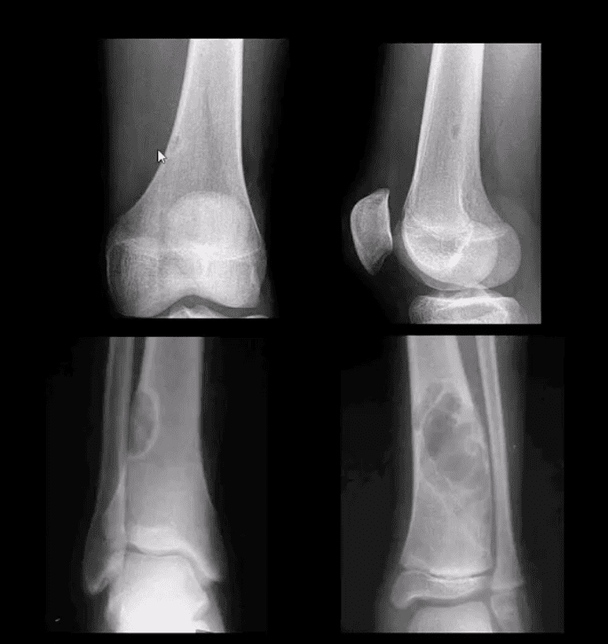
FCD & NOF or more appropriately Fibroxanthoma of the bone are benign bone processes that m/c seen in children. DDx based on the size with FCD presenting as <3-cm and NOF >3cm lesion composed of a fibrous heterogeneous matrix. FCD are asymptomatic and may regress in many cases. Some may progress to NOF. Location: identified in the knee region as an eccentric cortical based lesion.
FCD must be DDx from an avulsive irregularity d/t repeated stress along Linea aspera by extensors muscles
Dx: radiography
Management: leave-me-alone lesion. Occasionally NOF may progress and lead to pathologic fracture requiring orthopedic consult
Osteochondroma
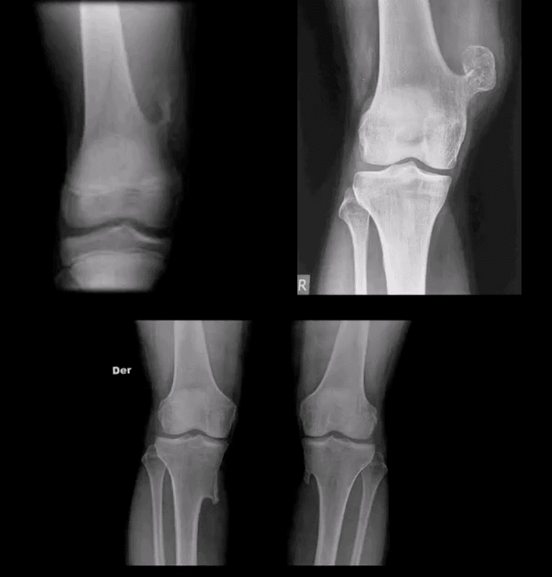
Osteochondroma: m/c benign bone neoplasm. Knee is the m/c location. Contains all bone elements with a cartilaginous cap. Presented as pedunculated or sessile bone exostosis pointing away from the joint.
1% malignant degeneration to chondrosarcoma if solitary lesion and 10-15% in cases of HME
Other complications: fracture (top left image) pseudoaneurysm of the Popliteal artery, adventitious bursa formation
Hereditary Multiple Exostosis (HME)– autosomal dominant process. Presents with multiple osteochondromas (sessile-type dominates). May lead to limb deformities (Madelung deformity, coxa valga) reactive ST pressure, malignant degeneration
Dx: radiography, MRI helps to Dx malignant degeneration to chondrosarcoma by changes in size and activity of cartilaginous cap (>2-cm in adults may manifest malignant degeneration). MRI will also help with Dx of regional complications
HME & Knee Pain
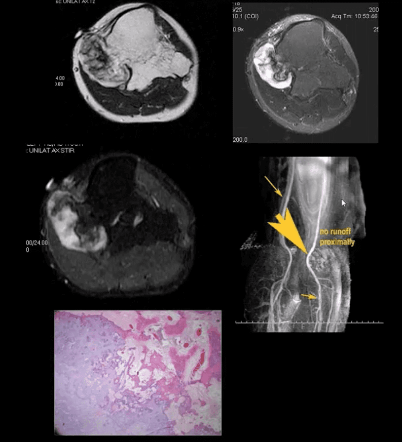
37-y.o male with HME and knee pain. Axial T1, T2 and STIR MRI slices at the popliteal region. Large cartilaginous cap and possible compression of the popliteal artery by osteochondroma. MRA was performed to evaluate popliteal A. pseudoaneurysm (large arrow). Pathology specimen obtained from the cartilaginous cap showed increased cellularity suggestive of malignant degeneration. Operative care was planned
Giant Cell Tumor (GCT) aka Osteoclastoma
GCT- is a relatively common primary benign bone neoplasm. Age 25-40. M>F slightly.
GCT is the M/C benign sacral tumor. In 50% of cases, GCT occurs about the knee.
GCT is histologically benign, but lung Mets may develop esp. if in distal radius and hands, often termed Malignant GCT
<1% unresponsive/recurring GCTs may undergo malignant transformation to high-grade bone sarcoma
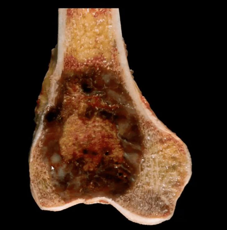
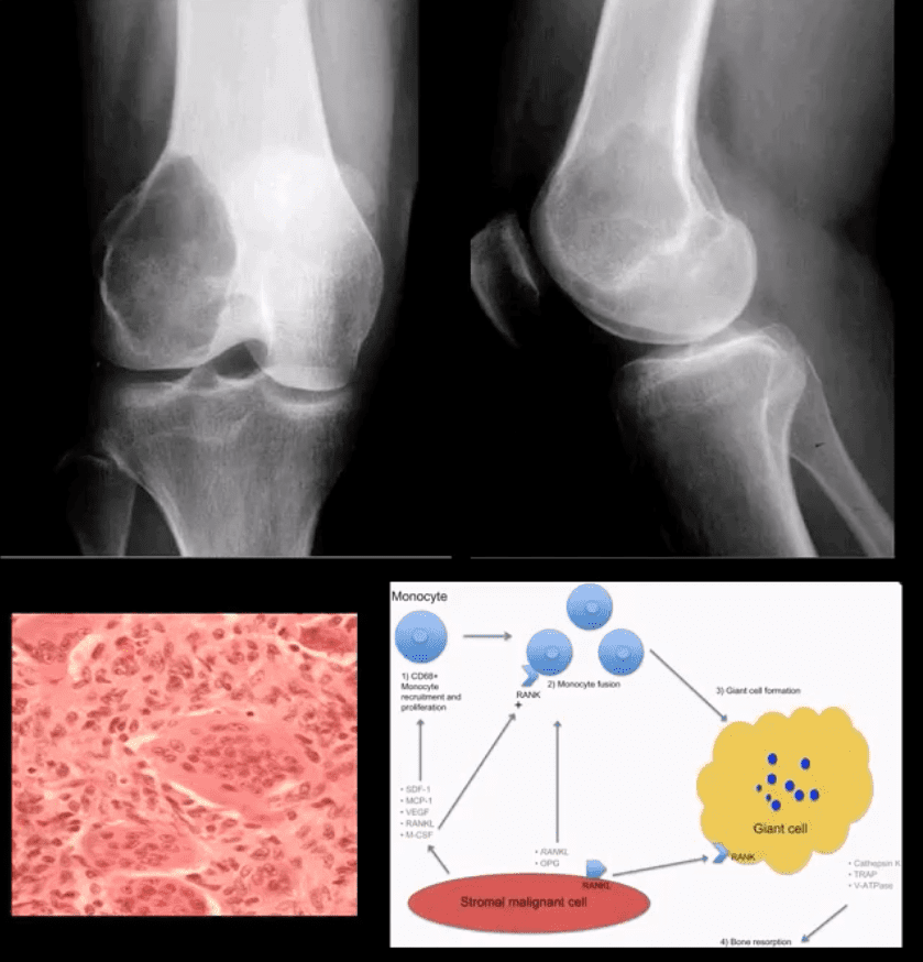
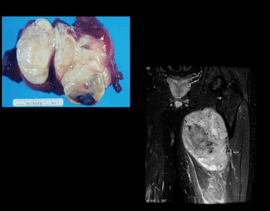
Pathology: histologically composed of osteoclasts-multinucleated giant cells with stromal cells derived from precursors monocyte-macrophage type. Produces cytokines and osteolytic enzymes. GCT may contain blood and associated with secondary Aneurysmal Bone Cyst (ABC)
Clinically: knee pain unresponsive to conservative care. Pathologic Fx may occur
Imaging: always begins with radiography followed by MRI and surgical biopsy that are crucial to Dx.
Rx: operative with curettage and cementing, a surgical appliance may be used if pathological fx present and cortical breach. In more severe cases other options available
Radiologic-Pathologic Dx
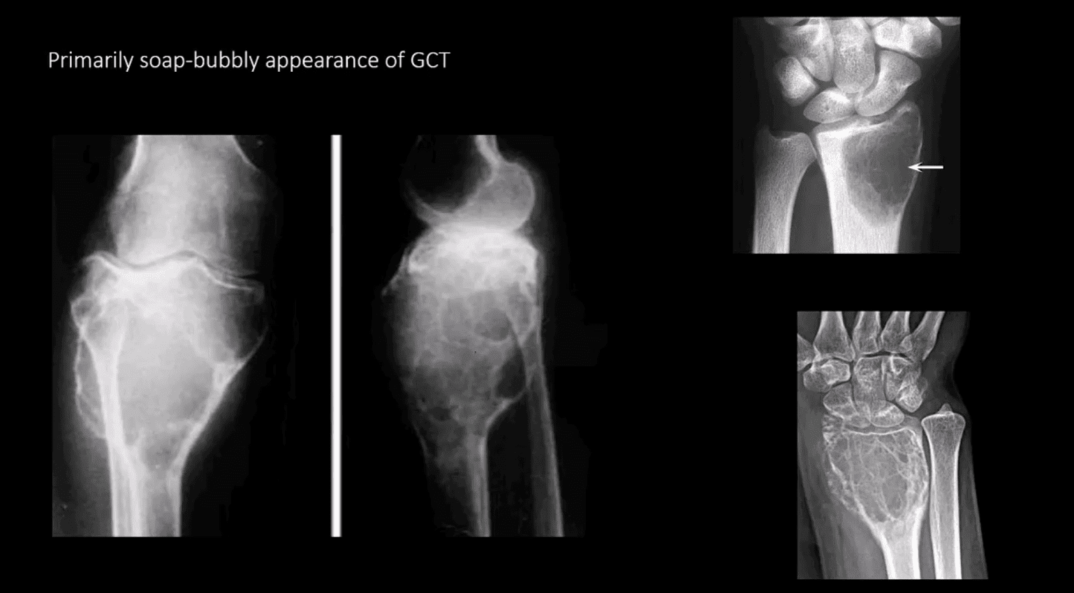
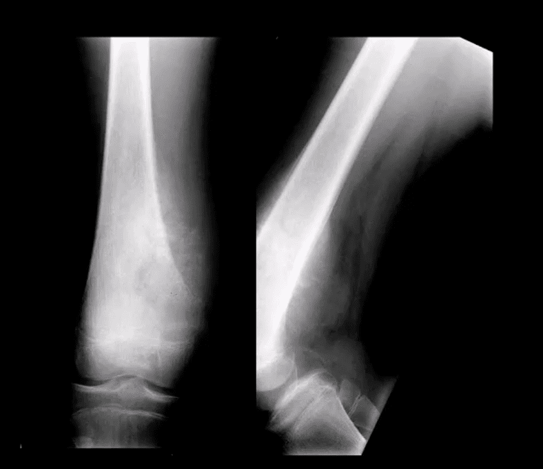
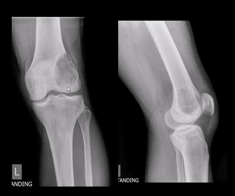
Radiologic-pathologic Dx: osteolytic and soap-bubbly lesion typically involving metaphysis and into epiphysis (classic key feature) with subarticular extension. Zone of transition is generally narrow but occasionally in aggressive lesions wide zone of transition may be seen.
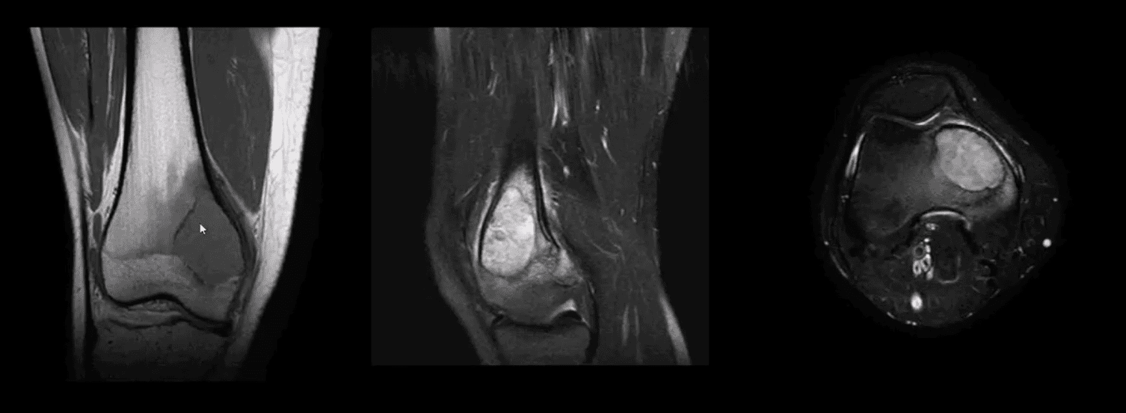
MRI: low T1, highT2/STIR, characteristic fluid-fluid levels noted that are present in GCT and ABC. Histology is crucial to Dx.
DDx: ABC, Brown cell tumor of HPT (osteoclastoma), Telangiectatic Osteosarcoma
Radiological rule: if the physeal growth plate is present Dx of GCT is taken off the list in favor of chondroblastoma and vice versa.
Primarily Soap-Bubbly Appearance of GCT
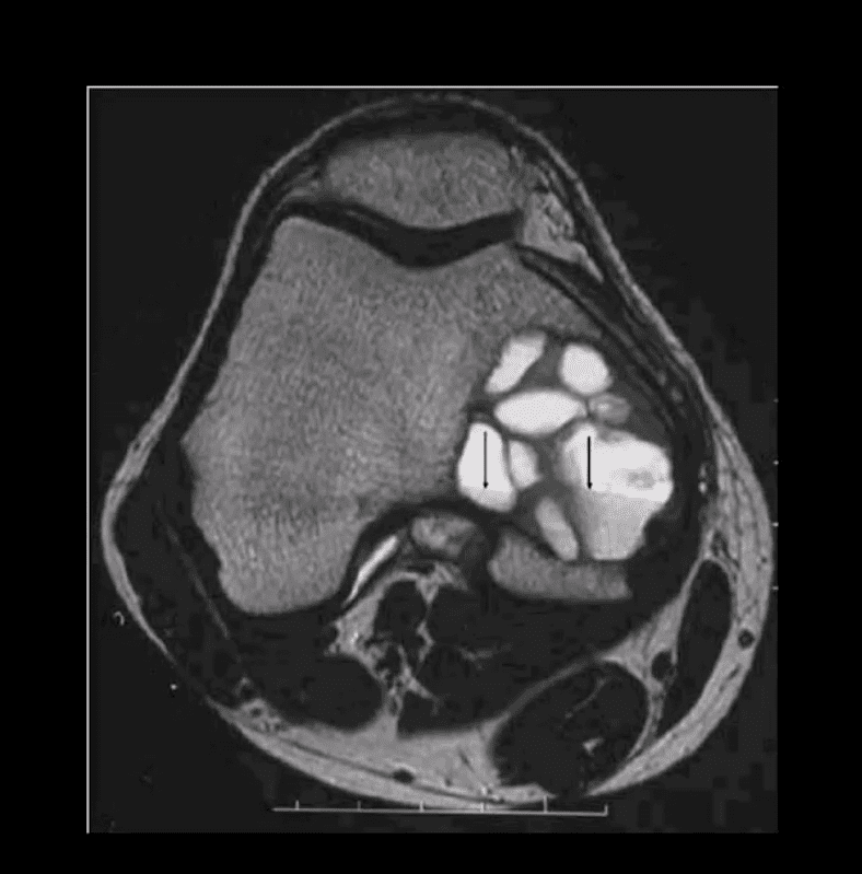
Coronal, Fat-Sat Sagittal & Axial MRI Slices of GCT
T1 coronal, T2 fat-sat sagittal and T2 axial MRI slices of GCT. Typically: low T1, highT2/STIR and fluid-fluid levels
Characteristic MRI Appearance of GCT
Fluid-fluid levels d/t different composition of blood degradation products
Important DDx: ABC
Malignant Neoplasms About the Knee
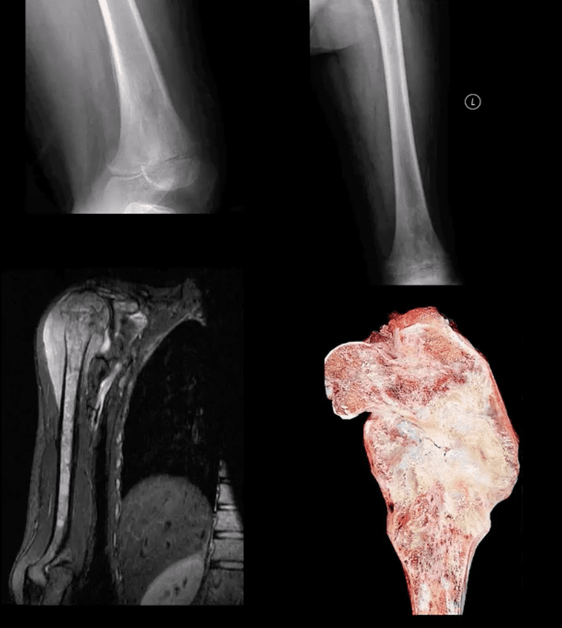
In children and very young adults, m/c primary malignant neoplasm is central aka intramedullary (osteogenic) osteosarcoma (OSA). Second peak of OS: >70 y.o d/t Paget�s (1%) and/or post radiation OSA.
The knee is the m/c location of OSA (distal femur, prox. Tibia)
A 2nd m/c malignant pediatric primary is Ewing sarcoma.
In adults >40 y.o. the m/c primary is Multiple Myeloma (MM) or Solitary Plasmacytoma
Overall m/c bone neoplasms in adults d/t bone Mets from lung, breast, prostate, renal cell, thyroid (discussed)
Dx: clinical and radiological with surgical biopsy
Imaging is crucial to Dx. 1st step x-radiography. MRI+ gad C is vital
CT scanning occasionally helps to evaluate pathological fracture
Central (Intramedullary) Osteosarcoma (OSA)
m/c age: 10-20. m/c location: knee, males>females. Increased risk in some
congenital syndromes and mutation of the retinoblastoma gene: Rothmund-Thompson AR syndrome.
Early Dx is important d/t 10-20% present with Lung Mets at Dx. Prognosis depends on stages. Early stages with local bone invasion and no
mets 76% of survival.
Rx: limb salvage procedures preferred with 8-12 weeks of chemo, amputation if encased neurovascular tissue, path Fx, etc.
Imaging: radiography and MRI.
Clinically: bone pain, Inc. Alkaline Phosphatase
Chest CT if lung Mets considered
Classic Rad Features of OSA
Osteoid forming a sclerotic mass with aggressive hair-on-end/speculated/sun-burst periosteal reaction, Codman’s triangle and soft tissue invasion. Order MRI for staging and extent. Chest CT is crucial for Lung Mets dx.
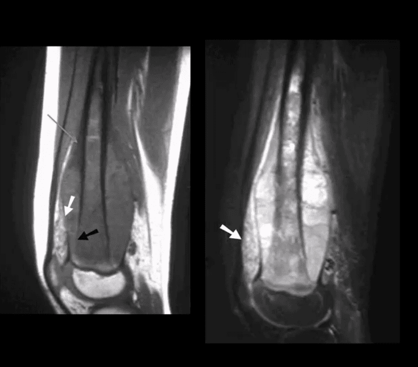
MRI is Crucial for Dx/Staging
Note sagittal T1 (left) and STIR (right) MR slices: large mass extending from distal femoral metaphysis to remaining shaft. A low signal on T1 and high on STIR d/t marrow invasion with edema, hemorrhaging and tumor invasion. Local ST invasion seen (white arrows). Periosteal lifting and Codman�s triangle (green arrow) are additional signs of aggressive neoplasm.
Note an interesting feature that the epiphysis is spared d/t physeal plate serving temporarily as an additional barrier to the tumor spread.
Ewing Sarcoma
Ewing sarcoma: age: 2-20, uncommon in black patients. 2nd m/c highly malignant bone neoplasm in children that typically arises from medullary cavity (Round cell tumors). Key symptom: bone pain that may mimic infection (ESR/CRP/WBC) Considered PNET Key Rad Dx: aggressive moth-eaten/permeative lucent lesions in the shaft of long bones with sizeable soft tissue invasion/typical onion skin periostitis. May produce saucerisation May affect flat bones. May appear as sclerotic in 33%. Early lung Mets (25-30%) bone-to-bone Mets Poor prognosis if delayed Dx. Imaging steps: 1st step x-rad, MRI is v. important followed by a biopsy. CXR/CT PET-CT Rx: combined rad-chemo, operative.
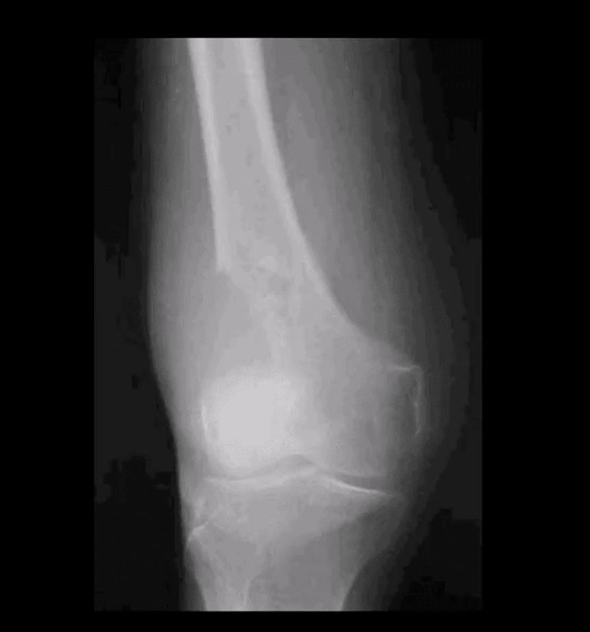
Note aggressive expansile osteolytic lesion in the distal femur metaphysis into epiphysis. No periosteal reaction present. Following further work up with abdominal and chest CT scanning, Dx of Renal cell carcinoma was established
Distal Mets into lower extremity are more common with lung, renal cell, thyroid and breast CA.
Renal cell and Thyroid will typically present with aggressive osteolytic expansile mass aka �blowout Mets.�
In general, imaging approach should consist of Radiographic knee series, followed by MRI if x-rays are unrewarding
Tc99 Bone scintigraphy is the modality of choice to evaluate metastatic bone disease
Soft Tissue Neoplasms About the Knee
Malignant fibrous histiocytoma (MFH) reclassified as Pleomorphic Undifferentiated Sarcoma (PUS) is the m/c S.T. sarcoma. MFH is aggressive biologically with poor prognosis M>F (1.2:1) 30-80 with a peak in a 6th decade. 25-40% of all adults sarcomas m/c extremities. Retroperitoneum next (worst prognosis d/t late Dx and large growth w/o symptoms) Clinically: painful, hard mass typically about the knee or thigh. Histology: poorly differentiated/undifferentiated malignant fibroblasts, myofibroblasts, and other mesenchymal cells Imaging: MRI is the modality of choice with T1, T2, T1+C. Typically appears as an aggressive heterogeneous mass intermediate to low signal on T1 and high signal on T2 with areas of necrosis and enhancement on T1+C. May appear misleadingly encapsulated w/o true capsule Management: operative with radiation and chemotherapy. Tumor depth is crucial for prognosis. 80% 5-year survival if <5cm deep in ST and 50% if >5-cm deep in ST.
Synovial Sarcoma
Synovial sarcoma: common malignant ST neoplasm esp. in younger patients or older children/adolescents. M/C found in knee area Clinically: can present slowly as a palpable mass in the extremity often ignored d/t slow growth Imaging is the key: radiography may reveal ST. density/mass. Some synovial sarcomas may show calcification and mistaken for Myositis Ossificanse or heterotopic bone formation MRI with T1, T2 and T1+C are Dx modality of choice. Other modalities: US, CT are non-specific DDx: MFH Management: operative, chemo-radiation Prognosis: variable depending on size, invasion, metastasis
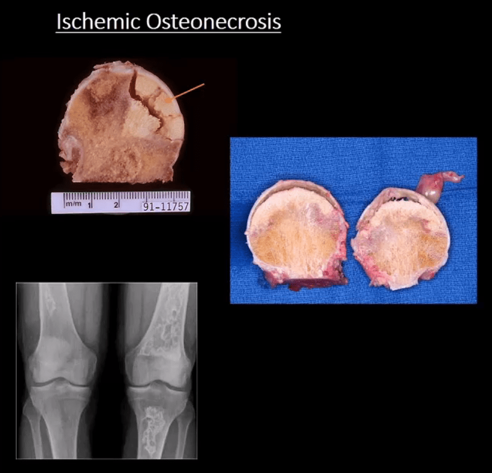
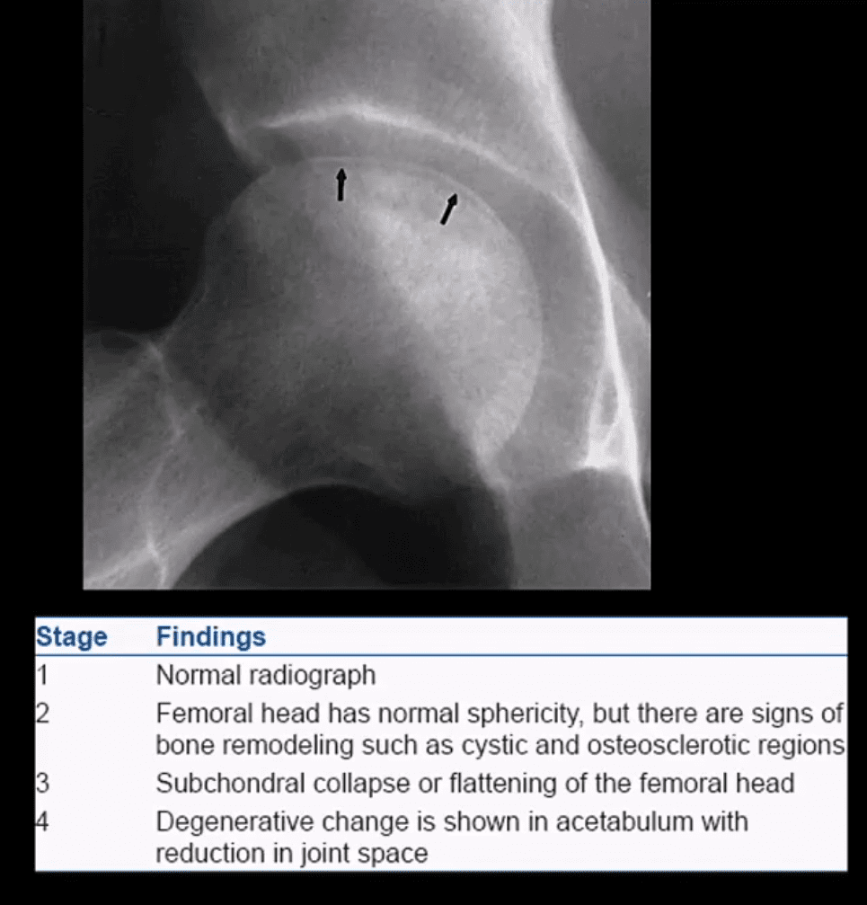
Ischemic Osteonecrosis (More accurate term) aka avascular necrosis AVN: this term describes subarticular (subchondral) bone death
Intramedullary bone infarct: depicts osteonecrosis within the medullary cavity of the bone (above x-ray image)
Causes: m/c: trauma, systemic corticosteroids, diabetes, vasculitis in SLE. The list is long. Other vital causes: Sickle cell disease, Gaucher disease, alcohol, caisson disease, SCFE, LCP, etc.
Pathology: ischemia and bone infarct with resultant devitalized center surrounded by ischemia and edema with normal bone on the outer periphery (MRI double line sign)
Sub-articular necrotic bone eventually collapses and fragments leading to progressive bone and cartilage destruction and rapidly progressing DJD
Early Dx often missed but crucial to prevent severe DJD
M/C Sites
Hips, shoulders, talus, scaphoid bone. Many peripheral idiopathic AVN sites are known by their eponyms (e.g., Kienbock aka AVN of the lunate bone, Preisier aka scaphoid AVN)
Radiography is insensitive to early AVN and may only present as subtle osteopenia
Some of the early appreciable rad features are increased patchy bone sclerosis followed by sub-articular bone collapse or “crescent sign” signifying stage-3 on Ficat classification (above)
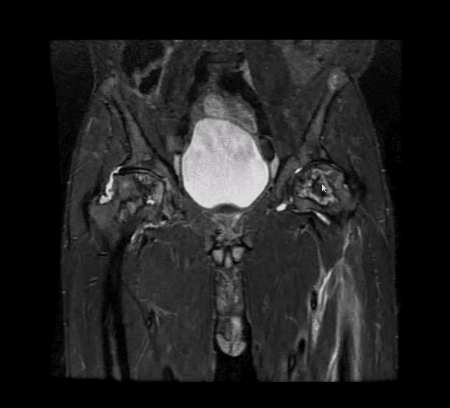
Earliest detection and early intervention can be achieved by MRI (most sensitive modality)
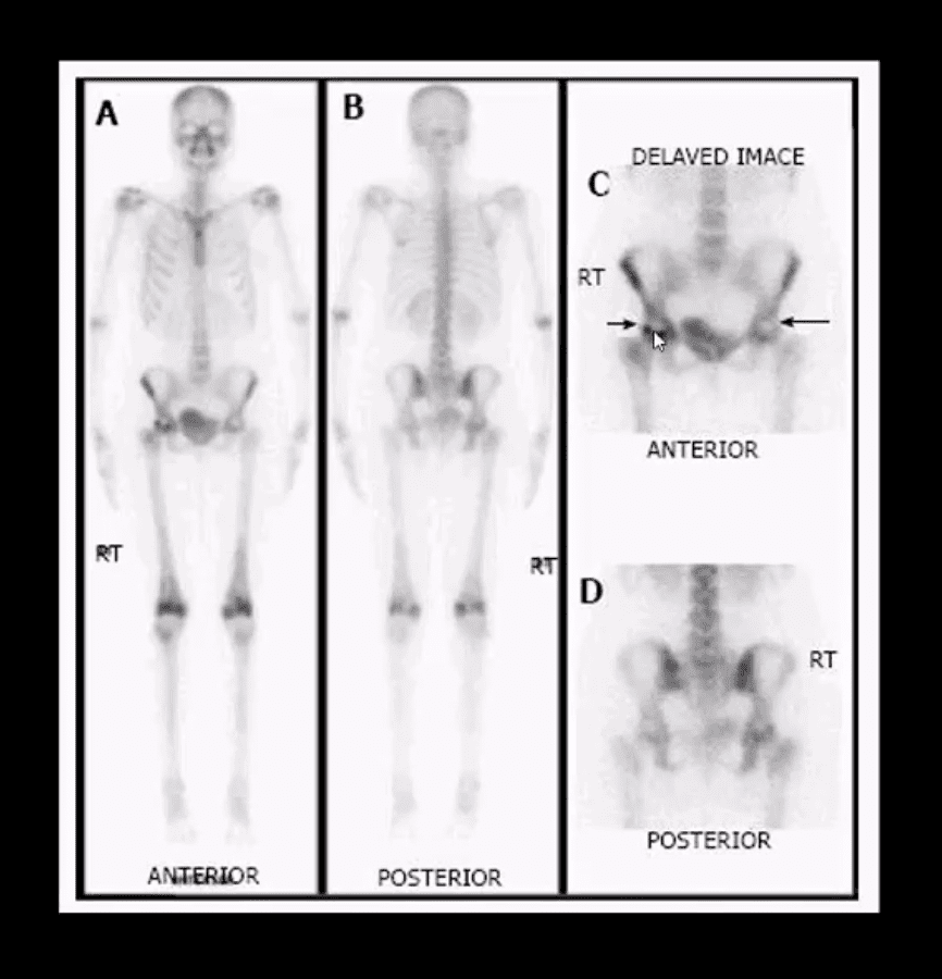
If MRI contraindicated or unavailable, 2nd most sensitive modality is radionuclide bone scan (scintigraphy)
X-ray and CT scanning are of equal value
Coronal MRI Slice
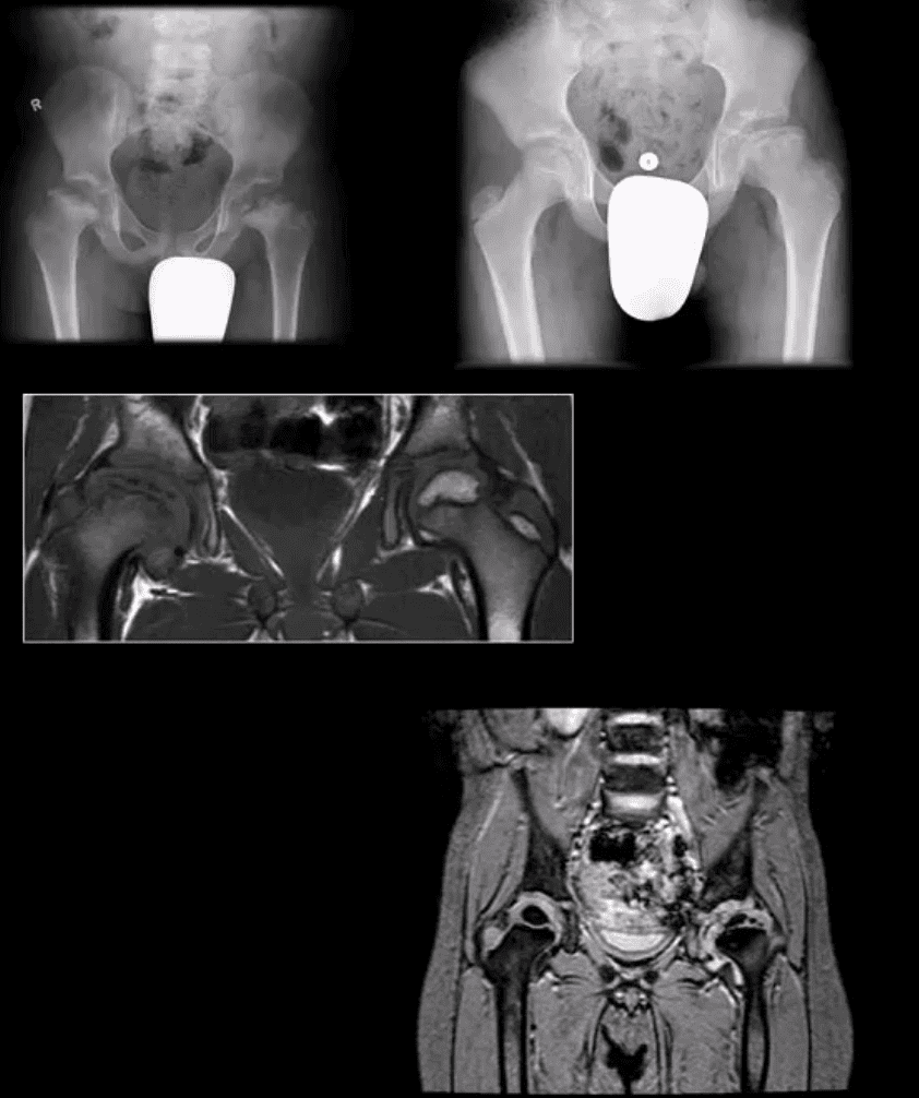
Fluid sensitive, sensitive coronal MRI slice revealing bill ischemic osteonecrosis of the femoral head
MRI findings: l
Tc99-MMDP Radionuclide Bone
Bone scan reveals a central area of photopenia (cold spot) d/t necrotic fragment surrounded by increased osteoblastic activity as increased uptake of Tc-99 MDP in the right hip
The patient is a 30-year-old female with breast cancer and chemotherapy treatment who suddenly presented with right hip pain
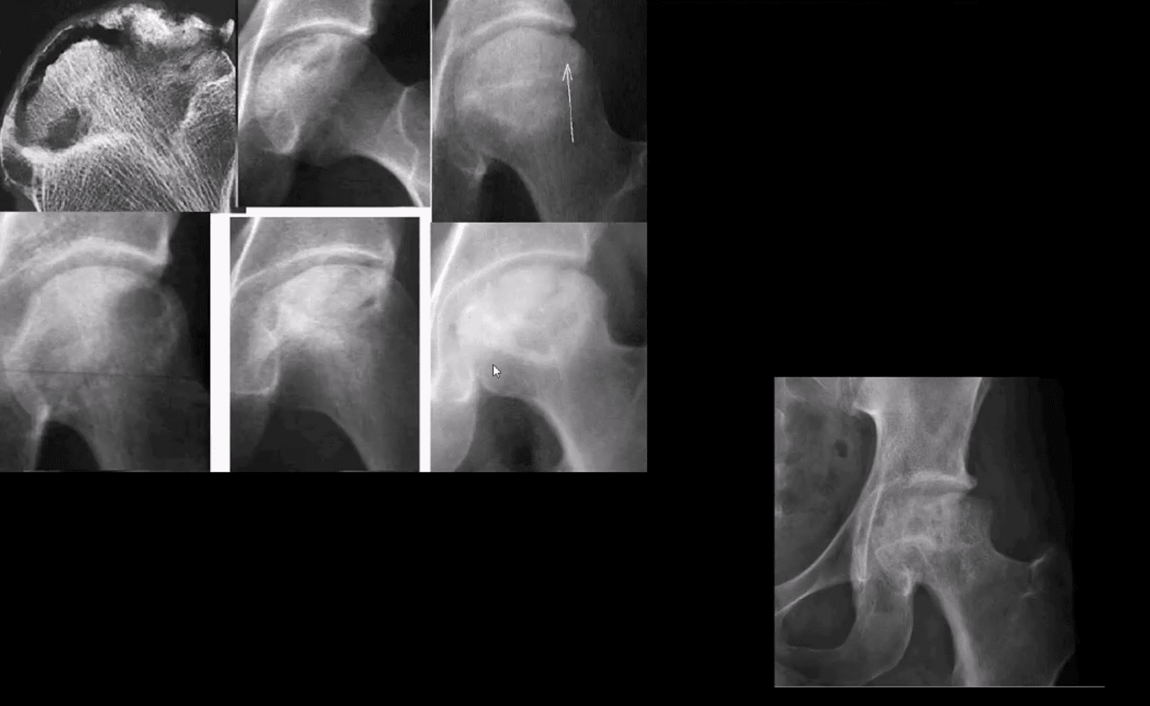
Radiographic Progression of AVN
Later stages present with articular collapse, subarticular cysts, increased patchy sclerosis and complete flattening of the femoral head with resultant severe DJD. Rx: THA
Management
Early imaging Dx with MRI or bone scintigraphy is essential
Referral to the Orthopedic surgeon
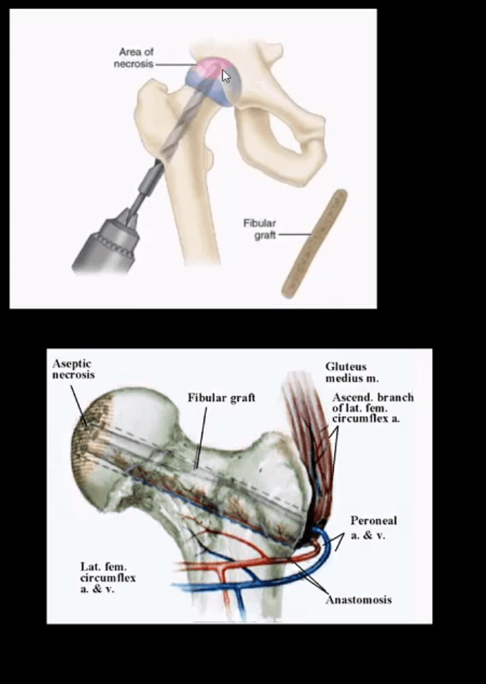
Core decompression (above) can be used to revascularize the affected bone during earlier stages but produces mixed results
Delayed changes of AVN: THA as IN severe DJD cases
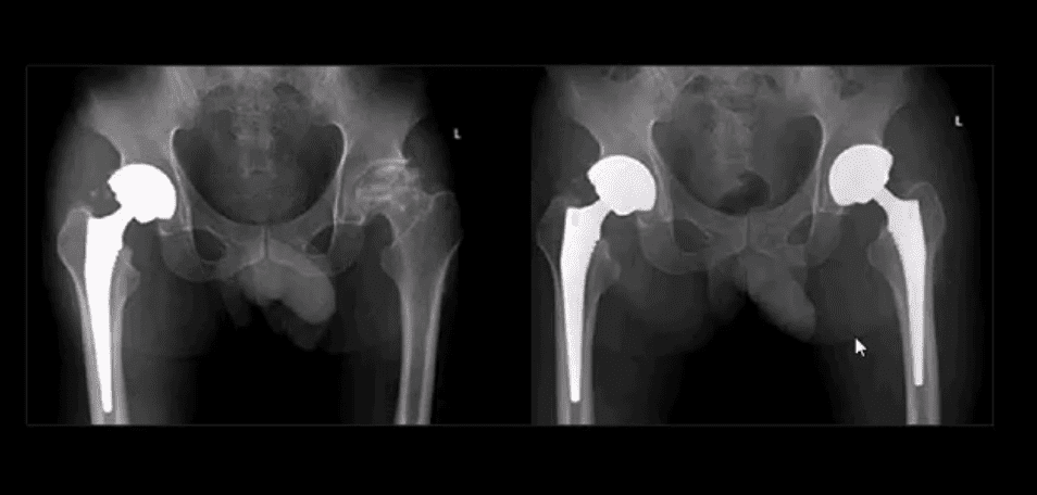
B/L THA
B/L THA in the patient with ischemic osteonecrosis of the right and later left hip
When B/L hip AVN is present, typically consider systemic causes (corticosteroids, diabetes)
Inflammatory Arthritis Affecting the Hip
Consider common systemic inflammatory condition such as RA and AS/EnA
Hip RA may develop in 30% of patients with RA
Key features to DDx inflammatory arthritis vs. DJD is symmetrical/uniform aka concentric joint loss often leading to axial migration and Protrusion Acetabule in advanced cases
Key element between RA vs. AS: the presence of RA bone erosion w/o productive bone changes or enthesitis in AS d/t inflammatory subperiosteal bone proliferation, whiskering/fluffy periostitis (collar-type enthesitis circumferentially affecting head-neck junction)
Dx: Hx, PE, labs: CRP, RH, anti-CCP Ab (RA)
CRP, HLA-B27, RF- (AS)
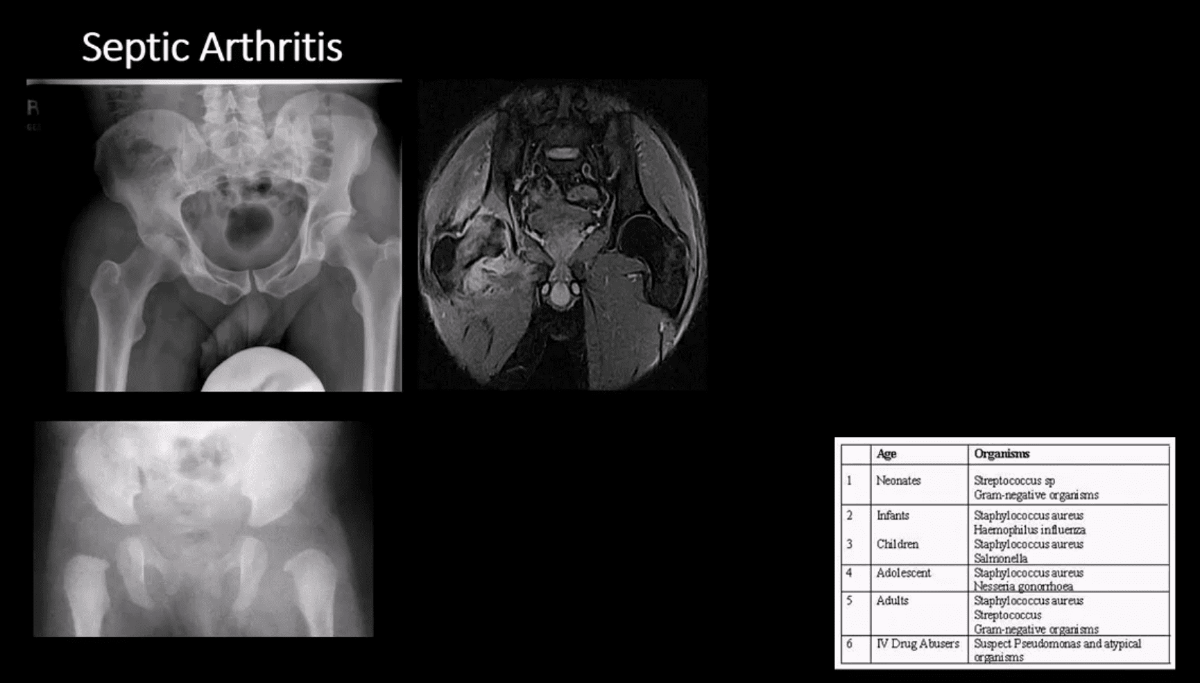
Septic Arthritis
Gonococcal infections, iatrogenic causes, I.V. drug use, and some others
Routes:�hematogenous, adjacent spread, direct inoculation (e.g., iatrogenic)
Clinically: pain and reduced ROM presented as monoarthritis, generalized signs/symptoms. CBC, ESR, CRP changes. ARthrocentesis and culture are crucial
M/C pathogen Staph. Aureus & Neisseria Gonorrhea
1st step: radiography, often unrewarding in the early stage. Later (4-10 days) indistinctness of the white cortical line at the femoral articular epiphysis, loss of joint space, effusion as a widening of the medial joint area (Waldenstrom sign)
MRI – best at early DX: T1, T2, STIR, T1+C may help with early. Early I.V. antibiotics crucial to prevent rapid joint destruction
Slipped Capital Femoral Epiphysis (SCFE)
Important to diagnose but easily missed potentially leading to Ischemic Osteonecrosis of the femoral head aka AVN
Presents typically in overweight children (more often boys), age over eight years. Greater incidence in African-American boys
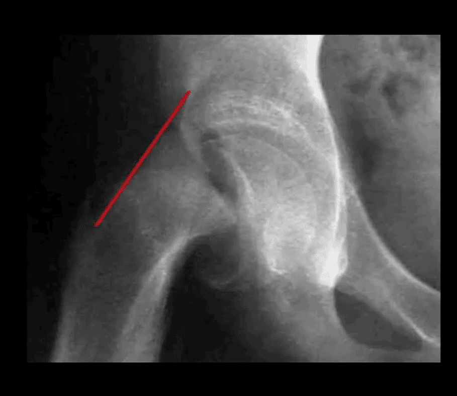
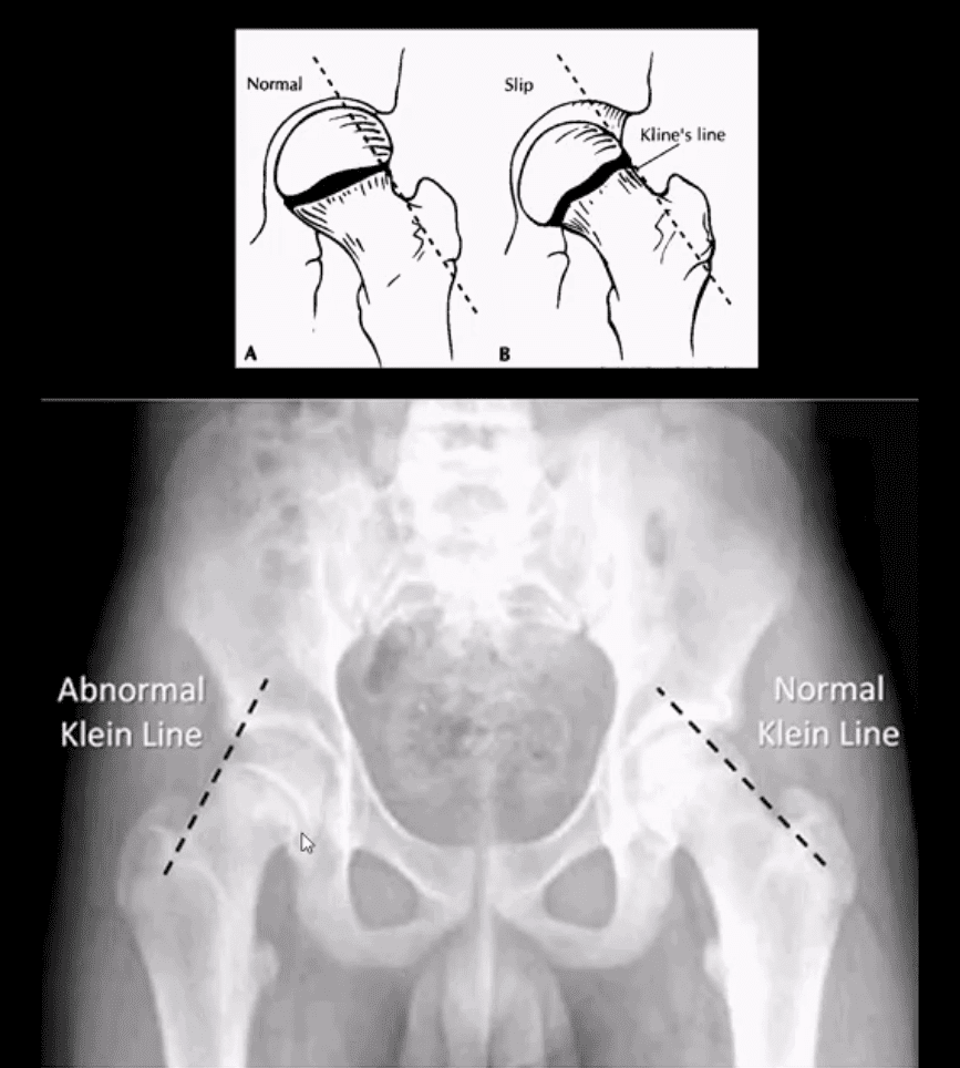
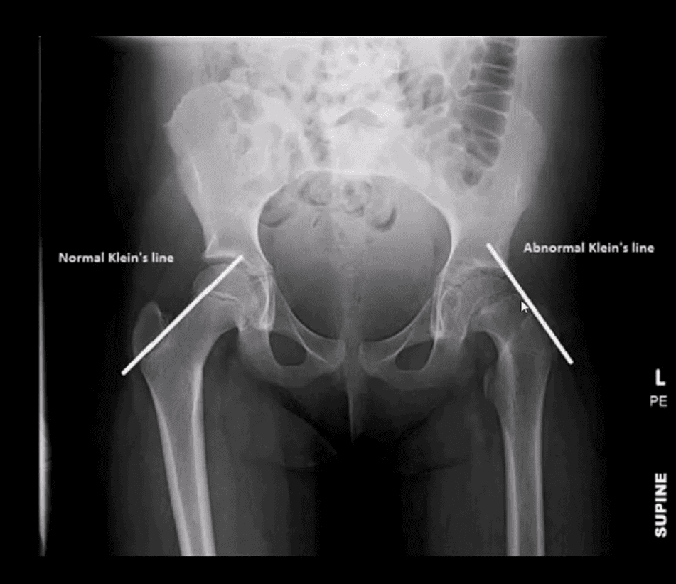
1st step: radiography, especially look for a widened physeal growth plate (so-called pre-slip). Later, slip and disturbed Klein’s line (above image). MRI – best modality for early Dx and early intervention
The frog lateral view often demonstrates the medial slip better than the AP view
Clinically Limping Child or Adolescent
M>F (10-18 years). African-Americans are at greater risk. 20% of cases of SCFE are B/L. Complications: AVN >>DJD
Radiography:�AP pelvis, spot, and frog leg may reveal slippage as Klein line failed to cross through the lateral aspect of the femoral head
Additional features: physis may appear widened
MRI w/o gad, is required for the earliest Dx and prevention of complications (AVN)
Normal and Abnormal Klein Line
Consistent with SCFE. The physis is also widened. Dx: SCFE
Urgent referral to the Pediatric Orthopedic surgeon
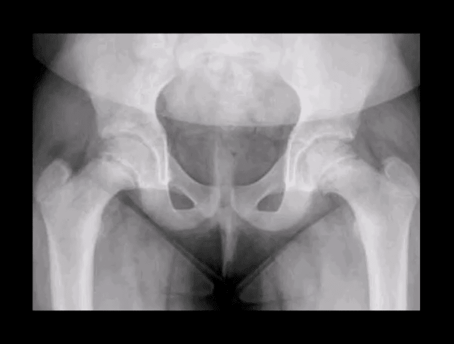
Subtle Changes in Left Hip
Note suspected subtle changes in the left hip that may require MR examination to confirm the Dx
Delay in care may result in major complications
Perthes’ Disease
aka Legg-Calves-Perthes Disease (LCP)
Refers to Osteochondritis of the femoral head with osteonecrosis likely d/t disturbed vascularization of the femoral head
Presents typically in children (more often boys) aged under eight years as atraumatic “limping child.” 15% may have B/L Perthe’s
Imaging steps: 1st step x-radiography, followed by MRI especially in stage 1 (early) w/o x-ray abnormalities
Unspecific signs: joint effusion with Waldenstrome sign+ (>2-mm increase in medial joint space compared to the opposite side). Past approach: Fluoroscopic Arthrography (replaced by MRI)
Pathologic-Radiologic Correlation: in well-established cases, the femoral head characteristically becomes sclerotic, flattened and fragmented due to avascular necrosis (AVN). Later on, an occasional Coxa Magna changes may develop (>10% femoral head enlargement)
Management: symptoms control, bracing. Boys at younger ate show better prognosis d/t more immaturity and better chances of bone/cartilage repair mechanisms. In advanced cases, operative care: osteotomy, hip arthroplasty in adulthood if advanced DJD develops
Common Neoplasms & Other Conditions Affecting Hip/Pelvis
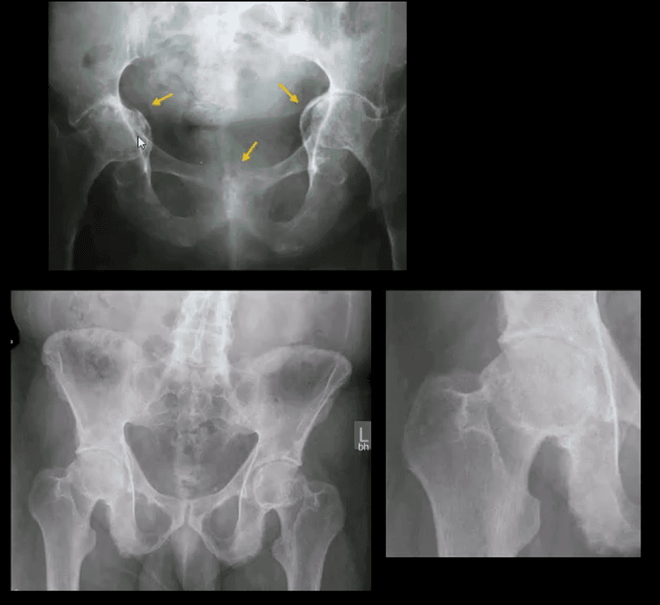
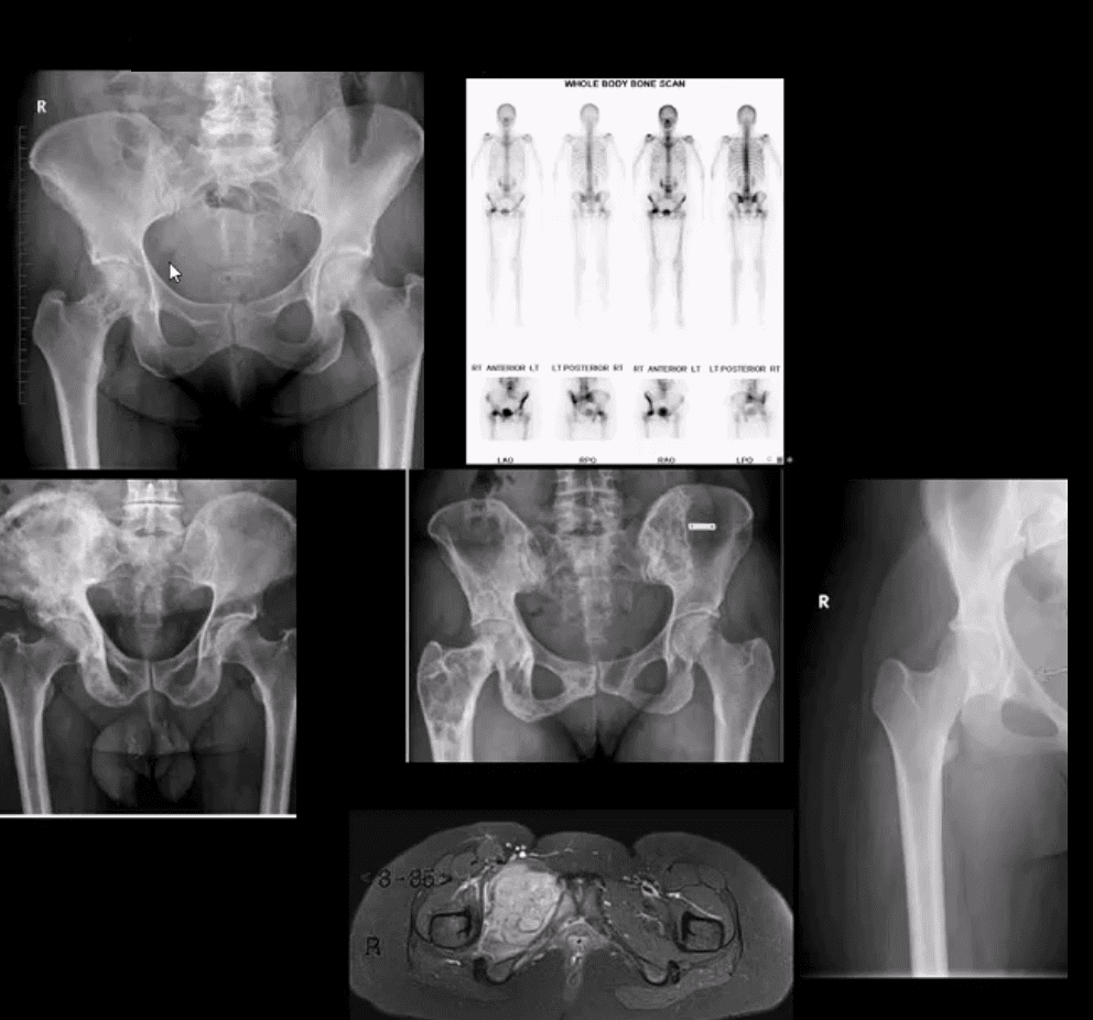
M/C hip & pelvis neoplasms in adults: bone metastasis ( above far left), 2nd m/c Multiple Myeloma (M/C primary bone malignancy in adults). Tips: remember Red Marrow distribution. Less frequent: Chondrosarcoma
Paget’s disease of bone (above-bottom left image) is m/c detected in the pelvis and Femurs
Children and young adults ‘limping child’ benign neoplasms: Fibrous Dysplasia (above middle image), Solitary Bone Cyst (21%), Osteoid Osteoma, Chondroblastoma. Malignant pediatric neoplasms: m/c Ewing Sarcoma (above middle right and bottom images) vs. Osteosarcoma. >2y.o-consider Neuroblastoma
Imaging: 1st step: radiography followed by MRI are most appropriate.
If Mets are suspected: Tc99 bone scintigraphy is most sensitive
Multiple Myeloma
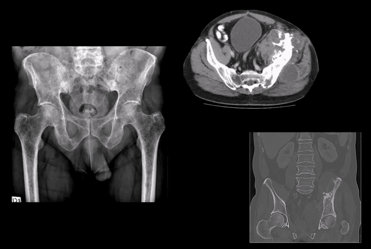
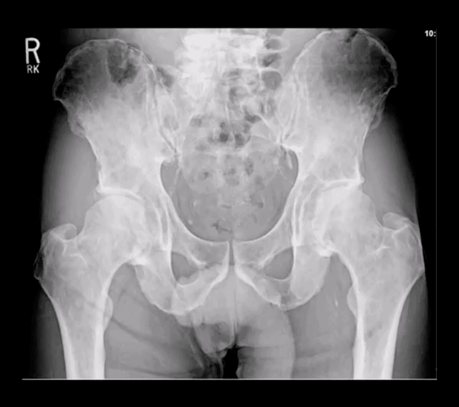
Multiple Myeloma in a 75-y.o male (AP pelvis view)
Chondrosarcoma in a 60-y.o male (axial and coronal reconstructed CT+C slices in the bone window)
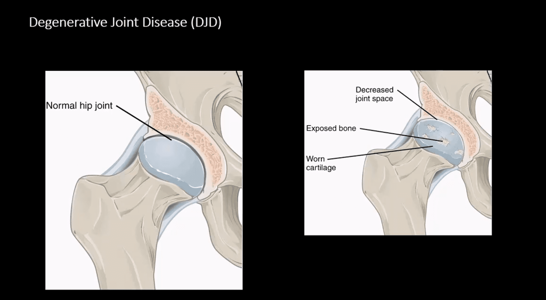
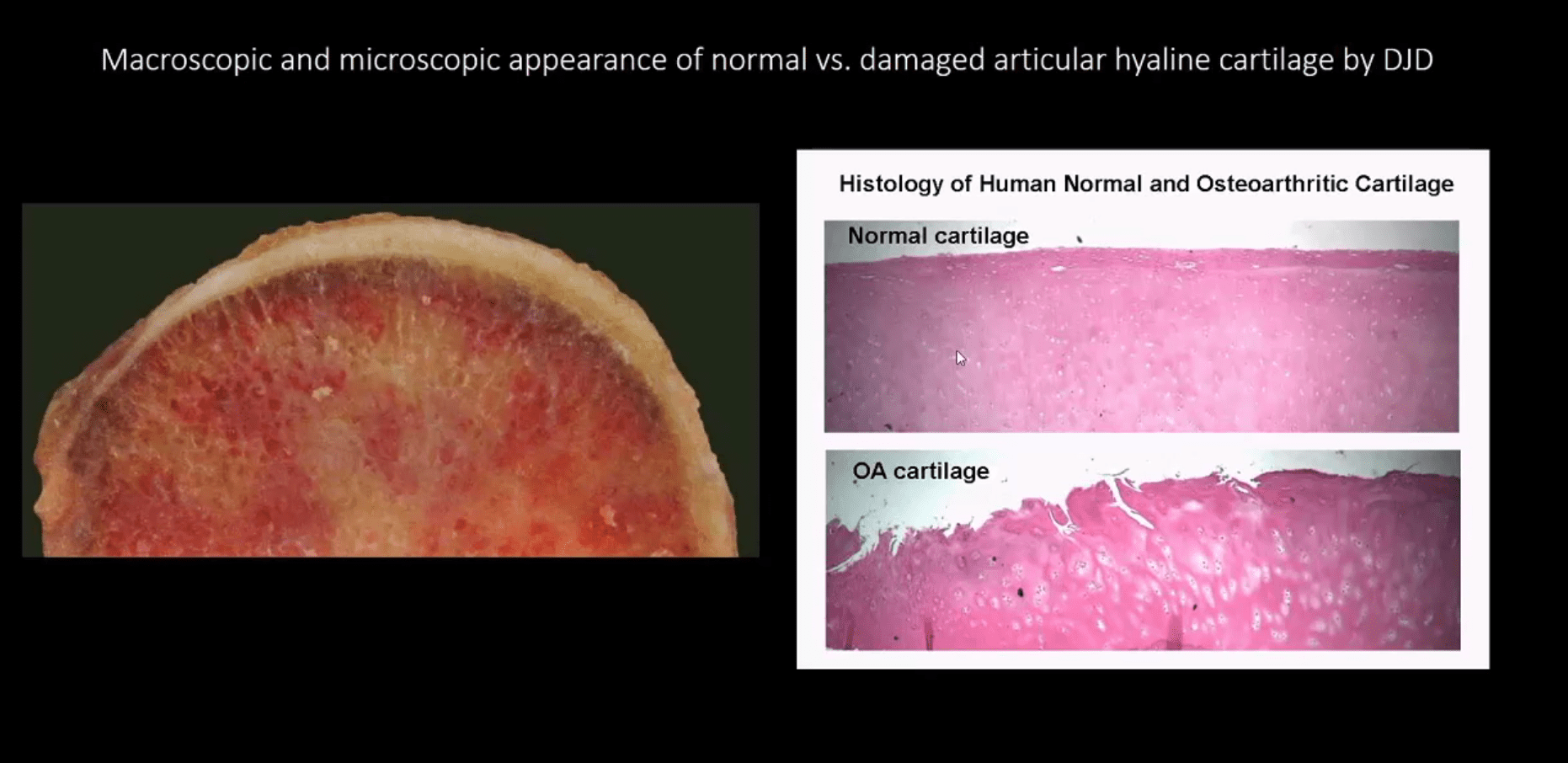
Macroscopic & Microscopic Appearance of Normal vs. Damaged Articular Hyaline Cartilage by DJD
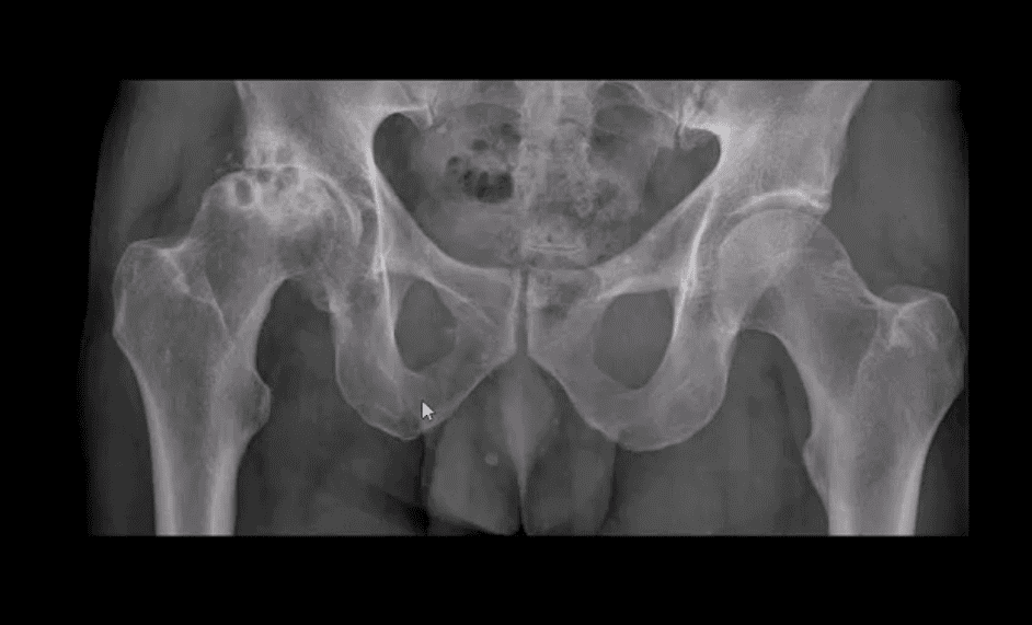
Hip Osteoarthritis (OA) aka Osteoarthrosis
Symptomatic and potentially disabling DJD
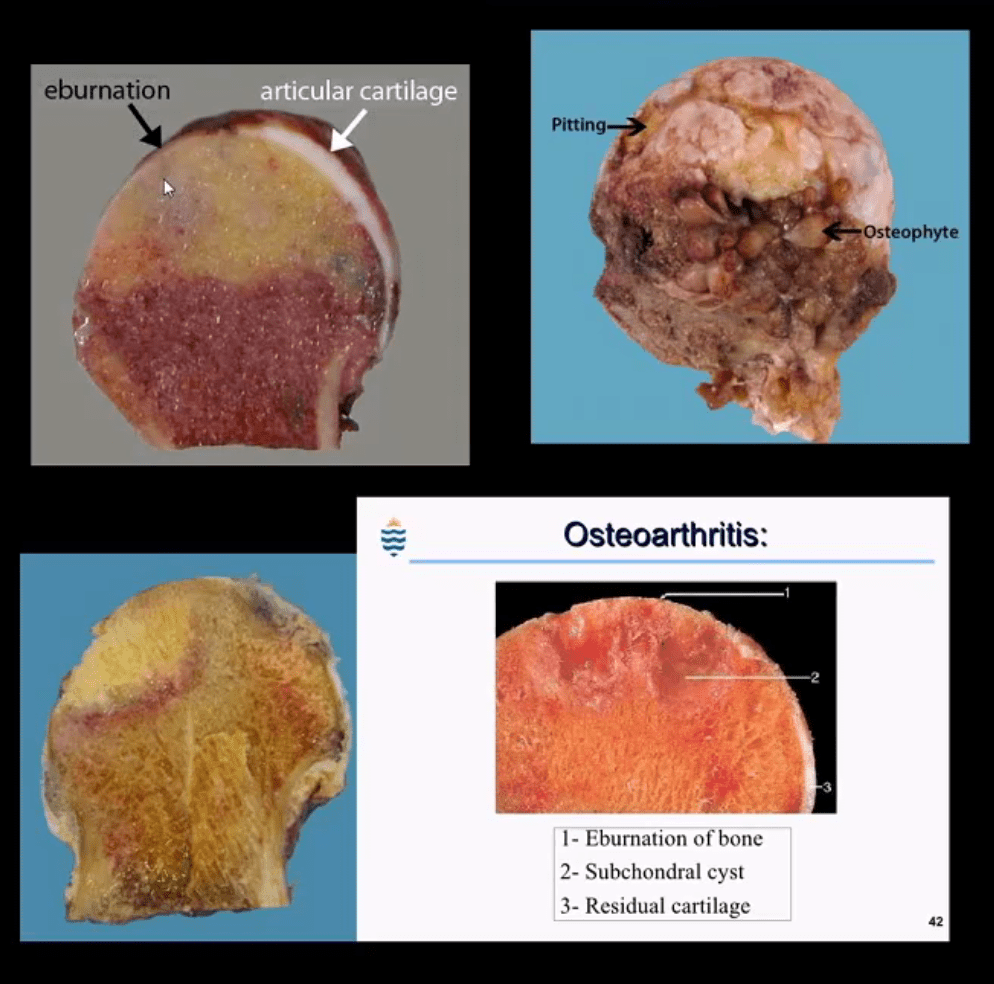
Progressive damage and loss of the articular cartilage causing denudation and eburnation of articular bone
Cystic changes, osteophytes, and gradual joint destruction
Develops d/t repeated joint loading and microtrauma
Obesity, metabolic/genetic factors
Secondary Causes: trauma, FAI syndrome, osteonecrosis, pyrophosphate crystal deposition, previous inflammatory arthritis, Slipped Capital Femoral Epiphysis, Leg-Calves-Perthes disease in children, etc.
Hip OA, 2nd m/c after knee OA. Women>men
88-100 symptomatic cases per 100000
Radiography is the Modality of Choice for the Dx and Grading of DJD
Special imaging is not required unless other complicating factors exist
The acetabular-femoral joint is divided into superior, axial and medial compartments/spaces
Normal joint space at the superior compartment should be 3-4-mm on the AP hip/pelvis view
Understanding the pattern of hip joint narrowing/migration helps with the DDx of DJD vs. Inflammatory arthritis
In DJD, m/c hip narrowing is superior-lateral (non-uniform) vs. inflammatory axial (uniform)
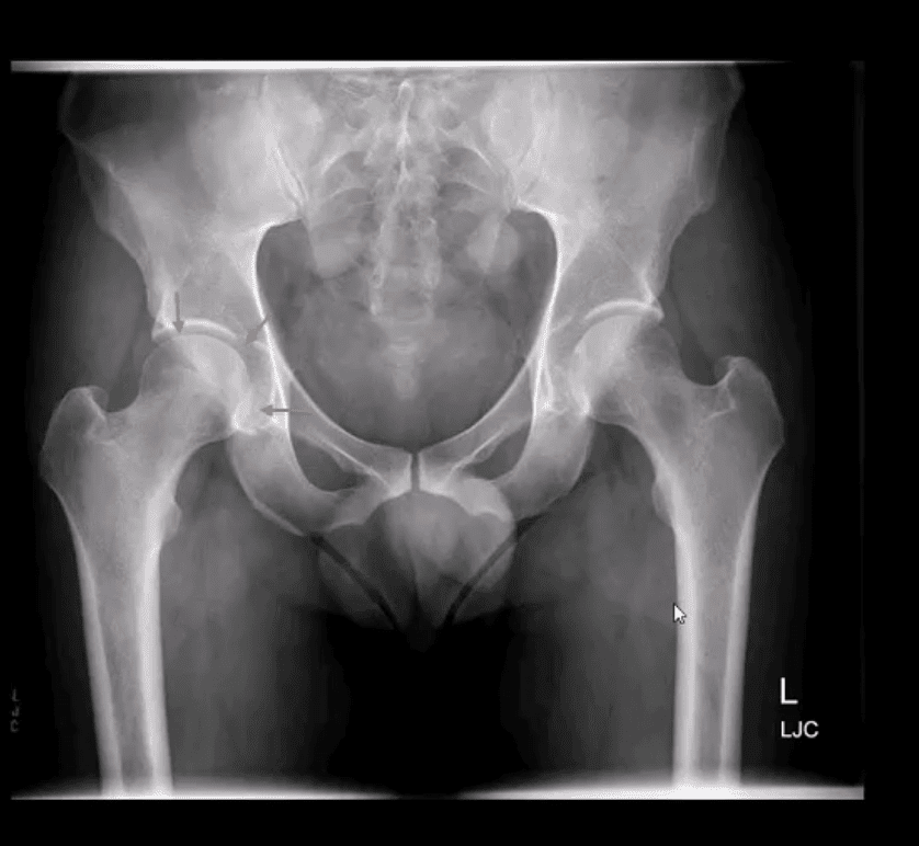
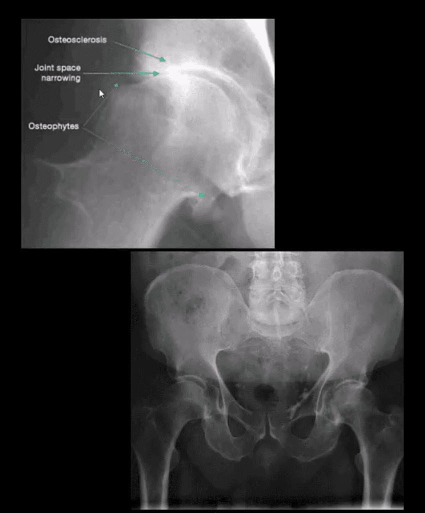
AP Hip Radiograph Demonstrates DJD
With a non-uniform loss of joint space (superior migration), large subcortical cysts and subchondral sclerosis
Radiographic features:
Like with any DJD changes: radiography will reveal L.O.S.S.
L: loss of joint space (non-uniform or asymmetrical)
O: osteophytes aka bony proliferation/spurs
S: Subchondral sclerosis/thickening
S: Subcortical aka subchondral cysts “geodes.”
Hip migration is m/c superior resulting in a “tilt deformity.”
Radiographic Presentation of Hip OA May Vary Depending On Severity
Mild OA: mild reduction of joint space often w/o marked osteophytes and cystic changes
During further changes, collar osteophytes may affect femoral head-neck junction with more significant joint space loss and subchondral bone sclerosis (eburnation)
Cyst formation will often occur along the acetabular and femoral head subarticular/subchondral bone “geodes” and usually filled with joint fluid and some intra-articular gas
Subchondral cysts may occasionally be very large and DDx from neoplasms or infection or other pathology
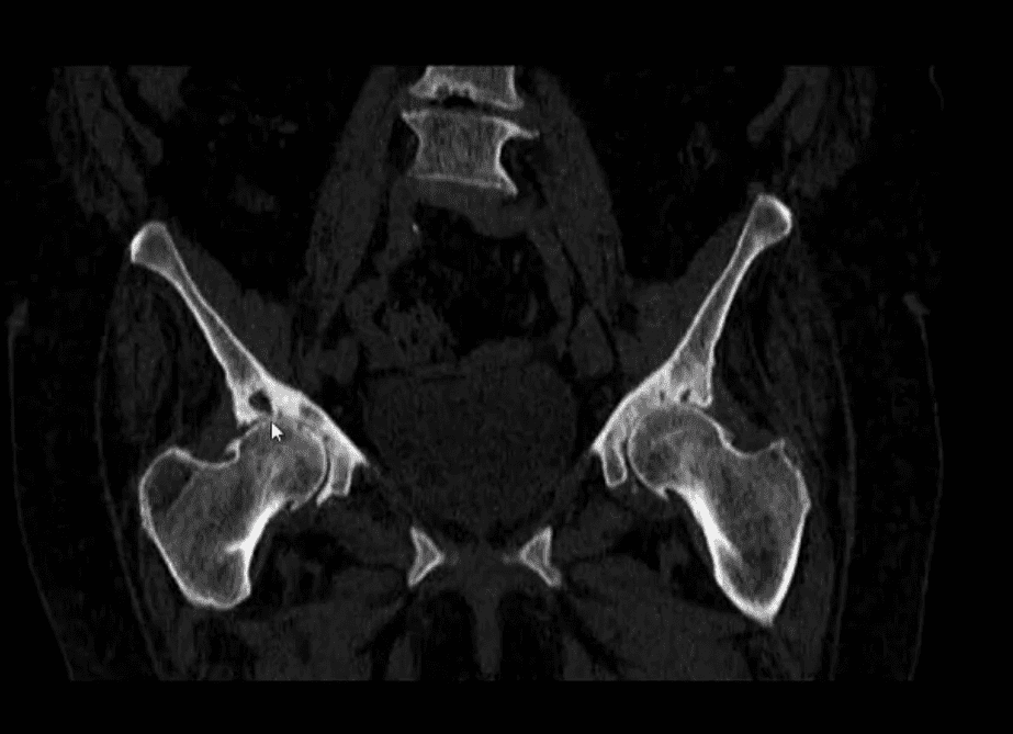
Coronal Reconstructed CT Slices in Bone Window
Note moderate joint narrowing that appears non-uniform
Sub-chondral cysts formation (geodes) are noted along the acetabular and femoral head subchondral bone
Other features include collar osteophytes along head-neck junction
Dx: DJD of moderate intensity
Referral to the Orthopedic surgeon will be helpful for this patient
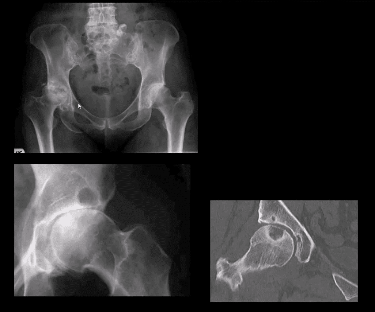
AP Pelvis (below the first image), AP Hip Spot (below the second image) CT Coronal Slice
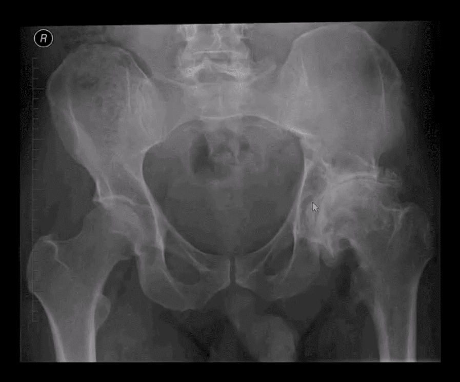
Note multiple subchondral cysts, severe non-uniform joint narrowing (superior-lateral) and subchondral sclerosis with osteophytes
Advanced hip arthrosis
Severe DJD, Left Hip
When reading radiological reports pay particular attention to the grading of hip OA
Most severe (advanced) OA cases require total hip arthroplasty (THA)
Refer your patients to the Orthopedic surgeon for a consultation
Most mild cases are a good candidate for conservative care
Hip Arthroplasty aka Hip Replacement
Can be total or hemiarthroplasty
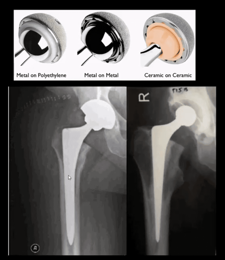
THA can be metal on metal, metal on polyethylene and ceramic on ceramic
A hybrid acetabular component with polyethylene and metal backing is also used (above right image)
THA can be cemented (above right image) and non-cemented (above-left image)
Non-cemented arthroplasty is used on younger patients utilizing porous metallic parts allowing good fusion and bone ingrowth into the prosthesis
Failed THA May Develop
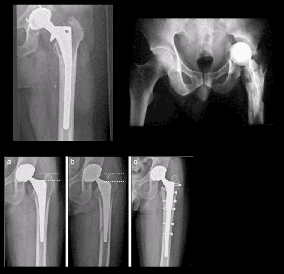
Most develop within the first year and require revision
Femoral stem may fracture (above left)
Postsurgical infection (above right)
Fracture adjacent to the prosthesis (stress riser)
Particle disease
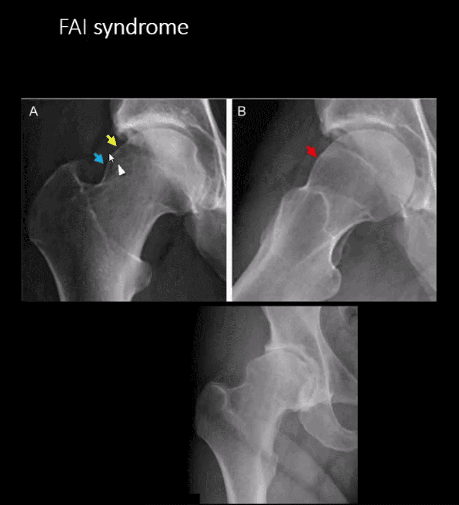
Femoroacetabular Impingement Syndrome
(FAI): abnormality of normal morphology of the hip leading to eventual� cartilage damage and premature DJD
Clinically:�hip/groin pain aggravated by sitting (e.g., hip flexed & externally rotated). Activity related pain on axial loading esp. with hip flexion (e.g., walking uphill)
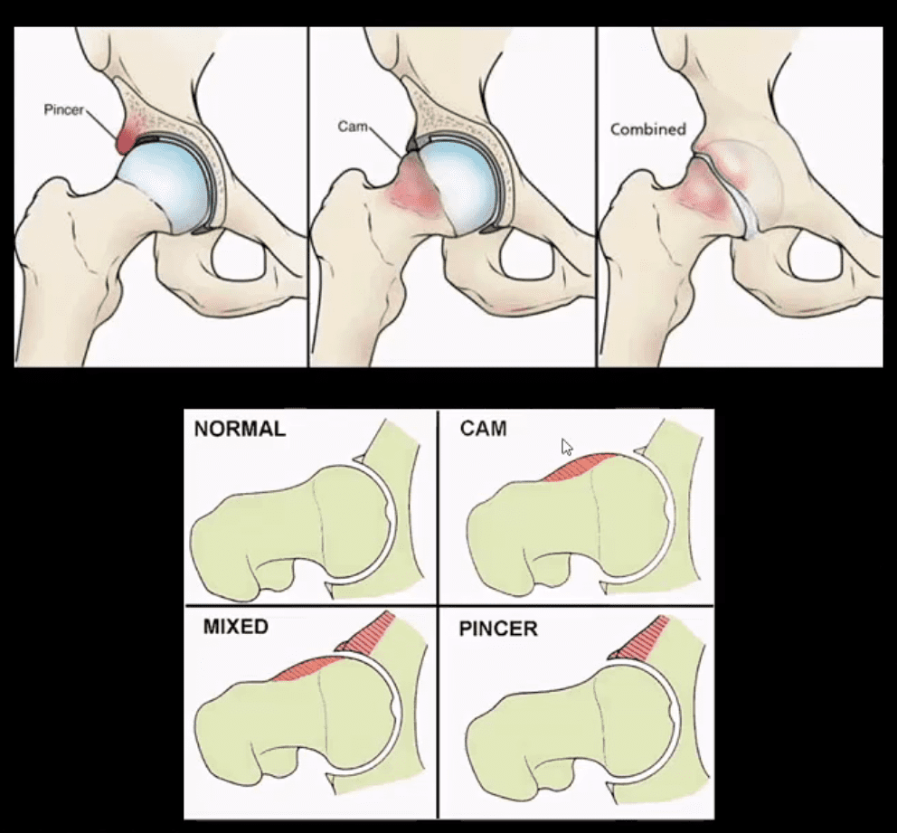
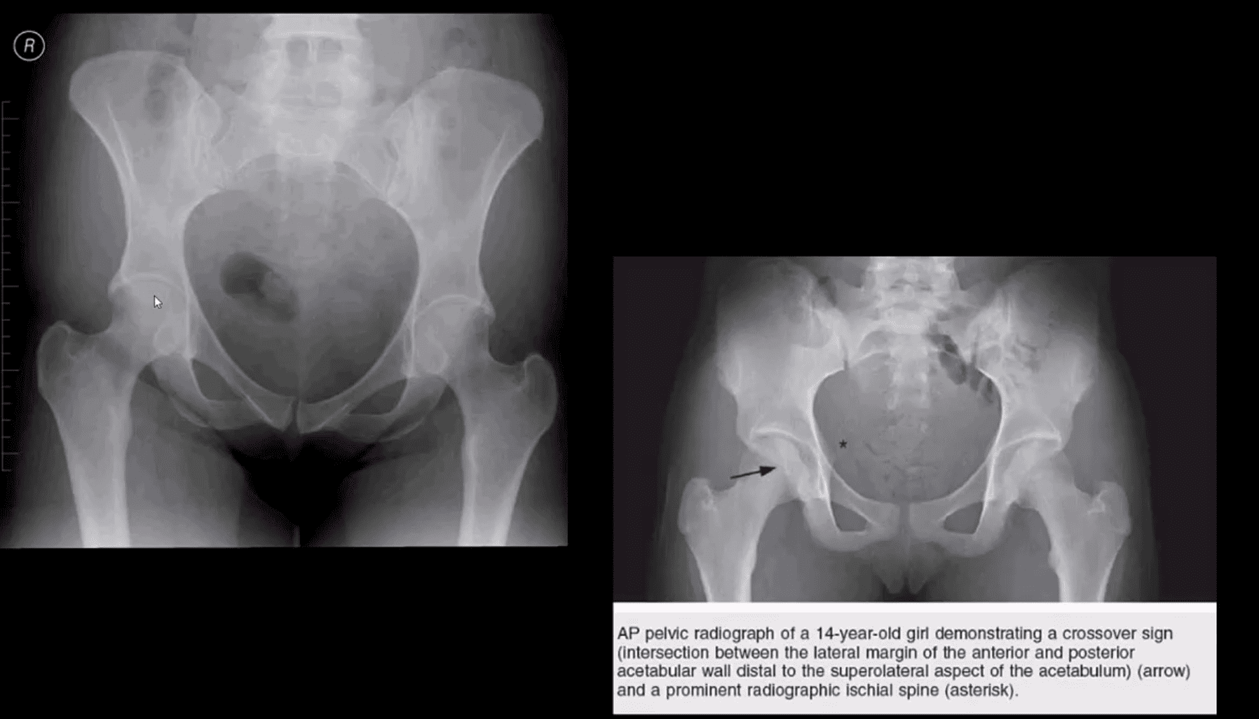
Pincer-type�acetabulum: > in middle age women potentially� many causes
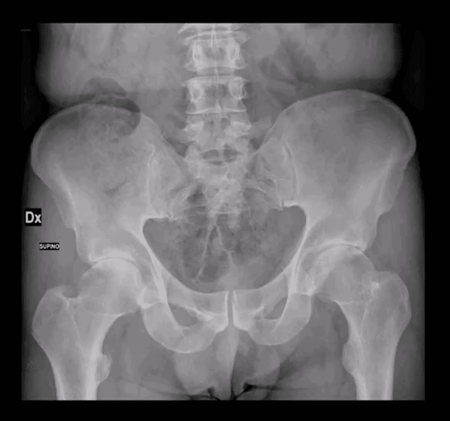
CAM-type deformity:�> in men in 20-50 m/c 30s
Mixed type (pincer-CAM) is most frequent
Up until the 90s, FAI was not well-recognized
FAI Syndrome
CAM-type FAI syndrome
Radiography can be a reliable Dx tool
X-radiography findings:�osseous bump on the lateral aspect of femoral head-neck junction. Pistol-grip deformity. Loss of normal head sphericity. Associated features: os acetabule, synovial herniation pit (Pit’s pit). Evidence of DJD in advanced cases
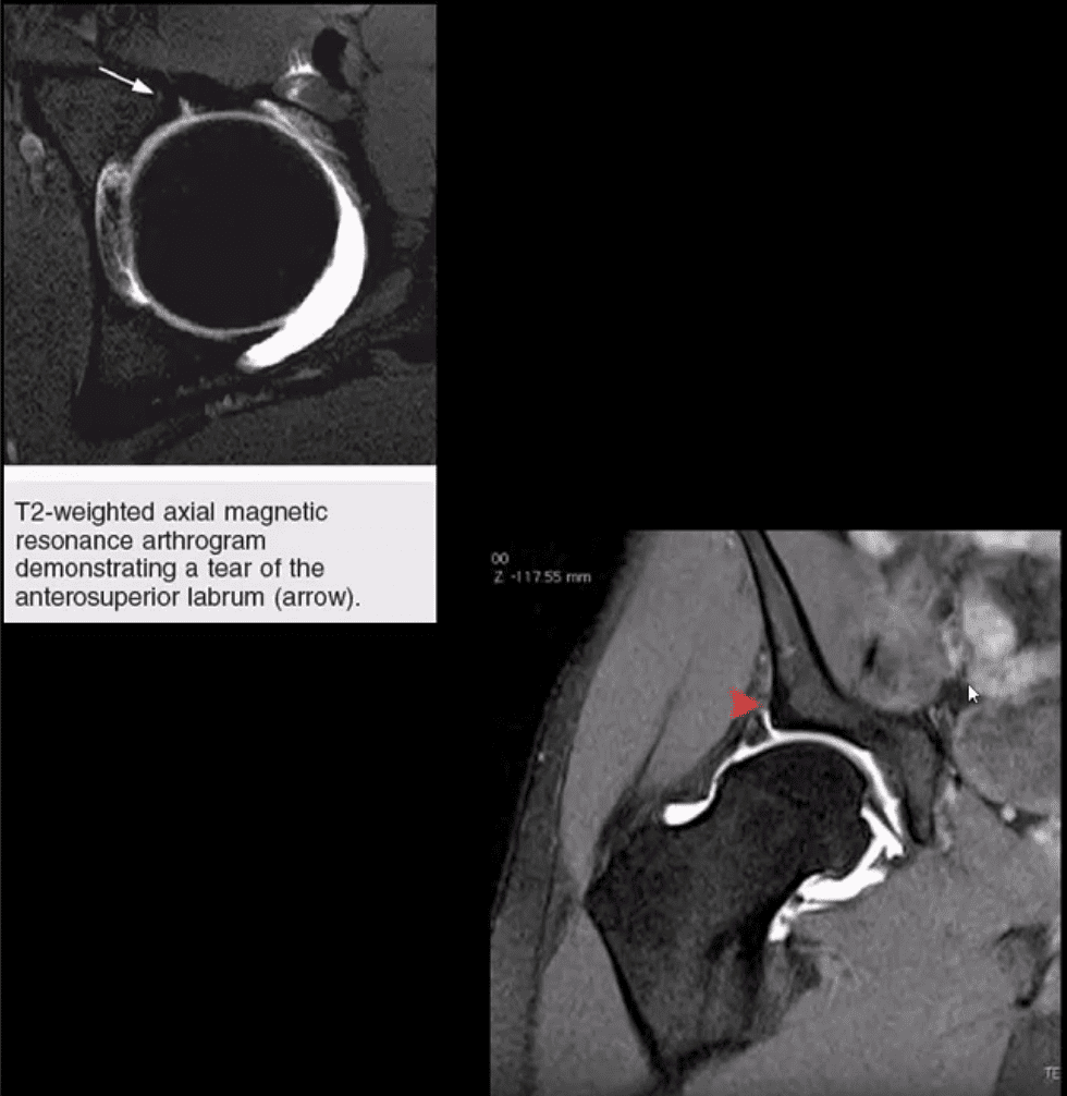
MRI and MR arthrography (most accurate Dx of labral tear) can aid the diagnosis of labral tear and other changes of FAI
Referral to the Orthopedic surgeon is necessary to prevent DJD progression and repair labral abnormalities. Late Dx may lead to irreversible changes of DJD
AP Pelvis: B/L CAM-type FAI syndrome
Pincer-Type FAI with Acetabula Over-Coverage
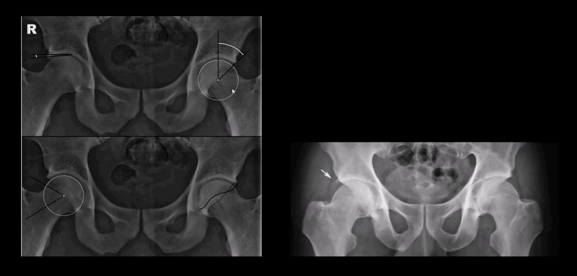
Key radiographic signs: “Cross-over sign” and abnormal center-edge and Alfa-angle evaluation methods
Dx of FAI
Center-edge angle (above the first image) and Alfa-angle (above the second image)
B/L CAM-type FAI with os acetabule�(above right image)
MR Arthrography
Labral tear and CAM-type FAI syndrome on axial (above left) and coronal T2 W (above right) MR arthrography
Note acetabula labral tear. Referral to an orthopedic surgeon is required. For more information:
The vast majority of clinically suspected bone Mets are found in the axial skeleton and proximal femurs/humeri
Radiography is the most cost-effective and readily available initial imaging tool to investigate bone Mets but often fails early metastatic detection
Tc99 bone scintigraphy is the most sensitive and cost-effective imaging modality to demonstrate metastatic foci
MR imaging may help� regional identification of bone Mets especially if x-radiography is unrewarding
Significant limitations of MRI: inability to perform a whole-body MRI scan
Cost and other contraindications such as cardiac pacemakers and cochlear implants may be another limiting factor
Marrow Based Neoplasms
Malignancy originating from the marrow cells are often referred to as “round-cell tumors.”
Multiple Myeloma (MM)
Lymphoma
Ewing’s sarcoma
The last two are less frequent than MM
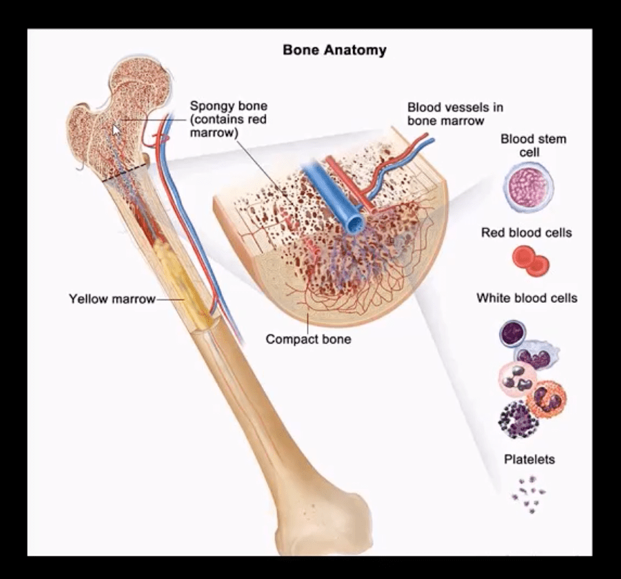
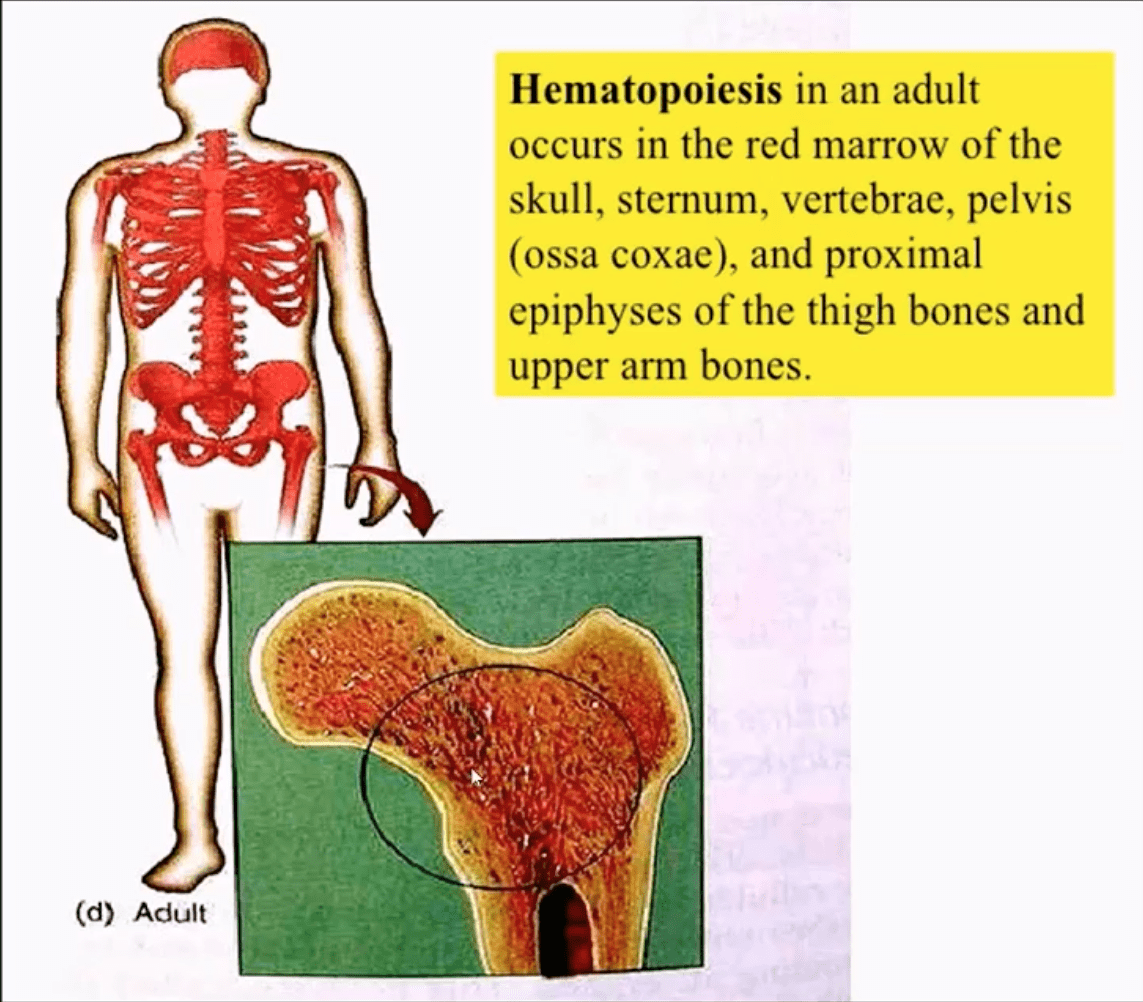
Red marrow in adults is in the axial skeleton and proximal femurs/humeri d/t gradual marrow “retraction” following the childhood
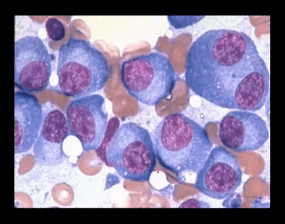
Note bone marrow biopsy histopathology specimen of MM with abnormal plasma cells replacing regular marrow residents (above image)
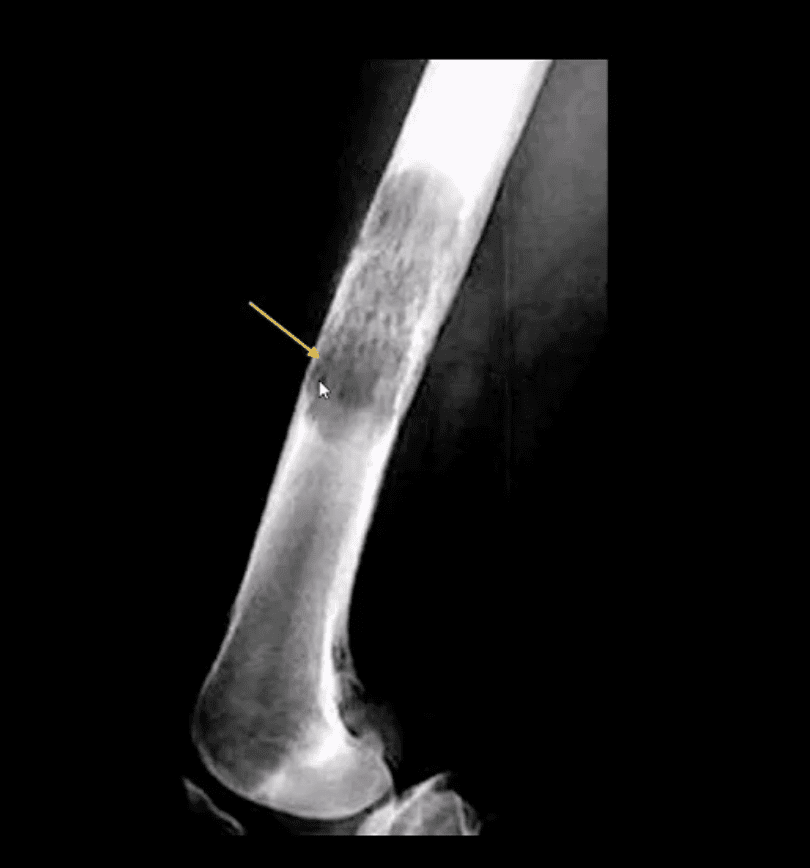
Multiple Myeloma (MM) is the most common primary bone neoplasm in adults>40s. Etiology is unknown, but many theories exist (e.g., genetic, environmental, radiation, chronic inflammation, MGUS)
MM: malignant proliferation of plasma cells >10% of red marrow, with subsequent replacement of normal marrow cells by myeloma cells and overproduction of monoclonal antibodies paraproteins (M protein) with heavy chains IgG (52%), IgA (21%), IgM (12%) and light chains kappa or lambda aka Bence-Jones proteins
Clinical Presentation of MM
MM is occasionally detected as unexplained anemia on routine blood studies for unrelated complaints
Common MSK symptoms: Bone pain/Pathologic fractures
Diagnostic imaging plays an essential role during the Dx of MM
Bone marrow aspiration biopsy, blood tests, and serum protein electrophoresis may be used
Imaging approach: bone pain is investigated with initial x-radiographs if radiographs are unrewarding MR imaging may help to reveal bone marrow abnormality. MRI is recommended as myeloma survey
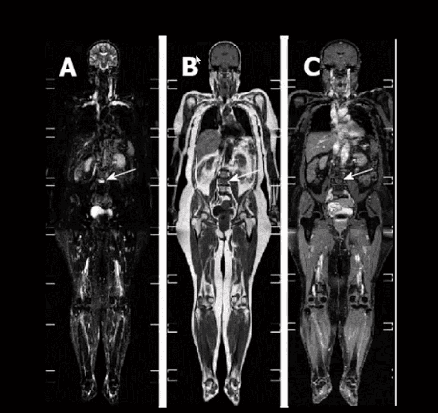
Currently, MRI protocol known as “whole body myeloma scan” consisting of T1, T2-fat suppressed, and T1+C coronal sequences can detect MM in the skull, spine, pelvis, ribs and femurs/humeri. This technique is much more superior to radiographic “skeletal myeloma survey.”
Tc99 bone scintigraphy is not typically used for MM because over 30% of MM lesions are “cold” or photopenic on radionuclide bone scan d/t highly lytic nature of MM with osteoclasts outpacing osteoblasts.
A radiographic skeletal survey is considered more sensitive than bone scintigraphy in MM
PET-CT scanning of MM is gaining popularity due to the high level of detection of multiple sites of MM
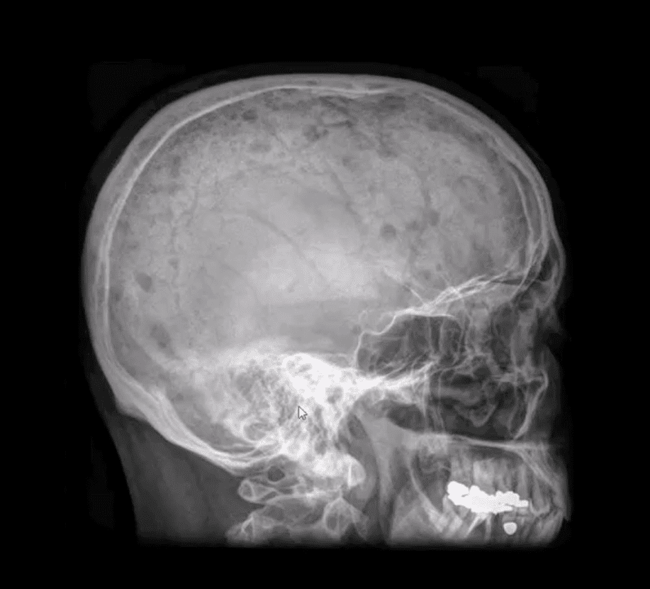
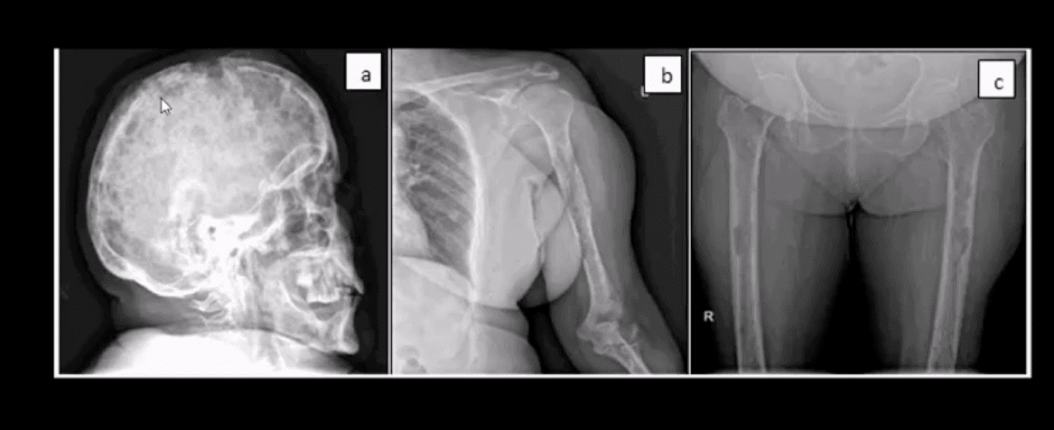
Radiographic Dx of MM: consists of identification of characteristically localized focal osteolytic “punched out” or “moth-eaten” lesions of variable sizes following the distribution of adults red marrow
Note rad abnormality is known as “raindrop skull” is characteristic of MM
Radiographic appearance of MM may vary from “punched out” round radiolucencies to “moth-eaten” or permeating osteolytic lesion producing endosteal scalloping (yellow arrow)
Pelvis and femurs are commonly affected by MM and present radiographically as round lytic punched out or moth-eaten lesions
N.B. Occasionally MM may pose radiographic dilemma by presenting as generalized osteopenia in the spine that can be difficult to differentiate from age-related osteoporosis
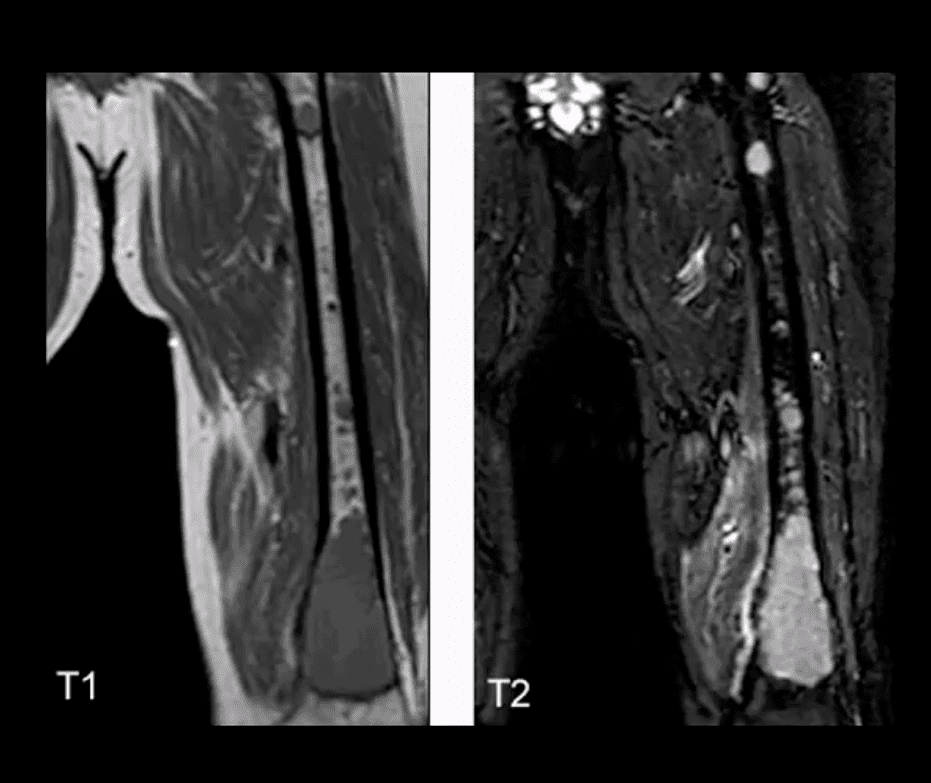
MR imaging of MM reveals� marrow changes with low signal on T1, a high signal on fluid-sensitive sequences and bright contrast enhancement on T1+C gad d/t increased vasculature and high activity of� MM cells
Example of full-body MRI of “whole body myeloma scan” with T2-fat suppressed (A), T1 (B) and T1+C (C) pulse sequences produced in coronal slices
Note multiple foci of bone marrow changes in the spine pelvis and femurs
Miscellaneous Neoplasms of the Spinal Column
Chordoma: is relatively uncommon but considered the m/c primary malignant neoplasm that only affects the spine. D/t slow growth is often misdiagnosed for a considerable length of time as LBP
Pathology: derives from malignant transformation of notochordal cells presented as mucoid, gelatinous mass containing physaliphorous cells
Demo:�M: F 3:1 (30-70S). 50%-sacrococcygeal, 35% spheno-occipital 15%-spine
Clinically: asymptomatic for a long time until non-specific LBP, changes in bladder & bowel, neurological signs are less common d/t midline “outward” growth & inferior to S1. Local invasion worsens prognosis. 60%-survive 5-years, 40%-10-years, Mets are delayed, poor prognosis d/t local invasion. >50% can be id. on DRE.
Imaging:�x-rays often tricky d/t overlying gas/feces. CT is >sensitive to id the bone mass and internal calcifications. MRI: T2 bight signal, T1 heterogeneously low and high d/t mucus/blood decomposition, MRI best detects local invasion and essential for care planning. Rx:� complete excision is often impossible d/t local vascular invasion.
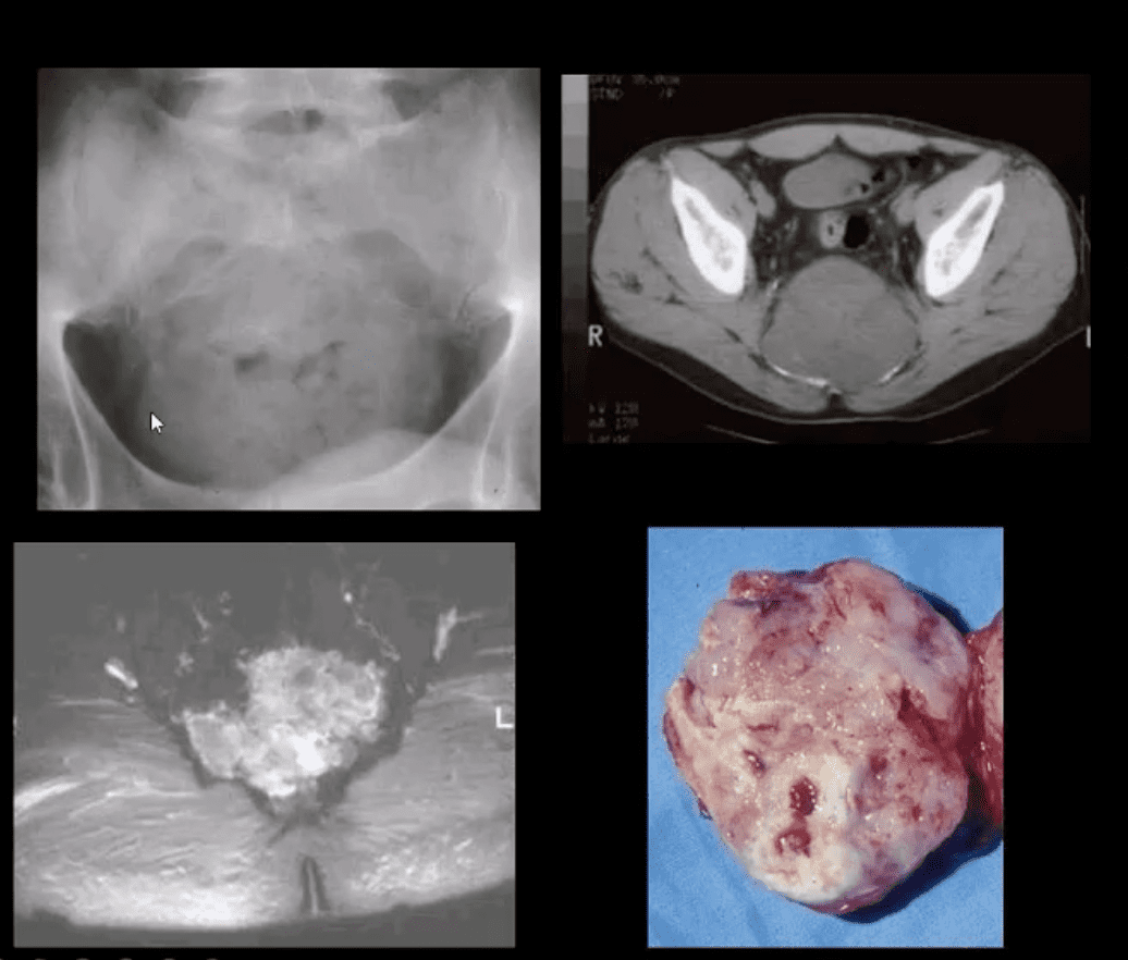
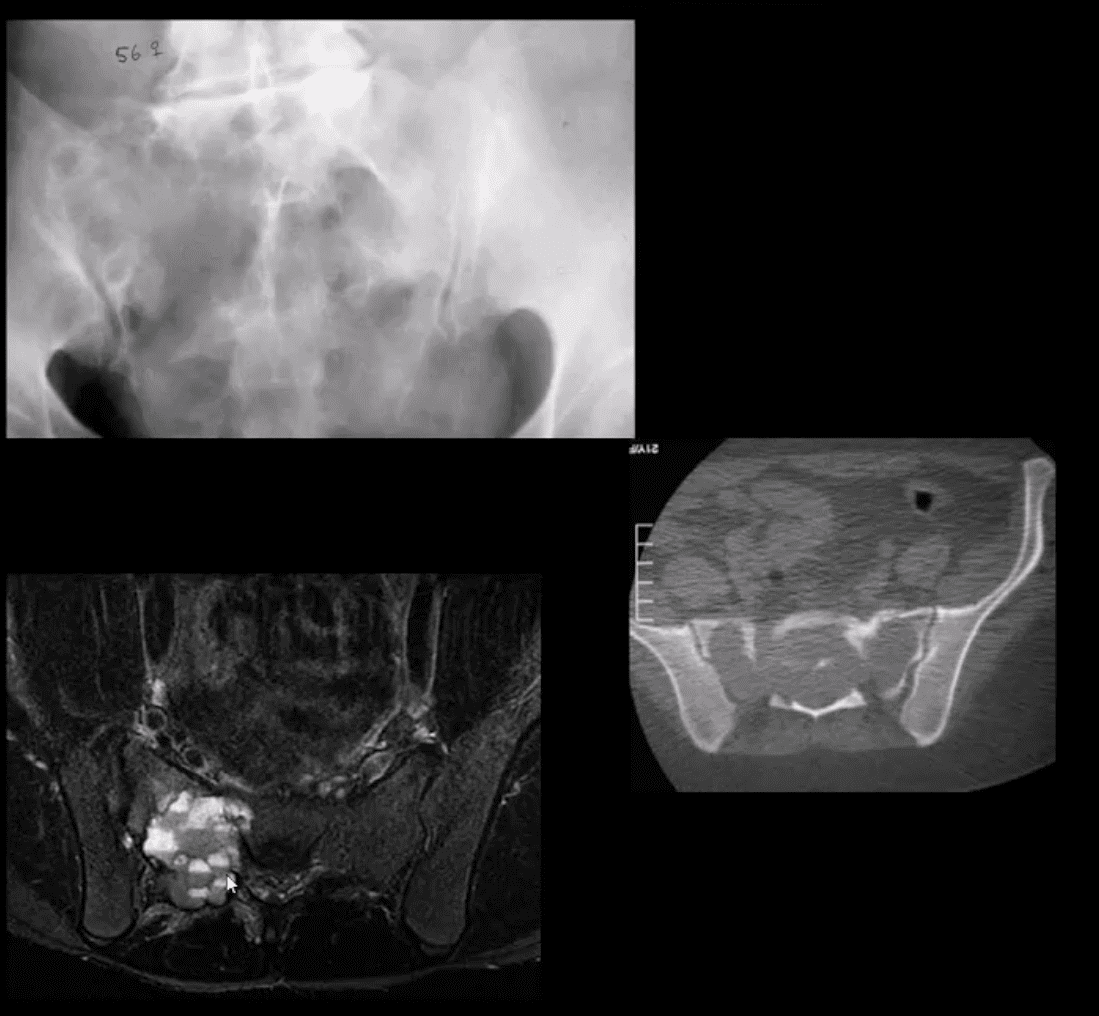
Giant cell tumor (GCT):�2nd most common primary sacral tumor. It is a histolgically benign neoplasm containing multinucleated Giant cells of Monocyte-Osteoclast origin
Imaging Dx:�x-radiography is the 1st step usually in response to complaints of LBP. Often challenging to id on x-rays d/t bowel gas/feces
Key rad feature: osteolytic expansile lesion noted by destruction of sacral arcuate lines. CT may id the lesion better. MRI is the modality of choice following x-rays. MRI: T1 low to intermediate signal. Heterogeneously high d/t edema with areas of low signal on T2 d/t blood degradation and fibrosis. Characteristic fluid-fluid levels may be noted especially if ABC develops within a GCT. Rx: operative. Prognosis is less favorable than GCT in long bones d/t lung Mets (deposits) in 13.7%
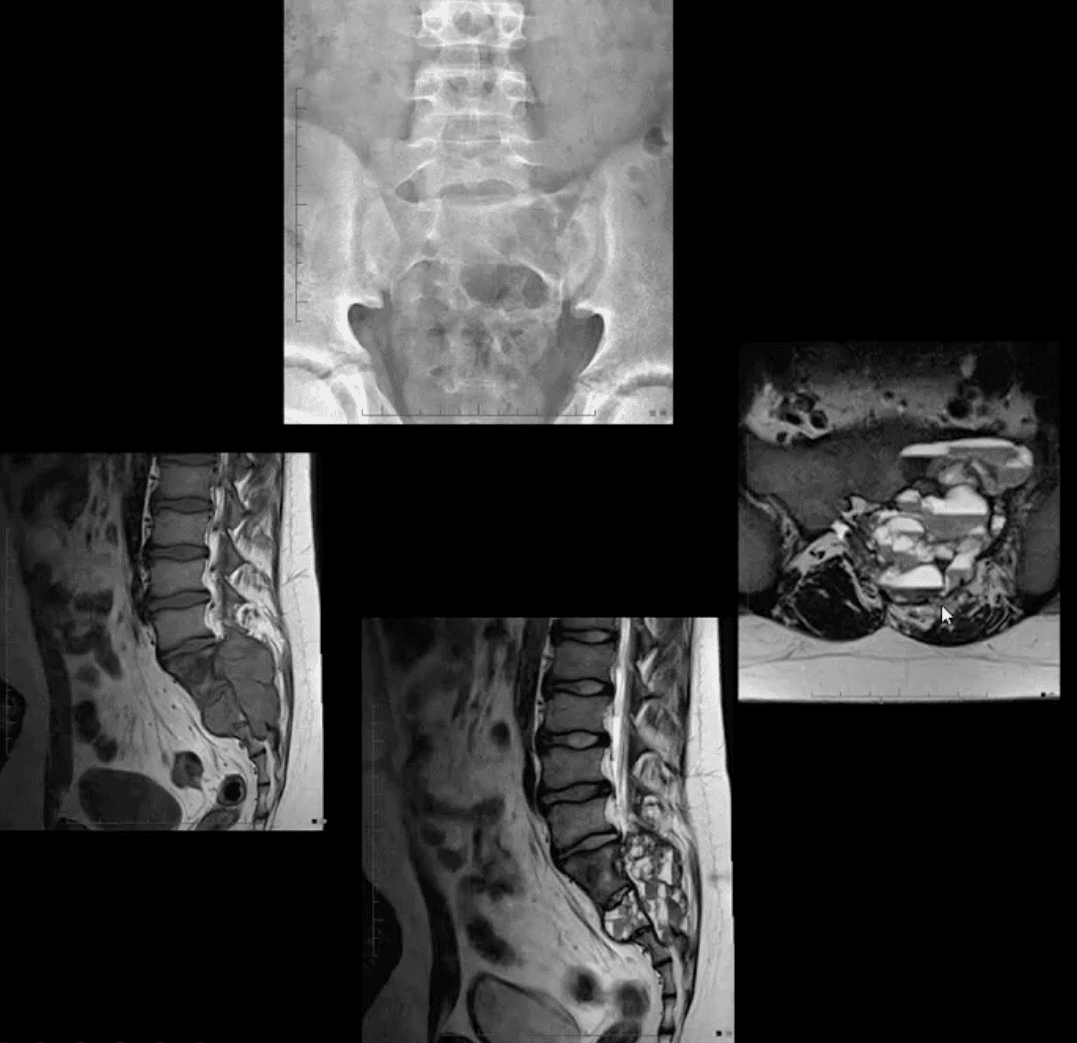
Aneurysmal Bone Cysts (ABC) are benign expansile tumor-like bone lesions (not a true neoplasm) composed and filled with numerous blood-filled channels. Thus the term “blood sponge.” ABC is m/c id in children and adolescents
Unknown etiology: trauma and pre-existing bone neoplasm (e.g., GCT) often reported. Clinically: pain that may be progressive d/t rapid nature of ABC expansion. In the spine, ABC m/c affects posterior elements and presented as expansile, soap-bubbly or lytic lesion.
DDx: can be broad, but Osteoblastoma and GCT are the top DDxs.
Imaging: x-rays demo expansile mass in posterior elements, CT is more sensitive than x-rays, MRI will demo characteristic fluid-fluid levels and mixed high and low signal d/t edema and blood decomposition/aging with some septations.
N.B. MRI fluid-fluid levels are not exclusive to ABC, and DDx includes GCT, osteoblastoma, telangiectatic osteosarcoma.
Rx: operative curettage and bone grafting, fibrosing agents. Recurrence 10-30%.
Metastatic Bone Disease (aka Mets) or “Secondaries.” Are the most common malignant bone neoplasms affecting the spine, aka spinal neoplasms (>70%) and the rest of the skeleton in adults.
5-Primaries are m/c involved:
Breast (16-37%)
Lung (12-15%)
Thyroid (4%)
Renal (3-6%)
Prostate (9-15%)
Spine, pelvis, proximal femurs & proximal humeri are m/c affected in that particular order of frequency
Thoracic & upper Lumbar spine considered the m/c site of spinal Mets
Pathophysiology & Etiology of Metastasis
Malignant cells a very good at evading immune detection and elimination
They gain�access to circulation expressing Vascular Endothelial Adhesion Molecules (e.g., integrines & selectins)
Once reaching their target organs, malignant cells stimulate the production of various vasogenic growth factors and by exiting blood vessels invade their target tissues
Lung, Liver, and Bone are particularly at risk due to the character of their blood supply
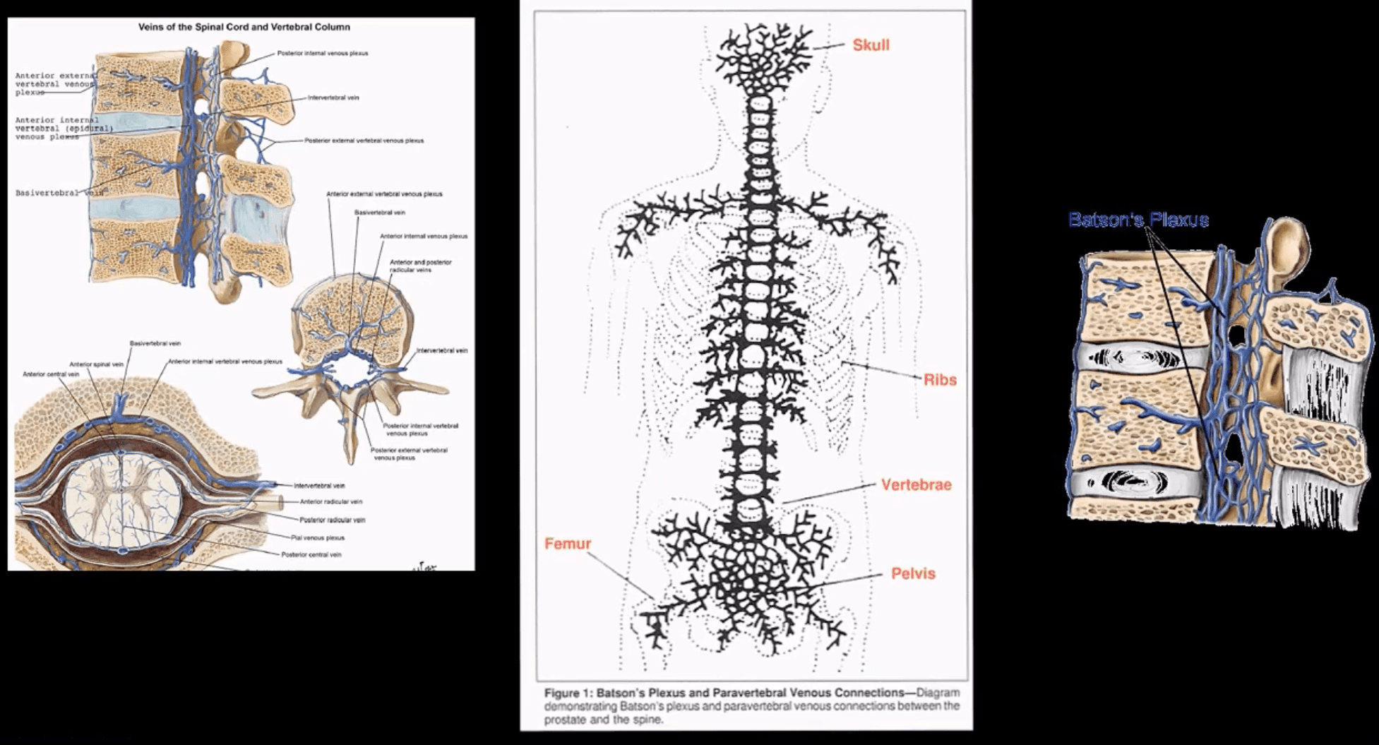
Baston venous plexus-is a network of valveless freely communicating� veins connecting axial skeleton/meninges and proximal femurs/humeri with abdomino-pelvic and thoracic cavities
The risk of Mets is increased during daily variations in the intra-abdominal and intra-thoracic pressure
In adults, the axial skeleton is involved in hematopoiesis, and it is particularly vulnerable to metastatic deposits via an abundant network of sinusoids within a spongy bone
The vast majority of bone Mets will be detected in the axial skeleton
Clinical Presentation
Back pain often mimicking “mechanical back pain” is the m/c and often misleading symptom
Chiropractors and other manipulators should be particularly aware of this dangerous pitfall.
Nocturnal pain or pain unresponsive to NSAID may be reported in more advanced cases
Advanced cases may also present with a neurological deficit due to pathologic vertebral fractures and spinal cord/nerves compression
Metastatic hypercalcemia may occasionally develop in severe cases and considered a medical emergency that potentially presents with confusion, muscle weakness, and renal signs
Imaging plays a significant role in the Dx and management of bone metastasis
Lab tests are of limited value, but hypercalcemia and alkaline phosphatase (Alk Phos) may be elevated
In some cases, a bone biopsy may be used to confirm bone Mets
When Bone Mets are Detected, Patients Prognosis is Significantly Worsened
Median survival:
Thyroid – 48 – months
Prostate – 40 – months
Breast – 24 – months
Renal Cell – may vary, can be as low as 6 – months
Lung – 6 – months
Imaging Diagnosis
Begins with radiography investigating a clinical complaint of back/bone pain
If radiographs are unrewarding or equivocal, unique imaging modalities are required
MRI may help to show marrow replacement by Mets foci but limited to specific regions
Tc99 radionuclide bone scan (scintigraphy) is considered one of the most sensitive and reliable imaging steps in evaluating bone Mets
Bone scintigraphy is good at detecting both lytic and blastic Mets
However, very aggressive/vascular osteolytic Mets and Multiple Myeloma often appear “cold” or photopenic on bone scan due to greater stimulation/activation of osteoclasts which “outpace” osteoblasts ability to uptake the radiopharmaceutical
CT scanning is an excellent modality to show bone destruction, but it is not widely used during bone Mets Dx especially if radiography, bone scintigraphy, and MRI provide adequate information about the process
CT scanning may be particularly helpful with delineation of pathological fractures
General Radiographic Features of Bone Mets
Osteolytic (lytic), osteoblastic (blastic) aka sclerotic Mets or misec Mets can be identified radiographically
However, it takes between 30-50% of lamella (cortical) bone and 50-75% of trabecular (cancellous) or spongy bone to be destroyed before it can be detected on plain film radiographs
This can make early radiographic detection of bone Mets very difficult, requiring particular imaging modalities (e.g., MRI)
Also, bowel gas/fecal matter and numerous soft tissue densities in the abdomino-pelvic and thoracic cavities may pose challenges of bone Mets detection
Different tumors often manifest with different metastatic appearance, depending on tumor activity and release of cytokines (IL6, IL11), endothelin 1 or other growth factors that will be responsible for either osteolytic, osteoblastic or mixed Mets
For example: purely lytic bone Mets are noted in Lung, Thyroid, and Renal cell CA (very vascular)
Breast CA may present with 60% of blastic Mets
Prostate CA presents with 90% of blastic Mets
Other blastic Mets may derive from urinary bladder, melanoma and GI adenocarcinomas
Sclerotic foci may also represent as previously treated primaries
Very vascular� Mets like Renal cell and Thyroid may present with markedly� lytic and expansile foci often called “blow out Mets.”
Mets found distal to elbows and knees (acro-metastasis) are commonly associated with Lung CA
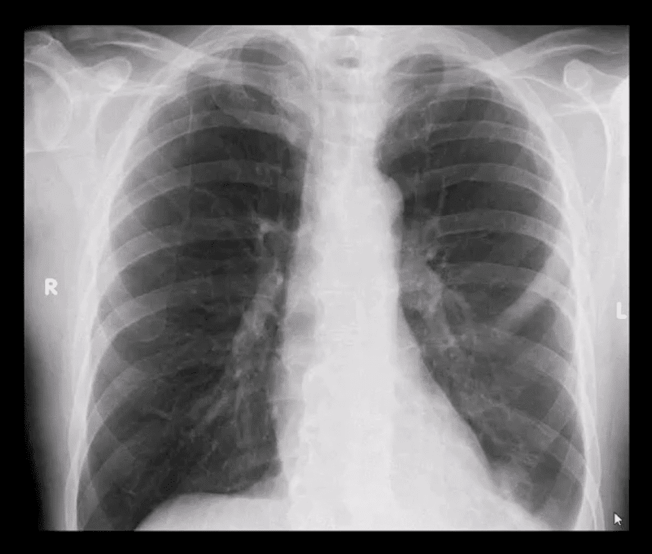
PA chest view of a routinely screened patient with a known Hx of Prostatic adenocarcinoma
Note sclerotic lesion identified in the left posterior Rib 5
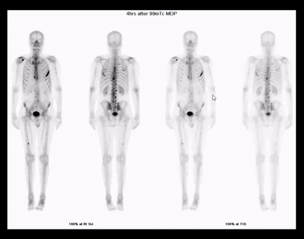
What imaging modality is required next?
Radionuclide bone scan should be suggested
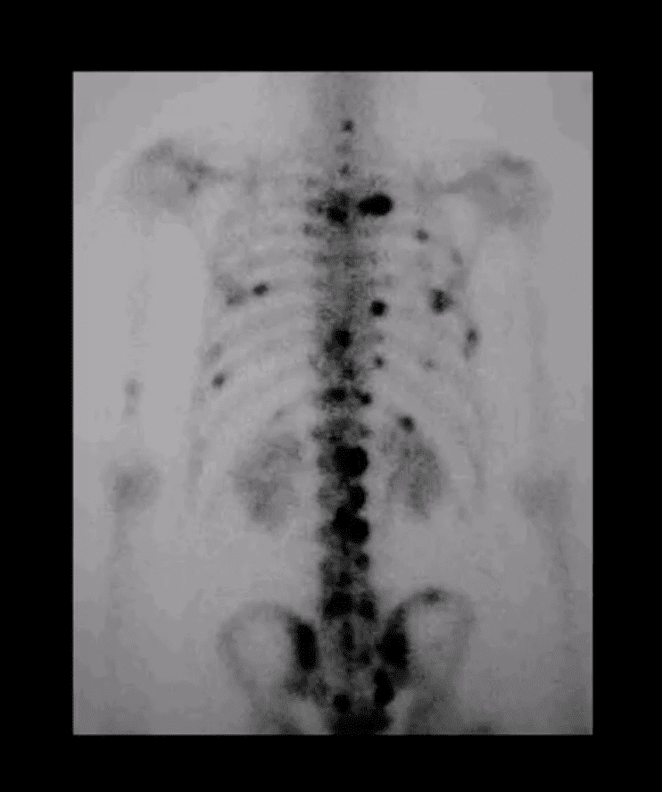
Multiple foci of high uptake of the Tc99 radiopharmaceutical
This is due to Mets and increased osteoblastic activity in the thoracic and lumbar spine, ribs and other sites of the skeleton
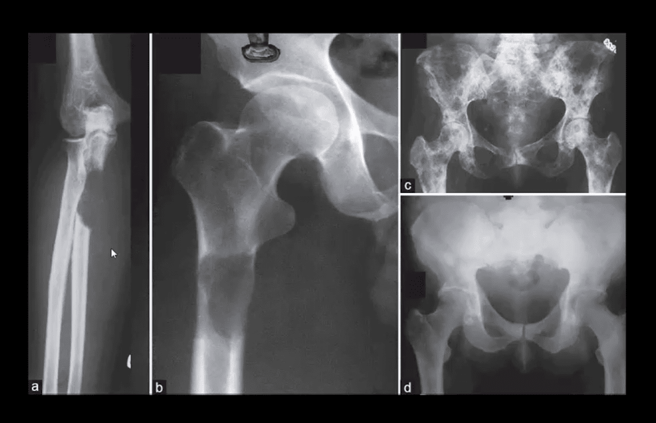
Comparison of purely lytic (a and b) versus blastic (d) and mixed (c) Mets
What primaries to consider?
Frog leg view of the hip
Clinical Dx: Prostatic adenocarcinoma
Note diffuse blastic Mets in the proximal femur
Hx: severe shoulder and arm pain unrelieved by rest
Rad DDx: Mets, Myeloma or less frequently Lymphoma
This classic DDx is used by the majority of Radiologists when aggressive osteolytic bone lesions are noted
The patient had a known Hx of Breast CA
A 51-year-old female with Breast CA
Large lytic destructive lesion in the distal femoral metaphysis characteristic of aggressive osteolytic Mets
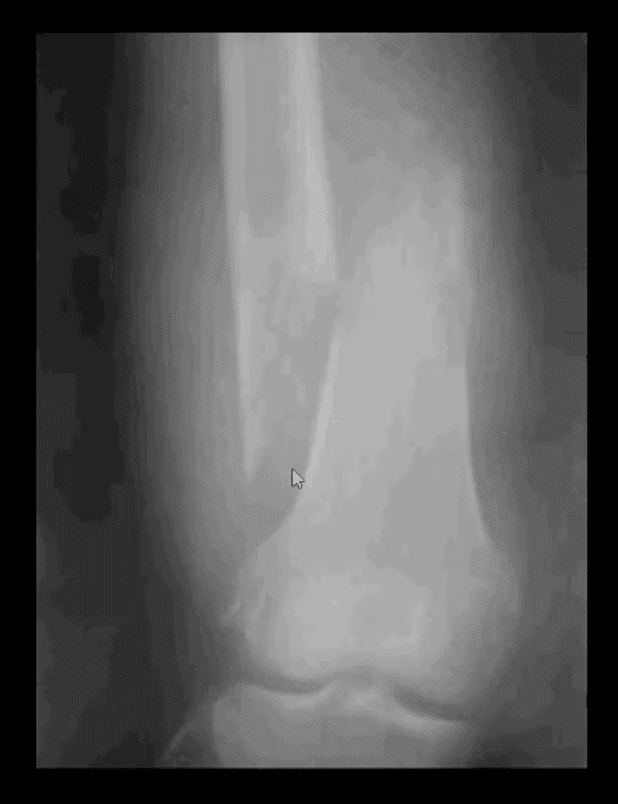
Sudden onset of severe leg pain and inability to stand in a 53-year-old female with Breast CA
Dx: Pathological fracture through the distal femoral shaft
Pathological Mets fractures in the spine and extremities are dreaded by most Oncologists due to higher association with severe complications and poor clinical prognosis
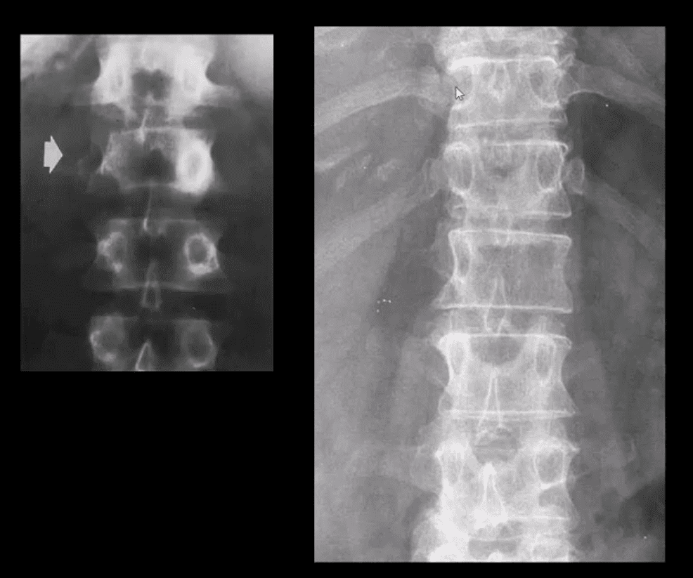
Radiographic Dx of vertebral Mets should be suspected if a “missing pedicle sign” aka “winking owl sign” is noted
DDx: pedicle agenesis (above left) shows hypertrophy and sclerosis of a contralateral pedicle d/t increased mechanical stress
Pedicle Mets are often thought of as the m/c initial site of spinal Mets
Vertebral Body Pathologic Fracture (VERTEBRA PLANA)
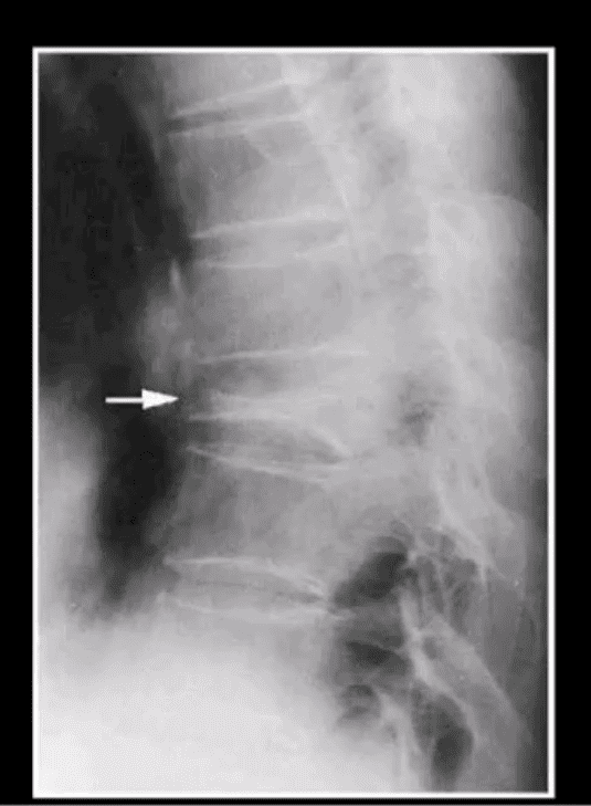
Isolated compression fracture at the T8 segment noted (above arrow)
The loss of the posterior and anterior height suggest an underlying pathologic condition for which the differential diagnosis includes:
Differentiating Pathological Fx of the vertebral body from an osteoporotic insufficiency Fx can be a significant challenge
Close inspection of the posterior body height is helpful but often not reliable
In metastasis, the posterior body is collapsed
In OSP, the posterior body may be maintained appearing more as anteriorly wedge fracture
MR imaging and/or radionuclide bone scan need to be performed
A skeletal radiographic survey may be used occasionally for the evaluation of bone Mets especially in well-established cases
It includes bilateral AP & lateral Thoracic and Lumbar views, AP pelvis, humeri, femurs, and the skull
Availability of special imaging has supplanted the use of skeletal radiographic survey
However, in a clinical practice skeletal radiographic study of Multiple Myeloma may still be used primarily if the diagnosis was previously established
Technetium-99 (99mTc) bone scintigraphy is very sensitive and cost-effective study:
For the detection/localization of Mets and often an assessment of their biologic activity and response to treatment
This modality is a well-established part of the workup for known as well as unknown primaries
It may also help with determination of lesions that will be most accessible and easy to biopsy
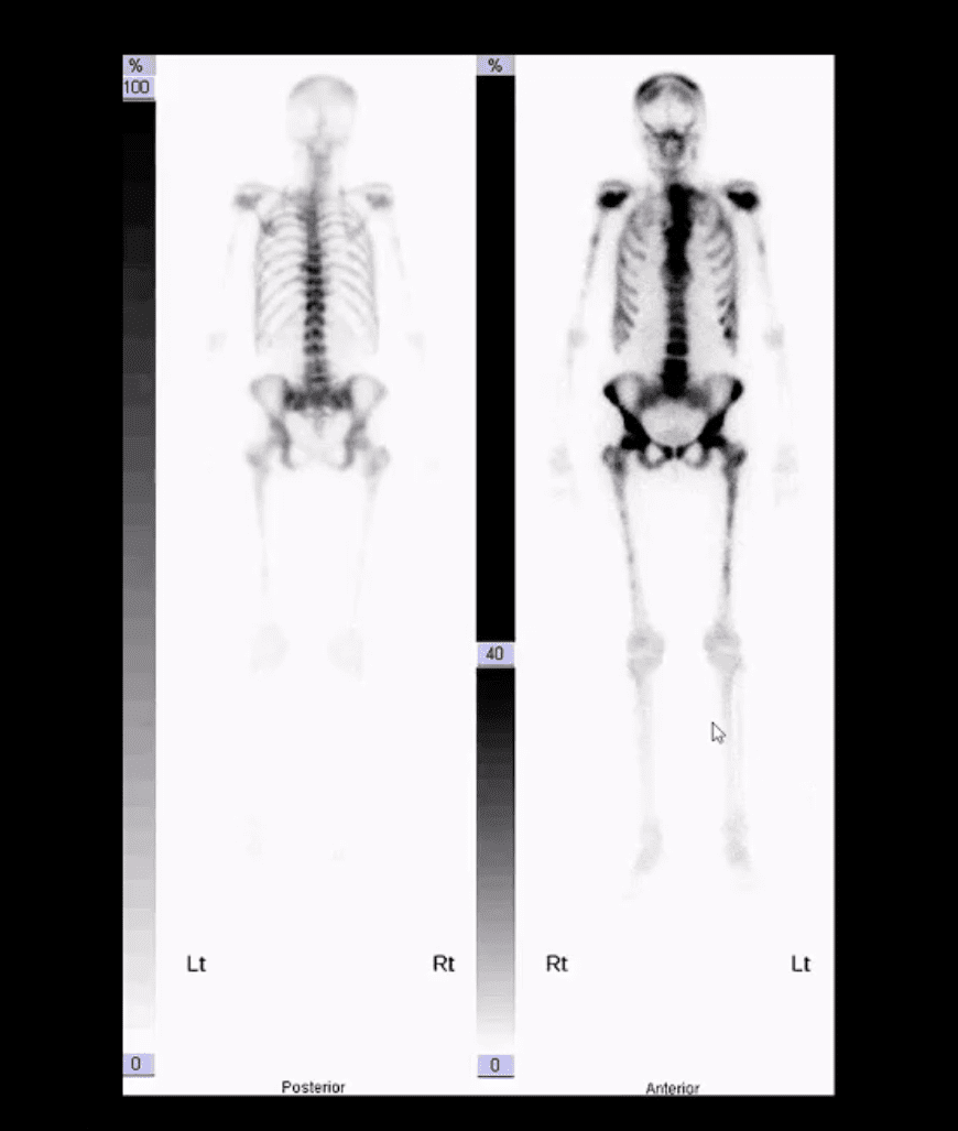
When the burden of Mets is significantly high as shown in the case above
The radiotracer uptake is being almost entirely taken in by metastatic lesions
No material is left for the kidneys to excrete
This is known as a “super scan”
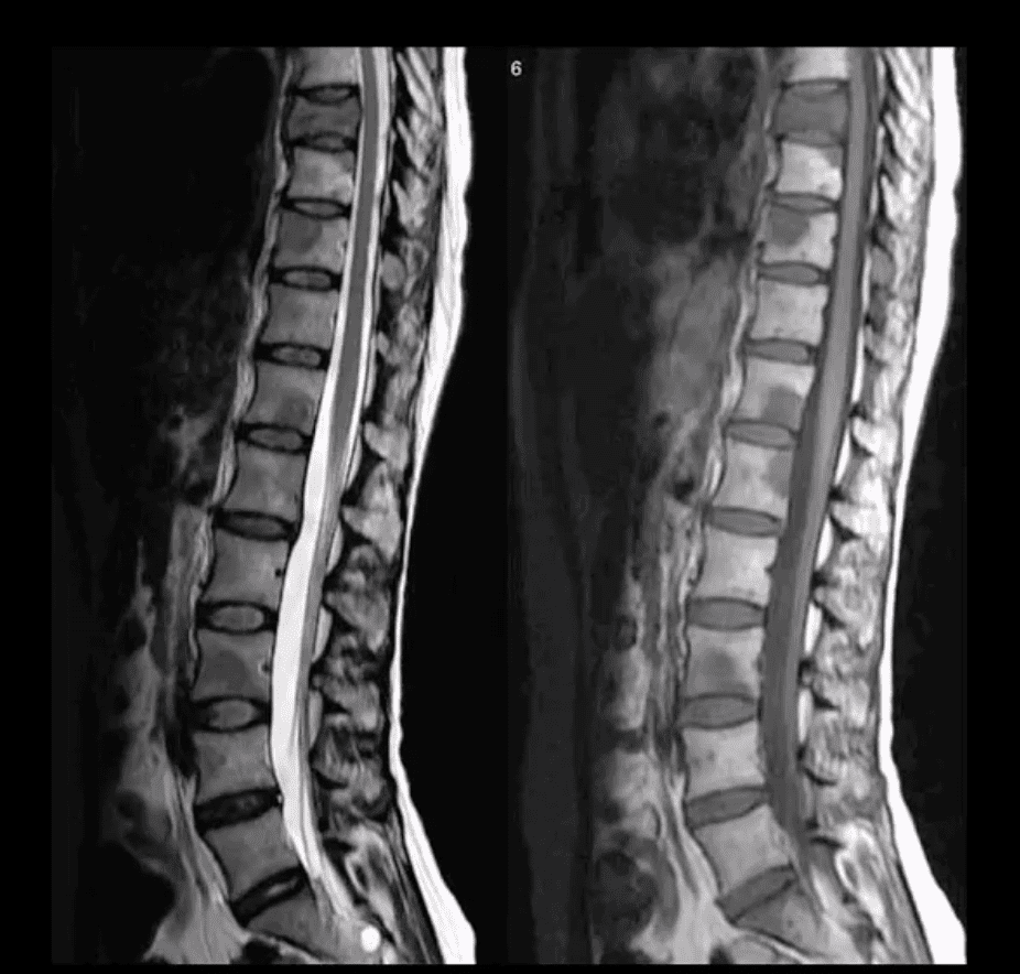
Sagittal Lumbar and Lower Thoracic MRI. Multiple metastasis are noted on T1 (above right) and T2 (above left)� WI as hypointense foci of marrow replacement of the vertebral bodies in a patient with Hx of Prostate CA
MR imaging protocol with T1, T2, and T1+C gad can be used in many cases if x-radiography is unrewarding or questionable
�MRI can reveal bone marrow changes due to bone marrow replacement by Mets and surrounding edema
Typically blastic Mets appear as abnormally decreased signal intensity (hypointense) lesions on T1 and T2 pulse sequences
Purely lytic Mets often appear as hypo-intense on T1 and hype-intense on T2
Increased gadolinium uptake may also be evident on T1+C fat suppressed sequence d/t increased vascularity of malignant foci especially in very aggressive vascular neoplasms
IFM's Find A Practitioner tool is the largest referral network in Functional Medicine, created to help patients locate Functional Medicine practitioners anywhere in the world. IFM Certified Practitioners are listed first in the search results, given their extensive education in Functional Medicine