The Q or quadriceps angle is a measurement of pelvic width that is believed to contribute to the risk of sports injuries in women athletes. Can non-surgical therapies and exercises help rehabilitate injuries?
Quadriceps Q – Angle Injuries
The Q angle is the angle where the femur/upper leg bone meets the tibia/lower leg bone. It is measured by two intersecting lines:
One from the center of the patella/kneecap to the anterior superior iliac spine of the pelvis.
The other is from the patella to the tibial tubercle.
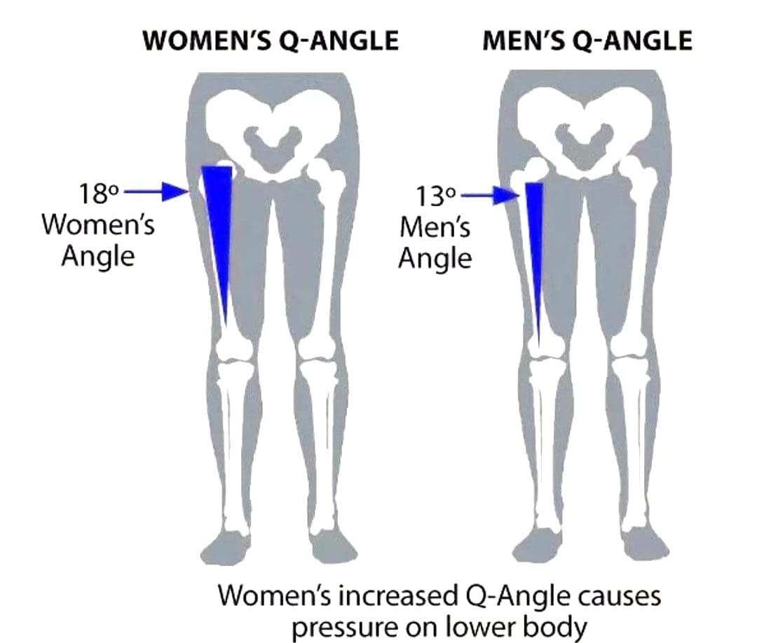
On average the angle is three degrees higher in women than men.
Women have biomechanical differences that include a wider pelvis, making it easier to give birth. However, this difference can contribute to knee injuries when playing sports, as an increased Q angle generates more stress on the knee joint, as well as leading to increased foot pronation.
Injuries
Various factors can increase the risk of injury, but a wider Q angle has been linked to the following conditions.
Patellofemoral Pain Syndrome
An increased Q angle can cause the quadriceps to pull on the kneecap, shifting it out of place and causing dysfunctional patellar tracking.
With time, this can cause knee pain (under and around the kneecap), and muscle imbalance.
Foot orthotics and arch supports could be recommended.
Some researchers have found a link, while others have not found the same association. (Wolf Petersen, et al., 2014)
Chondromalacia of the Knee
This is the wearing down of the cartilage on the underside of the kneecap.
An increased Q angle can be a factor that increases stress and causes the knee to lose its stability.
However, this remains controversial, as some studies have found no association between the Q angle and knee injuries.
Chiropractic Treatment
Strengthening Exercises
ACL injury prevention programs designed for women have resulted in reduced injuries. (Trent Nessler, et al., 2017)
The vastus medialis obliquus or VMO is a teardrop-shaped muscle that helps move the knee joint and stabilize the kneecap.
Strengthening the muscle can increase the stability of the knee joint.
Strengthening may require a specific focus on muscle contraction timing.
Closed-chain exercises like wall squats are recommended.
Glute strengthening will improve stability.
Stretching Exercises
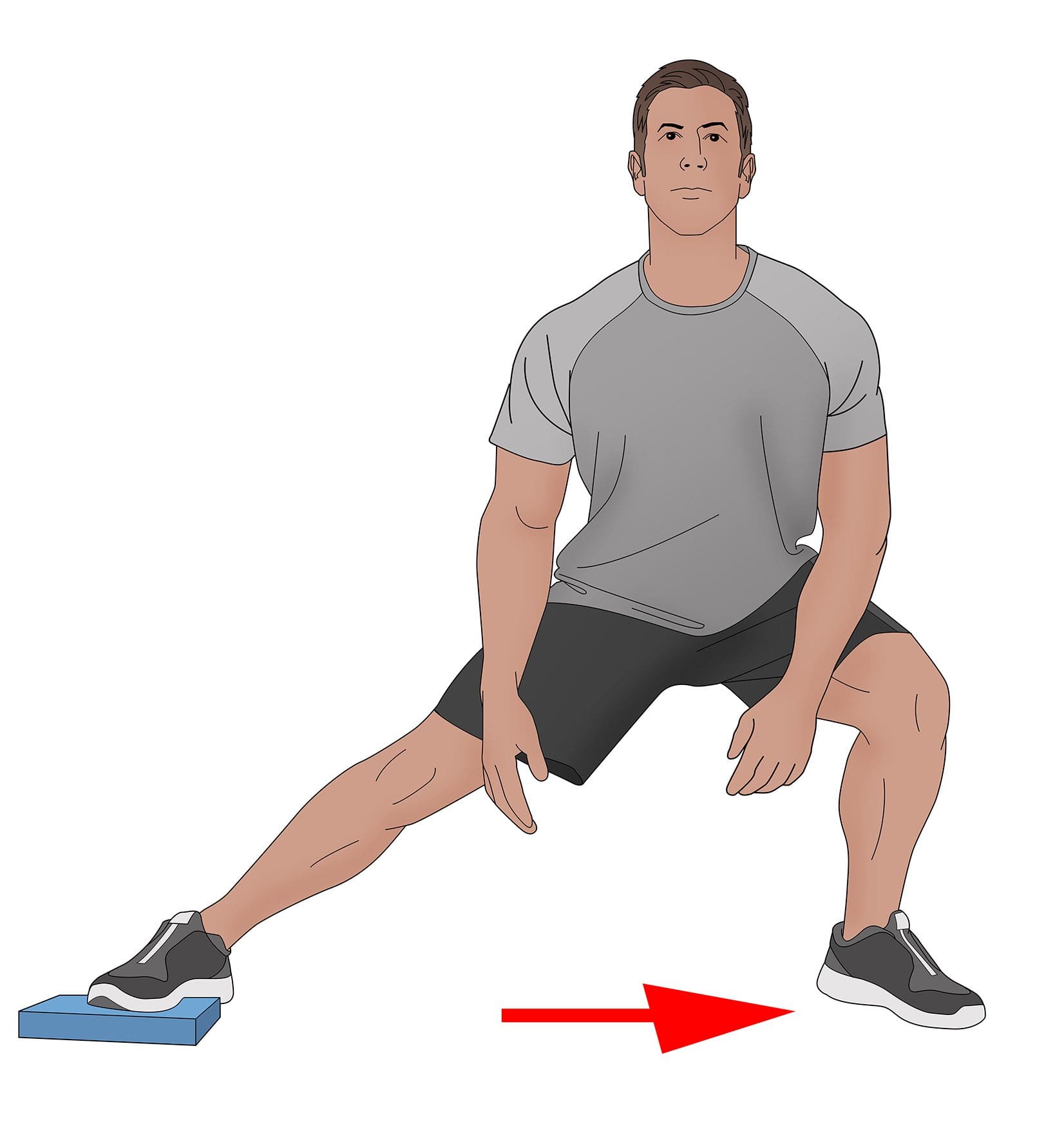
Stretching tight muscles will help relax the injured area, increase circulation, and restore range of motion and function.
Muscles commonly found to be tight include the quadriceps, hamstrings, iliotibial band, and gastrocnemius.
Foot Orthotics
Custom-made, flexible orthotics decrease the Q angle and reduce pronation, relieving the added stress on the knee.
A custom orthotic ensures that the foot and leg dynamics are accounted for and corrected.
Motion-control shoes can also help correct overpronation.
Knee Rehabilitation
References
Khasawneh, R. R., Allouh, M. Z., & Abu-El-Rub, E. (2019). Measurement of the quadriceps (Q) angle with respect to various body parameters in young Arab population. PloS one, 14(6), e0218387. https://doi.org/10.1371/journal.pone.0218387
Petersen, W., Ellermann, A., Gösele-Koppenburg, A., Best, R., Rembitzki, I. V., Brüggemann, G. P., & Liebau, C. (2014). Patellofemoral pain syndrome. Knee surgery, sports traumatology, arthroscopy: Official journal of the ESSKA, 22(10), 2264–2274. https://doi.org/10.1007/s00167-013-2759-6
Vaienti, E., Scita, G., Ceccarelli, F., & Pogliacomi, F. (2017). Understanding the human knee and its relationship to total knee replacement. Acta bio-medica : Atenei Parmensis, 88(2S), 6–16. https://doi.org/10.23750/abm.v88i2-S.6507
Mitani Y. (2017). Gender-related differences in lower limb alignment, range of joint motion, and the incidence of sports injuries in Japanese university athletes. Journal of Physical Therapy Science, 29(1), 12–15. https://doi.org/10.1589/jpts.29.12
Nessler, T., Denney, L., & Sampley, J. (2017). ACL Injury Prevention: What Does Research Tell Us? Current reviews in musculoskeletal medicine, 10(3), 281–288. https://doi.org/10.1007/s12178-017-9416-5
A nerve becomes pinched/compressed when added pressure is placed on it by surrounding structures that can include muscles, bones, ligaments, tendons, or a combination. This injures and damages the nerve causing function problems and symptoms and sensations in that area or other parts of the body that are supplied by that nerve. Medical practitioners refer to this as nerve compression or entrapment. Although compressed nerves are more commonly associated with the neck, arms, hands, elbows, and lower back, any nerve in the body can experience irritation, spasms, inflammation, and compression. The causes and treatment of a compressed nerve in the knee.
Compressed Nerve In The Knee
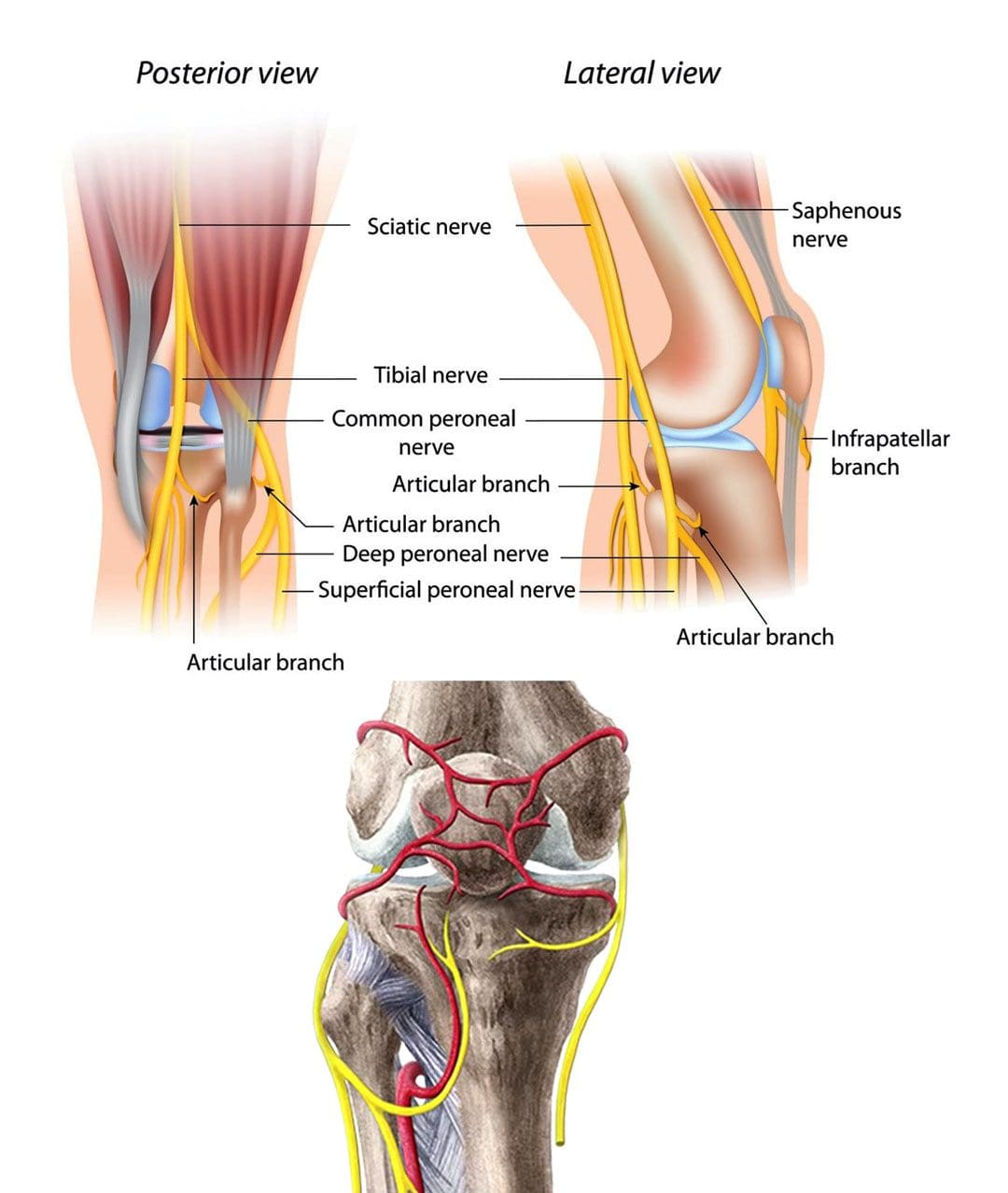
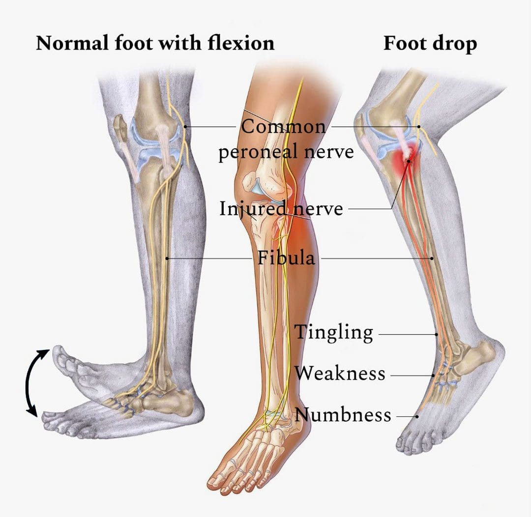
There’s only one nerve that goes through the knee that has an increased risk of getting compressed. It’s a branch of the sciatic nerve called the peroneal nerve. The nerve goes around the outside of the knee before traveling down the outside of the lower leg. At the bottom of the knee, it lies between the bone and skin, making it vulnerable to irritation or compression by anything that can put pressure on the outside of the knee.
Causes
Traumatic injuries over time can lead to pressure on the nerve from inside the knee. Common causes of a compressed nerve in the knee include:
Frequently Crossing Legs
Compression by the opposite knee, while the legs are crossed is the most common cause.
Knee Brace
A too-tight or strong brace can compress the leg and nerve.
Thigh-High Compression Stockings
Designed to maintain pressure on the legs, if too tight these stockings can compress the nerve.
Squatting Posture For Long Periods
This position places pressure on the side of the knee.
Fractures
A fracture of the large lower leg bone/tibia or sometimes the small bone/fibula near the knee can entrap the nerve.
Lower Leg Cast
The portion of the cast around the knee can be tight and compress the nerve.
Tell the doctor if a cast or brace feels tight or is causing numbness or pain in the leg.
Knee-High Boots
The top of a boot can land right below the knee and be too tight pinching the nerve.
Knee Ligament Injury
The nerve can become compressed due to bleeding or inflammation from an injured ligament.
Knee Surgery Complications
This is rare, but the nerve can inadvertently get pinched during knee replacement surgery or an arthroscopic procedure.
Prolonged Bed Rest
When lying down the legs tend to rotate outward and the knees flex.
In this position, the mattress can place pressure on the nerve.
Tumors or Cysts
Tumors or cysts can develop right on top or next to a nerve irritating and compressing the area.
Abdominal or Gynecologic Surgery
The equipment used to keep the legs rotated outward and the knees flexed for gynecologic and abdominal surgeries can compress the nerve.
Symptoms
The peroneal nerve supplies sensation and movement to the outside of the lower leg and the top of the foot. When compressed, it becomes inflamed, which causes the symptoms of a compressed nerve. Usually, only the lining/myelin sheath around the nerve is what gets injured. However, when the nerve gets damaged, the symptoms are similar but more severe. Common symptoms include:
Weakness that limits the ability to lift the foot toward the leg aka dorsiflexion.
This causes dragging the foot when walking.
The ability to turn the foot outward and extend the big toe is also affected.
Symptoms can be felt on the outside of the lower leg and on the top of the foot and include:
Tingling or pins and needles sensations.
Numbness.
Loss of sensation.
Pain.
Burning.
For individuals that have had a pinched nerve for two or more weeks, the muscles supplied by the nerve can begin to waste away or atrophy.
Symptoms can be intermittent or continuous depending on the cause.
The other common cause is a pinched nerve in the lumbar/lower spine.
When this is the cause, sensations, and pain will present in the lower back or the back and outside of the thigh.
Diagnosis
A doctor will look at medical history and perform an examination to make a diagnosis, determine the cause, and lay out a personalized treatment plan. The nerve in the knee can be felt as it travels around the top of the tibia, so a doctor may tap on it. If there is shooting pain down the leg, a pinched nerve may be present. Tests a doctor may order can include:
Knee X-ray
Shows any bone fractures or abnormal masses.
Knee MRI
Can confirm the diagnosis
Shows masses within the nerve.
Shows details of fractures or other problems in the bones.
Electromyogram – EMG
Tests electrical activity in the muscles.
Nerve Conduction Test
Tests the signal speed of the nerve.
Treatment
Treatment is aimed at reducing pain and improving mobility.
Over-the-Counter Pain Medication
OTC medication can reduce inflammation and improve symptoms short term.
Ice and Heat
Applying either heat or ice for 15 to 20 minutes at a time can provide relief from the symptoms.
An ice pack can make symptoms worse if it adds more pressure on the nerve.
Chiropractic and Physical Therapy
Chiropractic and physical therapy can release the compressed nerve, realign the structures, strengthen the muscles, and provide gait training.
Orthotic Boot
If walking gait is affected because the foot cannot bend, an orthotic boot can help.
This is a support that maintains the foot in a neutral position to walk normally.
Corticosteroid Injection
A corticosteroid injection can reduce inflammation and relieve pressure on the nerve.
Surgery
The nerve can suffer permanent damage if it has been pinched for a long time.
If that happens, surgery cannot repair the damage.
A doctor can perform surgery to correct a fracture, tumor, or other invasive problem causing a compressed nerve.
If conservative treatment doesn’t work, a peroneal nerve decompression procedure can be done to remove the pressure.
If surgery is needed, symptoms can disappear immediately, but it takes around four months to recover and rehabilitate.
Injury Rehabilitation
References
Krych, Aaron J et al. “Is peroneal nerve injury associated with worse function after knee dislocation?.” Clinical orthopedics and related research vol. 472,9 (2014): 2630-6. doi:10.1007/s11999-014-3542-9
Lezak B, Massel DH, Varacallo M. Peroneal Nerve Injury. [Updated 2022 Nov 14]. In: StatPearls [Internet]. Treasure Island (FL): StatPearls Publishing; 2023 Jan-. Available from: https://www.ncbi.nlm.nih.gov/books/NBK549859/
Soltani Mohammadi, Sussan, et al. “Comparing the squatting position and traditional sitting position for ease of spinal needle placement: a randomized clinical trial.” Anesthesiology and pain medicine vol. 4,2 e13969. 5 Apr. 2014, doi:10.5812/aapm.13969
Stanitski, C L. “Rehabilitation following knee injury.” Clinics in sports medicine vol. 4,3 (1985): 495-511.
Xu, Lin, et al. Zhongguo gu Shang = China Journal of Orthopedics and Traumatology vol. 33,11 (2020): 1071-5. doi:10.12200/j.issn.1003-0034.2020.11.017
Yacub, Jennifer N et al. “Nerve injury in patients after hip and knee arthroplasties and knee arthroscopy.” American Journal of physical medicine & Rehabilitation vol. 88,8 (2009): 635-41; quiz 642-4, 691. doi:10.1097/PHM.0b013e3181ae0c9d
Automobile accidents and collisions can cause knee and ankle injuries in various ways. Automobile crashes are considered high-energy collisions versus slip and fall traumas which are generally low-energy. However, a 30mph or under-collision can have serious and detrimental effects on the knees and ankles. The sudden forces can cause the knees to collide with the dashboard or push the feet and legs into the body, generating intense pressure and compressing the bones, muscles, and ligaments damaging soft tissues and bone structures from the impact. The Injury Medical Chiropractic and Functional Medicine Clinic Team can rehabilitate, realign, strengthen, and restore function to individuals with minor to severe auto collision injuries.
Knee and Ankle Injuries
Musculoskeletal motor vehicle crash/collision injuries affect the body’s movement. The impact can pull, tear, crush, and smash bones, muscles, tendons, ligaments, discs, and nerves. These injuries restrict the range of motion and can cause pain and sensation symptoms. The National Accident Sampling System reports 33% of injuries sustained during vehicle collisions are to the lower extremities.
Despite the knees and ankles having soft tissues that absorb and distribute the energy’s impact, the forces from the collision often happen instantly and unexpectedly, causing the individual to tense up, which overwhelms the structures.
Even panic stepping on the brake pedal can cause injury to the ankle and foot.
A passenger’s reflex of trying to resist forces can experience foot, ankle, and knee injuries from bracing off the vehicle’s floorboard.
Automobile collisions can cause strains, sprains, fractures, and dislocations.
Torn, Strained, or Sprained Knee
If the foot becomes planted on the floorboard while the body continues to move forward or sideways, the force can travel into the knee, causing twisting or shearing.
Depending on the injury type, the impact strength can damage different ligaments.
The ligaments resist forces that push the knee inwards/medially and outwards/laterally and slightly resist rotational forces.
When any of these ligaments are damaged, swelling, pain, and limited ranges of motion can result.
Putting weight on the affected leg can be difficult.
In some cases, the ligaments tear completely, necessitating surgical repair.
Once the individual can engage in mild activity, they can begin a rehabilitation program to restore function.
Recovery times vary based on the location and severity of the injury.
Fractured Knee or Ankle
When a fracture occurs in a joint, like the knees or ankles, surgical procedures may be necessary to repair the broken bone/s.
Broken bones can result in simultaneous damage and/or inflammation of the connective tissues that can cause the muscles to contract/tighten or atrophy during the recovery and healing phases.
Joints and bones are kept healthy with moderate movement and weight-bearing.
Fractures require immobilization of the affected area.
A physical therapy rehabilitation program can begin when the brace or cast comes off.
Targeted exercises and resistance will strengthen and stretch the joint to improve flexibility and promote healing through improved circulation.
Torn Meniscus
The meniscus is a C-shaped area of cartilage that rests between the thigh and shin bones.
It acts as a shock absorber.
The meniscus can become torn, resulting in pain, stiffness, and loss of motion.
This injury can heal independently with the right rest and therapeutic exercises.
A chiropractic auto collision specialist can diagnose the severity of the tear and provide the recommendations needed to rehabilitate and strengthen the knee.
If the tear is severe enough, surgery may be required.
Strained or Sprained Ankle
Strained tendons and sprained ligaments can result from the ankle being subject to tremendous force.
Strains and sprains vary in severity.
Both indicate that the connective tissue has been damaged or stretched beyond normal limits.
They can present with pain, inflammation, and problems moving the affected area.
With proper medical attention and rehabilitation, recovery is possible.
Torn Achilles Tendon
The Achilles tendon connects the calf muscle to the heel and is necessary for walking, running, physical activity, and bearing weight.
If the tendon gets torn, surgery will be required to reattach the muscle and tendon.
After recovery, the individual can begin physical therapy to work the tendon and muscle, slowly building strength and range of motion.
It is critical to do this with the supervision of an expert in musculoskeletal rehabilitation to avoid re-injury or developing new injuries.
Chiropractic Treatment
Any musculoskeletal motor vehicle injuries can result in intense pain that worsens with activity, inflammation, swelling, redness, and/or heat in the affected area. This is why correctly diagnosing the injury is essential if the condition is to be properly and thoroughly treated. A physical examination will vary based on the individual’s state and can include:
Strength assessment
Range of motion
Reflexes
Other variables to determine the underlying issues.
Diagnostic imaging such as X-rays, MRIs, and CT scans can help identify and clarify injuries’ extent, nature, and location and rule out problems.
A qualified healthcare professional will combine the data with medical history to develop an accurate diagnosis. Our ability to effectively treat accident individuals is based on applying clinical expertise in musculoskeletal diagnosis and care. Our medical team takes a practical approach to help individuals quickly heal from musculoskeletal injuries using the latest treatments possible. When you meet with one of our professionals, you will feel relaxed and confident that you have come to the right place.
From Injury To Recovery
References
Dischinger, P C et al. “Consequences and costs of lower extremity injuries.” Annual proceedings. Association for the Advancement of Automotive Medicine vol. 48 (2004): 339-53.
Fildes, B et al. “Lower limb injuries to passenger car occupants.” Accident; analysis and prevention vol. 29,6 (1997): 785-91. doi:10.1016/s0001-4575(97)00047-x
Gane, Elise M et al. “The impact of musculoskeletal injuries sustained in road traffic crashes on work-related outcomes: a protocol for a systematic review.” Systematic reviews vol. 7,1 202. 20 Nov. 2018, doi:10.1186/s13643-018-0869-4
Hardin, E C et al. “Foot and ankle forces during an automobile collision: the influence of muscles.” Journal of biomechanics vol. 37,5 (2004): 637-44. doi:10.1016/j.jbiomech.2003.09.030
Li, Wen-Wei, and Cheng-Chang Lu. “Knee deformity following a motor vehicle accident.” Emergency medicine journal: EMJ vol. 38,6 (2021): 449-473. doi:10.1136/emermed-2020-210054
M, Asgari, and Keyvanian Sh S. “Crash Injury Analysis of Knee Joint Considering Pedestrian Safety.” Journal of biomedical physics & Engineering vol. 9,5 569-578. 1 Oct. 2019, doi:10.31661/jbpe.v0i0.424
Torry, Michael R et al. “Relationship of knee shear force and extensor moment on knee translations in females performing drop landings: a biplane fluoroscopy study.” Clinical biomechanics (Bristol, Avon) vol. 26,10 (2011): 1019-24. doi:10.1016/j.clinbiomech.2011.06.010
A peroneal nerve injury/peroneal neuropathy can be caused by direct trauma to the outer knee with symptoms and sensations of numbness, tingling, pins-and-needles sensations, pain, or weakness in the foot that can cause a condition known as foot drop. Chiropractic can perform spinal manipulation, realignment, and decompression to restore the nerve’s function. They can also help with walking and mobility by providing muscle strengthening and stretching exercises to correct abnormal gait caused by foot drop and increase the range of motion in the ankle.
Peroneal Nerve Injury
The peroneal nerve begins near the sciatic nerve at the glutes/hip and buttocks. It travels down the back of the thigh to the knee, which wraps around the front of the leg and extends into the feet to the toes. It provides sensory input from the lateral aspect of the lower leg and the top of the foot. It also provides motor input to the muscles responsible for lifting the foot off the ground lifting the toes and ankles and turning the foot outwards.
Causes
Structural problems in the spine or misalignment can affect the functionality of the nervous system and lead to peroneal neuropathy. Traumatic nerve injury causes include musculoskeletal injury, peroneal nerve paralysis, compression, or laceration. Injuries by trauma and nerve compression include:
Compression of the nerve in the leg.
Knee dislocation.
Knee or hip replacement surgery.
Knee or leg fracture. Fractures of the tibia or fibula, especially in the areas closer to the knee, can injure the nerve.
Ankle fracture.
Blood clot.
Compression by a nerve sheath tumor or cyst.
Certain underlying medical conditions can cause symptoms of peroneal nerve injury. It is recommended to be evaluated by a medical professional who can diagnose and offer appropriate treatment options. Neurologic disorders that can cause similar symptoms:
Herniated lumbar disc
Multiple sclerosis
Parkinson’s disease
Amyotrophic lateral sclerosis – ALS or Lou Gehrig’s disease.
Metabolic syndromes – diabetes, alcohol abuse, exposure to toxins.
Symptoms
Nerve injury symptoms include:
Numbness, tingling, or loss of sensation in the top of the foot or outer part of the lower leg.
Inability to flex toes or ankles upward/dorsiflexion.
Inability to flex the ankle to take a step forward.
Inability to move the foot.
Weakness in foot eversion/rotating outward.
Flopping or slapping sounds when walking.
Gait changes – dragging the toes or lifting the knee higher than the other to raise the foot off the ground.
Tripping often.
Pain in the foot or lower leg.
Diagnosis
In diagnosing a peroneal nerve injury, a healthcare provider examines the leg and analyzes symptoms. Tests can include:
Imaging tests – CT scan, ultrasound, or MRI.
Magnetic resonance – MR – neurography is a specialized high-resolution MRI of the nerves.
An electromyogram measures how muscles react to nerve stimulation.
Treatment for a peroneal nerve injury depends on the severity and can be surgical or non-surgical. Non-surgical options include orthotic footwear, chiropractic care, and physical therapy. A physical therapy program could consist of the following:
Icing
Massage
Manual manipulation
Stretching
Strengthening exercises
Mobilization exercises
Balancing exercises
Ankle bracing
Ankle taping
Shoe inserts – splints, braces, or orthotics can improve gait.
Longo, Diego, et al. “The Muscle Shortening Maneuver: a noninvasive approach to treating peroneal nerve injury. A case report.” Physiotherapy theory and practice, 1-8. 31 Jul. 2022, doi:10.1080/09593985.2022.2106915
Milenković, S S, and M M Mitković. “Common peroneal nerve schwannoma.” Hippokratia vol. 22,2 (2018): 91.
Radić, Borislav et al. “PERIPHERAL NERVE INJURY IN SPORTS.” Acta clinica Croatica vol. 57,3 (2018): 561-569. doi:10.20471/acc.2018.57.03.20
Thatte H et al. (2022). Electrodiagnostic evaluation of peroneal neuropathy. ncbi.nlm.nih.gov/books/NBK563251/
T Francio, Vinicius. “Chiropractic care for foot drop due to peroneal nerve neuropathy.” Journal of bodywork and movement therapies vol. 18,2 (2014): 200-3. doi:10.1016/j.jbmt.2013.08.004
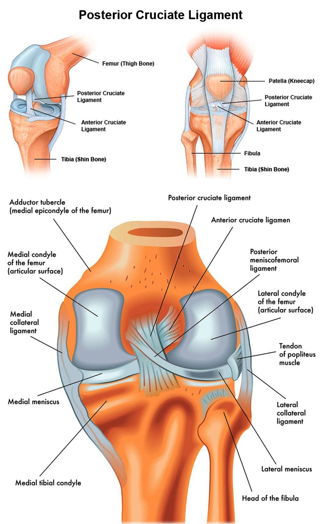
The body has around 1,000 ligaments that connect bones and joints. Ligaments are strong bands of tissue that support joint mobility and stabilize the muscles and bones. An injury to one or more ligaments can cause inflammation, swelling, discomfort, and instability. The PCL refers to the posterior cruciate ligament that runs along the back of the knee joint. This ligament connects the femur/thigh bone to the tibia/shinbone. Anyone can suffer from an injury to the posterior cruciate ligament. It can be caused by the knee hitting a dashboard in an automobile collision, a worker twisting or falling on a bent knee or a sports contact injury. The Injury Medical Chiropractic and Functional Medicine Clinic Team provide soft tissue work, trigger point therapy, and targeted non-surgical treatment through advanced therapy methods and technologies.
Posterior Cruciate Ligament
The posterior cruciate ligament – PCL is located inside the knee, just behind the anterior cruciate ligament – ACL. It is one of several ligaments that connect the femur/thighbone to the tibia/shinbone. The posterior cruciate ligament keeps the tibia from moving backward.
Injury
Posterior cruciate ligament injuries are far less common than ACL – anterior cruciate tears. PCL injuries make up less than 20% of all knee ligament injuries. It is more common for PCL tears to occur with other ligament injuries. A PCL injury can cause mild, moderate, or severe damage and is rated into four different categories:
Grade I
A partial tear is present in the ligament.
Grade II
There is a partial tear.
The ligament can feel loose.
Grade III
The ligament is completely torn.
The knee is unstable.
Grade IV
The PCL is injured.
Other knee ligaments are damaged.
Individuals with posterior cruciate ligament injuries can have short or long-term symptoms. Typically, long-term symptoms occur when an injury slowly develops over time. In mild cases, individuals may still be able to walk, and their symptoms may be less noticeable. Common symptoms associated with PCL injuries include:
Difficulty placing weight on the injured knee.
Stiffness.
Walking difficulties.
Difficulty descending stairs.
A wobbly sensation inside the knee.
Inflammation and swelling can be mild to severe.
Knee pain.
Pain that worsens over time.
Over time, tears could lead to the development of osteoarthritis.
There is an increased risk of extensive damage and chronic pain conditions if left untreated.
Chiropractic Care
The continued participation in work or activity following a mild injury is the primary reason individuals undergo therapy, injections, or surgical repairs. Knee injuries need immediate attention to prevent worsening or further damage. A chiropractor will examine the knee, check the range of motion and ask about symptoms. They may request imaging tests to determine the extent of the damage. These tests may include the following:
X-rays.
Magnetic resonance imaging.
CT scan.
During the physical examination, they will check all the structures of the injured knee and compare them to the non-injured knee. The wounded knee may appear to sag backward when bent or could slide back too far, specifically when beyond a 90-degree angle. Treatment depends on the severity of the injury. Common treatments include:
Crutches
Crutches may be recommended to limit the weight placed on the knee.
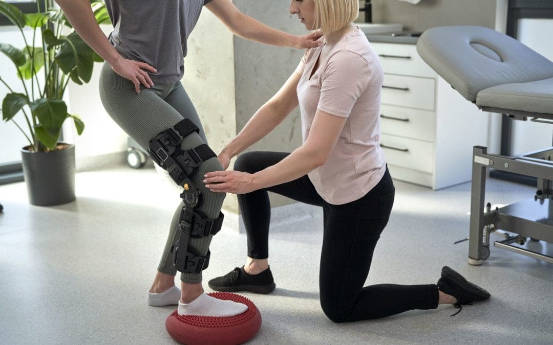
Knee Brace
A special brace can address instability and help prevent the tibia bone from sagging backward.
Gravity tends to pull the bone backward when lying down.
Chiropractic and Physical Therapy
As the swelling goes down, a carefully personalized rehabilitation program can begin.
This procedure is less invasive compared to traditional surgical methods.
Recovery time varies from person to person. If the injury is mild, it may only take around ten days to heal. If surgery was needed, recovery could take about six to nine months. Full recovery typically requires 6 to 12 months.
Best Knee Injury Chiropractor
References
American Academy of Orthopaedic Surgeons. Posterior Cruciate Ligament Injuries. (https://orthoinfo.aaos.org/en/diseases–conditions/posterior-cruciate-ligament-injuries) Accessed 7/26/21.
Bedi A, Musahl V, Cowan JB. Management of Posterior Cruciate Ligament Injuries: An Evidence-Based Review. Journal of the American Academy of Orthopedic Surgery. 2016 May;24(5):277-89. Accessed 7/26/21.
Lu, Cheng-Chang, et al. “Twelve Weeks of a Staged Balance and Strength Training Program Improves Muscle Strength, Proprioception, and Clinical Function in Patients with Isolated Posterior Cruciate Ligament Injuries.” International journal of environmental research and public health vol. 18,23 12849. 6 Dec. 2021, doi:10.3390/ijerph182312849
Pierce, Casey M et al. “Posterior cruciate ligament tears: functional and postoperative rehabilitation.” Knee surgery, sports traumatology, arthroscopy: official journal of the ESSKA vol. 21,5 (2013): 1071-84. doi:10.1007/s00167-012-1970-1
Schüttler, K F et al. “Verletzungen des hinteren Kreuzbands” [Posterior cruciate ligament injuries]. Der Unfallchirurg vol. 120,1 (2017): 55-68. doi:10.1007/s00113-016-0292-z
Zsidai, Bálint, et al. “Different injury patterns exist among patients undergoing operative treatment of isolated PCL, combined PCL/ACL, and isolated ACL injuries: a study from the Swedish National Knee Ligament Registry.” Knee surgery, sports traumatology, arthroscopy: official journal of the ESSKA vol. 30,10 (2022): 3451-3460. doi:10.1007/s00167-022-06948-x
Individuals dealing with aching knees is one of the most common health problems and affects people of all ages. The knee is the largest joint in the body, comprised of muscles, tendons, ligaments, cartilage, and bones. The knees support walking, standing, running, and even sitting. Constant use makes them highly susceptible to injuries and conditions. The knees are also surrounded by a complex network of nerves that transmit messages to and from the brain. Damage to the nerves from an injury or disease can create various symptoms of discomfort in and around the knee joint.
Knee Neuropathy
Causes
Knee discomfort symptoms can be brought on by injury, degenerative disorders, arthritis, infection, and other causes, including:
Rheumatoid arthritis
This is a chronic inflammatory disorder that causes the knees to swell and causes damage to the cartilage.
Osteoarthritis
This type of arthritis causes the cartilage to steadily wear away, causing damage to the joints and various symptoms.
Cartilage Issues
Overuse, muscle weakness, injury, and misalignments can cause compensating postures and movements that can wear down and soften the cartilage, generating symptoms.
Several factors can increase the risk of developing knee neuropathy, including:
Previous knee injury
Undiagnosed and untreated knee injury
Unhealthy Weight
Gout
Compromised leg muscle strength and/or flexibility
Symptoms
Symptoms associated with a knee injury or disorder can vary, depending on the severity and damage. Symptoms can include:
Joint stiffness
Swelling in the joint.
Reduced movement/flexibility in the joint.
Increased instability/feeling of weakness in the knee.
Changes in skin color around the knee joint, like increased redness or pale discoloration.
Numbness, coldness, or tingling in and/or around the joint.
Pain symptoms could be a dull ache or throbbing felt throughout the knee.
Sharp, stabbing discomfort in a specific area.
If left untreated, knee neuropathy could permanently affect the ability to walk and lead to partial or total loss of knee function and mobility. Doctors recommend taking note of the following:
What activity/s generates symptoms?
Where are the symptoms located?
What the pain feels like?
Treatments available for knee pain
Chiropractic treatment offers various approaches to addressing the pain caused by nerve damage. Standard treatment includes chiropractic adjustments, therapeutic massage, non-surgical decompression, stretching, posture and movement training, and nutritional anti-inflammatory plans. Our medical team specializes in non-surgical treatments that decrease symptoms and increase strength, flexibility, mobility, and restore function.
Knee Injuries Adjustment
References
Edmonds, Michael, et al. “The current burden of diabetic foot disease.” Journal of clinical orthopedics and trauma vol. 17 88-93. 8 Feb. 2021, doi:10.1016/j.jcot.2021.01.017
Hawk, Cheryl, et al. “Best Practices for Chiropractic Management of Patients with Chronic Musculoskeletal Pain: A Clinical Practice Guideline.” Journal of alternative and complementary medicine (New York, N.Y.) vol. 26,10 (2020): 884-901. doi:10.1089/acm.2020.0181
Hunter, David J et al. “Effectiveness of a new model of primary care management on knee pain and function in patients with knee osteoarthritis: Protocol for THE PARTNER STUDY.” BMC musculoskeletal disorders vol. 19,1 132. 30 Apr. 2018, doi:10.1186/s12891-018-2048-0
Kidd, Vasco Deon, et al. “Genicular Nerve Radiofrequency Ablation for Painful Knee Arthritis: The Why and the How.” JBJS essential surgical techniques vol. 9,1 e10. 13 Mar. 2019, doi:10.2106/JBJS.ST.18.00016
Krishnan, Yamini, and Alan J Grodzinsky. “Cartilage diseases.” Matrix biology: journal of the International Society for Matrix Biology vol. 71-72 (2018): 51-69. doi:10.1016/j.matbio.2018.05.005
Speelziek, Scott J A, et al. “Clinical spectrum of neuropathy after primary total knee arthroplasty: A series of 54 cases.” Muscle & nerve vol. 59,6 (2019): 679-682. doi:10.1002/mus.26473
As one of the stabilizers for the body, the knees are located between the thighs and legs, allowing flexion and extension. The knees help the hips by supporting the upper body’s weight and allowing the legs to move from one place to another without feeling pain. The knee has various muscles and ligaments surrounding the knee joint, allowing the leg to be bent when active. One of the muscles is located behind the knee, known as the popliteus, and supports the legs. However, minor injuries or actions can affect the knees causing the joint to be in a “lock” position and develop myofascial trigger points that can induce muscle spasms in the knees. Today’s article focuses on the popliteus muscle, how knee pain is associated with trigger points, and how to manage knee pain through various treatments. We refer patients to certified providers that incorporate multiple methods in the lower body extremities, like knee pain treatments correlating to myofascial trigger points, to aid many people dealing with pain symptoms along the popliteus muscles. We encourage and appreciate each patient by referring them to associated medical providers based on their diagnosis, especially when appropriate. We understand that education is an excellent source to asking our providers intricated questions at the patient’s request. Dr. Alex Jimenez, D.C., only utilizes this information as an educational service. Disclaimer
What Is The Popliteus Muscle?
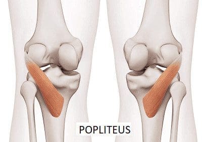
Have you been dealing with pain behind your knees? Do you have issues bending your knees when climbing up or down the stairs? Or do your back knee muscles start to twitch uncontrollably, causing muscle spasms? Many knee issues correlate with various factors that can affect the popliteus muscle and develop trigger points. The popliteus is a small muscle with a very important job as it is a major stabilizing muscle to the knees. The popliteus muscle originates from the lateral side of the femur and inserts itself into the posterior surface of the tibia. Some attachments are between the popliteus and lateral meniscus, allowing the knees to be in motion and providing flexion without pain and entrapment. Additional studies reveal that when a person exercises, the popliteus’s basic function helps bring about and maintain internal rotation of the tibia on the femur. The popliteus also helps prevent the foot from external rotation and allows the individual to stand correctly. However, injuries to the knee could overstretch the popliteus muscle and cause mobility issues to the knee flexion.
Knee Pain Associated With Trigger Points
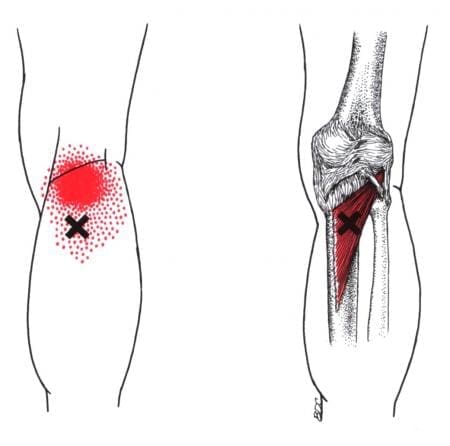
When dealing with knee pain, it could often be a joint disorder like osteoarthritis or a musculoskeletal condition like sciatica pain associated with the knee. These issues could be due to normal factors like constantly sitting down or bending down to lift heavy objects that cause the knees to buckle. However, when the popliteus muscle has been continuously overused from being bent, it can form tiny nodules known as trigger points to cause knee pain. Studies reveal that trigger points on the muscles surrounding the knee are often ignored during a clinical diagnosis. Trigger points cause referred pain to the surrounding muscles, accompanied by various sensory sensations like heaviness, tingling, and hypersensitivity to the popliteus muscle. In “Myofascial Pain and Dysfunction,” written by Dr. Travell, M.D. stated that one of the chief complaints that many patients often talk to their doctors about is the pain they feel in the back of their knees when they are in a crouch position. The book also states when normal actions like running or twisting have overloaded the popliteus muscle, it can cause trauma or strain to the popliteus muscle and tear the posterior cruciate ligament to the knees.
How To Find Trigger Points In The Popliteus- Video
Have you been having knee issues that make walking difficult for a long period? Do you feel like your knees are locking up constantly? What about feeling unstable when standing or carrying objects around? These issues that affect the knees are associated with trigger points along the popliteus muscles. The popliteus muscle is small, located at the back of the knees, and assists with knee flexion. When the popliteus muscle becomes overused, it can cause trigger points to form and cause knee issues. Studies reveal that various issues, like tendon injuries, are associated with repetitive mechanical stresses that can cause degenerative knee lesions. Any trauma or muscle strain can affect the knee’s function of flexing and bending without pain for trigger points to form along the popliteus muscles. The video above focuses on the popliteus muscle, where the trigger points are located, and where the referred pain patterns are situated in the knees. On the bright side, all is not lost, as various treatments offer ways to manage knee pain associated with trigger points.
Managing Knee Pain Through Various Treatments
When it comes to knee pain, many individuals will apply an ice or heat compress to allow the surrounding muscles to relax while reducing the pain and swelling. Other individuals use over-the-counter medicines to eliminate the pain for a few hours. While these work at managing knee pain, various treatments target trigger points and can help improve flexion mobility back to the knees. Studies reveal that muscle stretching on the popliteus muscle contributes to joint position sense to knee joint stability and function. Stretching the popliteus muscles can reduce the pain in the back of the knee while elongating the muscle fibers to manage trigger points from forming again. Other treatments that people can do to avoid trigger points from returning is to avoid walking or running in a lateral sloped area to prevent the knees from locking up. Incorporating these treatments to prevent knee issues and allow the knee to function properly.
Conclusion
The knees are one of the stabilizers in the body that are located between the thighs and legs, allowing flexion and extension. As a small muscle located in the back of the knees, the popliteus stabilizes the knees and enables them to be in motion without pain. However, when the popliteus muscle becomes overstretched and overused, it can develop trigger points in the popliteus that invoke referred pain to the surrounding muscles and cause the knees to lock up. To that point, it causes the body to be unstable and mimics knee pain issues. Fortunately, trigger points are treatable through various treatments that help relieve the pain and reduce the trigger points from returning. When these treatments are utilized on the knees, the surrounding muscles regain flexion mobility in the lower body.
References
English, S, and D Perret. “Posterior Knee Pain.” Current Reviews in Musculoskeletal Medicine, U.S. National Library of Medicine, 12 June 2010, https://www.ncbi.nlm.nih.gov/pmc/articles/PMC2941578/.
Ghaffarinejad, Farahnaz, et al. “Effect of Static Stretching of Muscles Surrounding the Knee on Knee Joint Position Sense.” British Journal of Sports Medicine, U.S. National Library of Medicine, Oct. 2007, https://www.ncbi.nlm.nih.gov/pmc/articles/PMC2465159/.
Hyland, Scott, and Matthew Varacallo. “Anatomy, Bony Pelvis and Lower Limb, Popliteus Muscle.” In: StatPearls [Internet]. Treasure Island (FL), StatPearls Publishing, 6 June 2022, https://www.ncbi.nlm.nih.gov/books/NBK526084/.
Mann, R A, and J L Hagy. “The Popliteus Muscle.” The Journal of Bone and Joint Surgery. American Volume, U.S. National Library of Medicine, Oct. 1977, https://pubmed.ncbi.nlm.nih.gov/908724/.
Sánchez Romero, Eleuterio A, et al. “Prevalence of Myofascial Trigger Points in Patients with Mild to Moderate Painful Knee Osteoarthritis: A Secondary Analysis.” Journal of Clinical Medicine, U.S. National Library of Medicine, 7 Aug. 2020, https://www.ncbi.nlm.nih.gov/pmc/articles/PMC7464556/.
Travell, J. G., et al. Myofascial Pain and Dysfunction: The Trigger Point Manual: Vol. 2:the Lower Extremities. Williams & Wilkins, 1999.
IFM's Find A Practitioner tool is the largest referral network in Functional Medicine, created to help patients locate Functional Medicine practitioners anywhere in the world. IFM Certified Practitioners are listed first in the search results, given their extensive education in Functional Medicine


 Posterior Cruciate Ligament